Ostracoda (Myodocopina) of Tulear Reef Complex, SW Madagascar
|
|
|
- Ruth Weaver
- 6 years ago
- Views:
Transcription


1 Ostracoda (Myodocopina) of Tulear Reef Complex, SW Madagascar LOUIS S. KORNICKER and BERNARD A. THOMASSIN SMITHSONIAN CONTRIBUTIONS TO ZOOLOGY NUMBER 595


2 SERIES PUBLICATIONS OF THE SMITHSONIAN INSTITUTION Emphasis upon publication as a means of "diffusing knowledge" was expressed by the first Secretary of the Smithsonian. In his formal plan for the institution, Joseph Henry outlined a program that included the following statement: "It is proposed to publish a series of reports, giving an account of the new discoveries in science, and of the changes made from year to year in all branches of knowledge." This theme of basic research has been adhered to through the years by thousands of titles issued in series publications under the Smithsonian imprint, commencing with Smithsonian Contributions to Knowledge in 1848 and continuing with the following active series: Smithsonian Contributions to Anthropology Smithsonian Contributions to Botany Smithsonian Contributions to the Earth Sciences Smithsonian Contributions to the Marine Sciences Smithsonian Contributions to Paleobiology Smithsonian Contributions to Zoology Smithsonian Folklife Studies Smithsonian Studies in Air and Space Smithsonian Studies in History and Technology In these series, the Institution publishes small papers and full-scale monographs that report the research and collections of its various museums and bureaux or of professional colleagues in the world of science and scholarship. The publications are distributed by mailing lists to libraries, universities, and similar institutions throughout the world. Papers or monographs submitted for series publication are received by the Smithsonian Institution Press, subject to its own review for format and style, only through departments of the various Smithsonian museums or bureaux, where the manuscripts are given substantive review. Press requirements for manuscript and art preparation are outlined on the inside back cover. I. Michael Heyman Secretary Smithsonian Institution
 of Tulear")

3 S M I T H S O N I A N C O N T R I B U T I O N S T O Z O O L O G Y N U M B E R Ostracoda (Myodocopina) of Tulear Reef Complex, SW Madagascar Louis S. Kornicker and Bernard A. Thomassin SMITHSONIAN INSTITUTION PRESS Washington, D.C. 1998
4 ABSTRACT Kornicker, Louis S., and Bernard A. Thomassin. Ostracoda (Myodocopina) of the Tulear Reef Complex, SW Madagascar. Smithsonian Contributions to Zoology, number 595, 134 pages, 86 figures, 2 tables, Twenty-four species (16 new), representative of all five myodocopid families, plus one left in open nomenclature, are described and illustrated from collections from the Tulear Reef Complex, SW Madagascar. Specimens were collected by personnel of the Station marine d'endoume et Centre d'oceanographie (CNRS-VRA n 41), Marseilles, France, between 1969 and The distribution of Myodocopina in the area and the faunal resemblances in the populations living in various parts of the reef, as indicated by Simpson Indices, are discussed. OFFICIAL PUBLICATION DATE is handstamped in a limited number of initial copies and is recorded in the Institution's annual report, Annals of the Smithsonian Institution. SERIES COVER DESIGN: The coral Montastrea cavernosa (Linnaeus). Library of Congress Cataloging-in-Publication Data Komickcr. Louis S., Ostracoda (Myodocopina) of Tulear Reef Complex, SW Madagascar / Louis S. Kornicker and Bernard A. Thomassin. p. cm. (Smithsonian contributions to zoology ; no. 595) Includes bibliographic references. I. Myodocopida Madagascar-Toliara. I. Thomassin, Bernard A. II. Title. III. Series. QL1.S54 no. 595 [QL ] 59Os-dc21 [595.3'3] % The paper used in this publication meets the minimum requirements of the American National Standard for Permanence of Paper for Printed Library Materials Z

5 Contents Page Introduction 1 Stations 1 Sampling 1 Disposition of Specimens 1 Abbreviations 1 Terminology 4 Acknowledgments 4 Distribution of Myodocopina in the Tulear Reef Complex 4 Discussion of Distribution 6 Biogeographic Comparisons 6 Lateral Eyes 6 Superorder MYODOCOPA Sars, Order MYODOCOPIDA Sars, Suborder MYODOCOPINA Sars, CYPRIDINIDAE Baird, CYPRIDININAE Baird, CYPRIDININI Baird, Codonocera Brady, Codonocera phoenix Kornicker, Paradoloria Hanai, Paradoloria vanhoeffeni (Muller, 1908) 8 Cypridinodes Brady, Cypridinodes strophinx, new species 9 Cypridinodes relax, new species 17 Cypridinodes parallax, new species 25 Skogsbergia Kornicker, Skogsbergia solox, new species 34 Skogsbergia plax Kornicker, Skogsbergia calyx Kornicker, Vargula Skogsberg, Vargula grex, new species 45 Pterocypridina Kornicker, Pterocypridina nex, new species 50 PHILOMEDIDAE Muller, PHILOMEDINAE Muller, Zeugophilomedes Kornicker, Zeugophilomedes sphinx, new species 56 PSEUDOPHILOMEDINAE Kornicker, Harbansus Kornicker, Harbansus flax, new species 68 RUTIDERMATIDAE Brady and Norman, RUTIDERMATINAE Brady and Norman, Rutiderma Brady and Norman, Rutiderma arx Kornicker, Rutiderma ferax, new species 83 Rutiderma exrex, new species 87


6 iv SMITHSONIAN CONTRIBUTIONS TO ZOOLOGY SARSIELLIDAE Brady and Norman, Key to the Subfamilies of the Sarsiellidae 92 DANTYINAE Komicker and Cohen, Dantya Komicker and Cohen, Key to the Species of Dantya 93 Dantya dux, new species 93 SARSIELLINAE Brady and Norman, Junctichela Komicker and Caraion, Key to the Species of Junctichela 97 Junctichela lex, new species 97 Chelicopia Komicker, Chelicopia fax, new species 102 Eurypylus Brady, Eurypylus matrix, new species 106 CYLINDROLEBERIDIDAE Muller, CYLINDROLEBERIDINAE Muller, Synasterope Komicker, Synasterope calix Komicker, Cylindroleberis Brady, 1868 Ill Cylindroleberis vibex Komicker, 1992 Ill Cylindroleberis vix Komicker, Parasterope Komicker, Paraster ope maddocksae, new species 117 Parasterope species A 122 Heptonema Cohen and Komicker, Heptonema latex Komicker, Appendix: Station Data with Species Collected 127 Literature Cited 132

 and in the lagoons of the Tulear and Ranobe bays, on fringing reefs (Songoritelo, Sarodrano), on lagoonal reefs (in the Tulear bay), and on")
7 Ostracoda (Myodocopina) of the Tulear Reef Complex, S W Madagascar Louis S. Kornicker and Bernard A. Thomassin Introduction This work reports on the Ostracoda in the suborder Myodocopina collected by the junior author and other scientists between 1969 and 1972 in the Tulear Reef Complex, SW Madagascar. Samples were collected using scuba-diving bucket-sampling and hydropneumatic sucking, dredging in lagoons, and shoveling in intertidal zones. Collections were made on the Great Reef (barrier reef) and in the lagoons of the Tulear and Ranobe bays, on fringing reefs (Songoritelo, Sarodrano), on lagoonal reefs (in the Tulear bay), and on littoral and cay beaches (in Ranobe lagoon and on Nosy Ve Island) (Figure 1). The distribution of soft-bottom carcinological fauna in the area was described initially by Thomassin (1974) and was more extensively described, including their place in the various "biocoenoses" colonizing soft bottoms, by Thomassin (1978b, 1983). The senior author was given the Ostracoda for identification in October Six species (five new) in the subfamily Cyclasteropinae in that collection were described previously by Kornicker (1981), but for completeness they are listed in Table 1 and Appendix 1. The pelagic ostracodes of the Tulear region have been reported by Leveau (1957), the podocopid ostracodes of the Nosy Be' area of Madagascar have been partly described by Louis S. Kornicker, Department of Invertebrate Zoology, National Museum of Natural History, Smithsonian Institution, Washington, DC Bernard A. Thomassin, Centre d'oceanologie de Marseille (CNRS- URA n 41), Station marine d 'Endoume, Universite d 'A ix-marseille II, traverse de la Rue de la Baterrie-des-Lions, Marseille, FRANCE. Review Chairman: Austin B. Williams, National Marine Fisheries Service, Systematics Laboratory, National Museum of Natural History, Smithsonian Institution. Reviewers: Anne C. Cohen, Los Angeles County Museum of Natural History, California; Rosalie F. Maddocks, University of Houston, Texas. Maddocks (1966, 1968, 1969a, 1969b, 1973, 1976, 1988, 1990, 1991) and Jellinek (1989,1990, 1993), with some allusions and reassignments in Maddocks (1991, 1992), and those from near Tulear have been reported by Maddocks (1969b). STATIONS. BT-codes are station designators. The 96 stations from which myodocopids were collected range in depth from littoral to 60 m, but only one (BT-330) is deeper than 36 m. The localities of stations from which ostracodes are reported are presented in chronological order in the Appendix. Station localities are from a station list (in French) that is filed in the Division of Crustacea, National Museum of Natural History, Smithsonian Institution. The locality names in the Appendix are mostly as they appear in the station list, but some have been Anglicized. Maps showing station localities have been presented in Thomassin (1978b, 1983). SAMPLING. As stated above, several methods of collecting were used to obtain sediment samples from which the ostracodes were later removed (Thomassin, 1978a, 1978b). Quantitative samples were sieved on 1.4 x 1.4 mm mesh and, for sea-grass beds and any lagoonal dredgings, on 2 x 2 mm mesh; the larger mesh may have resulted in loss of some juveniles and small adults. The collecting methods for individual samples are listed in the Appendix. Rose bengal was used to stain organic matter in later samples to make sorting more efficient. DISPOSITION OF SPECIMENS. Holotypes have been deposited at the Museum National d'histoire Naturelle, Paris, France, and these have been assigned MNHN numbers. Specimens deposited in the collections of the former United States National Museum (USNM), now the National Museum of Natural History, Smithsonian Institution, have been assigned USNM numbers. Unnumbered specimens also have been deposited in the Museum National d'histoire Naturelle, Paris. Except when indicated otherwise, specimens are undissected and are preserved in alcohol. ABBREVIATIONS. In the figures, Arabic numerals indicate limbs 1-7, as well as individual joints of each limb (the


8 SMITHSONIAN CONTRIBUTIONS TO ZOOLOGY transects on the outer fo#f 23 1O'S lf8ty bnnd P assa 9 e 23 20'S Nosy Vato Northern pass Tropic of Capricorn 23 30'S Mozambique Channel Southern/pass NOSY TAFAR A REEF* SARODRANO' REEF St. Augustine Bay Nosy Ve Cayrm*f j43«40'e FIGURE 1. Collecting area in vicinity of the Tulear Reef Complex. (Adapted from fig. 1 in Thomassin, 1974.)
; 2 = Spur-and-Groove Zone (6-24 m); 3 = Microatoll Flats (generally")
9 NUMBER 595 TABLE 1. Distribution of Myodocopina species at selected localities: Grand Reef Outer Slope: 1 = Coral Flagstone (21-36 m; BT-330 at 60 m); 2 = Spur-and-Groove Zone (6-24 m); 3 = Microatoll Flats (generally in turbid zone); 4 = Muddy-Sandy Accumulations of Reef Flats; 5 = Hydraulic Sand-Banks on Inner Edge of Reef Flat or on Lagoonal Slopes (4-10 m); 6 = Residual Pools of Reef Flat (6-12 m); 7 = Lagoonal Bottoms and enclosed Lagoon Bottoms ( m); 8 = Nosy Ve Cay Beaches (infralittoral). 1969/1972 are collecting years. 9 = NE Mozambique Channel* (includes only species also collected in present study area). (+ = species present; - = species absent.) Species Localities 1969/ 1972 CYPRIDINIDAE Codonocera phoenix Paradoloria vanhoeffeni Cypridinodes parallax Cypridinodes relax Cypridinodes strophinx Skogsbergia calyx Skogsbergia plax Skogsbergia solox Vargula grex Pterocypridina nex PHILOMEDIDAE Zeugophilomedes sphinx Harbansus flax RUTIDERMATIDAE Rutiderma arx Rutiderma ferax Rutiderma exrex SARSIELLIDAE Dantya dux Junctichela lex Chelicopia fax Eurypylus matrix CYLINDROLEBERIDIDAE CYLINDROLEBERIDINAE Synasterope calix Cylindroleberis vibex Cylindroleberis vix Parasterope maddocksae Parasterope species A Heptonema latex CYCLASTEROPINAEf Alphaleberis alphathrix Amboleberis antyx Cycloleberis galatheae Tetraleberis maddocksae Tetraleberis tanzania Asteropterygion thomassini Total number of species Total number of genera /20 19/ * From Komicker (1992) {Benthedi cruise 1977). t From Komicker (1981).


10 SMITHSONIAN CONTRIBUTIONS TO ZOOLOGY location of the numeral indicating whether a limb or joint is indicated). Roman numerals I-IV indicate the endites. Arrows on illustrations indicate anterior of specimen. The following abbreviations are used in the appendix, illustrations, and legends: am ant ap av bas Bo br CO ex e end ep, epip esop ex fu gird go hrt HS im iv le lft 11 IP lv me mnd mo mv nabs ov P pv precx prot rt SC sens t ul Y-scl central adductor muscle attachments antenna anterior process anterior view basale Bellonci organ bristles copulatory organ coxale edge of shell endopodite epipodite esophagus exopodite furca girdle genital organ heart sediment hydraulic air-sucker inner margin of infold inside view lateral eye left lower lip lamellar prolongation lateral view medial eye mandible mouth medial view not all bristles shown outside view parasite posterior view precoxale protopodite right sediment collected with bag, bucket, or shovel sensory bristle of 5th joint of 1 st antenna testis upper lip Y-sclerite TERMINOLOGY. Reef terms used herein are from Thomassin (1974:297). The names "Ankaradanva," "Antseteky," and "Andeteky" are native names for parts of the Grand Recif (Figure 1). "Tumuli" refers to mounds built by endofauna (40 cm high, about 1 m diameter). "Grand Recif" or "Great Reef is the main barrier reef in the area. "Grande Vasque" is an enclosed lagoon, a large pool, 1.8 km long, 0.3 km wide. It is part of the general lagoon (channel) now enclosed in the barrier reef by the building of several inner coral patches. The bottom at about m depth is a mixed silty sand or soft mud with a field of tumuli-and-funnels built by large enteropneusta and large squills (Lysiosquilla maculata). "Petite Vasque" is a residual pool, 10 m deep, just behind the actual boulder tract. It is the residual part of an outer pass in the reef front. "Boulder tracts" are formed by broken coral material produced by the upper part of the grooves on wave-beaten reefs (except on Songoritela and South Ifaty reefs) (Thomassin, 1974:298). "Hydraulic dunes" are accumulations of shifting medium and fine well-sorted sands deposited on groove floors in more or less continuous patches (10-20 m long, 3-5 m wide), which are constantly disturbed by surges and undertow-streams (Thomassin, 1974:298). "Melobesians" is a group of calcareous algae. "Rhodoliths" are the "stones" (sometimes "rolling stones") made by some calcareous algae in different environments: outer reef coral slabstone, passage bottoms under strong currents, or in sheltered environments, such as microatoll flats. ACKNOWLEDGMENTS. We wish to thank personnel at the Station marine d'endoume et Centre d'oceanographie, Marseilles, France, for assistance in collecting the specimens upon which this study is based. We are grateful to several people who assisted in preparation of this paper: the final drawings were inked by Jack Schroeder, Jack Schroeder Associates; shaded drawings of carapaces were rendered by Carolyn Gast (Smithsonian Institution, retired); the map (Figure I) was drawn by Molly K. Ryan (Smithsonian Institution), and Elizabeth Harrison-Nelson (Smithsonian Institution) prepared the "Literature Cited" section, catalogued specimens, and helped in many other tasks. We thank Rosalie Maddocks (University of Houston) and Anne C. Cohen (Natural History Museum of Los Angeles County) for critically reviewing the manuscript and making many helpful comments. We also thank Diane M. Tyler, Smithsonian Institution Press, for editing and preparing the manuscript. Distribution of Myodocopina in the Tulear Reef Complex The species collected at the various stations are listed in the Appendix. Thomassin (1978b) listed the geomorphic and sedimentologic features of the reef area as well as the faunistic assemblages belonging to various biocoenoses, using reef terminology proposed by Battistini et al. (1975). The latter publication contains an excellent schematic diagram (fig. 16) of coral reefs of S.W. Madagascar. The following account lists the species of ostracodes that were collected in the various parts of the reef described by Thomassin. The stations in each subdivision have been grouped according to locality or type of substrate, and the particular station-group is shown as a superscript following the name of each species collected there. Among the outer reefs in the coral reef complex of Tulear are the Grand Recif de Tulear (or Great reef), Nosy Tafara Reef (in the middle of the Southern Passage), and Sarodrano Reef, which now looks like fringing reef because a sand spit is built upon its flat. The Ifaty Reef (northern) is in the "coral reef complex of Ifaty-Ranobe," characterized by low terrigenous
 by a spur-and-groove zone, and below it (12-36 m) by a coral flagstone with scattered coral growths and spreads of")
. c, Gravel substrate: BT-221, 21 m.")
11 NUMBER 595 alluvial components in contrast to sediments off Tulear. OUTER REEF SLOPE. Generally, the outer reef slope is characterized in its upper levels (0-25 m) by a spur-and-groove zone, and below it (12-36 m) by a coral flagstone with scattered coral growths and spreads of heterometric sediment. Coral Flagstone: a, Sediment (sand) substrate: BT-172, 29 m; BT-211, 12 m; BT-230, 21 m; BT-240 and 240B, 36 m; BT-261, 26 m. b. Bottom of melobesians or rhodoliths: BT-184, 31 m; BT-186, 34 m; BT-222, 24 m). c, Gravel substrate: BT-221, 21 m. Amboleberis antyx*, Asteropterygion thomassini*, Codonocera phoenix* bc, Cyclolebehs galatheae* b, Cylindroleberis vibeip, Dantya dux 0, Eurypylus matrix*, Paradoloria vanhoeffeni* b, Pterocypridina nex 3, Rutiderma arx* b, Skogsbergia plax b, Synasterope calix* b, Vargula grex b. Spur-and-Groove Zone (6-27 m): a, Sediment present only in bottom grooves: Sta BT-135, 18 m; BT-191, 6 m; BT-213, 12 m; BT-219, 10 m./), Sediment present as patches: BT-161, 13 m; BT-197, 18 m; BT-198, 14 m; BT-223, 11 m; BT-237, 17 m. c, Sand formed in large ripples: BT-164, 9 m. d, Gravels and gravelly sand: BT-201 and BT-202,20 m; BT-218, 7 m; BT-224 and 224B, 17 m; BT-837, 7 m. e. Sand and coral fragments: BT-225, 9 m./ Sedimentary pockets with nodules of melobesians: BT-227, 27 m; BT-228, 17 m; BT-231, 24 m. g. Layer of coarse sediment with ripple marks at base of coral growths: BT-236, 15 m. Alphaleberis alphathhx* b, Asteropterygion thomassini d - { '%, Codonocera phoenix*- bde - f>g, Cyclolebehs galatheae*^' g, Cylindroleberis vibex {, Eurypylus matrix^, Paradoloria vanhoejfeni* f ' g, Parasterope maddocksae b ' (, Junctichela lex 0, Rutiderma arx 0 -^, Rutiderma exrex f, Skogsbergia calyx f, Skogsbergia plax?, Skogsbergia solox 6, Synasterope calix*- b s, Zeugophilomedes sphinx*^. MICROATOL FLATS. On the outer reef flats and boulder tracts, sediment is absent except in small holes containing gravel and where sand is trapped in coral pieces and slabs. Stations in the rubble embankment of South Lovobe, Great Reef, are BT-788, BT-790, BT-795, and BT-797, 10 m, which contained Paradoloria vanhoeffeni and Rutiderma exrex. In the inner moat of inner reef flats are holes, gullies, and patches of sand between coral-built formations. Welldeveloped microatoll flats, which generally occur in turbid waters, have small amounts of coarse sediment (often composed of fragments of Halimeda) among coral patches: a, Beloza Reef: BT-255; BT-256; BT-257. b, Songoritelo Reef: BT-262; BT-263; BT-264. c, Sarodrano Reef: BT-270; BT-272; BT-274. Asteropterygion thomassini abc, Cyclolebehs galatheae*- Harbansusflax*, Paradoloria vanhoeffeni abc, Parasterope species A a - Rutiderma arx bc, Synasterope calix*- c. MUDDY-SANDY REEF FLATS. On the barrier reef flat and Nosy Ve Cay flat are muddy-sandy accumulations: a, Seagrass bed on reef flat: BT-676; BT-678, BT-683; BT-851, BT-876 (with marl); BT-880. b. Zone of sand mounds without seagrass: BT-691. c, Same locale as "6" except with Halophila: BT-693. Asteropterygion thomassin^b- c, Cypridinodesparallax 11, Cylindroleberis vibex 0, Cylindroleberis vix, Paradoloria vanhoeffeni 3, Rutiderma arx b ' c, Skogsbergia calyx*, Synasterope calix*. No Myodocopina were collected in the coarser sands of channels in sea-grass beds. SANDY BANKS OF INNER EDGE OF REEF FLATS. Hydraulic sand-banks occur on the inner edge of reef flats and on lagoonal slopes: a, Inner slope (Tulear Bay and Nosy Ve Lagoon): BT-191, 6 m; BT-600, 5-6 m; BT-602, 6 m; BT-822, 7-8 m; BT-841, 7 m; BT-848A; BT-883, 7 m. b. Inner Slope of enclosed lagoon (Grand Vasque): BT-761,5 m; BT-777A, 7 m; BT-778, 10 m; BT-779, 4 m; BT-811, 5 m; BT-814, 3-5 m. Alphaleberis alphathrix* b, Asteropterygion thomassini* b, Codonocera phoenix**, Cypridinodes parallax*- b, Cypridinodes relax b, Cypridinodes strophinx* b ', Cylindroleberis vibex*, Cylindroleberis vix b, Heptonema latex*, Paradoloria vanhoeffeni, Skogsbergia plax*, Synasterope calix* b, Tetralebehs tanzania a, Zeugophilomedes sphinx*. RESIDUAL POOLS. Such pools occur in the Great Reef: a, Pool #1 (sand, 12 m): BT-706; BT-709. b, Pool #2 (sand, 6-12 m): BT-700 and BT-701, m; BT-715, 6 m; BT-836, (depth not given), c, Pool #3 (sand, 8 m): BT-712. Alphaleberis alphathrix b, Asteropterygion thomassini 0, Codonocera phoenix*, Cylindroleberis vix*' bx, Cypridinodes parallax*^, Cypridinodes strophinx b, Paradoloria vanhoeffeni', Rutiderma ferax b ', Synasterope calix b. LAGOONAL BOTTOMS. The lagoonal bottoms of the "Grande Vasque" of the Great Reef, Tulear Bay, and the Foly Reef pool have fairly similar environments: a, Tulear Bay: BT-615, BT-616, BT-617, BT-620, BT-621, BT-622, all at 8-13 m in Southern Pass (coarse and medium sands); BT-623 (behind Southern Pass area); BT-726, BT-730, both at 12 m (behind Southern Pass area); BT-734, BT-737, BT-738, BT-741, all at 12 m (Southern Pass), b, Grand Vasque (enclosed lagoon): BT-212, 12 m; BT-770, 18 m; BT-771, 17 m; BT-773, 7-8 m; BT-813, 18.5 m. c, Foly Reef pool: BT-719, 7 m; BT-720 and BT-721, 4-5 m. Alphaleberis alphathrixb, Asteropterygion thomassini*, Chelicopia fax*, Codonocera phoenix*, Cylindroleberis vix*- b, Cypridinodes parallax*, Cypridinodes relax*, Cypridinodes strophinx b, Paradoloria vanhoeffeni*, Rutiderma arx*, Synasterope calix 0, Tetralebehs maddocksae 0, Tetralebehs tanzania. Nosy Ve cay has sandy beaches: a, Eastern Beach, infralittoral: BT-852 (coarse sand with crushed corals), b. Western Beach: BT-870 (in mats of Phyllochaetopteridea). Alphaleberis alphathhx*, Cypridinodes parallax*, Paradoloria vanhoeffeni*, Rutiderma arx 0, Rutiderma exrex 0.


12 SMITHSONIAN CONTRIBUTIONS TO ZOOLOGY DISCUSSION OF DISTRIBUTION The distributions of species in various geomorphic areas are listed in Table 1, and the faunal resemblances of species and genera in the designated areas, as indicated by the Simpson Index (Kornicker, 1975a:31; 1992:4) as calculated from species and genera, are presented in Table 2. The Simpson Indices (S.I.) suggest that the myodocopid population of the outer reef (Coral Flagstone (1) and Spur-and-Groove Zone (2)) in wave-beaten zone, differs considerably from that of the Lagoonal Bottoms (7) in more sheltered conditions (S.I. = 38 to 46), and that the Muddy-Sandy Reef Flats (4), the Sandy-Banks of the Inner Edge of the Reef Flats (5), the Residual Pools of the Reef Flat (6), and the Lagoonal Bottoms (7) have more similar myodocopid populations (S.I. = 62 to 89). Fairly similar populations are also present on the Coral Flagstone (1), Spur-and-Groove Zone (2), and Microatoll Flats (3) (S.I. = 69 to 71). Cohen (1989:316) also showed that myodocopid faunal composition at the species and family levels differed between a shallow (1.5 m) lagoon site and a deeper (18-30 m) forereef site on a Belize barrier reef. Species collected in 1969 (23 species) and 1972 (20 species) are compared in Table 1. Only 12 species were collected in both years (S.I. 60), but this is most likely the result of having collected in different habitats of the reef each year rather than a change in populations with time. Biogeographic Comparisons Thirty-one species (including one left in open nomenclature and six described previously (Kornicker, 1981)) of myodocopid ostracodes were collected in the Tulear Reef Complex, which is fewer than the 51 species reported by Cohen (1989:328) from the Caribbean Belize barrier reef area, but more than the 21 species reported from the Great Bahama Bank by Kornicker (1958). Kornicker (1992, table 1) recorded 23 species from shelf depths in the NE Mozambique Channel, which lies off NW Madagascar. Only 10 of the species present there were also collected in the Tulear Reef Complex (S.I. = 43). Of the 22 genera collected in the present study area, 14 are also present at shelf depths in the NE Mozambique Channel (total genera 17) (S.I. = 82). Three additional genera were collected at shelf depths in the NE Mozambique channel: Cypridina (two species), Metasarsiella (one species), and Neomuelleriella (one species). The eight genera collected in the Tulear Reef Complex but not recorded from shelf depths in the NE Madagascar Channel are Cypridinodes (three species), Pterocypridina (one species), Zeugophilomedes (one species), Junctichela (one species), Parasterope (two species, including one left in open nomenclature), Alphaleberis (one species), Amboleberis (one species), and Tetraleberis (two species); of these, only Alphaleberis is presently known only from Madagascar. Lateral Eyes The relationship between development of lateral eyes and water depth has been discussed by Kornicker (1975a:42; 1989:9; 1992:4; Kornicker and Poore, 1996). At shelf depths in the NE Madagascar Channel all species possessed lateral eyes (Kornicker, 1992:4). At the Tulear Reef Complex, in relatively shallow water, the adult female Zeugophilomedes sphinx is without lateral eyes, although the adult male has welldeveloped lateral eyes. The unique specimen of Harbansus flax in the collection, an A-l female, has small unpigmented processes that may represent lateral eyes. The remaining species have well-developed lateral eyes. Superorder MYODOCOPA Sars, 1866 The Myodocopa include two orders: Myodocopida Sars, 1866, and Halocyprida Dana, 1853, of which only the former is in the present collection. TABLE 2. Simpson Indices of faunal resemblances between localities based on species (top diagonal) and on genera (bottom diagonal). (Numbers on 1st line correspond to localities in left column.) Species Localities Coral Flagstone 2. Spur-and-Groove Zone 3. Microatoll Flats 4. Muddy-Sandy Reef Flats 5. Sand-Banks Inner Edge Reef Flat/Lagoonal Slopes 6. Residual Pools Reef Flat 7. Lagoonal Bottoms 8. Nosy Ve Cay Beaches _ _


.")
13 NUMBER 595 Order MYODOCOPIDA Sars, 1866 The Myodocopida include a single suborder Myodocopina Sars, Suborder MYODOCOPINA Sars, 1866 The Myodocopina includes five families, all represented in the present collection: Cypridinidae Baird, 1850a, Philomedidae Muller, 1906, Rutidermatidae Brady and Norman, 1896, Sarsiellidae Brady and Norman, 1896, and Cylindroleberididae Muller, CYPRIDINIDAE Baird, 1850 The Cypridinidae include two subfamilies: Cypridininae Baird, 1850a, and Azygocypridininae Kornicker, Only the former is in the present collection. FIGURE 2. Codonocera phoenix Kornicker, 1992, adult male, USNM , complete specimen from left side, length 2.25 mm. CYPRIDININAE Baird, 1850 The Cypridininae include two tribes: Cypridinini Baird, 1850a, and Gigantocypridinini Hartmann, 1974 (in Hartmann and Puri, 1974). Only the former is in the present collection. CYPRIDININI Baird, 1850 The Cypridinini include 22 genera of which six are in the present collection. Codonocera Brady, 1902 TYPE SPECIES. Codonocera cruenta Brady, COMPOSITION AND DISTRIBUTION. Indo-Pacific and Australasian waters. Only two species, C. pusilla Muller, 1906, and C. phoenix Kornicker, 1992, have been reported from the western Indian Ocean. male (left valve lost); 8 specimens. BT-715: 1 juvenile. BT-738: 1 specimen. BT-841: 1 specimen. DISTRIBUTION. Northern Mozambique Channel at depths of m (Kornicker, 1992:34). Madagascar: see "Material." SUPPLEMENTARY DESCRIPTION OF ADULT MALE. Carapace similar to that described by Kornicker (1992:34) (Figure 2). Carapace Size (length, height in mm): USNM , 2.25, Seventh Limb: One limb of USNM with six bristles in proximal group, 3 on each side (other limb with distal part missing). (Male described by Komicker (1992:38) has 4 or 5 bristles in proximal group.) Codonocera phoenix Kornicker, 1992 FIGURES 2-4 Codonocera phoenix Komicker 1992:34, figs HOLOTYPE. MNHN Os 432, adult female in alcohol. TYPE LOCALITY. Mozambique Channel, Glorioso Islands, depth 250 m (Kornicker, 1992:34). MATERIAL. BT-161: USNM , 1 specimen. BT-172: 32 specimens. BT-191: 3 specimens. BT-201: 1 specimen. BT-213: 1 specimen. BT-221: 2 specimens. BT-222: 1 specimen. BT-224: 7 specimens. BT-225: 5 specimens. BT-227: 3 specimens. BT-230: 15 specimens. BT-231: 1 juvenile. BT-236: 4 specimens. BT-237: 4 specimens. BT-240: 1 juvenile. BT-261: USNM , partly dissected ovigerous female (left valve lost); USNM , partly dissected adult FIGURE 3. Codonocera phoenix Kornicker, 1992, ovigerous female, USNM , complete specimen from left side showing distribution of purple chromatophores, length 2.71 mm.

 FIGURE 5 Paradoloria vanhoeffeni. Komicker, 1992:55, figs.")
14 SMITHSONIAN CONTRIBUTIONS TO ZOOLOGY in the present collection, Paradoloria vanhoeffeni, was previously reported from Madagascar (Monod, 1932:3). The genera Paradoloria and Doloria differ mainly in a male character so that several species referred to either genera, but known only from the female, eventually may be transferred when the male becomes known (Komicker, 1992:13; Kornicker and Poore, 1996:38), and that could alter the presently conceived distribution of Paradoloria. The male is known for P. vanhoeffeni. Determining the distribution of Paradoloria and Doloria based only on males would be warranted. Paradoloria vanhoeffeni (Muller, 1908) FIGURE 5 Paradoloria vanhoeffeni. Komicker, 1992:55, figs. 30, 31 [see for additional synonymies]. FIGURE 4. Codonocera phoenix Komicker, 1992, ovigerous female, USNM , complete specimen from left side showing location of left lateral eye and 15 eggs in marsupium, length 2.71 mm. SUPPLEMENTARY DESCRIPTION OF ADULT FEMALE. Similar to that described by Komicker (1992:40) (Figures 3,4). Carapace Size (length, height in mm): USNM , 2.71,2.06. Seventh Limb: Both limbs of USNM with 7 bristles in proximal group, 3 or 4 on each side. (Female described by Komicker (1992:43) has 4 or 5 bristles in proximal group.) Eggs: USNM with 21 well-developed eggs (with lateral eyes and some appendages) in marsupium (location of 15 eggs shown in Figure 4); length of typical egg 0.33 mm. Remarks: The male and female from BT-261 has 6 or 7 bristles in the proximal group of the 7th limb, slightly more than the 4 or 5 bristles on the specimens described by Komicker (1992:38,43). This is interpreted to be the result of intraspecific variability. In the original description of C. phoenix, Komicker (1992:55) reported no ovigerous females, but 1 female had 40 unextruded eggs (20 on each side of the body) (the number was rechecked in the present study). The 21 eggs in the marsupium of USNM in present collection appear crowded, and it seems unlikely that the marsupium could hold 40 eggs of that size; this may reflect a specific difference. Poulsen (1962:319) reported 15 newly laid eggs in the marsupium of C. suensoni. According to A.C. Cohen (pers. comm., 1995) the number of eggs brooded is often fewer than those unextruded. HOLOTYPE. None selected. SYNTYPE LOCALITY. Benthic, off Simonstown, South Africa. MATERIAL. BT-172: 4 specimens. BT-186: 1 specimen. BT-216: 1 specimen. BT-219: 1 specimen and 1 partly dissected specimen. BT-222: 7 specimens. BT-227: 4 specimens. BT-230: 1 specimen and 1 partly dissected specimen. BT-236: 1 specimen. BT-240: 5 specimens and 1 partly dissected adult female (USNM ). BT-240B: 1 juvenile. BT-256: 1 empty carapace. BT-257: 1 partly dissected specimen. BT-259: 4 specimens. BT-261: 1 specimen. BT-263: 1 specimen. BT-272: 1 specimen. BT-274: 2 specimens. BT-616: 1 specimen. BT-621: 8 specimens. BT-622: 1 partly dissected adult male. BT-737: 1 juvenile. BT-761: 1 juvenile. BT-779: 1 specimen. BT-790: 2 specimens (1 with shell removed). BT-795: 1 specimen. BT-797: 1 specimen. BT-836: 1 juvenile. BT-851: 1 specimen. DISTRIBUTION. Off Simonstown, South Africa; east coast of Africa from Cape of Good Hope to Mombassa (35 S-4 S); Mozambique Channel (Komicker, 1992:55). Madagascar (Monod, 1932:3); herein, see "Material." Known depth range m. Paradoloria Hanai, 1974 TYPE SPECIES. Cypridina dorsoserrata Muller, COMPOSITION AND DISTRIBUTION. Circumglobal; the southernmost record is in the vicinity of Australia and the northernmost record is in the Bay of Biscay. Known depth range is intertidal to 2210 m (Komicker, 1992:55). One species FIGURE 5. Paradoloria vanhoeffeni (Muller, 1908), adult female, USNM , length 3.5 mm, right furcal lamella.

) (Kornicker, 1991:17). Three new species from Madagascar are described herein. Cypridinodes strophinx, new species FIGURES 6-10 ETYMOLOGY.")
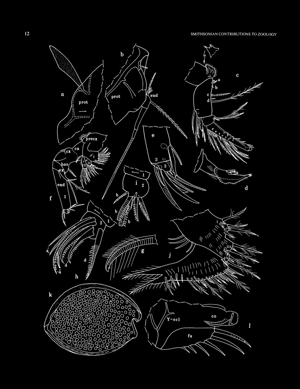
15 NUMBER 595 FIGURE 6. Cypridinodes strophinx Kornicker, new species, adult male, holotype, left valve, ov, length 2.52 mm. Cypridinodes Brady, 1902 TYPE SPECIES. Cypridinodes favus Brady, COMPOSITION AND DISTRIBUTION. Indo-Pacific and Australasian waters. Including a new species described herein, 13 species are referred to this genus, of which one (C. minuta Poulsen, 1962) has been reported from the western Indian Ocean, and two have been reported from the Red Sea (C. asymmetrica (Miiller, 1906) and C. dorsocurvata (Graf, 1931)) (Kornicker, 1991:17). Three new species from Madagascar are described herein. Cypridinodes strophinx, new species FIGURES 6-10 ETYMOLOGY. From the Greek strophinx (axle, pivot). HOLOTYPE. Adult male on slide and in alcohol. TYPE LOCALITY. BT-721. PARATYPES. BT-191: USNM , 1 partly dissected juvenile; USNM , 1 dissected juvenile. BT-719: USNM , 1 partly dissected ovigerous female on slide and in alcohol; USNM , 2 juveniles. BT-720: USNM , 1 juvenile. BT-721: USNM ,1 adult male, 10 additional specimens, and 4 valves. BT-773: USNM , 3 juveniles. BT-779: USNM , 2 ovigerous females plus 1 juvenile. BT-814: USNM , 2 juveniles, 1 ovigerous female, 1 adult female. BT-822: USNM , 1 ovigerous female; USNM , 2 juveniles. BT-836: USNM , 3 juveniles. BT-841: USNM , 1 juvenile. DISTRIBUTION. See type specimens, above. REMARKS. Although a few bristles of the 1st antenna of the male holotype were broken and, therefore, could not be completely described, the second adult male in the collection was left undissected for possible future reference. DESCRIPTION OF ADULT MALE (Figures 6-10a-e). Carapace with convex ventral and dorsal margins (Figure 6); anterior margin of rostrum straight or slightly convex (Figures 6, la, 8k); caudal process projecting posteriorly only slightly, and with straight posterior edge. Right valve only with lunate process ventral to incisur, with row of 21 bristles on inner surface just within outer edge of process (Figure 7 b); left valve with row of 14 similar bristles on anteroventral corner (Figure le). Each valve with low ridge just within valve edge, and low process on rostrum not extending past anterior edge of rostrum (Figure 6). Ornamentation: Surface with numerous closely spaced deep fossae (Figures 6, Sk); broad walls between fossae appear as reticulations (not shown) and have small platelets on surface (edges of platelets shown as slightly curved lines along folded edges in Figure 7c,/). Small round pores, some with minute bristle, others with minute processes (some bilobed) scattered over valve surface (some processes shown Figure Sk). Infold: Rostral infold with bristles (mostly bifurcate) paralleling valve edge, 1 bristle proximal to inner end of

, Iv; h.")
16 10 SMITHSONIAN CONTRIBUTIONS TO ZOOLOGY FIGURE 7. Cypridinodes strophinx Kornicker, new species, adult male, holotype: a,b, anterior right valve, iv; c, caudal process right valve, iv; d, detail of spine on list in c; e,f, anterior and posterior, respectively, of left valve, iv; g, left 1st antenna (nabs), Iv; h. tip right 1st antenna (nabs), mv; i. medial eye and Bellonci organ.
; anteroventral and ventral infold with narrow list with posterior end at ventral end of caudal processes; ventral list from anterior '/3 of valve length to caudal")
 narrow list (row terminates at anterior end of stout ventral list); left valve with 43 bristles along narrow anteroventral list and anterior end of slightly broader")
17 NUMBER incisur, and pair of additional bristles near inner end of incisur (Figure la, e); 2 small bristles just ventral to incisur, and 1 small bristle posterior to them at inner margin of infold (shown only in Figure la); anteroventral and ventral infold with narrow list with posterior end at ventral end of caudal processes; ventral list from anterior '/3 of valve length to caudal process of right valve stouter than list of left valve and appearing more strongly sclerotized; anteroventral and anterior part of ventral infold of right valve with single row of 30 bifurcate bristles along (or just distal to) narrow list (row terminates at anterior end of stout ventral list); left valve with 43 bristles along narrow anteroventral list and anterior end of slightly broader ventral list (posterior 9 bristles form 2 rows); posterior half of ventral infold of left valve with 3 or 4 short stout undivided bristles close to outer edge of infold. Infold of caudal process with anterior ridge bearing digitate striated processes (Figure lc,d,f), and several slender bristles (not all shown); on both valves or right valve only small bristle at inner edge of infold near ventral end of caudal process (Figure 7c,/); on left valve only ventral end of ridge forms low knob and infold near outer edge with 3 small bristles (2 posterior bristles shown in Figure If). Posterior edge of caudal process with 3-6 small bristles and many short straight pore canals (Figure 7c,/). Selvage: Lamellar prolongation (with smooth outer edge and minute surface pustules) present along dorsal edge of rostrum anterior to hinge and along anterior margin of rostrum. Prolongation along ventral edge of incisur broad, narrowly striated, and with surface pustules (Figure le (striations and pustules not shown)); prolongation absent along dorsal edge of incisur. Outer edge of prolongation along anteroventral and ventral margins smooth or minutely serrate, and broadly scalloped just posterior to anteroventral corner (Figure le); ventral prolongation slightly broader on right valve. Prolongation medial to lunate process of right valve (and also in similar location on left valve where lunate process absent) narrower than prolongation at either end of process (Figure lb,e), but minute serrations along edge similar (serrations not shown in Figure Ib.e). Prolongation absent along posterior edge of caudal process. Vestment: 3 or 4 small bristles present on vestment of posteroventral part of valves proximal to inner margin of infold (not shown; bristles not reported previously on vestment of Myodocopina). Carapace Size (length, height in mm): BT-721: holotype, 2.52, 1.86; USNM , 2.59, First Antenna (Figure lg,h): 1st joint bare. 2nd joint with ventral, medial, and lateral spines. 3rd joint with oblique distal margin, ventral and medial spines, 1 spinous proximal dorsal bristle, and 1 spinous ventral bristle at midlength. 4th joint with ventral spines, 1 subterminal spinous ventral bristle, and 1 terminal dorsal bristle. 5th joint without spines; sensory bristle broken on both limbs of holotype (probably same as that of adult female described herein); stump of bristle of left limb with 10 long slender filaments followed by 1 short slenderer filament. 6th joint with short bare tubular medial bristle near dorsal margin. 7th joint: a-bristle about twice length of bristle of 6th joint, with few indistinct spines; b-bristle broken on both limbs of holotype; stump of b-bristle of right limb with short proximal filament with large sucker (with marginal fringe) followed by slender filament with 5 minute suckers (Figure 1b); c-bristle with short proximal filament with large sucker (with marginal fringe) followed by 1 filament with 5 small suckers, then 1 pair of filaments (1 with 4 or 5 small suckers, other shorter with 2 proximal spines), and 6 single filaments (proximal with 2 spines). 8th joint: d- and e-bristles long, bare; f- and g-bristles long with tips missing, remaining parts with about 10 filaments, most with spines. Second Antenna: Protopodite with small medial bristle with indistinct spines (Figure 86); fulcrum sclerite (stippled) broad near midlength (Figure $a) (sclerite not part of protopodite). Endopodite 3-jointed (Figure Sb): 1st joint with 4 proximal bristles (1 long with indistinct spines and 3 short bare) and 1 long spinous bristle near midlength; 2nd joint long bare; 3rd joint short with long terminal filament. Exopodite: 1st joint with hairs along concave margin; bristle of 2nd joint reaching just past 9th joint, with 1 slender ventral spine followed by 11 stout ventral spines, and 4 scattered hair-like dorsal spines; bristles of joints 3-8 with natatory hairs, no spines; 9th joint with 4 bristles (1 short, 1 medium, 2 long), all with natatory hairs; joints 2-8 with stout basal spines increasing in length on distal joints (spine of 8th joint about twice length of 9th joint); 9th joint with lateral spine about length of joint; joints 2-8 with minute spines along distal edges. Mandible (Figure 8c -e (rings not shown on bristles of Figure 8c)): Coxale endite spinous with 2 stouter spines (with few marginal spines) at tip having small triangular peg between them (detail in Figure 8c); coxale with small bristle near base of endite (dashed in Figure 8c). Basale: ventral margin with 2 small spinous a-bristles, 1 small b-bristle with lateral base almost on margin, 2 c-bristles (longer with short marginal spines), and 2 d-bristles (longer with wreaths of long spines and short spines distal to last wreath; shorter with few indistinct short spines); dorsal margin with 1 distal and 2 terminal bristles, all with short spines; medial surface with few distal rows of indistinct spines near dorsal margin. Exopodite slightly longer than dorsal margin of 1st endopodial joint, hirsute, with diaphanous hirsute flair, and 2 bristles (proximal longer and with short spines, distal with few indistinct short spines) (Figure Sc.d). 1st endopodial joint with 4 ventral bristles (1 short bare medial, 1 medium slender with short spines, 2 long stout with wreaths of long spines and small spines distal to wreaths). 2nd endopodial joint narrows at about 2 li length (in vicinity of proximal ventral bristle); ventral margin spinous, with 2 single ringed bristles with slender tubular tips and ventral spines (distal bristle with very few indistinct spines) and paired terminal bristles (medial sclerotized, broader, longer, slightly sinuate; lateral ringed and with slender tubular tip); dorsal margin with 7 long spinous ringed bristles, 2 short

18 12 SMITHSONIAN CONTRIBUTIONS TO ZOOLOGY
 connected to protopodite of right 2nd antenna, lv; b, distal protopodite and endopodite left 2nd")
19 NUMBER FIGURE 8 (left). Cypridinodes strophinx Kornicker, new species, adult male, holotype: a, fulcrum sclerite (stippled) connected to protopodite of right 2nd antenna, lv; b, distal protopodite and endopodite left 2nd antenna, mv; c, right mandible (nabs), mv; d, exopodite right mandible, mv; e, tip right mandible, mv;/ left maxilla (nabs), lv; g, tip stout alpha-bristle 1st endopodial joint right maxilla, mv; h, tip left maxilla (nabs), lv; i. tip right maxilla (nabs), mv;/ left 6th limb, mv; k, complete specimen from left side; /, posterior of body from right side. ringed bristles with short slender spines, 5 short bristles with stout spines and proximal rings, and 7-9 short unringed bristles (with indistinct slender anterior spines) with bases medial to bases of the short bristles with stout spines (unringed bristles not shown in Figure 8c) (both short unringed bristles and short bristles with proximal rings located proximal to 4th long bristle); medial surface with rows of spines. 3rd endopodial joint (Figure Se) with short dorsal part bearing short bristle medial to long partly ringed claw, both bare; longer ventral part bearing 2 unringed claws (both with proximal ventral teeth; medial claw about '/3 longer than lateral claw), 2 long ringed ventral bristles (longer with proximal ventral spines, other bare), and 1 minute ringed bare ventral bristle (all 4 slender ringed bristles of 3rd joint with slender tubular tips). Maxilla: Endite I with 9 spinous and pectinate bristles (Figure 8/); endite II with 6 spinous and pectinate bristles; endite III with 6 bristles (1 plumose proximal, 5 spinous and pectinate terminal); all endites with long surface hairs. Precoxale with diaphanous flair with marginal fringe. Coxale with short ringed bare dorsal bristle. Basale with short ringed ventral bristle with indistinct spines. Exopodite elongate with 3 bristles (1 short bare subterminal; 2 long terminal (1 bare, other with few long spines)) (Figure 8/). 1st endopodial joint with 2 ringed alpha-bristles (1 long bare except for long hairs near hooked tip and with small tooth proximal to hooked tip (Figure $g), 1 shorter with long proximal spines), and 3 ringed beta-bristles (inner bristle short bare, 2 outer bristles with recurved marginal teeth). 2nd endopodial joint with 4 a-bristles (3rd pectinate and longest, 4th with indistinct hairs, others bare) (Figure 8/i), 3 pectinate ringed b-bristles (Figure 8/), 3 ringed c-bristles (inner short bare, others longer pectinate; middle bristle with proximal end slightly bulbous) (Figure 8/), and 3 stout pectinate d-bristles (posterior ringed, others unringed) (Figure Sh). Fifth Limb: Epipodite with 55 bristles. Endite I with 7 bristles, all with long spines; only 1 shown in Figure 9a); endite II with 6 bristles (1 minute bare spine-like posterior, 2 pectinate and with long proximal spines, others more slender spinous) (only 1 shown in Figure 9a); endite III with 7 bristles (1 minute bare spine-like posterior, 1 posterior proximal bristle stout pectinate with long proximal spines, 4 more slender pectinate, 1 anterior with long proximal and short distal spines) (Figure 9b). Anterior tooth of protopodite short with rounded tip (stippled in Figure 9a). Exopodite: anterior side of 1st joint with row of 3 closely spaced bristles (with long proximal and short distal spines), and 1 bristle (with long proximal and short distal hairs) closer to protopodial tooth (Figure 9a); main tooth comprising proximal smooth peg and 6 cuspate teeth (Figure 9b); bristle with long proximal hairs present proximal to smooth peg. 2nd joint with posterior c-bristle with short marginal spines (Figure 9b), anterior d-bristle with long proximal and short distal hairs (Figure 9a), 4 pectinate ringed a-bristles (Figure 9c), 5 pectinate ringed b'-bristles (Figure 9d), and 4 pectinate b"-bristles (2 posterior (proximal) bristles ringed and with larger teeth along middle part, next bristle ringed distally and with 2 very large teeth proximal to narrow ringed part, last bristle with 1 very large tooth proximal to narrow ringed part) (Figure 9e). 3rd joint (Figure 9a,b): inner lobe with few short spines and hairs, 1 proximal short ringed bare bristle, and 2 longer bare terminal bristles (outer ringed only in proximal '/3, inner completely ringed); outer lobe hirsute, with 2 ringed bristles (shorter bare, longer with short spines). 4th and 5th joints fused, hirsute, with total of 4 or 5 spinous ringed bristles (Figure 9a, b). Sixth Limb (Figure 8/): Hirsute, with 4 epipodial bristles. Endite I with 3 bristles (2 short hirsute medial, 1 longer terminal); endite II with 1 stout hirsute medial bristle, 2 long terminal bristles and 1 small medial bristle between them; endites III and IV each with 1 short hirsute medial bristle, and 2 long terminal bristles with minute bristle between them. End joint posteriorly extended, with 19 anteroventral bristles (most with bases on medial side near margin), 4 posteroventral bristles with bases on medial side some distance from ventral margin, and 2 broad plumose posterior bristles. (All bristles ringed; rings not shown in Figure 8/.) Seventh Limb (Figure 9g,h): Terminal segment with 4 bristles on ventral margin (with 3-5 bells) and 3 proximal to teeth and distal to jaw (1 on one side, 2 on other, each with 3-5 bells). Proximal bristles comprise 7 on ventral side and 8 or 9 on dorsal jaw side; all bristles with 3 bells. Total number of bristles 22 or 23. Stout dorsal jaw with diaphanous flange with serrate margin and lineations perpendicular to margin (edges of few serrations appear to continue to edge of jaw forming narrow spines, but could not resolve with certainty) (Figure 9h). Comb with 6 long teeth (terminal tooth longest) and 9 shorter square-tipped teeth (4 or 5 on each side); small spine-like process projecting inward just proximal to arc formed by teeth (Figure 9h). Furca (Figures 8/, 9/): Each lamella with 6 articulated claws; all claws with row of teeth along posterior edge (not shown); some teeth slightly larger than others on claws 1-5; claws 1-3 and probably others with medial row of teeth (not shown); medial row of claw 1 with about 32 teeth of which distal 10 teeth larger; anterior edge of claws bare; right lamella anterior to left by about width of base of claw 1. Bellonci Organ (Figure li): Lemon-shaped with small process at tip. Eyes: Medial eye unpigmented, bare (Figure li). Lateral eye unpigmented, with 17 amber-colored ommatidia (Figure 10a).
, av; b, part of right 5th limb")

20 14 SMITHSONIAN CONTRIBUTIONS TO ZOOLOGY FIGURE 9. Cypridinodes strophinx Kornicker, new species, adult male, holotype: a, part of left 5th limb (nabs), av; b, part of right 5th limb (nabs), pv; c, a-bristles right 5th limb, pv; d, b'-bristles right 5th limb, pv; e. b"-bristles left 5th limb, av;/ endites I and II right 5th limb, pv; g, 7th limb; h, detail of tip of limb in g, i, left furcal lamella; j, upper lip from left side; k. anterior of body from left side; /, posterior of body from right side; m. left Y-sclerite.

: Complex lobes on each side of body anterior to furca (sclerotized parts stippled).")
21 NUMBER 15 Upper Lip (Figure 9j,k): Anterior unpaired part with 4 short pairs of processes followed by 1 longer pair (in lateral view each pair appears as single process). Posterior paired part with elongate pointed tusks without glandular processes (each tusk with few long anterior hairs and more abundant hairs forming row along medial and lateral posterior edges (proximal lateral hairs stouter)), and a posterior serrate process with 4 teeth; hirsute rounded process present between serrate process and mouth. Genitalia (Figures 8/, \0b-e): Complex lobes on each side of body anterior to furca (sclerotized parts stippled). Posterior of Body (Figure 9/): Slightly undulate dorsal to dorsal end of girdle, and evenly rounded ventral to dorsal end of girdle. Y-Sclerite (Figures 8/, 9m): Typical for subfamily. Pigmentation: Absent. DESCRIPTION OF ADULT FEMALE (Figure 10/-*). Carapace similar in shape and ornamentation to that of adult male, but larger (Figure lof.g). Right valve only with lunate process ventral to incisur, with row of 26 bristles on inner surface just within outer edge of process (not shown); left valve with row of 18 similar bristles on anteroventral corner (not shown). Infold: Ventral list of right valve stouter and appearing more strongly sclerotized than that of left valve. Infold of caudal process with anterior ridge bearing 32 or 33 digitate striated processes and several slender bristles. Bristles not counted on other parts of infold but appearing similar to those of adult male. Selvage: Similar to that of adult male. Carapace Size (length, height in mm): BT-719: USNM , 3.03, BT-779: USNM , 2 specimens, 2.81, 2.08; 2.87, BT-814: USNM , 2 specimens, 2.89, 2.14; 2.89, BT-822: USNM , 2.89, First Antenna: Joints 1-4 and 6 similar to those of adult male. Sensory bristle of 5th joint with 10 long slender filaments followed by 2 shorter and slenderer filaments and bifurcate tip (Figure loh). 7th joint: a-bristle about twice length of bristle of 6th joint, with few small spines; b-bristle about 3 times length of a-bristle and 2 /3 length of bristle of 5th joint, with 5 short filaments (some with spines); c-bristle almost twice length of bristle of 5th joint, with about 9 filaments (some with spines) and bifurcate tip. 8th joint: d- and e-bristles about same length as bristle of 5th joint, bare with blunt tips; f- and g-bristles similar to c-bristle. Second Antenna, Mandible, Maxilla, Fifth Limb (Figure 10/), Sixth Limb, Upper Lip, and Y-Sclerite: Similar to those of adult male. Seventh Limb: Terminal segment with 4 bristles on ventral margin (with 2-5 bells) and 3 proximal to teeth (1 on one side, 2 on other, each with 3-5 bells). Proximal bristles comprise 8 or 9 on ventral side and 10 or 11 on dorsal jaw side; all bristles with 3 bells. Total number of bristles 26. Stout dorsal jaw with diaphanous flange with serrate margin and lineations perpendicular to margin (Figure 10/). Comb with 6 long teeth (terminal tooth longest) and 9 or 10 shorter square-tipped teeth (4 or 5 on each side); short stout tooth projecting inward just proximal to arc formed by teeth (Figure 10/). Furca: Similar to that of adult male except with 6 or 7 claws. Bellonci Organ (Figure 10*): Oval with process at tip larger than that of adult male. Eyes: Medial eye unpigmented bare (Figure 10*). Lateral eye with 17 ommatidia with black pigment between ommatidia. Genitalia: None observed. Posterior of Body: Evenly rounded. Pigmentation: Differs from adult male in lateral eye having black pigmentation between ommatidia (may be preservation difference). Eggs: USNM with 11 eggs in marsupium; length of typical egg 0.58 mm. Remarks: The female carapace is considerably larger than that of the male (length mm compared to mm), which is unusual for most previously described species for which both sexes are known, but not for specimens referred to C. favus by Poulsen (1962:285) (female length mm, male length 4.2 mm). COMPARISONS. Only three species have been described previously having both a lunate process on the anteroventral corner of the right valve and all furcal claws articulated: C. favus (Brady, 1902), C. bairdii (Brady, 1866), and C. asymmetrica (Miiller, 1906). The carapace of C. favus differs from that of C. strophinx in having two large protuberances at midlength and midheight. C. bairdii is not sufficiently known to judge whether it is conspecific with C. strophinx, but it has been collected only in the vicinity of China, so this seems unlikely. The 5th limb of C. strophinx differs from that of C. asymmetrica described by Poulsen (1962:300) in having two of the four b"-bristles with one or two very large teeth (Figures 9b, 10/). The b"-bristles resemble those of C. minuta described by Poulsen (1962:305). C. minuta differs from C. strophinx in lacking a lunate process on the right valve as well as in having furcal claw 2 nonarticulated. The 1st antenna of the adult male C. strophinx differs from that of C. asymmetrica described by Poulsen (1962:297), but not that illustrated by Muller (1906, pi. VI:9), in having filaments with small suckers on the b- and c-bristles of the 7th joint. The tip of the 7th limb of C. strophynx bears a tooth-like process within the arc of the terminal comb not previously described or illustrated on other species of the genus (possibly the tooth is present in other species but difficult to see). The serrate flange on the dorsal jaw of the 7th limb of C. strophinx resembles that of a specimen of C. asymmetrica from the Java Sea, which Poulsen (1962:300) interpreted to be a variant of the usual toothed jaw. The furca of C. strophinx differs from that of C. dorsocurvata (Graf, 1931) in having claw 2 articulated.


22 16 SMITHSONIAN CONTRIBUTIONS TO ZOOLOGY FIGURE 10. Cypridinodes strophinx Kornicker, new species, adult male, holotype: a, left lateral eye and posterior edge of protopodite of 2nd antenna, Iv; b,c, posterior and anterior views, respectively, of copulatory organs; d.e, left and right copulatory limbs, respectively, lv. Ovigerous female, paratype, USNM :/ outline of complete specimen from right side, length 3.03 mm; g, detail of anterior from/; h, sensory bristle of 5th joint of left 1st antenna, mv;;', two b'-bristles of 2nd exopodial joint of left 5th limb, pv;/ tip 7th limb; k, medial eye and Bellonci organ.

23 NUMBER Cypridinodes relax, new species FIGURES ETYMOLOGY. From the Latin relax (loosen, slacken, unbend). HOLOTYPE. Ovigerous female on slide and in alcohol. TYPE LOCALITY. BT-620. PARATYPES. BT-620: USNM , ovigerous female on slide and in alcohol; USNM , partly dissected ovigerous female; USNM , partly dissected adult male. BT-779: USNM , adult male on slide and in alcohol. DISTRIBUTION. See type specimens, above. DESCRIPTION OF ADULT FEMALE (Figures ll-14a-/). Carapace with convex ventral and dorsal margins (Figure 1 la); anterior margin of rostrum convex (Figure lla.d): caudal process small, triangular, with straight or convex posterior edge (Figure 11/g); neither valve with lunate process ventral to incisur (Figure 1 \a-c,e); edge of anteroventral corner of valve with 0-7 short bristles (bristles shown on Figures 1 \e, \4d). Ornamentation: Surface appearing smooth without fossae or reticulations. Infold: Rostral infold with about 27 bristles, some bifurcate, and pair of additional bristles near inner end of incisur (Figure 1 Id); anteroventral and ventral infold with narrow list with posterior end at ventral end of caudal process (Figure 11/); ventral list from point at anterior '/3 of valve to caudal process stout on both valves, but stouter and closer to valve edge on right valve than on left; anteroventral infold of right valve (from incisur to anterior end of stout part of list) with single row of about 65 bristles; left valve with about same number of anteroventral bristles, but some distal to list, and with about 10 additional bristles along anterior end of stouter part of list (Figure 1 le; not all bristles shown). Infold of caudal process with digitate striated processes and several slender bristles (Figure 1 \f,g; not all shown); on right valve only, small bristle present at inner edge of infold near ventral end of caudal process (Figure 1 Ig). Posterior and dorsal edges of caudal process with 8-14 minute bristles and many short straight pore canals (Figure 11/g). Selvage: Lamellar prolongation with narrow striations and smooth outer edge present along dorsal edge of rostrum anterior to hinge and along anterior margin of rostrum (not shown). Prolongation absent along dorsal edge of incisur; prolongation along ventral edge of incisur broad, narrowly striated, and with indistinct pustules (striations and pustules not shown in Figure 1 \c,e); incisur narrow for short distance just anterior to anteroventral corner of valve, then broadens along ventral margin (Figure 1 lc); outer edge of prolongation along anteroventral and ventral margins minutely serrate; prolongation absent along posterior and dorsal edges of caudal process in vicinity of short straight pore canals. Hingement: Same as male. Carapace Size (length, height in mm): BT-620: holotype, 2.75, 1.92; USNM , 2.76, 1.95; USNM , 2.79, First Antenna (Figure \\h,i): 1 st joint bare. 2nd joint with spines. 3rd joint with oblique distal margin, few rows of lateral spines, spinous proximal dorsal bristle, and terminal ventral bristle. 4th joint with medial spines near ventral margin (not shown), 2 distal bristles (1 ventral, 1 dorsal). Sensory bristle of 5th joint with 9 long slender filaments followed by 3 shorter and slenderer filaments and bifurcate tip. 6th joint with short medial bristle. 7th joint: a-bristle longer than bristle of 6th joint, with few short spines; b-bristle about 4 times length of a-bristle, with 5 short filaments; c-bristle about V3 longer than bristle of 5th joint, with 9 filaments and bifurcate tip. 8th joint: d- and e-bristles about same length as sensory bristle of 5th joint, bare with blunt tips; f-bristle shorter than c-bristle, with 9 filaments and bifurcate tip; g-bristle similar to c-bristle. Second Antenna: Protopodite with small medial bristle (Figure 12a). Endopodite 3-jointed (Figure 12a): 1st joint with 4 proximal bristles (1 long, 3 short) and 1 long spinous bristle near midlength; 2nd joint long bare; 3rd joint short with long terminal filament. Exopodite: bristle of 2nd joint reaching well past 9th joint, with 1 slender ventral spine followed by 8 stouter spines, and 3 hair-like dorsal spines (Figure \2b); bristles of joints 3-8 with natatory hairs, no spines; 9th joint with 4 bristles (1 short dorsal, 1 medium, 2 long), all with natatory hairs; joints 2-8 with stout basal spines increasing in length on distal joints (spine of 8th joint about twice length of 9th joint); 9th joint with lateral spine about length of joint; joints 3-8 with minute spines along distal edges. Mandible (Figure \2c-e): Coxale endite spinous with 2 stout terminal spines with minute peg between them; coxale with small bristle near base of endite. Basale: ventral margin with 2 small a-bristles, 1 small b-bristle with base lateral, 2 c-bristles (longer with few indistinct short spines), and 2 d-bristles (longer with wreaths of long spines and short spines distal to last wreath); dorsal margin with 1 distal and 2 terminal bristles, all with short spines; medial surface with few rows of distal indistinct spines near dorsal margin. Exopodite about same length as dorsal margin of 1 st endopodial joint, hirsute, with 2 bristles (proximal slightly longer and with indistinct short spines). 1st endopodial joint with 4 ventral bristles (1 minute bare medial, 1 short medial with short spines, 2 long stout with short and long spines). 2nd endopodial joint narrows at about 2 /3 length (in vicinity of proximal ventral bristle); ventral margin spinous, with 2 single ringed bare bristles with slender tubular tips and paired terminal bristles (medial unringed sclerotized, broader, longer; lateral ringed and with slender tubular tip); dorsal margin with 7 long spinous ringed bristles (Figure 12c), 2 short ringed distal bristles with short marginal spines, 6 short proximal bristles with stout spines and proximal rings (Figure \2d), 10 or 11 short proximal unringed bristles (with indistinct slender anterior spines) with bases slightly medial to bases of the short bristles with stout spines, and 2 short bristles (with indistinct slender anterior spines and proximal rings) proximal to most bristles; medial surface with rows of spines. 3rd endopodial joint (Figure 12e) with short dorsal part bearing 2 ringed bare bristles (lateral bristle longer,

24 18 SMITHSONIAN CONTRIBUTIONS TO ZOOLOGY

, and longer ventral part bearing 2 unringed claws (medial about '/4 longer, both with proximal ventral teeth), 2")
25 NUMBER FIGURE (left) 11. Cypridinodes relax Kornickcr, new species, ovigerous female, holotype: a, complete specimen from right side, length 2.75 mm; b, anterior right valve (nabs), iv; c. anteriorright valve, ov; d.e. anteriorrightand left valves, respectively, iv; f.g, caudal process left and right valves, respectively, iv; h.i, left (Iv) andright (mv) 1st antennae (nabs), respectively. claw-like with pointed sclerotized tip; shorter bristle medial and with tubular tip), and longer ventral part bearing 2 unringed claws (medial about '/4 longer, both with proximal ventral teeth), 2 long ringed ventral bristles (with few proximal ventral spines) with tubular tips, and 1 minute ringed bare ventral bristle with tubular tip. (Rings and spines of many bristles not shown.) Maxilla (Figure \2f-k): Endite 1 with about 8 bristles (Figure 12/); endite II with 5 bristles (Figure 12g), endite III with 1 proximal and 6 terminal bristles (Figure 12g (proximal bristle not shown)). Precoxale with dorsal fringe of hairs (not shown). Coxale with ringed bare dorsal bristle (Figure 12/J). Basale with ringed ventral bristle with few indistinct spines (Figure 12/J). Exopodite elongate with 3 ringed bristles (1 short bare subterminal; 2 long terminal (1 bare, other with few long spines)) (Figure 12/J). 1st endopodial joint with 2 ringed alpha-bristles (1 long bare except for long hairs near hooked tip (with or without minute tooth proximal to hooked tip), 1 shorter with long proximal spines), and 3 ringed beta-bristles (inner bristle short slender bare, 2 outer bristles stouter and longer and with recurved marginal teeth) (Figure 12/). 2nd endopodial joint with 4 a-bristles (3rd from anterior pectinate, others bare) (Figure 12/), 3 pectinate ringed b-bristles, 3 ringed c-bristles (inner short bare, others with long curved teeth), and 3 stout pectinate d-bristles (posterior ringed, others unringed) (Figure \2k). (Rings not shown on all bristles.) Fifth Limb: With 3 endites (not all bristles shown on endites II and III in Figure 13a). Anterior process of protopodite short with rounded tip (stippled in Figure \3b,c). Exopodite: anterior side of 1st joint with row of 3 closely spaced bristles (inner ringed, short, slender, with long proximal hairs; middle and outer bristles stout, with distal rings and long proximal and short distal spines) and 1 or no bristle closer to protopodial process (Figure 13ft,c); main tooth comprising proximal triangular peg (with few indistinct spines at tip) and 6 cuspate teeth (Figure 13a); bristle with long proximal spines proximal to triangular peg. 2nd joint with posterior c -bristle with short spines (Figure 13a), anterior d-bristle with long proximal hairs (Figure \3c,d), 4 ringed pectinate a-bristles (Figure 13a) (the longest a-bristle could be a b"-bristle), 5 ringed pectinate b'-bristles (Figure 13e), and 4 b"-bristles (3 with very large teeth and mostly unringed) (Figure 13/). 3rd joint (Figure \3d): inner lobe with 3 bare bristles (1 proximal short ringed, 2 longer terminal, ringed only in proximal half, and with curved tips); outer lobe hirsute, with 2 ringed bristles with indistinct spines and slender tips. 4th and 5th joints fused, with total of 5 ringed bristles (3 on inner corner (1 with stout spines, others bare or with few indistinct spines), and 2 on outer corner (1 with stout spines, other bare or with few indistinct spines)) (Figure 13a"). Sixth Limb (Figure 13g): With 4 epipodial bristles. Endite I with 3 bristles (2 short medial, 1 long terminal); endite II with 5 bristles (2 short medial, 2 long terminal, and 1 minute medial near bases of long bristles); endites III and IV each with 1 hirsute medial bristle and 2 long terminal bristles with minute medial bristle between them. End joint hirsute, posteriorly extended, with 22 anteroventral bristles (many with bases on medial side), 5 posteroventral bristles with bases on medial side some distance from ventral margin, and 2 broad plumose posterior bristles (only proximal parts shown). Seventh Limb (Figure \3h-j): Terminal segment with 4 ventral bristles (with 3-5 bells) and 3 bristles proximal to teeth (1 on one side, 2 on other, each with 3-5 bells). Proximal bristles comprise 5 or 6 on ventral side and 7 on dorsal jaw side; all bristles with 3 bells. Total number of bristles 19 or 20. Stout dorsal jaw with marginal row of 7 stout pointed teeth (not all shown), and 4 slender indistinct teeth on each end of row. Comb with 7 long teeth (terminal tooth longest) and 8 shorter teeth (4 on each side), all teeth with rounded tips; small pointed process projecting inward within arc of teeth (Figure 13;). Furca (Figure 14a,ft): Each lamella with 7 claws; claw 2 either nonarticulated (Figure 14a) or weakly articulated (faint suture at base of claw (Figure 146)). All claws with row of teeth along posterior edges, some teeth slightly stouter than others; claws 1-5 with medial row of teeth; medial row of claw 1 with about 25 teeth of which distal 8 teeth larger; right lamella anterior to left by about width of base of claw 1. (Not all teeth shown on claws.) Bellonci Organ (Figures \3k, \4e,f): Cup-shaped or cylindrical. Eyes: Medial eye unpigmented, bare (Figures 13k, 14e,/). Lateral eye about '/4 larger than medial eye, with 16 amber-colored ommatidia with black pigment between them (Figures lla, 13/). Upper Lip (Figure 14c): Anterior part with 4 short pairs of processes followed by 1 longer pair (in lateral view each pair appears as single process). Posterior part with elongate pointed tusks without glandular processes (each tusk with row of stout lateral hairs near posterior edge), and a posterior proximal serrate process with 5 teeth; hirsute rounded process present between serrate process and mouth (not shown). Genitalia: Not observed. Posterior of Body: Evenly rounded, bare. Y-Sclerite (Figure 14a): Typical for subfamily. Pigmentation: Black pigment in lateral eyes. Eggs: Holotype with 38 colorless eggs (some with faint eyes and developing, incipient antennae visible, membrane absent) in marsupium, length (mm) of 3 typical eggs: 0.31, 0.30, USNM with 31 amber-colored eggs (without eyes) in marsupium, length of typical egg excluding membrane: 0.34 mm. USNM with 15 amber-colored

; e, detail from c;/ endite I maxilla; g, endites II and III of maxilla opposite that shown in/; h, left maxilla (nabs), Iv; /,")
26 20 SMITHSONIAN CONTRIBUTIONS TO ZOOLOGY FIGURE 12. Cypridinodes relax Kornicker, new species, ovigerous female, holotype: a, endopodite and distal protopodite right 2nd antenna, mv; b, bristle of 2nd exopodial joint of left 2nd antenna, mv; c, right mandible (nabs), mv; d, detail of 2nd endopodial joint in c showing short dorsal bristles (spines shown only on the six bristles having stout spines); e, detail from c;/ endite I maxilla; g, endites II and III of maxilla opposite that shown in/; h, left maxilla (nabs), Iv; /, alpha- and beta-bristles left maxilla, \\;j, a-bristles left maxilla, Iv; k, 2nd endopodial joint right maxilla (nabs), mv.
, pv; b, part of")

27 NUMBER FIGURE 13. Cypridinodes relax Kornicker, new species, ovigerous female, holotype: a, part of right 5th limb (nabs), pv; b, part of left 5th limb (nabs), av; c, part of right 5th limb (nabs), av; d, part of right 5th limb (nabs), pv; e, b'-bristles right 5th limb, pv;/ an a-bristle and four b"-bristles of right 5th limb, pv; g, left 6th limb, lv; h, tip of 7th limb; i,j, details from h; k, medial eye and Bellonci organ; /, lateral eye (black pigment stippled).


28 22 SMITHSONIAN CONTRIBUTIONS TO ZOOLOGY FIGURE 14. Cypridinodes relax Kornicker, new species, ovigerous female, holotype: a, right furcal lamella and right Y-sclerite; b, claws 1-3 of left furcal lamella showing weak suture at base of claw 2; c, upper lip, anterior to right. Ovigerous female, paratype, USNM , length 2.76 mm: d, anterior right valve, lv;e, medial eye and Bellonci organ, lv;/ same as e, oblique view. Adult male, paratype, USNM : g, complete specimen from right side, length 2.34 mm: h, anterior right valve, lv; i,j, caudal process left and right valves (not all spines shown), respectively, iv.
: Carapace similar in shape to that of adult female (Figure 14g).")
29 NUMBER eggs in marsupium, length of typical egg (with visible eyes and antennae) including transparent membrane: 0.41 mm; excluding membrane: mm. DESCRIPTION OF ADULT MALE (Figures \4g-j, 15): Carapace similar in shape to that of adult female (Figure 14g). Ornamentation: Surface appearing smooth without reticulations; small bilobed pores present but sparse. Infold: Rostral infold with about 20 bifurcate bristles, and pair of additional bristles near inner end of incisur; list similar to that of adult female. Infold of right valve (from incisur to anterior end of stout part of list) with about 49 bristles mostly along list but some distal to it; left valve with about 55 bristles similarly distributed plus about 7 bristles along anterior end of stouter part of list. Infold of caudal process with processes and several slender bristles (Figure \4i,j (not all processes or bristles shown)). Posterior and dorsal edges of caudal process with 6-8 minute bristles and many short straight pore canals (not shown). Selvage: Similar to that of adult female. Hingement: Bar dorsal to rostrum of right valve fits into elongate socket of left valve (not shown). Dorsal margin of right valve with bar with knob at posterior end (Figure 14/). Vestment: 3 or 4 widely separated small bristles present on vestment of posteroventral part of valve proximal to inner end of infold (not shown). Carapace Size (length, height in mm): BT-620: USNM , 2.38, BT-779: USNM , 2.34, First Antenna (Figure 15a): Joints 1, 2, 5, and 6 similar to those of adult female. 3rd joint with medial and ventral spines and 2 spinous bristles (dorsal proximal, ventral terminal). 4th joint with medial spines near ventral margin, stout ventral spines, and 2 distal bristles (ventral spinous, dorsal shorter with few indistinct spines). 7th joint: a-bristle longer than bristle of 6th joint, with short spines; b-bristle about 6 times length of a-bristle, with short proximal filament with large sucker (with marginal fringe) followed by a slender filament with small triangular process proximal to row of 5 or 6 small round suckers, then a second slender filament (with small triangular process proximal to row of 5 small round suckers) adjacent to a small bare filament, followed by a small bare filament; c-bristle with short stout proximal filament with large sucker (with marginal fringe) followed by a slender filament with row of 5 small round suckers (with triangular process proximal to row of 5 small round suckers), then a short bare filament adjacent to a longer slender filament, followed by 6 narrow filaments. 8th joint with d- and e-bristles long, bare; f- and g-bristles long stout, each with 10 or 11 filaments, some with 2-4 small spines. (3rd and 4th joints of male differ from those of female in having numerous ventral spines.) Second Antenna: Protopodite similar to that of adult female. Endopodite differs from that of adult female in having a minute round ventral protuberance adjacent to distal bristle of 1st joint. Exopodite: 1st joint with ventral and dorsal spines (on USNM spines better developed on right limb); exopodite otherwise similar to that of adult female. Mandible: 2nd endopodial joint: ventral margin with medial unringed terminal bristle longer than that of adult female (extending past 3rd joint) (Figure \5b). Dorsal margin with 7 long spinous ringed bristles, 2 short ringed distal bristles with short spines, 7 short proximal bristles with stout spines and proximal rings, 11 short proximal unringed bristles (with indistinct slender anterior spines) with bases slightly medial to bases of short bristles with stout spines, and 2 short bristles (with indistinct slender anterior spines and proximal rings) proximal to most bristles. Limb otherwise similar to that of adult female. Maxilla: All 3 bristles of exopodite with few long spines. Long alpha-bristle of 1 st endopodial joint of each limb with tooth proximal to tip. Anterior a-bristle of 2nd endopodial joint about 3 A length of adjacent bristle. Limb otherwise similar to that of adult female. Fifth Limb: Epipodite with 51 bristles. Anterior side of 1st endopodial joint of both limbs with row of 3 bristles plus 1 bristle near protopodial tooth. b"-bristles of 2nd exopodial joint similar to those of adult female (Figure 15c). Remaining bristles of exopodial joints similar to those of adult female. Sixth Limb: Endite I with 2 or 3 bristles (1 or 2 short proximal, 1 long terminal). End joint with 17 anteroventral bristles, 3 posteroventral bristles with bases on medial surface some distance from ventral margin, and 2 broad plumose posterior bristles. Limb otherwise similar to that of adult female. Seventh Limb (Figure \5d): Except for having only 6 proximal dorsal bristles, bristles similar to those of adult female. Total number of bristles 18 or 19. Stout dorsal jaw with marginal row of about 7 stout teeth (some alate), and 5 or 6 slender teeth at each end of row (exact number and shape of teeth difficult to resolve). Comb with 6 or 7 long teeth and 8 shorter teeth (4 on each side). Small pointed process projecting inward within arc of teeth. Furca (Figure \5e): Each lamella with 6 claws; claw 2 either nonarticulated or with indistinct suture at base of claw. Teeth on claws similar to those of adult female (not shown). Bellonci Organ (Figure 15/): More cylindrical than that of adult female. Eyes: Medial eye unpigmented, bare (Figure 15/). Lateral eye large (about same size as that of adult female), with 19 amber-colored ommatidia and black pigment between them (Figures 14g, 15g). Upper Lip (Figure 15/z): Similar to that of adult female. Genitalia (Figure ISe.i): Elongate lobes on each side of body anterior to furca. Posterior of Body (Figure 15/): With about 5 lobes dorsal to dorsal end of girdle, bare. Y-Sclerite (Figure 15/): Similar to that of adult female. Pigmentation: Black pigment in lateral eyes, and minute areas of brown pigment in upper lip.
, mv; b, tip of right")

30 24 SMITHSONIAN CONTRIBUTIONS TO ZOOLOGY FIGURE 15. Cypridinodes relax Kornicker, new species, adult male, paratype, USNM : a, tip of left 1st antenna (nabs), mv; b, tip of right mandible (nabs), mv; c, an a-bristle and four b"-bristles of 2nd exopodial joint right 5th limb, av; d, tip of 7th limb (nabs); e, right furcal lamella and right copulatory limb;/ medial eye and Bellonci organ; g. right lateral eye (black pigment stippled); h, upper lip from right side; i, right copulatory limb from right side;/ posterior of body from right side.
 along the edge of the bulge. The bulges are absent on C.")
31 NUMBER COMPARISONS. Cypridinodes relax is similar to C. minuta Poulsen, According to Poulsen (1962:304) both valves of C. minuta have a very shallow bulge ventral to the incisur, with about 23 hairs (bristles) along the edge of the bulge. The bulges are absent on C. relax, and no more than 5 marginal bristles were observed on valves of C. relax. Three of the b"-bristles of the 2nd exopodial joint of the 5th limb of C. relax bear a few very large teeth compared to only two bristles for C. minuta. The serrate process posterior to the tusks of the upper lip of C. minuta appears more rounded in lateral view than that of C. relax. The valves of both C. relax and C. acuminata Skogsberg, 1920, are without bulges ventral to the incisur, but the latter species is much larger and has many more bristles on the 7th limb. The right valve of C. relax differs from that of C. strophinx in lacking a lunate process ventral to the incisur. The b"-bristles of the 5th limb of C. parallax, new species, differ from those of C. relax in not having a few very large teeth. Cypridinodes parallax, new species FIGURES ETYMOLOGY. Arbitrary combination of letters derived from the Greek parallaxis (to vary, decline, wander). HOLOTYPE. Adult male on slide and in alcohol. TYPE LOCALITY. BT-709. PARATYPES. BT-616: USNM , ovigerous female. BT-620: USNM , adult female on slide and in alcohol; USNM , partly dissected ovigerous female; USNM , adult female plus juvenile. BT-621: USNM , 3 ovigerous females plus 1 juvenile. BT-622: USNM , 3 adult males; USNM , partly dissected adult male; USNM , 4 ovigerous females; USNM , partly dissected ovigerous female; USNM , 3 ovigerous females, 1 adult female, and 1 juvenile. BT-623: USNM , carapace (containing only lateral eye) in alcohol. BT-676: USNM , 1 ovigerous plus 1 adult female. BT-715: USNM , partly dissected adult male. BT-726: USNM , juvenile. BT-730: USNM , 1 adult male plus 1 juvenile. BT-737: USNM , adult male. BT-761: USNM , partly dissected juvenile on slide and in alcohol. BT-777A: USNM , juvenile. BT-778: USNM , partly dissected adult male; USNM , partly dissected adult male; USNM : partly dissected adult female; USNM , 9 specimens (2 ovigerous females, 3 adult females, 1 adult male, 3 juveniles). BT-822: ovigerous female plus 2 juveniles. BT-836: USNM , partly dissected juvenile. BT-851: USNM , partly dissected adult female. BT-883: USNM , partly dissected adult male. DISTRIBUTION. See type specimens, above. DESCRIPTION OF ADULT MALE (Figures 16-20a). Carapace with convex ventral and dorsal margins (Figure 16a, b); anterior margin of rostrum straight or slightly convex; caudal process projecting posteriorly only slightly and with posterior edge convex (left valve (Figure 16/)) or straight (right valve (Figure 16/)). Right valve with slight lateral overhang or narrow lunate process ventral to incisur (Figures \6a,b,e,h, lib), and with row of bristles on inner surface just within outer edge of process (missing bristles dashed in Figure I6h, and bristles not shown in Figure 17b); left valve without lunate process but with lateral overhang along anteroventral corner bearing 5-18 bristles on inner side (Figure 17a). Each valve with low process on rostrum not extending past anterior edge of rostrum (Figure 16a). Ornamentation (Figure 16/): Surface with shingle-like appearance on well preserved specimens but appearing smooth on many specimens, and with small round pores, many bilobed; some pores with minute bristle or process. Infold: Rostral infold with 20 bristles (mostly bifurcate) paralleling valve edge, 1 bristle proximal to inner end of incisur, and pair of bristles at inner end of incisur; 2 small bristles just ventral to inner end of incisur, and 1 small bristle posterior to them at inner margin of infold (not shown in Figure 16g). Anteroventral infold with narrow list with posterior end at ventral end of caudal process; ventral list from anterior '/3 of valve length to caudal process of right valve slightly stouter than list of left valve and appearing more strongly sclerotized (amber-colored); anteroventral and anterior part of ventral infold of right valve with single row of 31 bifurcate bristles along or just distal to narrow list (list absent in vicinity of anterior 4 bristles) (row terminates at anterior end of stout ventral list) (Figure 16/J); left valve with 38 bristles along narrow anteroventral list and anterior end of slightly broader ventral list (list absent in vicinity of anterior 4 bristles); posterior half of ventral infold of left valve with 3 small widely separated bristles; small bristle at inner edge of infold near ventral end of caudal process (Figure 16/). Infold of caudal process with anterior ridge bearing pointed processes (Figure 16/; not all shown in Figure 16/); on left valve only ventral end of ridge forms low knob (Figure 16/); posterior edge of caudal process with 5 or 6 minute bristles and numerous short straight pore canals (Figure \6i,j). Selvage: Lamellar prolongation with smooth outer edge present along dorsal edge of rostrum anterior to hinge and along anterior margin of rostrum. Prolongation along ventral edge of incisur broad, narrowly striated; prolongation absent along dorsal edge of incisur. Prolongation medial to lunate process of right valve and also in similar location on left valve (lunate process absent) narrower than prolongation at either end and does not extend past edge of valve (Figure \7b), with minute hairs along edge in vicinity of incisur and minutely serrate or smooth elsewhere (hairs and serrations not shown). Prolongation absent along posterior edge of caudal process. Vestment: 4 small widely separated bristles present on vestment of posteroventral part of valve proximal to inner margin of infold (not shown).


32 26 SMITHSONIAN CONTRIBUTIONS TO ZOOLOGY FIGURE 16. Cypridinodes parallax Kornicker, new species, adult male, paratype, USNM : a. complete specimen from right side, length 2.59 mm; b, anterior right valve, ov; c, b"-bristles of 2nd exopodial joint left 5th limb, pv; d, right furcal lamella. Adult male, holotype: e. complete specimen from right side, length 2.57 mm;/ surface structures from right valve near central adductor muscle attachments, ov; g,h, rostrum and anteroventral curvature, respectively, right valve, iv; ij, caudal process right and left valves (not all spines shown in j), respectively, iv.
: 1 st joint bare. 2nd joint with ventral, dorsal, medial, and lateral spines.")
 but")
33 NUMBER Carapace Size (length, height in mm): BT-622: USNM , 2.59, 1.76; USNM , 3 specimens: 2.65, 1.79; 2.50, 1.62; 2.57, BT-709: holotype, 2.57, BT-715: USNM , 2.27, BT-778: USNM , 2.56, BT-883: USNM , 2.76, First Antenna (Figure 1 lc,d): 1 st joint bare. 2nd joint with ventral, dorsal, medial, and lateral spines. 3rd joint with oblique distal margin, ventral, dorsal, and medial spines, 1 subterminal spinous ventral bristle, and 1 proximal spinous dorsal bristle. 4th joint with ventral, dorsal, and medial spines, 1 terminal spinous ventral bristle, and 1 short subterminal dorsal bristle (dorsal bristle missing on both limbs of holotype (Figure 17c) but present on USNM ). 5th joint without spines; sensory bristle with 9 long slender filaments followed by 3 shorter and slenderer filaments and 1 small subterminal filament. 6th joint with short tubular medial bristle (with few spines) near dorsal margin. 7th joint: a-bristle about twice length of bristle of 6th joint, with few small spines; b-bristle with short proximal filament with large sucker (with marginal fringe) followed by a slender filament with 5-7 small suckers, then a 2nd slender filament (with 7 or 8 small suckers) adjacent to a small bare filament, followed by a small bare filament; c -bristle with short proximal filament with large sucker (with marginal fringe) followed by a slender filament with 5 or 6 small suckers, then a small bare filament just proximal to a longer slender filament with 7 small suckers, followed by 6 narrow filaments. 8th joint: d- and e-bristles long, bare; f- and g-bristles long and stout, each with 10 filaments, some with 1 or 2 spines. Second Antenna: Protopodite similar to that of C. strophinx. Endopodite with small round ventral process near distal bristle on 1st joint (Figure 17e), otherwise similar to that of C. strophinx. Exopodite: 1st joint with spines on both ventral and dorsal margins; bristle of 2nd joint with slender proximal ventral spine followed by 7 stout spines, and 4 scattered hair-like dorsal spines; exopodite otherwise similar to that of C. strophinx. Mandible (Figure 17/): Basale with many rows of medial spines. 2nd endopodial joint: 2 single ventral bristles bare; dorsal margin with 7 long spinous bristles, 2 medium bristles with indistinct short spines, 2 short bristles with stout spines and proximal rings, and 7 or 8 short unringed bristles with indistinct anterior spines. 3rd endopodial joint with ringed stout claw-like dorsal bristle with sclerotized pointed tip, 2 stout unringed claws (about same length) with row of ventral spines along proximal 2 /3 (spines along distal x li of row stouter), and 4 slender ringed bristles (longest ventral bristle with minute proximal ventral spines). Limb otherwise similar to that of C. strophinx. Maxilla (Figure ISa-e): Endite I with 8 spinous and pectinate bristles (Figure 18a); endite II with about 6 spinous and pectinate bristles; endite III with a proximal plumose bristles and about 6 terminal spinous and pectinate bristles (Figure 186). Coxale with long dorsal bristle with short indistinct spines (Figure 18c). Basale with short ventral bristle with short indistinct spines. Exopodite elongate with 3 bristles (1 short with long proximal spines, 1 long bare, 1 long with few long spines). 1st endopodial joint with 2 ringed alpha-bristles (1 long bare, except for long hairs near hooked tip, and with small tooth proximal to hooked tip (detail in Figure 18c); 1 shorter with proximal spines) and 3 ringed beta-bristles (inner bristle short bare, 2 outer bristles with recurved marginal teeth). 2nd endopodial joint with 4 a-bristles (3rd from anterior pectinate along posterior edge, 4th with 3 indistinct proximal anterior spines, others slenderer bare) (Figure 18e), 3 pectinate ringed b-bristles (Figure 18*/), 3 pectinate ringed c-bristles (middle bristle with slightly bulbous proximal end) (Figure 18a"), and 3 stout ringed pectinate d-bristles (Figure 18e). (Rings of bristles not always shown.) Fifth Limb: Epipodite with about 50 bristles. Endite I with 7 bristles with long spines; endite II with about 5 bristles (1 posterior small weakly developed, 1 anterior fairly short with short spines, others longer, stouter, spinous, and pectinate); endite III with about 5 spinous bristles. Anterior process of protopodite short with rounded tip (sclerotized part stippled in Figure 18/). Exopodite: anterior side of 1st joint with row of 3 closely spaced bristles with long proximal and short distal spines, and 1 bristle (with long proximal hairs) closer to protopodial process (Figure 18/); main tooth comprising proximal peg (with few minute spines at tip) and 6 cuspate teeth (Figure 1 Sh); bristle with long proximal hairs present proximal to peg. 2nd joint with posterior c-bristle with few stiff marginal spines (Figure 18A), anterior d-bristle with long proximal and short distal hairs (Figure \$h), 4 pectinate ringed a-bristles (Figure 18/), 5 pectinate ringed b'-bristles (Figure 18g), and 4 pectinate ringed b"-bristles (posterior bristle ringed in distal half, with few small teeth proximal to rings, then teeth become slightly stouter except near tip where teeth are slenderer but about same length; next bristle with rings just distal to midlength, with small teeth proximal to rings, then larger teeth to near tip, where small teeth form row; next 2 bristles ringed in distal '/3, with few small teeth proximal to rings, then larger teeth to near tip (distal teeth larger), where small teeth form row) (Figure 16c; not clearly shown in Figure 18/). 3rd joint (Figure 18A): inner lobe with short spines and hairs, 1 proximal short ringed bristle with few marginal hairs, and 2 longer bare terminal bristles (outer strongly ringed in proximal 2 /3 then weakly ringed; inner ringed in distal 2 li); outer lobe hirsute, with 2 ringed bristles (shorter bare, longer with short spines, both with long terminal spine). 4th and 5th joints fused, hirsute, with total of 5 spinous ringed bristles (Figure ISh). (Rings not shown on all bristles.) Sixth Limb (Figure 19a): Hirsute, with 4 epipodial bristles. Endite I with 3 bristles (2 short hirsute medial, 1 longer terminal); endite II with 2 stout hirsute medial bristles, 2 long terminal bristles, and 1 small medial bristle between terminal bristles; endites III and IV each with 1 hirsute medial bristle, 2 long terminal bristles, and minute bristle between terminal bristles. End joint posteriorly extended, with 13 anteroventral bristles, 3 posteroventral bristles with bases on medial side

, iv; c,d, left 1 st antenna (nabs, and 8th joint not")
34 28 SMITHSONIAN CONTRIBUTIONS TO ZOOLOGY FIGURE 17. Cypridinodes parallax Kornicker, new species, adult male, holotype: a, anterior left valve (infold bristles not shown), iv; b, anterior right valve showing lamellar prolongation of selvage (striated) (infold bristles and outer bristles not shown), iv; c,d, left 1 st antenna (nabs, and 8th joint not shown), mv; e, endopodite right 2nd antenna, mv;/ left mandible, mv.

, lv; d, tip of left maxilla (nabs), mv; e, tip of right maxilla (nabs), lv;/ 2nd exopodial")
35 NUMBER FIGURE 18. Cypridinodes parallax Kornicker, new species, adult male, holotype: a, endite I right maxilla, mv; b, endite III right maxilla, lv; c. right maxilla (nabs), lv; d, tip of left maxilla (nabs), mv; e, tip of right maxilla (nabs), lv;/ 2nd exopodial joint left 5th limb (nabs), pv; g, b'-bristles 2nd exopodial joint right 5th limb, pv; h, left 5th limb (nabs), pv; /, right 5th limb (nabs) (only distal tooth of 1st exopodial joint shown), av.


36 30 SMITHSONIAN CONTRIBUTIONS TO ZOOLOGY a m FIGURE 19. Cypridinodes parallax Kornicker, new species, adult male, holotype: a, left 6th limb, mv; b, tip of 7th limb; c. jaw of 7th limb opposite that shown in b; d, posterior of body from left side; e, medial eye and Bellonci organ;/ right lateral eye; g, upper lip from left side; h, posterior serrate process on right side of upper lip; i, anterior view of unpaired anterior part of upper lip; j.k, tip of left (lv) and right (mv) copulatory limbs, respectively; /, right copulatory limb showing internal structures; m, posterior of body from left side (tip of 7th limb not shown).
: Terminal segment with 4 bristles on ventral margin (with 3-5 bells) and 3 proximal to teeth (1 on one side, 2 on other, each with 3-5 bells).")
37 NUMBER some distance from ventral margin, and 2 broad plumose posterior bristles. Seventh Limb (Figure \9b,c,m): Terminal segment with 4 bristles on ventral margin (with 3-5 bells) and 3 proximal to teeth (1 on one side, 2 on other, each with 3-5 bells). Proximal bristles comprise 4-6 on ventral side and 6-8 on dorsal jaw side; all bristles with 3 bells. Total number of bristles Stout dorsal jaw with marginal row of about 21 teeth (6 or 7 slender teeth on each end of row of about 9 stouter teeth) (Figure I9b,c). Comb with about 7 long teeth (terminal tooth longest) and 6 shorter teeth (3 on each side), all teeth with rounded tips; small pointed tooth projecting inward within arc of teeth (stippled in Figure 196). Furca (Figures I6d, 19a 1 ): Each lamella with 5 or 6 claws (usually 6); claw 2 nonarticulated; all claws with lateral and medial rows of teeth along posterior edges (not shown); lateral row of claw 1 with groups of small teeth (separated by a slightly longer tooth) in proximal half and larger teeth in distal half; medial row of claw 1 with small teeth in proximal half and about 12 large teeth in distal half; lateral and medial rows of claws 2-5 with groups of small teeth separated by slightly larger tooth; claws 2-4 with slender distal spines along anterior margin (not shown); right lamella anterior to left by about width of claw 1. Bellonci Organ (Figure 19e): Broader distally, with triangular tip bearing indistinct parallel rings. Eyes: Medial eye bare, with brown pigment (Figure \9e). Lateral eye larger than medial eye, with black pigment and about 16 amber-colored ommatidia (Figures \6a,e, 19/). Upper Lip (Figure 19g-/): Anterior part with 4 short pairs of processes (distal pair fused) and 1 longer fused pair (Figure 19g,/). Posterior part with elongate pointed tusks without glandular processes (each tusk with few long anterior hairs and more abundant hairs along medial and lateral posterior edges (proximal lateral hairs stouter)), and a posterior serrate process with 6 or 7 teeth (posterior 5 teeth larger) (Figure \9g,h); hirsute rounded process present between serrate process and mouth; canals within tusks with light brown pigment (Figure 19*). Genitalia (Figure \9d,j-l): Complex lobes with stout spines and slender bristles (some sclerotized parts stippled). Posterior of Body (Figure \9m): Undulate with 7 welldeveloped lobes dorsal to dorsal end of girdle, and evenly rounded ventral to dorsal end of girdle. Y-Sclerite (Figure I9d): Typical for subfamily. Pigmentation: Brown pigment in medial eye; black pigment in lateral eyes; light brown pigment in upper lip. Ectozoa: Protistans with three lobes were observed on the anterior part of the carapace of USNM (Figure 20a). Similar protistans were observed on the carapace of a juvenile (USNM ) of undetermined sex (Figure 206). DESCRIPTION OF ADULT FEMALE (Figure 21). Shape similar to that of adult male (Figure 21a). Lunate process of right valve of USNM narrower than that of male FIGURE 20. Cypridinodes parallax Kornicker, new species, protistans attached to outer surface of right valve: a, adult male, paratype, USNM ; b, juvenile (sex unknown), paratype, USNM holotype and with only 21 bristles along edge (Figure 21 b), but process of female USNM similar in width to that of male; left valve without lunate process but with lateral overhang (about half width of lunate process of right valve) bearing 16 bristles on inner side (Figure 21c). Ornamentation, Infold (Figure 2\b,c), and Selvage: Similar to those of adult male. Carapace Size (length, height in mm): BT-620: USNM , 2.82, BT-622: USNM , 4 specimens: 2.81, 1.88; 2.71, 1.92; 2.65, 1.85; 2.68, 1.89; USNM , 2.83, BT-851: USNM , 2.82, First Antenna: Joints 1-3 similar to those of adult male. 4th joint differs from adult male holotype in having a short spinous terminal dorsal bristle (this bristle probably present on other males). 5th and 6th joints similar to those of adult male. 7th joint: a-bristle almost twice length of bristle of 6th joint, with few small spines; b-bristle almost 4 times length of a-bristle, with 5 short filaments, some with few spines; c-bristle at least '/3 longer than bristle of 5th joint, with 10 filaments (6 proximal short with 1-3 spines; 3 distal longer and without spines, 1 small bare subterminal). 8th joint: d- and e-bristles longer than b-bristle, bare with blunt tips; f- and g-bristles long and stout, tips missing on both limbs of USNM , with 10 filaments on longest stumps, most with 2 or 3 minute spines. Second Antenna: Similar to that of adult male.

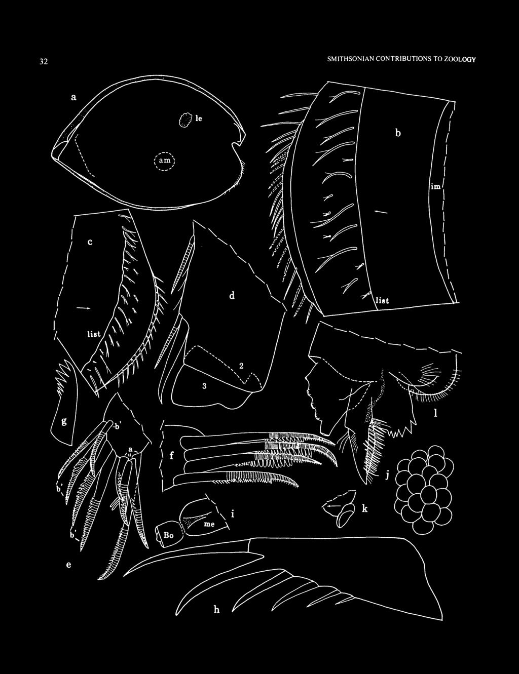
38 32 SMITHSONIAN CONTRIBUTIONS TO ZOOLOGY a
, mv; e, a- and b'-bristles of 2nd exopodial joint left 5th limb, pv;/ b\"-bristles of")
39 NUMBER FIGURE (left) 21. Cypridinodes parallax Kornicker, new species, adult female, paratype, USNM : a, complete specimen from right side, length 2.82 mm; b,c anterior of right and left valves, respectively, ventral to incisur, iv; d, tip of left mandible (nabs), mv; e, a- and b'-bristles of 2nd exopodial joint left 5th limb, pv;/ b"-bristles of 2nd exopodial joint left 5th limb, pv; g, dorsal jaw of 7th limb; h, left furcal lamella (teeth not shown);;, medial eye and Bellonci organ;/ right lateral eye, lv; k, genitalia of left side; /, upper lip from left side. Mandible: 2nd endopodial joint: ventral margin with medial spine of distal pair shorter than that of adult male (Figure 2\d); dorsal margin with 7 long spinous bristles, 2 medium bristles with indistinct spines, 2 short bristles with stout spines, and 8 short unringed bristles with indistinct anterior spines. Limb otherwise similar to that of adult male. Maxilla: Endite III with 1 plumose proximal bristle and 5 terminal spinous and pectinate bristles. Limb otherwise similar to that of adult male. Fifth Limb (Figure 2\e,f): Except for teeth along middle section of b"-bristles being better defined and larger than those of adult male (Figure 21/), limb similar to that of adult male. Sixth Limb: End joint with 18 anteroventral bristles, 3 or 4 posteroventral bristles with bases on medial side some distance from ventral margin, and 2 broad plumose posterior bristles. Limb otherwise similar to that of adult male. Seventh Limb: Terminal bristles similar to those of adult male. Proximal bristles comprise 6 or 7 bristles on ventral side and 8 or 9 bristles on dorsal jaw side, each with 3 bells. Total number of bristles 22. Stout dorsal jaw with marginal row of 7 stout teeth and 6 shorter teeth (3 on each side) (Figure 2 Ig). Comb with about 5-7 long teeth (terminal tooth slightly longer) and 8 shorter teeth (4 on each side), all teeth with rounded tips; small pointed process projecting inward within arc of teeth. Furca (Figure 21 h): With 6 claws on each lamella; teeth on claws similar to those of adult male (not shown). Bellonci Organ (Figure 21 i): Wider than that of adult male. Eyes: Medial eye bare with brown pigment and smaller than that of adult male (Figure 21 /). Lateral eye about same size as that of adult male, with about 19 ommatidia and black pigment (Figure 2\a,j). Upper Lip (Figure 21/): Similar to that of adult male. Genitalia (Figure 21 ): USNM with attached spermatophore on each side of body anterior to furca. Posterior of Body: Evenly rounded, bare. Y-Sclerite and Pigmentation: Similar to that of adult male. Eggs: USNM with 13 eggs (eyes not developed) in marsupium, length of typical egg including transparent membrane: 0.37 mm, excluding membrane: mm. USNM , 1 specimen with about 20 elongate eggs without transparent membranes, each egg with 2 black lateral eyes, length of 1 egg: 0.44 mm. USNM with 27 elongate eggs in marsupium, each with 2 black eyes visible and appendages partly formed; length of 1 egg without visible transparent membrane: 0.34 mm; length of 1 egg including transparent membrane: 0.43 mm, length excluding transparent membrane: 0.43 mm. VARIABILITY. The width of the lunate process ventral to the incisure of the right valve of both sexes varies considerably. The maximum width observed is present in the male holotype (Figure \6h), but this width was observed in only four additional specimens (USNM (A-l male), (juvenile), (adult female), (adult female)). A narrower and more common width is present in USNM (adult female) (Figure 21 b), but in many specimens the process barely overhangs the valve edge. It is possible that more than one species has been included in C. parallax, but all specimens whose appendages were examined have similar furcae and 5th limbs. COMPARISONS. The mandible of C. parallax differs from that of C. strophinx in having two rather than five short proximal dorsal bristles with stout spines on the 2nd endopodial joint, and in having on the 3rd endopodial joint two unringed claws of equal or subequal length rather than a medial claw about '/3 longer than the lateral claw; also, the dorsal claw of the 3rd joint is more bristle-like (completely rather than partly ringed). The 2nd endopodial joint of the maxilla of C. parallax differs from that of C. strophinx in having rings on all three d-bristles. The 5th limb of C. parallax differs from those of C. strophinx, C. relax, and C. minuta in not having a few unusually large teeth on b"-bristles of the 2nd exopodial joint. The furca of C. parallax differs from that of C. strophinx in having a nonarticulated 2nd claw. The right valve of some specimens of C. parallax differs from those of C. relax and C. minuta in having a narrow lunate process ventral to the incisur, but in many specimens the process is barely indicated by slight overhang, and the valves of those could be confused with those of C. relax and C. minuta. Cypridinodes parallax is very close to C. galatheae Poulsen, 1962:301, which is known from a single male collected off Singapore. Males of the two species differ as follows: the sensory bristle of the 5th joint of the 1st antenna of C. parallax has nine long proximal filaments compared to 12 for C. galatheae; the bristle of the 2nd joint of the 2nd antenna of C. parallax bears seven stout ventral spines compared to for C. galatheae; the terminal dorsal jaw of the 7th limb of C. parallax bears about 21 teeth compared to eight or nine teeth for C. galatheae. Skogsbergia Kornicker, 1974 TYPE SPECIES. Skogsbergia minuta Poulsen, 1962, by subsequent designation (Kornicker, 1974:3). COMPOSITION AND DISTRIBUTION. The genus is circumglobal between about 60 N and 10 S and has a known depth range of m. Three species have been described from the western Indian Ocean (5. calyx Kornicker, 1992; S. iota Kornicker, 1992; S. plax Kornicker, 1992); a new species from Madagascar is described herein.

40 34 SMITHSONIAN CONTRIBUTIONS TO ZOOLOGY Skogsbergia solox, new species FIGURES ETYMOLOGY. From the Latin solox (coarse, rough, bristly). HOLOTYPE. Dissected adult male on slide and in alcohol. TYPE LOCALITY. BT-218. PARATYPES. BT-216: USNM : 4 adult males, 1 adult female with large unextruded eggs, plus 18 specimens. BT-218: USNM , undissected adult male in alcohol; USNM , adult female on slide and in alcohol; USNM , 1 adult male, 3 adult females with large unextruded eggs, plus 3 specimens. NONTYPES. BT-218: USNM , female without valves; USNM , juvenile without valves. DISTRIBUTION. See type specimens, above. DESCRIPTION OF ADULT MALE (Figures 22-24). Carapace elongate with convex ventral and dorsal margins, and broad caudal process delimited from margin of valve dorsal to caudal process by either slight (Figure 24g) or distinct angle (Figure 22a, e); anterior edge of rostrum convex, with narrow lateral projection and minute medial projection in vicinity of inferior corner, both projections reach past valve edge (Figure 22b); narrow ridge extends from inner end of incisur to minute protuberance at anteroventral corner of valve (Figure 24g). Surface smooth. Infold: Rostral infold with 8 bristles forming outer row, 3 bristles proximal to outer row, 2 closely spaced bristles near ventral edge of rostrum just anterior to inner end of incisur, and 2 minute bristles at inner end of incisur (Figure 22b). Anteroventral infold with small bristle at inner margin near incisur (Figure 22b). Narrow list extending from anteroventral infold, continuing along ventral margin of valve, and then broadening to form wide shelf anterior to caudal process; anteroventral infold and anterior part of ventral infold with 9 long divided bristles followed by about 28 shorter divided bristles (Figure 22c, a 1 ); anteroventral infold between narrow list and inner margin of infold with 7 short bristles (Figure 22a 1 ); posterior half of ventral infold with about 6 widely spaced bristles along list. Broad list at anterior end of caudal process with pore canals, some exiting on minute protuberances (Figure 22e). Selvage: Lamellar prolongation present along anterior and ventral margins, broadest and with indistinct narrow striations (not shown) at inferior corner of rostrum and dorsal edge of incisur (Figure 22c), divided at inner end of incisur, and absent along edge of caudal process. Prolongation double along anterior half of ventral margin, with proximal layer about '/2 width of other layer (Figure 22c). Carapace Size (length, height in mm): BT-216: USNM , 4 specimens: 1.43, 0.86; 1.42, 0.87; 1.38, 0.81; 1.46, BT-218: 3 specimens: USNM , 1.26, 0.86; holotype, 1.31, 0.75 (flexible carapace measured after body removed and may be distorted); USNM , 1.33, First Antenna (Figure 22/,'g): 1st and 2nd joints bare. 3rd joint shorter on medial side than on lateral side; with minute subterminal ventral bristle and short dorsal bristle at midlength. 4th joint with 2 minute bristles (ventral terminal, dorsal at distal 3 /4 measured on medial side). Sensory bristle of short 5th joint with 9 long proximal filaments (only proximal parts shown), 3 shorter and slenderer distal filaments, and 1 short subterminal filament. Short 6th joint with short spinous medial bristle near dorsal margin. 7th joint: a-bristle slightly shorter than bristle of 6th joint, with few short indistinct spines; b-bristle shorter than sensory bristle of 5th joint, with short stout proximal filament with large sucker, followed by short slender filament with 2 small suckers, then filament with 2 small suckers and 2 filaments with marginal spines; stem distal to filaments with marginal spines; c-bristle longer than sensory bristle of 5th joint, with short stout filament with large sucker (about 'A larger than sucker of b-bristle), followed by slender filament with 2 small suckers, then filament with 2 small suckers, 6 slender bare filaments, and short subterminal filament. 8th joint: d- and e-bristles slightly shorter than b-bristle, bare with blunt tips; f- and g-bristles with abundant long thin proximal filaments followed by about 5 short filaments and short subterminal filament; f-bristle slightly shorter than c-bristle, g-bristle about same length as c-bristle. Second Antenna: Protopodite with distal medial bristle (Figure 22//). Endopodite single jointed, with 4 proximal bristles (3 short, 1 long) and 1 long terminal filament (Figure 22h). Exopodite: bristle of 2nd joint reaching to about 9th joint; ventral margin of bristle with long proximal hairs followed by 7 stout spines, dorsal margin with 4 slender spines (Figure 22/); bristle of 3rd joint long with proximal natatory hairs followed by 7 slender ventral spines and natatory hairs; remaining bristles obscured but with natatory hairs, no spines; 9th joint with 4 bristles (3 long, 1 short dorsal); joints 3-8 with basal spines; spine of 3rd joint about '/3 length of 4th joint; spine of 8th joint about twice length of 9th joint; 9th joint with lateral spine about 2 /3 length of spine of 8th joint. Mandible (Figure 23a,b): Coxale endite well developed, spinous, with 2 stout terminal spines and small peg between them, and with small bristle near base (Figure 23a). Basale: ventral margin with 3 small a-bristles, 1 small b-bristle, 3 small c-bristles, and 1 long spinous d-bristle; dorsal margin with 1 slender bristle proximal to joint midlength and 2 terminal bristles. Exopodite hirsute, about 3 /4 length of dorsal margin of 1st endopodial joint, with 2 bristles (distal about '/3 length of proximal). 1st endopodial joint with 4 ventral bristles (1 minute bare, 1 short and 1 long, both with short spines, and 1 long with FIGURE (right) 22. Skogsbergia solox Kornicker, new species, adult male, holotype: a, complete specimen from right side, length 1.31 mm; b-d, anterior right valve, iv; e, posterior right valve, iv;/ right 1st antenna (nabs), mv; g, proximal part b- and c-bristles right 1st antenna, mv; h, distal protopodite and endopodite left 2nd antenna, mv; i, bristle of 2nd exopodial joint left 2nd antenna, mv;/ medial eye and Bellonci organ from right side; k, left lateral eye.


41 NUMBER \-

, av;/ part of right 5th limb (nabs), pv; g, left lamella of")
42 36 SMITHSONIAN CONTRIBUTIONS TO ZOOLOGY FIGURE 23. Skogsbergia solox Kornicker, new species, adult male, holotype: a,b, proximal and distal parts of right mandible, mv; c, left maxilla (nabs) lv; d, b-d-bristles 2nd exopodial joint left maxilla, lv; e, part of left 5th limb (nabs), av;/ part of right 5th limb (nabs), pv; g, left lamella of furca (teeth not shown), lv; h, left Y-sclerite.

43 NUMBER long spines). 2nd endopodial joint: ventral margin with distal spines, and distal pointed bristles forming 3 groups of 1, 1, and 2 bristles; dorsal margin with 5 long bristles, 2 medium bristles (1 proximal to long bristles, and 1 adjacent to distal long bristle), 2 short bristles (1 with stout spines) with bases on or near dorsal edge of joint, and row of 6 short bare bristles with bases just medial to dorsal edge (only 3 bristles shown in Figure 23b). 3rd endopodial joint with 3 stout claws (dorsal claw shorter; medial ventral claw with 10 or 11 proximal ventral spines, lateral ventral claw with 9 proximal ventral spines (not shown), and dorsal claw with about 2 proximal ventral spines (not shown)) and 4 ringed bristles. Maxilla (Figure 23c,d): Endite I with 12 spinous bristles (not shown); endite II with 6 spinous and pectinate bristles (not shown); endite III with 1 proximal bristle and 5 distal bristles (latter not shown). Usual dorsal bristle on coxale not observed but could be obscured or broken. Basale with 1 short dorsal bristle, 1 long ventral bristle, and 1 short medial bristle (Figure 23c). Exopodite hirsute with 1 subterminal bristle with long spines and 2 longer terminal bristles (bristle closest to subterminal bristle spinous, other bare). 1st endopodial joint with evenly rounded hyaline cutting tooth (stippled in Figure 23c), 2 slender bare ringed alpha-bristles, and 2 ringed beta-bristles (outer stouter and pectinate, inner bare) (Figure 23c). 2nd endopodial joint with 4 ringed bare a-bristles (Figure 23c), 3 pectinate b-bristles (outer ringed, others unringed), 3 c-bristles (2 stout pectinate, 1 minute ringed bare medial), and 3 stout pectinate d-bristles (outer ringed distally, others unringed) (Figure 23d). Fifth Limb (Figure 23e,f): Endite I with 6 spinous bristles; endite II with 5 spinous bristles; endite III with 7 spinous bristles (2nd bristle from posterior side bare spine-like) (only 1 bristle shown on each endite in Figure 23e). Protopodite with long slender anterior tooth (stippled in Figure 23e); 1st exopodial joint: main tooth with stout triangular smooth proximal peg followed by 6 pectinate teeth (distal tooth with about 8 similar cusps) (Figure 23/); bristle proximal to peg with 2 long hairs, 1 on each side; anterior side with 2 or 3 adjacent bristles (1 or 2 slender with long proximal spines; 1 stout with long spines and pectinate) and 1 bristle (with long proximal spines) close to anterior protopodial tooth (Figure 23e). 2nd exopodial joint with 4 pectinate unringed a-bristles, 2 ringed pectinate b'-bristles, 4 ringed pectinate b"-bristles, 1 c-bristle with long proximal spines (Figure 23/), and 1 d-bristle with long proximal spines (Figure 23e). 3rd exopodial joint: inner lobe with 1 proximal bristle with few long hairs, and 2 terminal bristles (inner ringed bare, outer unringed with 2 minute spines near midlength); outer lobe hirsute with 2 terminal bristles (inner bristle with short spines, outer bristle about '/2 length of inner bristle, with long proximal spines). 4th and 5th exopodial joints fused, hirsute, with 2 pairs of spinous terminal bristles (1 with long proximal spines, others with short spines) with or without (Figure 23e) very low spinous node between them. Sixth Limb (Figure 24a): With 3 short bare epipodial bristles. Endite I with 2 short spinous proximal medial bristles and 2 longer spinous terminal bristles; endite II with 2 short spinous proximal medial bristles, 1 long spinous terminal bristle, and 1 short spinous subterminal medial bristle; endite III with 1 long spinous proximal medial bristle and 4 spinous terminal bristles; endite IV with 1 long spinous proximal medial bristle and 6 spinous terminal bristles. End joint with spinous bristles and 2 posterior plumose bristles, few long medial hairs, and row of long stout lateral spines along distal margin. Seventh Limb: With total of 18 bristles: 6 or 7 bristles in proximal group (3 or 4 on each side), each with 4-6 bells and long terminal process; terminal group with 11 or 12 bristles (5 or 6 on each side), each with 1-7 bells. Comb with middle tooth (sclerotized and with rounded tip) and on each side 3 slightly shorter teeth with rounded tips, and 2 stout teeth with flat tips (Figure 24b). Tooth opposite comb with minute distal cusps (tooth closed against comb on both limbs of holotype and partly obscured). Furca (Figure 23g): Each lamella with 7 claws; claw 2 nonarticulated and stouter than claw 1 but shorter; claw 5 stouter and longer than claw 4; all claws with teeth along posterior edge (not shown); claw 1 with large distal medial teeth (not shown); right lamella anterior to left by width of base of claw 1. Bellonci Organ (Figure 22/): Short, cylindrical, bare, with indistinct striations near tip. Eyes: Medial eye unpigmented, bare (Figure 22/). Lateral eye about same size as medial eye, with about 24 ommatidia, and without pigment between ommatidia (Figure 22A). Upper Lip (Figure 24c,d): Anterior unpaired part with large glandular openings; each side of paired posterior part with short lateral lobe with 2 glandular openings followed by smaller lateral lobe with 1 glandular opening. Genitalia (Figure 24e,f): Elongate lobes on each side of body anterior to furca (sclerotized parts stippled). Basal sclerite Y-shaped, distal hood very short, with toothed tip. Anterior of Body (Figure 24c,d): With truncate anterior process dorsal to upper lip. Posterior of Body: Evenly rounded and bare ventral to dorsal end of girdle, slightly undulate dorsal to dorsal end of girdle, but without distinct lobes. Y-Sclerite (Figure 23/?): Ventral branch represented by small prong. REMARKS. The variation in the width of the posterior extension of the caudal process is greater than usually present in species of the genus, and it is possible that specimens with narrow caudal processes and those with broad caudal processes are not conspecific. Because of the similarity in appendages of both narrow and broad forms it is expedient to include them in one species. The size range of the male is also greater than usually present in species of the genus.


44 38 SMITHSONIAN CONTRIBUTIONS TO ZOOLOGY a FIGURE 24. Skogsbergia solox Kornicker, new species, adult male, holotype: a, right 6th limb, mv; b, tip 7th limb (nabs); c,d, anterior process and upper lip from right and left sides; e, left and right copulatory organs;/ left copulatory organ. Adult male, paratype, USNM : g. complete specimen from left side, length 1.26 mm.
.")
45 NUMBER DESCRIPTION OF ADULT FEMALE (Figure 25). Carapace larger than that of adult male and with dorsal end of caudal process not forming angle with margin of valve dorsal to process (Figure 25a-c). Infold: Not examined in detail, but in general similar to that of adult male. Carapace Size (length, height in mm): BT-216:1.62, BT-218: USNM , 1.66, 0.98; USNM , 3 specimens: 1.62, 0.99; 1.49, 0.86; 1.59, First Antenna (Figure 25d,e): 1 st joint bare. 2nd joint with medial and lateral spines (spines not shown in Figure 25d). 3rd joint shorter on medial side than on lateral side, with minute terminal ventral bristle and short proximal dorsal bristle. 4th joint with short terminal ventral bristle and longer subterminal dorsal bristle. Sensory bristle of 5th joint with 9 long proximal filaments, 3 shorter and slenderer distal filaments, and 1 short subterminal filament. 6th joint with short spinous medial bristle (with few indistinct spines) near dorsal margin. 7th joint: a-bristle about same length as bristle of 6th joint, with few indistinct spines; b-bristle shorter than sensory bristle of 5th joint, with 5 marginal filaments, some with spines; c-bristle longer than sensory bristle of 5th joint, with 10 short proximal filaments, some with few spines. 8th joint: d- and e-bristles slightly shorter than sensory bristle of 5th joint, bare with blunt tips; f-bristle slightly shorter than c-bristle, with 9 short filaments, most with spines; g-bristle similar to c-bristle. Second Antenna: Protopodite with spinous distal medial bristle (Figure 25/). Endopodite single jointed, with 4 proximal bristles (2 short bare, 1 medium bare, 1 longer spinous) and 1 long terminal filament (Figure 25/). Exopodite: bristle of 2nd joint reaching to about 9th joint; ventral margin of bristle with 7 or 8 stout spines, dorsal margin with few small indistinct spines; bristles of 3rd and 4th joints with proximal hairs and 11 ventral spines followed by natatory hairs; bristles of joints 5-8 with natatory hairs; 9th joint with 4 bristles (2 long, 1 medium, 1 short dorsal) with natatory hairs; joints 3-8 with basal spines; spine of 3rd joint about 3 A length of 4th joint; spine of 8th joint about twice length of 9th joint; lateral spine of 9th joint about 2 /3 length of spine of 8th joint. Mandible: Similar to that of adult male. Maxilla: Coxale with dorsal bristle with long proximal hairs. 1st endopodial joint: cutting tooth undulate, and inner beta-bristle with small spines (tooth stippled in Figure 25g). 2nd endopodial joint: 2 posterior of the 4 a-bristles with few anterior spines at midlength. Limb otherwise similar to that of adult male. Fifth Limb: Endite I with 7 spinous bristles (male with 6). 1st exopodial joint: bristle proximal to smooth peg of main tooth with 3 long proximal hairs (male with 2 hairs); anterior side with row of 3 bristles (male with 2 or 3). 3rd exopodial joint: both bristles of outer lobe with long proximal and short distal spines (inner bristle of male without long spines). Fused 4th and 5th joints without node between pairs of terminal bristles (adult male either with or without node). Limb otherwise similar to that of adult male, and differences noted above probably not significant. Sixth Limb: With 4 short bare epipodial bristles. Endite I with 2 short spinous proximal medial bristles and 1 long spinous terminal bristle; endite III with 1 long spinous medial bristle and 5 or 6 spinous terminal bristles. End joint with 16 spinous bristles. Limb otherwise similar to that of adult male. Seventh Limb: With total of 18 or 19 bristles: 7 in proximal group (3 or 4 on each side); terminal group with 11 or 12 bristles (5 or 6 on each side). Limb otherwise similar to that of adult male. Furca (Figure 25h): Claw 2 about same length as claw 1. Furca otherwise similar to that of adult male. Bellonci Organ (Figure 25/): Broadens distally and with flat tip. Eyes: Medial eye unpigmented bare (Figure 25/). Lateral eye about same size as medial eye, with 30 ommatidia when viewed under coverslip (Figure 25k), but only 24 apparent when viewed laterally and not under coverslip (Figure 24/), and with black pigment between ommatidia. (Lateral eye of adult male without black pigment, possibly faded during preservation.) Upper Lip (Figure 25/): Similar to that of adult male. Genitalia (Figure 25«): Small oval with attached spermatophore on each side of body anterior to furca. Anterior of Body (Figure 25e,l): Distal edge of anterior process more rounded than that of adult male. Posterior of Body and Y-Sclerite (Figure 25m): Similar to those of adult male. COMPARISONS. The carapace of 5. solox differs from those of 5. calyx Kornicker, 1992, and 5. hesperidea (Muller, 1906) in having a broadly rounded rather than triangular caudal process. The adult male 1 st antenna of 5. solox differs from those of S. iota Kornicker, 1992, and S. plax Kornicker, 1992, in having abundant filaments on the f- and g-bristles. The furca of 5. solox differs from those of 5. costae Kornicker, 1974, and 5. megalops (Sars, 1872) in having claw 2 nonarticulated, and from that of 5. squamosa Kornicker, 1974, in having claw 3 articulated. Skogsbergia solox differs from 5. galapagensis Kornicker and Iliffe, 1989a, in having a well developed lateral eye. The carapace of the adult male S. solox differs from S. minuta Poulsen, 1962, in being larger; the mandible differs in having a basale with three rather than two a-bristles, three rather than two c-bristles, one short dorsal bristle with stout spines on the 2nd endopodial joint, and in having the shortest bristle of the 3rd endopodial joint slender rather than broad; the 7th limb differs in having 18 rather than 12 bristles; the furca differs in having on each lamella seven rather than eight claws, and in having claw 2 with a pointed rather than rounded tip. The adult male S. solox differs from that of S. curvata Poulsen, 1962, in being smaller, in having a 1st antenna with a shorter ventral bristle on the 3rd joint, shorter ventral and dorsal


46 40 SMITHSONIAN CONTRIBUTIONS TO ZOOLOGY a n FIGURE 25. Skogsbergia solo.x Kornicker, new species, adult female, paratype, USNM : a, complete specimen from right side, length 1.66 mm; b. anterior left valve, ov; c, posterior right valve, ov; d, left 1 st antenna (nabs), mv; e, part of anterior of body from left side (nabs);/ distal protopodite and endopodite left 2nd antenna, mv; g. cutting tooth (stippled) and beta-bristles 1st endopodial joint left maxilla, lv; h, right lamella of furca (teeth not shown); i, medial eye and Bellonci organ from left side;y, right lateral eye; k, lateral eye under cover slip at twice magnification of/: /. anterior process and upper lip from left side; m, left Y-sclerite; n, left genital organ and spermatophore.

 are not curved. The adult male S. solox differs from those of 5. lerneri (Kornicker, 1958) and S.")
47 NUMBHR 41 bristles on the 4th joint, and two rather than three small suckers on filaments of the b- and c-bristles of the 7th joint; the mandible differs in having three rather than two c-bristles on the basale, and the three claws of the 2nd endopodial joint are pectinate rather than bare; the end joint of the male 6th limb differs in having rather than 18 bristles; the male furca differs in not having strongly curved claws and in having seven rather than eight claws, and the Bellonci Organ differs in being cylindrical rather than pear-shaped. The claws of the furca of the female 5". curvata (Poulsen, 1962:169) are not curved. The adult male S. solox differs from those of 5. lerneri (Kornicker, 1958) and S. menezi Kornicker, 1970, in being smaller and in having seven rather than eight claws on each lamella; S. lerneri also has five rather than four proximal bristles on the endopodite of the 2nd antenna. The adult male S. solox differs from that of S. strophinx Kornicker, 1991, in having a smaller lateral eye and seven rather than eight claws on each lamella of the furca. Bristles along the anterior list of each valve ventral to the incisur of the male S. solox are unusually long (Figure 22c,d) and might be useful in identification. Skogsbergia plax Kornicker, 1992 FIGURE 26 Skogsbergia plax Kornicker, 1992:73, figs HOLOTYPE. MNHN Os 440, undissected adult male in alcohol. TYPE LOCALITY. Mozambique Channel, Glorioso Islands, depth 24 m (Kornicker, 1992:73). MATERIAL. BT-222: USNM , 1 adult male and 2 juveniles. BT-837: USNM , adult male on slide and in alcohol. BT-848A: USNM , adult female with small unextruded eggs. DISTRIBUTION. Northern Mozambique Channel at depths of 6-24 m (Kornicker, 1992:73). Madagascar: see "Material." Known depth range 7-24 m. SUPPLEMENTARY DESCRIPTION OF ADULT MALE (Figure 26a-g). Carapace shape similar to that described by Kornicker (1992:75), but slightly larger (Figure 26a). Carapace Size (length, height in mm): BT-222: USNM , 1.06, BT-837: USNM , 1.05, Kornicker (1992:75) gave length range of mm, and height range of mm. Including Madagascar specimen, length range mm, height range mm. Lateral Eye (Figure 26a,e): With about 32 ommatidia; without brown pigment between ommatidia present on specimens described by Kornicker (1992:77) (possibly a preservation artifact). SUPPLEMENTARY DESCRIPTION OF ADULT FEMALE (Figure 26h). Carapace shape similar to that described by Kornicker (1992:73), but slightly larger (Figure 26h). Carapace Size (length, height in mm): BT-848A: USNM , 1.46, Kornicker (1992:73) gave length 1.36 mm, height 0.81 mm. Seventh Limb: Limb coiled, similar to 7th limb illustrated by Kornicker (1992,fig. 42g). Lateral Eye (Figure 26/z): Large with many ommatidia and brown pigment between ommatidia. Skogsbergia calyx Kornicker, 1992 FIGURES 27,28 Skogsbergia calyx Kornicker, 1992:59, figs HOLOTYPE. MNHN Os 437, adult female on slide and in alcohol. TYPE LOCALITY. Mozambique Channel, Glorioso Islands, depth 26 m (Komicker, 1992:59). MATERIAL. BT-227: USNM , ovigerous female on slide and in alcohol. BT-683: undissected specimen (length 2.14 mm, height 1.27 mm). DISTRIBUTION. Northern Mozambique Channel at a depth of 26 m (Kornicker, 1992:59). Madagascar: see "Material." Known depth range intertidal to 27 m. CORRECTION. During the present study I noticed some errors in the original description of the species (Kornicker, 1992:59), which I would like to correct here. The "3rd endopodial joint" in the description of the 5th limb (p. 63) should be "3rd exopodial joint." "USNM " in the descriptions of the 1 st and 2nd antennae, mandible, and 6th limb should be "MNHN Os 437." In Figure 33/ (maxilla) the bristle labeled "c" adjacent to the anterior b-bristle should be labeled "b." SUPPLEMENTARY DESCRIPTION OF ADULT FEMALE (Figures 27, 28). Dorsal edge of caudal process of left valve with broad swelling (Figure 21a,c,e) not present on right valve (Figure 27d,f). Infold: Caudal process: ridge at anterior end with 7-9 small bristles; infold posterior to ridge with 7-10 short bristles (Figure 21e,f). Carapace Size (length, height in mm): USNM , 2.13, First Antenna: Sensory bristle of 5th joint with 8 long proximal filaments followed by 3 shorter and slenderer filaments and very short subterminal filament. Second Antenna: Endopodite unjointed, with 1 long, 1 medium, and 2 short proximal bristles, and 1 very long terminal filament (Figure 28a). Mandible: Dorsal margin of 2nd endopodial joint with 4 long bristles, 3 spinous cleaning bristles, and 2 short slender proximal bristles (Figure 286). Maxilla: Endite I with 10 or 11 bristles. Cutting tooth of 1st endopodial joint rounded (Figure 28c). Middle b-bristle of 2nd endopodial joint with 3 or 4 anterior and 2 posterior teeth (in Figure 28c, a-, c-, and d-bristles not shown).


48 42 SMITHSONIAN CONTRIBUTIONS TO ZOOLOGY a FIGURE 26. Skogsbergia plax Kornicker, 1992, adult male, USNM : a, complete specimen from right side, length 1.05 mm: b, right lamella of furca; c, posterior of body from right side; d, medial eye and Bellonci organ from right side; e, right lateral eye;/ anterior process and upper lip from right side; g, part of anterior of body from left side. Adult female, USNM : h, complete specimen from left side, length 1.46 mm.


49 NUMBER FIGURE 27. Skogsbergia calyx Komicker, 1992, ovigerous female, USNM : a, complete specimen from left side, length 2.13 mm; b,c, anterior and posterior, respectively, left valve, ov; d, posterior of complete specimen from right side; e,f, caudal process of left and right valves, respectively, iv.

; h, right lateral eye, lv; i.")
50 44 SMITHSONIAN CONTRIBUTIONS TO ZOOLOGY FIGURE 28. Skogsbergia calyx Kornicker, 1992, ovigerous female, USNM : a, distal protopodite and endopodite right 2nd antenna, mv; b, 2nd endopodial joint right mandible, mv; c, tip of left maxilla (nabs), lv; d, bristle row of I st exopodial joint 5th limb, av; e, 1 st and 2nd exopodial joints right 5th limb, pv (drawn without cover slip):/ tip of 7th limb; g, posterior of body from right side (not all teeth of furcal claws shown); h, right lateral eye, lv; i. left lateral eye (drawn at twice magnification of A), mv;y", medial eye and Bellonci organ; k, upper lip and anterior process from right side; /, anterior of body from left side.
; anterior bristle near protopodial tooth with short")
51 NUMBER Fifth Limb: Epipodite with 51 bristles. Endites I, II, and III with 7, 5, and 7 bristles, respectively. Small inner bristle of row of 3 bristles on anterior side of 1st exopodial joint with long proximal and short distal spines (Figure 28a 1 ); anterior bristle near protopodial tooth with short marginal spines. 2nd exopodial joint with 4 b'-bristles (longest with 17 cusps), and 3 b"-bristles (longest with 25 cusps) (Figure 28e); d-bristle short bare. Outer lobe of 3rd exopodial joint with 2 terminal bristles with long proximal and short distal spines. Sixth Limb: With 3 or 4 epipodial bristles with tubular tips. End joint with 20 bristles (3 short slender (with short marginal spines) near anterior end, 14 longer and stouter bristles with long proximal and short distal spines, followed by 3 longer plumose bristles). Seventh Limb: Proximal group with 6 or 7 bristles (3 on comb side, 3 or 4 on peg side) each with 4-6 bells; 12 bristles (6 on each side) in distal group, all on terminal segment, each with 1-7 bells. Sclerotized ridge within comb indistinct, spines not observed. Process opposite comb with 2 adjacent ridges each with 6 minute terminal teeth (Figure 28/). Furca (Figure 2Sg), Upper Lip (Figure 2Sk,l), Genitalia (Figure 28g), Anterior of Body (Figure 2Sk,l), Posterior of Body (Figure 28g), Y-Sclerite (Figure 28g), and Pigmentation: Similar to those described by Kornicker (1992:59-63). Bellonci Organ (Figure 28/): Short, with length and height equal. Eyes: Medial eye unpigmented bare (Figure 28/,/). Lateral eye larger than medial eye; right eye of USNM with 26 ommatidia visible when eye viewed laterally and eye not under cover slip (Figure 28//); left eye of same specimen with 33 ommatidia when count made with eye under cover slip (Figure 28/). Ommatidia light amber color; in transmitted light all ommatidia, except ommatidia along outer edge, covered with matrix of brown pigment (stippled area in Figure 2Sh,i). Eggs: USNM with 6 fairly well-developed elliptical eggs in marsupium, but lateral eyes not visible; length of typical egg 0.27 mm. DISCUSSION. The description above of USNM is concerned mainly with characters that differ from those in the original description of the species (Kornicker, 1992:59). The larger carapace of the present specimen and the shorter Bellonci organ suggests that it might not be S. calyx, but until additional specimens are available to determine intraspecific variability, I find it expedient to refer the specimen to 5. calyx. The dorsal margin of the caudal process of the left valve of USNM has a swelling fairly similar to that on the right valve of Skogsbergia hesperida (Muller, 1906). This swelling was not reported in the original description of S. calyx, but the tip of the caudal process of the left valve of the holotype appears to be broken off (see Kornicker, 1992, fig. 32f), and that may account for the apparent lack of a swelling on the holotype. I did not reexamine the holotype, which is in France, but I reexamined a paratype, USNM , a juvenile (A-2 instar), and observed a swelling on the dorsal margin of the caudal process of the left valve. TYPE SPECIES. Cypridina Vargula Skogsberg, 1920 (Vargula) norvegica Baird, COMPOSITION AND DISTRIBUTION. Including a new species described herein, the genus contains about 50 species; two have been described from the northern Mozambique Channel at depths of m (Kornicker, 1992:81). Vargula grex, new species FIGURES ETYMOLOGY. From Latin grex (flock, herd). HOLOTYPE. Instar V female on slide and in alcohol. TYPE LOCALITY. BT-186. PARATYPES. None. DISTRIBUTION. Only collected at type locality. DESCRIPTION OF INSTAR V FEMALE (Figures 29-31). Carapace elongate (Figure 29a); tip of rostrum with 3 or 4 minute digitations (Figure 29c); several digitations also along anteroventral margin (Figure 29a); in side view, dorsal end of narrow caudal process delimited from valve edge dorsal to process by very slight concavity (Figure 29a). Infold: Rostral infold with 9 or 10 bristles (Figure 29c); inner edge of incisur with 2 bristles (1 long, 1 short); 2 small bristles ventral to inner end of incisur and 2 longer divided bristles along anteroventral edge (1 shown in Figure 29c); anteroventral infold plus ventral infold to about valve midlength with about 28 bristles; list broadens from posterior '/4 of ventral margin to about ventral half of posterior margin; broad list with fairly straight distal edge, several indistinct minute bristles (2 shown in detail in Figure 29b), and numerous pore canals (Figure 29b). Selvage: Lamellar prolongation of selvage broad and striate along lower margin of incisur, narrow and without striae elsewhere, but prolongation not observed along posterior edge of caudal process. Central Adductor Muscle Attachments (Figure 29d): Comprising about 15 oval attachments. Carapace Size (length, height in mm): Holotype, 1.43, First Antenna (Figure 29e,f): 1st and 2nd joints bare. 3rd joint short with 2 bristles (1 ventral, 1 dorsal). 4th joint with 2 bristles (1 ventral, 1 dorsal). Sensory bristle of 5th joint with 8 long proximal filaments, 2 shorter and slenderer distal filaments, and bifurcate tip. 6th joint with short medial bristle near dorsal margin. 7th joint: a-bristle slightly longer than bristle of 6th joint; b-bristle about twice length of a-bristle, with about 5 short filaments; c-bristle about x b longer than sensory bristle of 5th joint, with 8 slender filaments and bifurcate tip. 8th joint: d- and e-bristles about twice length of b-bristle, bare with blunt tips; f-bristle with tip missing (7 slender filaments on remaining part); g-bristle similar in length to c-bristle, with 8 slender filaments, some with spines, and bifurcate tip; joint with minute lateral papilla proximal to d- and e-bristles.


52 46 SMITHSONIAN CONTRIBUTIONS TO ZOOLOGY sens FIGURE 29. Vargula grex Kornicker, new species, instar V female, holotype: a, complete specimen from left side, length 1.43 mm; b, posterior left valve, iv; c, anterior left valve, iv; d, central adductor muscle attachments left valve, ov; e. left 1st antenna (nabs), Iv;/ distal part of right 1st antenna (nabs), Iv; g, distal protopodite and endopodite left 2nd antenna, mv; h,i, left mandible, mv.
, mv; b, endites left maxilla, Iv; c, a-bristles, 1st endopodial joint right maxilla, lv; d,")
, av; g,h, parts of left 5th limb (nabs), pv. posterior steps not well defined as on illustrated left tusk. Genitalia: None observed.")
53 48 SMITHSONIAN CONTRIBUTIONS TO ZOOLOGY /I FIGURE 30. Vargula grex Kornicker, new species, instar V female, holotype: a, left maxilla (nabs), mv; b, endites left maxilla, Iv; c, a-bristles, 1st endopodial joint right maxilla, lv; d, cutting tooth (stippled) and beta-bristles 2nd endopodial joint left maxilla, mv; e, alpha-bristles 1st endopodial joint and b-d-bristles 2nd endopodial joint, left maxilla, mv;/ part of right 5th limb (nabs), av; g,h, parts of left 5th limb (nabs), pv. posterior steps not well defined as on illustrated left tusk. Genitalia: None observed. Anterior of Body: Small rounded knob just ventral to base of 1 st antenna (Figure 29e). Posterior of Body (Figure 3 \g): posterodorsal corner. Bare with evenly rounded
; d.")
: Holotype with minute eggs in ovaries. COMPARISONS. The mandible of V. grex differs from those of V. arx Kornicker (1992:81) and V.")
54 NUMBER FIGURE 31. Vargula grex Kornicker, new species, instar V female, holotype: a, dorsal bristle of coxale right maxilla, lv; b, left 6th limb, mv; c, left lamella of furca (teeth not shown); d. part of anterior body from left side; e, detail left lateral eye;/ upper lip from left side; g, posterior of body from left side. Y-Sclerite (Figure 3 lg): Typical for subfamily. Eggs (Figure 3 lg): Holotype with minute eggs in ovaries. COMPARISONS. The mandible of V. grex differs from those of V. arx Kornicker (1992:81) and V. sagax Kornicker (1992:87) in having a ventral finger-like terminal process on the 2nd endopodial joint, and a bristle with a proximal bulge near the ventral edge of the 3rd endopodial joint. Vargula arx also differs from V. grex in lacking lateral eyes. Vargula plicata Poulsen (1962:189) is known only from the adult male and, therefore, is not directly comparable with the A-l female V. grex. The length of the adult male V. plicata is 1.31 mm (Poulsen, 1962:190), whereas the length of the A-l female V. grex is 1.43 mm. The middle claw of the 3rd endopodial joint of the mandible ofv.plicata is shorter than the other two (Poulsen, 1962:191), whereas all three are of similar length in V. grex. Other differences occur in the lengths and spinosities of the bristles of the basale and 1st endopodial joint of the mandibles of the two species. Vargula plicata is known only from the Celebes Sea (Poulsen, 1962:189). It is possible that when equivalent stages and sex of V. plicata and V. grex are known they will be shown to be conspecific. The 3rd endopodial joint of the mandible of Sheina orri Harding (1966:374) bears a bristle with a bulbous base similar to that of V. grex, but the bristle on 5. orri bears a hirsute pad at the tip that is not present on the equivalent bristle of V. grex (Kornicker, 1986b:641, 642). The coxale endite of the mandible of V. grex is not reduced as it is in S. orri (Kornicker, 1986b:641). The generic placement of V. grex should be considered tenuous until the adult male is known.

); joints 3-8 with basal spines longer on distal joints; basal spine of")
55 NUMBER Second Antenna (Figure 29g): Protopodite with short distal medial bristle with indistinct spines. Endopodite 3-jointed: 1st joint with 1 long and 2 short proximal bristles and 1 long distal bristle; 2nd joint elongate with small terminal bristle; 3rd joint elongate with long terminal filament. Exopodite: bristle of 2nd joint reaching 8th joint, with about 13 stout ventral spines; bristle of 3rd joint long, with natatory hairs and proximal ventral spines; bristles of joints 4-8 with natatory hairs; 9th joint with 4 bristles (2 long and 1 medium with natatory hairs, 1 short dorsal (hairs if present obscured)); joints 3-8 with basal spines longer on distal joints; basal spine of 8th joint slightly longer than 9th joint; 9th joint with lateral spine longer than joint; minute spines usually present along distal edges of joints 2-8 not observed (obscured). Mandible (Figure 29h,i): Coxale endite spinous, with 2 closely spaced terminal spines larger than adjacent spines and without peg between them; small bristle present near base (Figure 29h). Basale without spines; ventral margin with 2 fairly long bare a-bristles (1 about 2 /3 length of other); 1 small bare b-bristle (with base on lateral surface), 2 bare c-bristles (proximal short, distal long); and 2 d-bristles (proximal medium length bare, distal long with long spines in middle part and bare in terminal '/4-'/3); c- and d-bristles separated by wide space; dorsal margin with 1 short bristle at 3 /4 length, and 2 slender subterminal bristles (medial bristle shorter). Exopodite about 3 /4 length of dorsal margin of 1st endopodial joint, with distal hairs and 2 slender subterminal bristles (distal bristle shorter). 1st endopodial joint with 4 ventral bare bristles. 2nd endopodial joint: ventral margin with 2 small separated distal bristles and 1 stout finger-like subterminal process (bristle not observed lateral to process, possibly obscured); dorsal margin with 8 proximal bristles (5 long, 2 medium, 1 short spinous). 3rd endopodial joint with 3 stout bare claws and 4 bristles (1 lateral near dorsal edge (type of tip and exact length of bristle obscured), 2 small medial near ventral edge, 1 lateral near ventral edge with stout proximal bulge and rings only in narrower distal part) (Figure 29/). Maxilla: Endite I with 10 spinous and pectinate bristles (Figure 306); endite II with 4 spinous and pectinate bristles (Figure 30b); endite III with 1 proximal bristle (Figure 30a) and 4 terminal spinous and pectinate bristles (Figure 30b) (not all marginal spines and teeth shown). Coxale with fringe of dorsal hairs and plumose dorsal bristle (Figure 31a; not shown in Figure 30a). Basale with slender ventral bristle (Figure 30a). Exopodite well developed, with 3 bristles (proximal and middle bristle with long marginal hairs) (Figure 30a). 1st endopodial joint with few hairs, 2 ringed alpha-bristles, both with fairly long marginal hairs (Figure 30a,e), and 2 ringed beta-bristles, only the smaller with marginal hairs (Figure 30a,d), and cutting tooth with 3 cusps (Figure 300,0"). 2nd endopodial joint with 4 short stout ringed a-bristles with minute marginal teeth (Figure 30a,c), 3 b-bristles (shortest unringed, others ringed) with marginal teeth (teeth on middle bristle longest; shortest bristle with only few distal teeth) (Figure 30a,e), 3 c-bristles (1 minute) (Figure 30e; minute c-bristle not shown in Figure 30a), and 3 d-bristles (only outer bristle ringed) (Figure 30a,e). Fifth Limb: With 3 endites (bristles not counted). Epipodite with 42 bristles. Protopodite with long curved anterior tooth (stippled in Figure 30/). 1st exopodial joint: anterior side with 2 stout bristles and 2 slender weaker bristles closer to protopodial tooth (Figure 30/); main tooth with smooth proximal peg and 5 stout cuspate teeth (proximal tooth with 9 small cusps, distal tooth with 15); spinous ringed bristle proximal to main tooth (Figure 30g). 2nd exopodial joint with 3 stout unringed pectinate a-bristles, a total of 7 or 8 ringed pectinate b'- and b"-bristles, 1 stout posterior ringed c-bristle with long proximal hairs (Figure 30g), and 1 long anterior d-bristle with long hairs (Figure 30/). 3rd exopodial joint: inner lobe with 3 stout ringed bristles (proximal bristle with long hairs); outer lobe hirsute, with 2 stout ringed terminal bristles (Figure 30h). 4th and 5th exopodial joints fused, hirsute, with total of 3 bristles. Sixth Limb (Figure 3\b): Epipodite with 4 bare bristles. Endite 1 with 1 small medial spinous bristle and 1 longer terminal bristle (broken off on both limbs of holotype); endite II with 3 spinous bristles (2 small medial, 1 long terminal) (right limb of holotype aberrant in having two 2nd endites, one with 1 small medial bristle, and other with 3, both with 1 terminal bristle); endite III with 3 spinous terminal bristles (long bristle on each side of short bristle); endite IV with 3 terminal bristles (1 short, 2 long). Ventral margin of skirt with 5 anterior bristles (with long proximal and short distal spines) separated by space from 3 bristles (anterior with long proximal and short distal spines, 2 posterior with long hairs); ventral margin of skirt with long medial hairs and stouter lateral hairs; medial surfaces of skirt and endites III and IV with long hairs. Seventh Limb: Each limb with 5 tapered proximal bristles (2 on comb side, 3 on peg side) each with 1 bell; terminus with 7 tapered bristles (4 on comb side, 3 on peg side) each with 1-3 bells. Terminus with 5 comb teeth opposite single peg. Furca (Figure 31c,g): Each lamella with 7 claws; claws 2 and 4 nonarticulated; claw 2 very slightly broader than claw 1; claw 3 much narrower than claw 4 and either same length or slightly longer; claw 1 of right lamella anterior to left by width of claw base; each claw with medial and lateral rows of teeth (not shown); distal medial teeth of claw 1 slightly larger, otherwise teeth of each claw similar in length. Bellonci Organ (Figure 3ld): Lemon-shaped with small terminal process. Eyes: Medial eye bare and with brown pigment (Figure 31a"). Lateral eye larger than medial eye, with 14 amber-colored divided ommatidia (Figure 31a',e); suture dividing each ommatidium more or less parallels long axis of eye; brown pigment present between ommatidia and coating all ommatidia except those along periphery. Upper Lip (Figure 31/): Unpaired part with many small glandular processes. Posterior paired part with 2 long tusks each with 2 small terminal processes and 2 steps on posterior edge (each step with 1 glandular process); medial surface of each tusk adjacent to proximal step with 2 small glandular processes (dotted in Figure 31/); tusks without hairs. Globose posterior part of lip with long hairs. Right tusk of holotype with
, and a new species is described herein.")
56 50 Pterocypridina Kornicker, 1975 TYPE SPECIES. Pterocypridina excreta Poulsen, 1962 (subsequent designation, Kornicker, 1975:142). COMPOSITION AND DISTRIBUTION. Eight species have been described (Kornicker, 1983a:5; Kornicker and Poore, 1996), and a new species is described herein. Species are known from southeastern North America, Singapore, Thailand, Australia, and Madagascar. The known depth range is m. The genus has not been reported previously from the Indian Ocean. Pterocypridina nex, new species FIGURES ETYMOLOGY. From the Latin nex (violent death). HOLOTYPE. Ovigerous female on slide and in alcohol. TYPE LOCALITY. BT-172. PARATYPES. None. DISTRIBUTION. Collected only at type locality. DESCRIPTION OF ADULT FEMALE (Figures 32-35). Carapace oval in lateral view with prominent rostrum with lateral process and small distinct caudal process with rounded tip (Figure 32a-g); valve slightly overhangs edge in vicinity of anteroventral corner (Figure 32e); distinct scalloped ridge present just within anterior end of valve ventral to incisur (Figure 326). Valve surface near dorsal edge of rostrum with indistinct shallow fossae (Figure 326). Infold: Rostral infold with 20 undivided bristles (Figure 32</); inner end of incisur with 2 closely spaced bristles, a minute bristle near outer edge, and 3 or 4 small bristles set back from edge (Figure 32d); anterior infold ventral to incisur with single row of 8-10 divided bristles ( Figure 32d,e); anterior '/6 of ventral margin with about 30 closely spaced, fairly long, divided spinous bristles; posterior 5 /6 of ventral margin with 2 or 3 widely spaced bristles. Infold of caudal process with anterior ridge bearing few minute spines; posterior edge of ridge straight or with few minute protuberances and forms anterior edge of pocket (Figure 32/g). Selvage: Lamellar prolongation of selvage broad and narrowly striate along inner part of ventral margin of incisur (Figure 32e); narrower prolongation along edge of rostrum and ventral valve margin; prolongation not observed along posterior edge of caudal process. Central Adductor Muscle Attachments (Figure 32A): With elongate and oval attachments. Hinge: Right valve with knob at anterior (Figure 32d) and posterior (Figure 32g) ends of hinge, and left valve with corresponding sockets (Figure 32c. f). Carapace Size (length, height in mm): Holotype, 2.64, First Antenna (Figure 33a): 1st joint bare. 2nd joint with few rows of medial spines near dorsal margin. 3rd joint short (dorsal edge longer on lateral side) with small proximal bare dorsal bristle and longer distal spinous ventral bristle. 4th joint SMITHSONIAN CONTRIBUTIONS TO ZOOLOGY with short subterminal bare dorsal bristle and short terminal spinous ventral bristle. Sensory bristle of 5th joint with 9 long proximal filaments followed by 3 slender filaments (with few spines) and short subterminal filament. 6th joint with short medial bristle (with small widely separated marginal spines) near dorsal margin. 7th joint: a-bristle similar to bristle of 6th joint; b-bristle shorter than sensory bristle of 5th joint, with about 5 short filaments (with few marginal spines) and minute spines on stem distal to filaments; c-bristle long with tip missing and 9 short filaments (with few spines) on remaining part. 8th joint: d- and e-bristles long, bare, with blunt tips; f-bristle long stout with about 11 short filaments with few spines; g-bristle long stout with tip missing and 7 short filaments (with few marginal spines) on remaining part. Second Antenna: Protopodite with small distal medial bristle (Figure 336). Endopodite with single joint with 4 proximal bristles and long terminal filament (Figure 336). Exopodite: 1st joint with indistinct minute terminal medial bristle (Figure 33c); bristle of 2nd joint with long hairs on proximal 3 /4 and 6-8 slender ventral spines on distal '/4 (Figure 33c): bristles of joints 3 and 4 with short proximal hair-like spines and natatory hairs (Figure 33c); bristles of joints 5-8 and 4 bristles of 9th joint with natatory hairs, no spines (Figure 33a 1 ); joints 3-8 with basal spines stouter on distal joints (Figure 33d); basal spine of 8th joint longer than 9th joint; lateral spine of 9th joint about same length as joint (Figure 33*/); joints 3-8 with indistinct minute spines at distal dorsal corner (Figure 33c,d). Mandible (Figure 33e): Coxale endite with 2 stout terminal spines without peg between them; short bristle at base of endite. Basale: ventral margin with 2 short a-bristles, no b-bristle, 3 c-bristles (1 minute tubular), and 1 long spinous d-bristle; dorsal margin with 1 short bare bristle just distal to midlength and 2 spinous subterminal bristles. Exopodite about same length as dorsal margin of 1st endopodial joint, with hirsute tip and 2 subterminal bristles (proximal very long and with short spines, distal short bare). 1st endopodial joint with 4 ventral bristles (1 long with long spines, 1 long with short spines, 1 short with short spines, 1 minute bare). 2nd endopodial joint: ventral margin with 1 minute, tubular, unringed, subterminal, medial bristle; dorsal margin with 8 proximal bristles (5 long spinous ringed, 1 medium bare ringed, 1 short slender ringed, and 1 short stout unringed and with long spines); medial surface with indistinct rows of slender spines. 3rd endopodial joint embedded in tip of 2nd joint, with 3 stout subequal bare claws and 4 bristles (1 long and 2 short ringed, 1 minute unringed) with tubular tips. (Rings on most bristles not shown in illustrated limb.) Maxilla (Figure 34a-c): Endite I with 10 bristles; endite II with 6 bristles; endite III with 1 proximal and 5 terminal bristles (Figure 34a). Basale with long spinous terminal ventral bristle near base of exopodite, 1 short terminal medial bristle near midwidth, and 1 short bare dorsal bristle (this bristle could be interpreted to be on coxale). Exopodite with 3 bristles with long

, iv;/g, caudal process left and right")
57 NUMBER FIGURE 32. Pterocypridina nex Kornicker, new species, ovigerous female, holotype: a, complete specimen from right side, length 2.64 mm; b, anterior right valve, ov; c,d. anterior left and right valve, respectively, iv; e, anterior left valve (nabs), iv;/g, caudal process left and right valves, respectively, iv; h, freehand sketch of central adductor muscle ends projecting from right side of body; i, outline of genital organ and attached spermatophore.


58 52 SMITHSONIAN CONTRIBUTIONS TO ZOOLOGY \ FIGURE 33. Pterocypridina nex Kornicker, new species, ovigerous female, holotype: a, left 1st antenna (nabs), mv; b, distal protopodite and endopodite right 2nd antenna, mv; c, part of exopodite left 2nd antenna, mv; d, distal exopodite right 2nd antenna (nabs), Iv; e. left mandible, mv.


59 NUMBER FIGURE 34. Pterocypridina nex Kornicker, new species, ovigerous female, holotype: a, endites right maxilla, lv; b, right maxilla (nabs), lv; c, distal left maxilla (nabs), mv; d, distal 2nd exopodial joint right 5th limb, pv; e, endites left 5th limb, av;/ left 5th limb (nabs), av; g, right 5th limb (nabs), pv.
, and triangular cutting tooth.")
60 54 proximal and short distal spines (Figure 34b); proximal bristle close to tip of exopodite; middle bristle with more long spines than others. 1st endopodial joint with dorsal spines, 2 alpha-bristles with long spines, 2 beta-bristles (outer pectinate, inner with short spines), and triangular cutting tooth. 2nd endopodial joint with 4 a-bristles (2 anterior with few proximal spines, 3rd bare, 4th pectinate) (Figure 34b), 5 pectinate b-bristles (Figure 34c; only 3 shown in Figure 34b), 3 pectinate c-bristles (Figure 34c); and 3 coarsely pectinate d-bristles (outer with rings in distal half) (Figure 34c). Fifth Limb: Epipodite with about 50 bristles. Endite I with 5 spinous bristles (Figure 34e); endites II and III each with 6 spinous bristles (Figure 34e,g). Protopodite with stout sclerotized anterior tooth (stippled in Figure 34/). 1st exopodial joint: anterior side with row of 3 pectinate or spinous bristles (stoutest tooth strongly pectinate) (on illustrated limb middle bristle fused to stoutest bristle at base); an additional anterior bristle near protopodial tooth appears to be on endite III but could be on 1st endopodial joint (Figure 34/); main tooth with stout proximal triangular peg (with 2 minute terminal spines) and 6 cuspate teeth (Figure 4g); bristle proximal to peg with long proximal and short distal spines. 2nd exopodial joint with 3 stout unringed a-bristles with relatively few rounded cusps, a total of about 6 ringed pectinate b'- plus b"-bristles (Figure 34d), a c-bristle with long proximal hairs and short distal spines (Figure 34g), and an unusually short bare d-bristle (Figure 34/). 3rd exopodial joint (Figure 34g): inner lobe with short bare ringed proximal bristle and 2 longer ringed terminal bristles with long proximal spines; outer lobe hirsute, with 2 stout ringed bristles with long proximal and short distal spines. 4th and 5th joints fused, with total of 6 ringed bristles with long proximal spines (some bristles also with distal spines or teeth). (Rings not shown on most bristles.) Sixth Limb (Figure 35a,b): With 4 bare epipodial bristles. Endite I with 4 spinous bristles (2 short medial proximal, 2 long terminal); endite II with 4 or 5 bristles (2 or 3 short spinous medial, 1 small bare distal medial, 1 long spinous terminal); endite 111 with 5 spinous bristles (1 short medial proximal, 4 terminal (1 short, 3 long)); endite IV with 5 or 6 spinous bristles (1 short medial proximal, 4 or 5 terminal (2 long, 2 or 3 short)). End joint with spinous bristles, abundant medial hairs, and lateral spines along ventral edge. Limb with brown pigment filling veins (stippled in illustration). (Not all spines or rings shown on bristles.) Seventh Limb (Figure 35c): Each limb with bristles: 6-8 in proximal group (3 on comb side, 3-5 on jaw side), each with 4-7 bells; 13 or 14 in terminal group (6 or 7 on comb side, 7 on jaw side), each with 1-7 bells; 1 bristle on each side in terminal group longer than others. Terminal segment much wider than proximal segments; comb with 17 or 18 teeth: 1 or 2 long terminal teeth and on each side 3 shorter teeth with rounded tips and 5 slightly longer teeth with flat tips; jaw opposite comb with 6 small teeth on each side of tip. SMITHSONIAN CONTRIBUTIONS TO ZOOLOGY Furca (Figure 35d): Each lamella with 10 claws; claw 2 nonarticulated; claw 4 narrower than claw 5 but about same length; all claws with medial and lateral rows of teeth along posterior edges (not shown); few distal medial teeth of claw 1 larger than others, otherwise teeth on each claw similar in size; claws 1-3 with slender hairs along anterior edges (not shown); ventral edge of lamella following claws with row of minute lateral spines; right lamella anterior to left by width of base of claw 1. Bellonci Organ: Missing from holotype, probably broken off (Figure 35e). Eyes: Lateral eye with about 34 ommatidia but only 16 visible in lateral view (Figure 35/), with pigment appearing black in transmitted light but reddish brown in reflected light (stippled). Medial eye bare with brown pigment (stippled in Figure 35e). Upper Lip (Figure 35g): Unpaired anterior part with thin ventral edge bearing many small glandular processes; posterior half of unpaired part appearing more transparent than anterior half. Posterior paired part unusual, with glandular processes mostly on lateral side; very small indistinct lobe with single glandular process present proximal and slightly posterior to lateral glandular processes; posterior lobe with slender spines and hairs present proximally on each side of lip. Genitalia (Figure 32/): Oval; USNM with attached spermatophore. Anterior of Body (Figure 35g): With rounded process just ventral to attachment of 1 st antenna. Posterior of Body: Bare. Y-Sclerite (Figure 35h): Without usual ventral branch. Eggs: Holotype with 32 eggs in marsupium (5 shown in Figure 32a); length of typical egg 0.34 mm. COMPARISONS. The 2nd endopodial joint of the mandible of P. nex differs from other Cypridinidae in not having four distal ventral bristles, but instead has only one minute medial bristle (Figure 33e). Although both limbs of the single specimen on hand are similar, it is possible that the presence of only one bristle is an aberrancy. Pterocypridina nex is close to P. birostrata Poulsen, 1962, which is incompletely known because the upper lip, mandibles, and 6th limbs of the single female are in poor condition, and the female may not be adult (Poulsen, 1962: ). Pterocypridina nex differs from P. birostrata in having a furca with the 4th claw slenderer than the 5th, five rather than three b-bristles on the 2nd endopodial joint of the maxilla, a distal medial bristle on the protopodite of the 2nd antenna (Poulsen (1962:243) states that no bristle was observed on his specimen, so it may have been obscured rather than absent), but the most important difference may be that the bristle of the 2nd exopodial joint of the 2nd antenna of P. birostrata, according to Poulsen (1962:243), has proximally five or six stout spines and distally a few scattered longer hairs; stout spines are absent on the bristle of P. nex, which has many long proximal hairs and six to eight distal short slender ventral spines.


61 NUMBER FIGURE 35. Ptewcypridina nex Kornicker, new species, ovigerous female, holotype: a, right 6th limb, mv; b, epipodite left 6th limb, mv; c, tip of 7th limb; d, right furcal lamella (teeth not shown); e, medial eye (Bellonci organ broken off);/ right lateral eye (stippled area red in reflected light, black in transmitted light); g, anterior of body from right side; h, right Y-sclerite. PHILOMEDIDAE Miiller, 1906 PHILOMEDINAE Miiller, 1906 This family includes two subfamilies, Pseudophilomedinae and Philomedinae. Both are in the collection. This subfamily includes eight genera of which only Zeugophilomedes is in the collection.
, Red Sea, and Z.")
62 56 Zeugophilomedes Kornicker, 1983 TYPE SPECIES. Philomedes muhichelatus Kornicker, 1958 (Kornicker, 1983c). COMPOSITION AND DISTRIBUTION. Including a new species described herein, the genus includes six species, of which two have been reported from the Indian Ocean and Red Sea: Z polae (Graf, 1931:5;), Red Sea, and Z. arostratus (Kornicker, 1967a:2, 1967b: 14), Indian Ocean and Red Sea. Euphilomedes graffi (Hartmann, 1964:37) (Red Sea) and E. debilis (Brady, 1902:186) (Sri Lanka) may, when more completely known, be referred to Zeugophilomedes. Zeugophilomedes sphinx, new species FIGURES ETYMOLOGY. From the Greek Sphinx (female monster of Thebes who asked riddles). HOLOTYPE. Ovigerous female on slide and in alcohol. TYPE LOCALITY. BT-164. PARATYPES. BT-164: USNM , adult female with large unextruded eggs. BT-224B: USNM , adult male. BT-841: USNM , 1 juvenile; USNM , adult male on slide and in alcohol; USNM , partly dissected A-l male on slide and in alcohol. DISTRIBUTION. See type specimens, above. DESCRIPTION OF ADULT FEMALE (Figures 36-39). Oval in lateral view with slight flattening in posterior V3 of dorsal margin in vicinity of hinge line, and shallow anterior concavity at midheight (Figure 36a,b,d-g). Anterior of rostrum broadly rounded and scalloped; incisur minute, more clearly visible from inside (Figures 36d,f,g, 31a,b). Anteroventral margin of valve scalloped. Edge of valve folded inward along dorsal margin, posterior margin, and posterior x li of ventral margin (Figure 36d-g); folded area with convex projection just dorsal to rostrum and also near posteroventral corner of valve (Figure 36</.g). Ornamentation: Surface of valve appearing scalloped in anterior half and reticulate in posterior half (Figure 36a,c). Lateral surface and valve margins with short bristles broadening distally and with flat or slightly rounded tips (Figures 36a,c-e. 31b); anterior, ventral, and posterior margins also with longer pointed bristles (not all shown in Figures 36a,d, 31a). A round clear spot present dorsal to central adductor muscle attachments (Figure 36a,d,h). Not all reticulations or bristles shown in illustrations. Infold: Infold narrow at midheight of anterior margin, broad posterior to rostrum and along ventral and posterior margins (Figures 36d,f.g, 31a,b); rostral infold with 4 or 5 spinous bristles (Figures 36d,f,g, 31b); infold ventral to incisur with 1 bristle (Figure 37a); anteroventral infold with 6-8 striae (Figure 36d,e); ventral infold with narrow list (Figure 36d,g); posterodorsal and dorsal infold with slender bristles mostly hidden by infolded edge of valve (Figure 36g). SMITHSONIAN CONTRIBUTIONS TO ZOOLOGY Selvage: Outer edge of rostrum with broad lamellar prolongation with fringe of very long hairs; lamellar prolongation of anteroventral margin and anterior 3 A of ventral margin with fringe of short or long hairs; lamellar prolongation along posteroventral corner with fringe of abundant very long hairs; lamellar prolongation not observed along posterior margin; lamellar prolongation divided at inner end of small incisur. Central Adductor Muscle Attachments (Figure 36a,h): Comprising about 23 ovoid scars. A small round muscle attachment present just ventral to round clear spot dorsal to adductor muscle attachments. A large round muscle attachment (attached muscle extends anteriorly and not part of central adductor muscles) partly covered by clear spot (Figure 36a). Hinge (Figure 36a,d,e,g): Posterior >/3 of dorsal margin with straight indented hinge delimited by minute ridge at each end (Figure 36e). Carapace Size (length, height in mm): Holotype, 1.32, USNM , 1.18, First Antenna (Figure 31c,d): 1st joint with few distal rows of minute spines. 2nd joint with long spines medially near ventral margin and proximally on dorsal margin, short terminal spines on dorsal margin, and 3 bristles (1 ventral, 1 dorsal, 1 lateral). 3rd joint with medial spines, 2 dorsal bristles (1 short bare, 1 long with long spines at midlength and short distal spines) near midlength, and 1 subterminal bare ventral bristle. 4th joint with ventral spines, 4 terminal ventral spinous bristles, and 2 separated distal dorsal bristles with long spines near midlength and short distal spines. Sensory bristle of 5th joint with 5 short proximal filaments, 2 (paired) short distal filaments, then 1 long filament and 2 short terminal filaments including stem. 6th joint minute, fused to 5th joint, with 1 short medial bristle near dorsal margin. 7th joint: a-bristle similar to bristle of 6th joint except with slightly longer spines; b-bristle with 1 short proximal filament, 2 (paired) short distal filaments, and 2 short terminal filaments including stem; c-bristle about same length as f-bristle, with 4 short proximal filaments, 2 (paired) short distal filaments, then 1 longer filament and 2 short terminal filaments including stem. 8th joint: d-bristle as long as bristle of 5th joint, bare with blunt tip (not shown); e-bristle small, slender, about 'A length of a-bristle (not shown); f-bristle longer than b-bristle, with 3 short proximal filaments, 2 (paired) short distal filaments, then 1 longer filament and 2 short terminal filaments including stem; g-bristle longer than f-bristle, with 2 short proximal filaments, 2 (paired) short distal filaments, then 1 longer filament and bifurcate tip. Second Antenna: Protopodite bare (Figure 37e). Endopodite 2-jointed (Figure 31e): 1st joint with 4 short bare bristles and 1 long bristle with long spines near midlength and short distal spines; 2nd joint with long subterminal bristle with long spines near midlength and short distal spines, and 1 small spine-like terminal bristle with minute spine at tip. Exopodite: 1st joint with small terminal medial spine; bristle of 2nd joint


63 NUMBER FIGURE 36. Zeugophilomedes sphinx Kornicker, new species, ovigerous female, holotype: a, complete specimen from right side (representative surface ornamentation shown), length 1.32 mm; b, anterior left valve, ov; c, detail of surface ornamentation left valve (anterior 'A at left, posterior '/3 at right), ov; d, right valve, iv; e,f, details from d; g, left valve, iv; h, central adductor muscle attachments and round clear spot dorsal to them.

, mv; d, distal left 1 st antenna (d- and e-bristles not shown); e.")
64 58 SMITHSONIAN CONTRIBUTIONS TO ZOOLOGY a FIGURE 37. Zeugophilomedes sphinx Kornicker, new species, ovigerous female, holotype: a.b, details of anterior left valve from Figure 36g, iv; c. left 1 st antenna (nabs), mv; d, distal left 1 st antenna (d- and e-bristles not shown); e. distal protopodite and endopodite right 2nd antenna, mv; / right 6th limb, mv; g, right furcal lamella and part of posterior of body; h. anterior of body from right side.

65 NUMBER 595 reaching past 9th joint, with ventral edges of rings on proximal 3 A developed as blunt spines, and with pointed ventral spines distal to blunt spines (spines much smaller near tip); bristle of 3rd joint slightly longer than bristle of 2nd joint but with similar ventral spines; bristles of joints 2 and 3 without natatory hairs; bristles of joints 4-8 long, with natatory hairs and few stout blunt proximal ventral teeth; 9th joint with 2 long bristles with natatory hairs and 4 very short bristles (bare or with short spines); joints 2-8 with minute spines along terminal edges. Mandible (Figure 38): Coxale endite spinous with or without small bristle near base. Basale: lateral and medial surfaces with long spines; dorsal margin with 1 long spinous bristle at midlength and 2 terminal bristles (lateral bristle shorter) (dorsal bristles not shown in Figure 38a); ventral margin and medial surface near margin with 10 or 11 short or medium spinous bristles (includes 3 or 4 in proximal ventral corner). Exopodite with spinous pointed tip reaching past midlength of dorsal margin of 1st endopodial joint, with 2 subterminal bristles. 1 st endopodial joint: medial surface with rows of spines; ventral margin with 4 spinous bristles (2 long, 1 medium, 1 short). 2nd endopodial joint: medial surface with rows of spines; ventral margin with spines and 2 distal groups of spinous bristles (2 in proximal group, 3 in distal (2 stout unringed or weakly ringed, 1 shorter ringed)); row of several stout medial spines adjacent to each ventral group (Figure 38c); dorsal margin and vicinity with 10 long and short bristles (bare or with short indistinct spines). 3rd endopodial joint with 2 equal-length stout claws (with ventral teeth except near tip) and 3 ringed bristles (bare or with short indistinct spines). Maxilla: Endites (Figure 39a,6): I with 9 bristles; II with 4 bristles; III with 1 proximal (shown in Figure 39b) and 6 distal bristles (Figure 39a). Precoxale and coxale with hairs near dorsal margin (Figure 39b); coxale without dorsal bristle. Basale with 3 bristles (1 ventral (with long spines), 1 dorsal, 1 medial) (Figure 39b). Exopodite short with 3 bristles (2 long stout, 1 short slender) (Figure 39d). 1st endopodial joint with dorsal and distal medial spines, 1 long alpha-bristle with long proximal and short distal spines, and 2 slender beta-bristles (Figure 396). 2nd endopodial joint with 3 a-bristles (not shown), 3 unringed pectinate claws, and 4 ringed spinous or pectinate bristles (Figure 39c). Fifth Limb: With 3 endites. Exopodite: 1st joint: anterior side with 2 bristles at midwidth, both with long spines at midlength, inner bristle also with short distal spines (Figure 39e); main tooth with 4 constituent teeth (distal tooth large, blunt, with low rounded proximal cusp and large anterior part; middle 2 teeth with pointed cusps along dorsal margin; proximal tooth bare, pointed), and spinous bristle proximal to proximal tooth (Figure 39e,/). 2nd joint with large flat tooth (inner margin of tooth obscured on holotype) (Figure 39e); posterior side (not shown) with long stout proximal bristle and 2 distal adjacent bristles (1 long stout, other small) (usual small 3rd bristle either absent or obscured on holotype). 3rd joint with 3 spinous bristles (2 long, 1 short) on inner lobe and 2 long bristles (with long spines) on outer lobe. 4th and 5th joints fused, with total of 7 long spinous bristles. Sixth Limb (Figure 37/): With 2 small spinous epipodial bristles. Endites: I with 3 spinous bristles (2 small medial and 1 long terminal); II with 4 spinous bristles (1 short medial, 3 long terminal); III with 7 spinous terminal bristles; IV with 6 spinous terminal bristles; III and IV with rows of minute spines on lateral surfaces. End joint narrow, not produced posteriorly, with 13 or 14 bristles (4 longest plumose, others with long proximal and short distal spines); medial surface with many rows of short spines; lateral surface with few rows of minute spines. Seventh Limb: Proximal group with 3 bristles, 1 or 2 on each side, each bristle with 2 or 3 bells. Terminal group with 5 bristles, 2 or 3 on each side, each bristle with 3 or 4 bells. All bristles with marginal spines. Terminus with comb of about 8 slender teeth opposite 2 spinous pegs (Figure 39g). Furca (Figure 37g): Each lamella with claws: 4 primary claws (2 anterior claws (claws 1 and 2) articulated; 2 posterior claws nonarticulated) and secondary claws (4 between both 2nd and 3rd and 3rd and 4th primary claws; and 2-4 following 4th primary claw). Primary claws 1 and 2 with 2 rows of fairly large teeth along posterior margins; nonarticulated primary claws bare; secondary claws with few proximal teeth along anterior and posterior margins; anterior margin of right lamella with many small spines; fewer anterior spines on left lamella; right lamella slightly anterior to left. Bellonci Organ (Figure 31 h): Elongate with short bare middle segment and long distal segment with indistinct spines visible at high magnification ( 15 ocular, 100 objective) and rounded tip with minute diaphanous spine. Eyes: Medial eye unpigmented (Figure 37/J). Lateral eye absent. Upper Lip (Figure 31h): Projecting anteriorly, with minute spine-like process at tip, and many slender spines. Y-Sclerite (Figure 39h): Straight, with broad dorsal end with inset clear triangular area. Eggs: Holotype with 7 eggs in marsupium; length of typical egg 0.28 mm. DESCRIPTION OF ADULT MALE (Figures 40-42). Carapace more elongate than that of adult female (Figure 40a. b); width of edge folded inward along posterior margin narrower than that of adult female (Figure 406). Ornamentation (Figure 40a,g,h): Similar to that of adult female. Infold: Rostral infold with 5 spinous bristles (Figure 406, fg); anteroventral infold with few indistinct striae and 1 bristle (Figure 406; striae not shown in Figure 40/g); posteroventral and posterior infold with many slender bristles (Figure 40h). Selvage: Similar to that of adult female. Central Adductor Muscle Attachments (Figure 40a-a'): More closely spaced than those of adult female and located more posteriorly (Figure 40a, 6). Hinge (Figure 406,h): More horizontal than that of adult female. 59


66 60 SMITHSONIAN CONTRIBUTIONS TO ZOOLOGY \ FIGURE 38. Zeugophilomedes sphinx Kornicker, new species, ovigerous female, holotype: a, part of left mandible, mv h r rioht m*r.h;ki,» ~.. mandible, mv; b.c. right mandible, mv.

, av;/ main tooth right 5th limb,")
67 NUMBER FIGURE 39. Zeugophilomedes sphinx Kornicker, new species, ovigerous female, holotype: a, endites right maxilla, mv; b, right maxilla (nabs), mv; c, 2nd endopodial joint right maxilla, mv; d, exopodite left maxilla, mv; e, distal left 5th limb (nabs), av;/ main tooth right 5th limb, av; g, tip 7th limb; h, posterior of body from left side (Y-sclerite stippled).


68 62 SMITHSONIAN CONTRIBUTIONS TO ZOOLOGY clear spot " (y o ooo Ooo o 0 O o o FIGURE 40. Zeugophilomedes sphinx Kornicker, new species, adult male, paratype, USNM : a, complete specimen from left side (representative surface ornamentation shown), length 1.23 mm; b, left valve, iv; c,d, central adductor muscle attachments of right valve drawn from outside and inside, respectively; e,f, anterior right valve drawn from outside and inside, respectively; g,h, anterior and posterior left valve, respectively, iv.
.")
69 NUMBER 595 Carapace Size (length, height in mm): USNM , 1.23, USNM , 1.30, First Antenna (Figure A\a,b): 1st joint with rows of small distal medial spines. 2nd joint with rows of long medial spines distally and near ventral margin, few rows of distal lateral spines near dorsal margin, and 3 spinous bristles (1 ventral, 1 dorsal, 1 lateral). 3rd joint with few rows of minute medial spines, 1 bare ventral bristle, and 2 dorsal bristles (1 short bare, I long with long spines at midlength and short distal spines). 4th joint with 6 spinous bristles (4 ventral, 2 dorsal). 5th joint wedged ventrally between 4th and 6th joints; sensory bristle with stout proximal part (about x k length of bristle) with abundant thin filaments (not all shown) and slender distal part with short proximal filament, 2 (paired) smaller distal filaments, and bifurcate tip (Figure 416). 6th joint long, with spinous medial bristle near dorsal margin. 7th joint: a-bristle slightly stouter than bristle of 6th joint and with slightly longer marginal spines; b-bristle about x h longer than a-bristle, with 3 short proximal filaments, 2 short (paired) distal filaments, and bifurcate tip (Figure 416); c-bristle long (about twice length of combined joints 2-8) stout, with 12 short marginal filaments and bifurcate tip. 8th joint (fused to 7th joint): d-bristle slightly longer than b-bristle, bare with blunt tip; e-bristle short, slender, about 'A length of a-bristle (Figure 416); f-bristle long (about twice length of joints 2-8, slightly longer than c-bristle) stout, with 13 short marginal filaments and bifurcate tip; g-bristle about same length as d-bristle, with 2 short proximal filaments, 3 or 4 short distal filaments, and bifurcate tip. (c- and f-bristles folded back laterally on both limbs of USNM and USNM ; bristles not shown in Figure 41a,6.) Second Antenna: Protopodite bare (Figure A\c,d). Endopodite 3-jointed (Figure 41e): 1st joint short with 5 short bare proximal bristles and 1 long bristle (near midlength) with long spines near midlength and short spines distally; 2nd joint elongate with 3 spinous bristles at distal 2 /3; 3rd joint elongate, reflexed on 2nd joint, with 1 short proximal bristle, 2 short slender subterminal bristles, and at tip about 5 ridges formed by rows of minute spines (middle spine longer and stouter). Exopodite (Figure 41/): 1st joint with minute straight, terminal, medial spine; 2nd joint short with short ventral bristle with small ventral spines; 3rd joint twice length of 2ndjoint, with long ventral bristle with natatory hairs; joints 4-8 short, each with long bristle with natatory hairs; 9th joint with 5 bristles (3 long with natatory hairs, 2 small (bare or with few short hairs)); joints 2-8 with few minute indistinct terminal spines (lateral and at distal dorsal corner) (not shown). Mandible (Figure 42a,b): Coxale endite a small sclerotized structure with 2 terminal prongs and few short proximal spines; no bristle but several minute papillae present near base of endite (Figure 42a). Basale: lateral and medial surfaces with long spines; dorsal margin with long spinous bristle at distal 2 /3 and 2 terminal (lateral bristle shorter, both ringed); ventral margin and medial and lateral surfaces near ventral margin with I1 bristles (2 long with long proximal and short distal spines; 9 short either bare or with few short spines (2 short bristles unringed, others ringed). Exopodite with spinous pointed tip reaching to about midlength of dorsal margin of 1st endopodial joint, with 2 subterminal bristles. 1st endopodial joint: medial surface with rows of spines; ventral margin with 4 spinous bristles (2 long, 2 shorter (1 long and 1 short with long spines, others with only short spines)). 2nd endopodial joint: medial surface with rows of spines; ventral margin with spines and 2 groups of spinous weakly ringed distal bristles (2 in proximal group, 3 in distal group (1 shorter than others)); row of stout medial spines near distal group (Figure 426); dorsal margin and vicinity with 8 ringed bristles (rings not shown) (bristles bare or with short spines (spines not shown)). 3rd endopodial joint with 2 stout claws (with slender ventral teeth except near tip, and either ringed or weakly ringed) of equal length, 1 short unringed dorsal claw with upturned tip, and 3 ringed bristles (bare or with indistinct spines). Maxilla (Figure 42c,d): Small and with bristles difficult to resolve (not all shown). Fifth Limb (Figure 42e): Small; outer lobe of 3rd exopodial joint with 2 long stout plumose bristles. (Most bristles difficult to resolve and not all shown.) Sixth Limb (Figure 42/): With 2 small ringed epipodial bristles. Endites: I with 3 short spinous bristles (1 medial, 2 terminal); II with 4 spinous bristles (1 medial, 3 terminal); III with 7 spinous terminal bristles; IV with 6 spinous terminal bristles; endites III and IV with few rows of minute spines on lateral surface. End joint narrow, not produced posteriorly, with 14 bristles (1 with short spines, 5 with long proximal and short distal spines, remaining bristles plumose and generally longer than others); medial surface with many rows of short spines. (Not all rings, hairs, and spines shown on bristles.) Seventh Limb (Figure 43a): Proximal group with 3 bristles, 1 or 2 on each side, each bristle with 2 or 3 bells. Terminal group with 4 bristles, 2 on each side, each bristle with 3 or 4 bells. All bristles with marginal spines. Terminus with comb of about 9 spinous teeth opposite 2 spinous pegs. Furca (Figure 436): Each lamella with 12 or 13 claws: 4 primary claws (2 anterior claws (claws 1 and 2) articulated; 2 posterior claws nonarticulated) and 8 or 9 secondary claws (3 between both 2nd and 3rd and 3rd and 4th claws; 2 or 3 following 4th primary claw). Primary claws 1 and 2 with 2 rows of teeth along posterior margin (not shown); nonarticulated primary claws with row of slender teeth along proximal half of posterior margin (not shown); secondary claws with slender teeth along posterior margin and some with few teeth along anterior margin (not shown); anterior margin of right lamella with slender spines; right lamella with medial spines proximal to claw 1 (not shown); right lamella anterior to left by width of base of claw 1. Bellonci Organ (Figure 41a): Elongate with indistinct suture at proximal '/3 and broadening near rounded tip; no spines visible at magnification examined ( 15 ocular; 40 objective). 63
 and right lateral eye (not all ommatidia shown); b,")

70 SMITHSONIAN CONTRIBUTIONS TO ZOOLOGY a FIGURE 41, Zeugophilomedes sphinx Kornicker, new species, adult male, paratype, USNM : a, anterior of body from right side showing left 1 st antenna (nabs) and right lateral eye (not all ommatidia shown); b, distal left 1 st antenna, mv; c, protopodite of left 2nd antenna and left lateral eye from left side; d, distal protopodite and proximal endopodite left 2nd antenna, mv; e, endopodite left 2nd antenna, mv; / part of exopodite left 2nd antenna, mv; g, lateral eye.

, lv; d, distal right maxilla (nabs), Iv;")
71 NUMBER FIGURE 42. Zeugophilomedes sphinx Kornicker, new species, adult male, paratype, USNM : a,b, left mandible, mv; c, part of right maxilla (nabs), lv; d, distal right maxilla (nabs), Iv; e, distal 5th limb (nabs);/ right 6th limb, mv.


72 66 SMITHSONIAN CONTRIBUTIONS TO ZOOLOGY FIGURE 43. Zeugophilomedes sphinx Komicker, new species, adult male, paratype, USNM : a, 7th limb; b, posterior of body from right side (Y-sclerite stippled; teeth not shown on furcal claws); c, copulatory organ removed from body; d, part of body near posterior from left side (Y-sclerite stippled, only claw 1 of furca shown; only 1 muscle attachment shown; round clear spot shown dorsal to muscle attachment); e, posterior of body from left side.
.")
: Elongate with several lobes at tip (one with small hook-like process, one with few bristles, other hirsute). DESCRIPTION OF A-l MALE (Figure 44).")
73 NUMBER Eyes: Medial eye with brown pigment (stippled in Figure 41a). Lateral eye about same size as medial eye, with 20 ommatidia and brown pigment between ommatidia (Figure 4\a,c,g; not all ommatidia visible in Figure 41a,c). Upper Lip (Figure 41a): Rounded, spines not observed. Y-Sclerite (Figure 43b,d,e): Similar to that of adult female. Copulatory Organ (Figure 43c): Elongate with several lobes at tip (one with small hook-like process, one with few bristles, other hirsute). DESCRIPTION OF A-l MALE (Figure 44). Carapace similar in shape to that of adult female (Figure 44a). Ornamentation: Similar to that of adult female (some bristles shown in Figure 44a). Infold and Selvage: Not examined. Central Adductor Muscle Attachments: With round clear spot dorsal to adductor muscle attachments. Carapace Size (length, height in mm): USNM , 0.93,0.71. First Antenna: Joints 1-3 similar to those of adult female. 4th joint with 2 separated dorsal bristles (distal with long proximal and short distal spines, proximal with only short spines), and 2 terminal ventral bristles (1 or 2 additional ventral bristles may be missing). Sensory bristle of 5th joint with same number of filaments as on adult female. 6th joint similar to that a \ FIGURE 44. Zeugophilomedes sphinx Kornicker, new species, A-l male, paratype, USNM : a. complete specimen from left side, length 0.93 mm: b, distal left 1st antenna (nabs); c, protopodite and 1st endopodial joint left 2nd antenna, mv; d, distal endopodite left 2nd antenna, mv; e, endopodite right 2nd antenna, mv;/ medial eye and Bellonci organ from right side; g, right lateral eye (same magnification as/).

74 68 of adult female. 7th joint: a-bristle similar to that of adult female; b-bristle with 1 short proximal filament, 1 short distal filament, and 2 short terminal filaments; c-bristle with 5 single filaments, 2 distal (paired) filaments, then 1 longer filament, and 2 short terminal filaments including stem. 8th joint: bare e-bristle about ] /e length of bare d-bristle (Figure 44b); f- and g-bristles similar to those of adult female. Second Antenna: Protopodite bare (Figure 44c). Endopodite weakly 3-jointed (Figure 44c-e): 1st joint with 1 long and 3 short bristles, all ringed (rings not shown; bristles not shown in Figure 44c); 2nd joint elongate with 2 ringed (rings not shown) ventral bristles (1 short bare, 1 long with long proximal spines) near midlength; 3rd joint with 2 small unringed bristles (1 subterminal, 1 terminal). Exopodite: 1st joint with minute terminal medial spine; bristle of 2nd joint reaching to about 7th joint, with ventral edge of rings forming blunt proximal and pointed distal spines; bristles of joints 3-8 short, about same length as joints 2-9 combined, with spines similar to those of bristle of 2nd joint, without natatory hairs; 9th joint with 4 short bristles (2 longest with blunt ventral spines, others bare); joints 2-8 with minute spines along terminal edges. (Absence of natatory hairs on exopodial bristles indicates that juveniles are not capable of swimming.) Mandible: Coxale endite similar to that of adult female, without small bristle near base. Basale: ventral margin and medial surface near ventral margin with 10 short bristles (4 in proximal ventral corner); dorsal bristles similar to those of adult female. Exopodite and 1st endopodial joint similar to those of adult female. 2nd endopodial joint: dorsal margin with 8 long and short bristles; joint otherwise similar to that of adult female. 3rd endopodial joint similar to that of adult female. Spinosity of limb similar to that of adult female. Seventh Limb: Proximal group with 2 bristles, 1 on each side, each bristle with 1 or 2 bells. Terminal group with 4 bristles, 2 on each side, each bristle with 1 or 2 bells. All bristles strongly tapered (juvenile character). Tip of limb obscured. Furca: Each lamella with 11 claws; 4 primary claws (2 anterior claws (claws 1 and 2) articulated, remaining 2 posterior claws nonarticulated); and 7 secondary claws (2 between 2nd and 3rd and 3rd and 4th primary claws, and 3 following 4th primary claw; teeth on claw 1 large, especially 2 proximal teeth in medial row. Bellonci Organ (Figure 44/): Elongate, tapering to broadly rounded tip with minute terminal spine; with proximal suture better developed than that of adult; at high magnification ( 15 ocular, 100 objective) spines visible near suture. Eyes: Medial eye pigmented (Figure 44/). Lateral eye not identified with certainty; about half length of medial eye, without pigment, ommatidia indistinct (Figure 44g). Genitalia: Not observed. Y-Sclerite: Similar to that of adult female and male. SEXUAL DIMORPHISM OF MANDIBLE. Coxale endite of adult male smaller and without spines present on endite of SMITHSONIAN CONTRIBUTIONS TO ZOOLOGY female. Basale of male with two very long ventral bristles absent on female. Third endopodial joint of male with short dorsal claw absent on female. COMPARISONS. The carapace of the female Z sphinx resembles that of Z polae in having a minute incisur; they differ in that Z sphinx bears four or five bristles on the rostral infold compared to nine on Z polae. The carapace of Z sphinx is also larger than that of Z polae (female: length mm compared to mm; male: length mm compared to 1.12 mm) (Kornicker, 1967b:6, 10)). The anteroventral infold of Z sphinx bears one bristle compared to six for Z polae. Reticulations on the shell surface of Z sphinx are without the minute polygons described by Kornicker (1967b: 10) within reticulations of Z polae, but this could be a matter of preservation differences. The broad inward foldings of the dorsal and posterior edges of both valves of the female Z sphynx have not been reported previously on species of Zeugophilomedes. The e-bristle of the 8th joint of the 1st antenna of Z sphinx is only about V4-V2 the length of the a-bristle (Figures 4\b, 44b); whereas the e-bristle is long in Z polae (Kornicker: 1967b: 10). I reexamined the 1st antennae of a male (USNM , paratype) and female (USNM , holotype) of Z arostratus, and a male Z multichelatus (Kornicker, 1958) (USNM , paratype) and observed that the e-bristle on each specimen is long. The e-bristle of Z fonsecensis (Hartmann, 1959) is also long (Kornicker, 1984:131). PSEUDOPHILOMEDINAE Kornicker, 1967 This subfamily includes five genera (Kornicker, 1967c) of which one is in the present collection. Harbansus Kornicker, 1978 TYPE SPECIES. Harbansus bradmyersi Kornicker, COMPOSITION AND DISTRIBUTION. Including the new species described herein the genus includes 19 species plus two left in open nomenclature (Kornicker, 1992:97; 1995:19). The genus is cosmopolitan, and two species have been reported previously from the Mozambique Channel (Kornicker, 1992:97). The known depth range is m. Harbansus flax, new species FIGURES ETYMOLOGY. From the Anglosaxony7ea;t (linen). HOLOTYPE. A-1 female on slide and in alcohol. TYPE LOCALITY. BT-259. PARATYPES. None. DISTRIBUTION. Collected only at type locality. DESCRIPTION OF A-l FEMALE (Figures 45-47). Each
.")
. Single bristles present along valve margins and distributed sparsely elsewhere (Figure 45c).")
75 NUMBER 595 valve with overhanging rostrum, well-developed caudal process, poorly defined lateral ribs, and numerous fairly large shallow fossae (Figure 45a-d). Anterior edge of rostrum with minute tubercles (Figure 45d,e). Rostrum with narrow rib (with scalloped dorsal edge) near midheight extending almost to anterior margin of rostrum, and a ridge with scalloped dorsal edge close to dorsal margin of rostrum (Figure 45d). Single bristles present along valve margins and distributed sparsely elsewhere (Figure 45c). (All surface bristles except those along the ventral margin disappeared on valves of holotype kept in glycerin for 9 years; because of this it is not certain whether a few divided bristles might have been initially present; bristles shown in Figure 45c were drawn by Carolyn Gast in about 1984; bristles of the infold were not affected by the glycerine.) Infold: Infold of rostrum with row of 4 bristles and paired bristles at inner end of incisur (Figure 45e); 1 small bristle ventral to inner end of incisur. Infold of caudal process with row of 5 setose bristles along anterior edge of "pocket" (Figure 45/). Anteroventral infold with 4 or 5 ridges parallel to valve edge. Selvage: Wide lamellar prolongation divided at inner end of incisur. Carapace Size (length, height in mm): Holotype, 0.87, First Antenna (Figure 45g,h): 1 st joint bare. 2nd joint with dorsal spines but without bristles. 3rd joint short with ventral spines and 3 bristles (2 dorsal, 1 medial near ventral margin). 4th joint elongate with indistinct ventral spines and 4 bristles (2 dorsal, 2 ventral). Sensory bristle of 5th joint with 1 short proximal filament, 1 distal filament, 3 subterminal filaments, and tip with 2 minute papillae (Figure 45h). 6th joint minute, fused to 5th, with short medial bristle near dorsal margin (Figure 45g/ 7th joint: a-bristle about same length as 5th joint, with minute indistinct marginal spines (Figure 45/;); b-bristle shorter than bristle of 5th joint, with 2 distal filaments and tip with 2 minute papillae; c-bristle same length as sensory bristle, with 3 short proximal filaments, 3 subterminal filaments, and tip with 2 minute papillae. 8th joint: d- and e-bristles same length as c-bristle, bare with blunt tips; f-bristle about same length as c-bristle, with 2 short proximal filaments, 2 longer subterminal filaments, and tip with 2 minute papillae; g-bristle about same length as c-bristle, with 2 short proximal filaments, 3 longer subterminal filaments, and tip with 2 minute papillae. Second Antenna: Protopodite bare (Figure 46a). Endopodite 2-jointed (Figure 46a,d): 1st joint with 1 or 2 small bristles; 2nd joint with broadly rounded tip (tip either bare (left limb of holotype, Figure 46d) or with long terminal bristle (right limb of holotype, Figure 46a), and long ventral bristle with long spines near midlength. Exopodite (Figure 45b,c): 1st joint with minute medial bristle on distal margin (not shown); bristle of 2nd joint reaching past 9th joint, with 6 stout proximal ventral spines followed by slender ventral spines (Figure 46b); bristles of joints 3-8 with 6-8 stout ventral spines and distal natatory hairs (bristle of 5th joint shown in Figure 46c); bristle sutures more closely spaced distally (in part bearing natatory hairs); 9th joint with 2 short spinous bristles (ventral bristle with ventral spines (2 proximal spines stouter); dorsal bristle with few minute spines) (Figure 46c); joints 4-8 with stout basal spines (Figure 46c); joints 2-8 with indistinct minute spines along distal edge. (The right exopodite of the holotype is aberrant in having only 8 joints and 4 bristles on the terminal 8th joint; the long terminal bristle on the 2nd joint of the right endopodite is unusual for the genus and also could be an aberrancy.) Mandible (Figure 46e): Coxale endite spinous with bifurcate tip. Basale: medial surface with 4 small bristles near ventral margin (3 proximal, 1 near midlength); lateral surface with long hairs near ventral margin and 2 long spinous bristles; ventral margin with 1 long spinous terminal bristle; dorsal margin with 3 bristles (1 near midlength, 2 terminal). Exopodite about 2 h length of dorsal margin of 1 st endopodial joint, hirsute near tip, with 2 bristles (terminal bristle about '/2 length of proximal bristle). 1 st endopodial joint with 3 ventral bristles (2 long with wreaths of long spines, 1 short with small spines). 2nd endopodial joint: ventral margin with 2 pairs of distal bristles (medial bristle of proximal pair longer than others); dorsal margin with 5 or 6 bristles near midlength (2 with bases on lateral side; proximal marginal bristle (present only on right limb of holotype) and distal medial bristle short). 3rd endopodial joint with 3 unringed claws (dorsal claw minute and indistinct; large claws with minute ventral spines) and 3 ringed bristles). Maxilla: Endites I and II each with 6 bristles; endite III with 7 terminal bristles (Figure 46/). Basale with 3 long terminal bristles (1 spinous ventral, 1 bare dorsal (this could be on the coxale), and 1 spinous lateral at midwidth) (Figure 46g). Exopodite small, with 3 bristles (2 long, 1 short) (Figure 46g). Endopodite: 1st joint with 1 spinous alpha-bristle and 3 beta-bristles (Figure 46A); 2nd joint with 9 bristles including 3 claw-like bristles (Figure 46/). (The single limb studied is missing the proximal part of endite III so that the presence of a proximal bristle could not be determined, but one is usually present in members of the genus). Fifth Limb: Endite I with 1 long and 1 short bristle (Figure 47a); endite II with about 5 bristles; endite III with about 9 bristles. Exopodite: 1st joint with 2 anterior bristles near midwidth of distal margin and 1 small bristle near outer distal corner (Figure 41b)\ main tooth with 4 cuspate teeth (proximal tooth could be interpreted to be part of 2nd tooth) and short spinous proximal bristle (Figure 47a,b). 2nd joint with large squarish tooth with small node on inner edge and 4 posterior bristles (1 proximal and row of 3 distal (1 long bristle with small bristle on each side)) (Figure 47a,*). 3rd joint with 3 bristles on inner lobe and 2 on outer lobe (Figure 47a). 4th and 5th joints fused, with total of 5 or 6 bristles (Figure 47a). (Not all spines and rings on bristles shown.) Sixth Limb (Figure 47c): With 1 epipodial bristle. Endite I 69


76 70 SMITHSONIAN CONTRIBUTIONS TO ZOOLOGY a FIGURE 45. Harbansus flax Kornicker, new species, A-l female, holotype, length 0.87 mm: a.b, separated right and left valves, respectively, ov; c.d, posterior and anterior of right valve, respectively, ov; e,f, anterior and posterior left valve, respectively, iv; g.h, left 1st antenna. Iv.

, lv; d, endopodite left 2nd antenna, lv; e, right")
77 NUMBER g ex FIGURE 46. Harbansus flax Kornicker, new species, A-l female, holotype: a, distal protopodite and endopodite right 2nd antenna, mv; b, bristle 2nd exopodial joint right 2nd antenna, mv; c, part of exopodite left 2nd antenna (nabs), lv; d, endopodite left 2nd antenna, lv; e, right mandible, lv;/ endites right maxilla, lv; g-i, right maxilla, mv;y, 7th limb.
, pv; b, distal right 5th limb (nabs), av; c, left 6th limb, mv; d, left furcal")
 each with 5 spinous terminal bristles.")
78 72 SMITHSONIAN CONTRIBUTIONS TO ZOOLOGY FIGURE 47. Harbansus flax Kornicker, new species, A-l female, holotype: a, left 5th limb (not all spines of bristles shown), pv; b, distal right 5th limb (nabs), av; c, left 6th limb, mv; d, left furcal lamella; e, medial eye and proximal part of Bellonci organ (broken);/ dorsal view anterior of body with 1st and 2nd antennae removed; g, left Y-sclerite. small with 3 bristles; endite II elongate with 3 spinous terminal bristles; endites III and IV broad (about same width) each with 5 spinous terminal bristles. End joint not prolonged posteriorly, with 7 bristles (5 spinous anterior, 2 plumose posterior). (Not all spines and rings shown on bristles.) Seventh Limb (Figure 46/): Each limb with 2 proximal bristles (1 on each side) with distal spines but no bells, and 4 terminal bristles (2 on each side) with distal spine and 2 bells; distal bell with very long clapper. All bristles taper distally. Terminus with comb with 3 recurved teeth opposite 1 recurved peg- Furca (Figure 47d): Each lamella with 6 claws; claws 1,2, and 4 stout; claws 3,5, and 6 shorter, slenderer, and with broad base; all claws with short teeth along posterior edges; claw 1 with long slender distal medial tooth); right lamella anterior to left by width of base of claw 1. Bellonci Organ (Figure 47e): Distal end missing from USNM , stump with 1 visible suture. Eyes: Medial eye bare unpigmented (Figure 47e). Lateral eyes of holotype not visible in lateral view, but a small unpigmented process observed on each side of anterior of body viewed dorsally (after 1st and 2nd antennae were removed) may be lateral eyes (Figure 47/). Upper Lip (Figure 47/): With minute spines or processes at anterior tip. Genitalia: None observed.


79 NUMBER Posterior of Body: Bare. Y-Sclerite (Figure 47g): With ventral branch. COMPARISONS. The 1st antenna of H. flax differs from previously described species of the genus in lacking a dorsal bristle on the 2nd joint. It is possible that lack of that bristle on H.flax is a juvenile character; however, except for the absence of the bristle on one of the limbs of an A-l female of//, thrix, which was considered an aberrancy by Kornicker (1992:101) because other bristles also were missing from the limb, known juveniles of other species do not lack the bristle. However, if future collections of adults of H. flax in Madagascar show adults to have a dorsal bristle on the 2nd joint, H. flax should probably be referred to H. thrix, which it resembles in other characters. RUTIDERMATIDAE Brady and Norman, 1896 RUTI DERM ATI NAE Brady and Norman, 1896 Rudder ma Brady and Norman, 1896 TYPE SPECIES. Rutiderma compressa Brady and Norman, COMPOSITION AND DISTRIBUTION. The genus contains about 30 species of which two (R. arx Kornicker, 1992, and R. rex Kornicker, 1992) were collected in the western Indian Ocean. The former species as well as two new species are in the present collection. Rutiderma arx Kornicker, 1992 FIGURES Rutiderma arx Kornicker, 1992:124, figs HOLOTYPE. MNHN Os 272, ovigerous female in alcohol. TYPE LOCALITY. Mozambique Channel, Glorioso Islands, depth 26 m (Kornicker, 1992:124). MATERIAL. BT-164: 3 adult males. BT-184: 1 juvenile. BT-202: 1 early instar. BT-211: USNM , adult male; MNHN, 5 juveniles. BT-224: USNM , 1 Instar IV male. BT-231: USNM , ovigerous female on slide and in alcohol; USNM , partly dissected adult male; USNM , adult male. BT-262: adult male. BT-263: USNM , adult male. BT-264: adult male. BT-270: USNM , partly dissected adult female; USNM , ovigerous female on slide and in alcohol; MNHN, 5 juveniles and adult females plus 22 adult males. BT-272: USNM , partly dissected adult male; MNHN, 2 adult males. BT-274: USNM , partly dissected ovigerous female; MNHN, 10 juveniles and adult females (including 2 ovigerous) plus 12 adult males. BT-622: 1 late instar. BT-691: 1 juvenile in alcohol. BT-693: adult male. BT-870: adult female. DISTRIBUTION. Glorioso Islands, depth m. Madagascar: see "Material"; depth, reef flat to 31 m. REMARKS. The variability observed in this species is FIGURE 48. Rutiderma arx Kornicker, 1992, ovigerous female, USNM , length 1.42 mm. greater than previously noted for other species of the genus, and it is possible that more than 1 species has been included in R. arx. I could not, however, find sufficient consistent morphological characters to warrant further subdivisions in the specimens on hand. Nevertheless, several specimens from Madagascar are described below to assist in splitting the species, if further study indicates that it is warranted. Variability within the species is discussed following the present descriptions. SUPPLEMENTARY DESCRIPTION OF ADULT FEMALE USNM (Figures 48-51,57e,f). Carapace oval in lateral view with very slight indication of incisur and well-developed caudal process (Figures 48, 49a); edge of valve in medial view with slight flattening in vicinity of incisur but without indentation. Right valve (Figure 49a) with posterodorsal protuberance absent on left valve. Ornamentation (Figure 48): Surface with distinct small round fossae and abundant minute pits between them, weakly developed low radial ribs along anterior and anteroventral margins, and scattered bristles; bristles more abundant along anterior and ventral margins; edge of valve between inner margin of selvage and valve edge with bristles forming medial row comprising 1 small bristle between longer bristles (not shown). Infold: Rostral infold with 5-7 bristles forming row paralleling anterodorsal margin; 1 minute bristle present at outer edge near ventral end of rostrum; anteroventral infold striate with 5 or 6 bristles forming row paralleling anteroventral margin. Infold of caudal process with pocket with 8 or 9 bristles along dorsal edge (Figure 49a); 3 additional bristles anterior to pocket seemingly a continuation of bristles forming row along dorsal edge of pocket (2 shown in Figure 49a); 1 small bristle in pocket near ventral end of dorsal edge of pocket of left valve,

80 74 SMITHSONIAN CONTRIBUTIONS TO ZOOLOGY but not on right valve (Figure 49a). Posterior infold with small bristle near inner edge dorsal to caudal process (Figure 49a). Selvage: Lamellar prolongation of selvage divided at inner end of small incisur; prolongation with marginal hairs along anterior edge of rostrum, anteroventral margin of valve, and anterior 3 A of ventral margin of valve, bare elsewhere; marginal hairs longest along anterior margin of rostrum and anteroventral margin of valve. Central Adductor Muscle Attachments (Figure 49b): Consisting of 16 ovoid scars at midheight of valve and just anterior to midlength. Carapace Size (length, height in mm): 1.42, First Antenna (Figure 49c): 1st joint with few distal medial spines. 2nd joint with row of proximal dorsal spines, row of distal lateral spines, and 2 spinous bristles (1 dorsal, 1 lateral). 3rd joint fused to 4th, with 3 bristles (1 ventral bare, 2 dorsal spinous); 4th joint with 3 spinous bristles (2 ventral, 1 dorsal). 5th joint with row of lateral spines at base of sensory bristle; sensory bristle with 2 small proximal filaments, several indistinct distal hairs, and terminal spine. 6th joint minute, fused to 5th, with short spinous medial bristle near dorsal margin. 7th joint: a-bristle spinous, slightly longer than bristle of 6th joint; b-bristle about '/3 shorter than a-bristle, with slight protuberance at midlength and terminal papilla; c -bristle same length as sensory bristle of 5th joint, with small proximal filament, several indistinct distal hairs, and terminal papilla adjacent to indistinct hair-like spine. 8th joint: d-bristle slightly longer and wider than e-bristle, both shorter than c-bristle and with blunt tips; f-bristle with 2 small proximal filaments, 1 minute distal spine, and terminal papilla; distal x h of bristle crooked, with narrowly spaced rings and unusually slender; g-bristle with 2 fairly long proximal filaments and terminal papilla adjacent to hair-like spine; distal '/3 crooked, similar to f-bristle; lateral side of fused 7th and 8th joints with 2 minute processes (similar to those of R. arx illustrated by Komicker (1992, fig. 74b). (Rings not shown on bristles.) Second Antenna: Protopodite bare; edge of sclerite near endopodite with faint parallel lines (dashed in Figure 49a"). Endopodite comprising single joint with 4 small ringed bare proximal anterior bristles (Figures 49d, 51e,f). Exopodite: 1st joint with small terminal tubular bristle near ventral margin; bristles of joints 2-5 fairly short (about same length as combined joints 1-9) with minute blunt spines (distal 7 spines more widely spaced and longer) and recurved tips; bristles of joints 6-8 long (about twice length of bristles of joints 1-6) and with natatory hairs and straight tips; 9th joint with 6 bristles (3 long and 1 medium (dorsal) with natatory hairs, and 2 small dorsal either bare or with few small spines); all bristles of 9th joint with straight tips. Mandible (Figure 49e): Coxale endite bifurcate, with both branches pectinate; proximal prong with long hairs near base. Basale: dorsal margin with 3 bristles at midlength; ventral margin with 6 bristles (2 proximal unringed pectinate, 2 proximal ringed either bare or with small indistinct spines, 2 at midlength ringed and with small spines). Exopodite absent. 1st endopodial joint triangular, with medial spines forming rows, and with 2 small ventral bristles. 2nd endopodial joint: medial surface with spines forming rows; dorsal margin with spines and 4 spinous bristles; ventral margin with 2 terminal a-bristles (medial of these minute), terminal b-bristle in form of small process with small, terminal, weakly ringed, tubular bristle; c-bristle terminal, stout, claw-like, unringed, with proximal dorsal tooth and serrate dorsal margin; and 2 d-bristles terminal, spinous, with bases on lateral side. 3rd endopodial joint: 3 slender ringed bare a-bristles with tubular tips; b-bristle claw-like, unringed with ventral teeth perpendicular to bristle; c-bristle stout, claw-like, unringed, with small distal tooth on dorsal margin, and small teeth along ventral margin except along tip. Maxilla (Figure 50a,b): With 3 endites (Figure 50a): I with 3 stout unringed pectinate claws and 3 spinous ringed bristles; II with 2 stout unringed pectinate claws and 3 spinous ringed bristles; III with 3 stout unringed pectinate claws, 2 spinous ringed distal bristles, and 1 spinous ringed proximal bristle. Precoxale with long dorsal hairs. Coxale with short bare ringed dorsal bristle. Basale with 3 spinous ringed bristles (1 dorsal, 1 medial at midheight, 1 ventral (not shown in Figure 50a)). 1 st endopodial joint with few medial rows of spines near dorsal margin, 1 spinous ringed alpha-bristle with short spines, and 1 ringed beta-bristle with long proximal and short distal spines. 2nd endopodial joint with 2 spinous a-bristles, 2 stout unringed pectinate claws, 1 spinous ringed bristle dorsal to claws, and 2 spinous ringed bristles ventral to claws. Exopodite small with 3 bare ringed bristles (1 short, 2 long) (Figure 50b). (Rings not shown on all bristles.) Fifth Limb: With 3 endites (Figure 50e): I with 3 ringed bristles; II with 6 ringed bristles; III with 5 ringed bristles (4 long, 1 short). 1st endopodial joint (Figure 52c) with 4 constituent teeth (proximal tooth small with low subterminal bulge; 2nd to 4th tooth larger and with marginal teeth); 1 spinous ringed bristle proximal to proximal tooth, and 1 bare ringed tubular bristle proximal to distal tooth. 2nd exopodial joint comprising large flat sclerotized tooth with 3 smooth lobes forming inner margin, 1 long spinous ringed bristle on inner margin, 2 smaller ringed posterior bristles near inner lobe, 1 small spinous ringed posterior bristle near outer proximal corner, and 2 rows of minute spines adjacent to small bristle (Figure 50a 1 ). Inner lobe of 3rd exopodial joint with 3 spinous or pectinate ringed bristles; outer lobe with 2 short spinous ringed bristles (Figure 50/). 4th and 5th exopodial joints fused, with total of 4 spinous bristles (Figure 50/). (Rings not shown on all bristles.) Sixth Limb (Figure 50g): With 2 small ringed epipodial bristles with few indistinct marginal hairs. With 4 endites: I with 3 small spinous ringed bristles; II with 2 ringed bristles (1 long bare, 1 short spinous); III and IV each with 3 ringed bristles. End joint with 6 bristles (anterior 3 with 2 or 3 long


81 NUMBER sens FIGURE 49. Rutiderma arx Kornicker, 1992, ovigerous female, USNM : a, posterior right valve, iv; b, central adductor muscle attachments left valve, iv; c, left 1 st antenna, lv; d, distal protopodite and endopodite right 2nd antenna, mv; e, left mandible, mv.


82 76 SMITHSONIAN CONTRIBUTIONS TO ZOOLOGY FIGURE 50. Rutiderma arx Kornicker, 1992, ovigerous female, USNM : a, right maxilla, mv; b, exopodite left maxilla, Iv; c, 2nd exopodial joint left 5th limb, pv; d. 1st exopodial joint right 5th limb, pv; e, endites right 5th limb, av; f, distal left 5th limb, pv; g, left 6th limb, mv.
; e.")
; lateral surface with long spines. (Rings of bristles not shown.")
83 NUMBER FIGURE 51. Rutiderma arx Kornicker, 1992, ovigerous female, USNM : a, 7th limb; b, right furcal lamella; c, anterior of body from left side; d, posterior of body from right side (Y-scIerite stippled); e. part of posterior of body from left side showing Y-sclerite and genital organ. spines followed by many short spines, or with only short spines; next with long proximal and short distal spines; posterior 2 plumose); lateral surface with long spines. (Rings of bristles not shown.) Seventh Limb (Figure 51a): 4 bristles in proximal group, 2 on each side, each bristle with 2 or 3 (usually 3) bells and distal marginal spines; 6 bristles in terminal group, 3 on each side, each bristle with 2-5 bells (2 bristles with 2 bells, 4 with 5) and distal marginal spines. Terminus with opposing combs of 3 or 4 indistinct alate teeth. Furca (Figure 5\b): Each lamella with 3 stout claws followed by 3 smaller claws; long hairs along margins of lamellae following short claws; claws with slender teeth along posterior margins; claws 1 and 2 also with slender hairs along anterior margin; claws 1-3 with medial row of long hairs near bases; anterior margin of right lamella with few long spines; right lamella anterior to left by width of base of claw 1. Bellonci Organ (Figure 51c): Elongate with short broad part near midlength then tapering to rounded tip with terminal spine. Eyes: Lateral eye small, unpigmented, with 4 ommatidia (Figure 51c). Medial eye with brown pigment (stippled) along dorsal edge, with 2 long and 4 short filaments along dorsal edge (Figure 51c). Upper Lip (Figure 51c): Simple, rounded in lateral view. Posterior of Body (Figure 5\d): Hirsute. Genitalia (Figure 5\d,e): A small indistinct oval on each side of body anterior to furca and Y-sclerite.
 in marsupium; the embryo much smaller than surrounding transparent membrane; length of 1 extruded egg, including membrane, 0.")
.")
84 78 SMITHSONIAN CONTRIBUTIONS TO ZOOLOGY Y-Sclerite: Right sclerite without ventral branch (Figure 51a 1 ); left sclerite with indistinct diaphanous ventral branch (Figure 51e). Eggs: With 2 well-developed eggs (with some appendages formed) in marsupium; the embryo much smaller than surrounding transparent membrane; length of 1 extruded egg, including membrane, 0.37 mm; excluding membrane, 0.25 mm. SUPPLEMENTARY DESCRIPTION OF ADULT FEMALE USNM (Figures 52-54a-/ 57c,d). Shape similar to that of USNM (Figure 52a). Ornamentation: Differs from USNM in having low ridges and radial ribs (Figure 52a). (These are more clearly visible with body removed from shell and valves separated and examined in reflected light; they are not visible in transmitted light.) a FIGURE 52. Rutiderma arx Kornicker, 1992, ovigerous female, USNM : a, complete specimen from left side (representative fossae shown), length 1.41 mm; b,c. anterior and posterior left valve, respectively, iv; d, posterior right valve, iv; e. central adductor muscle attachments left valve, iv.
\ left valve only with small pointed process near posterior end of dorsal edge")
85 NUMBER Infold: Rostral infold with row of 4 or 5 bristles (Figure 52b); minute bristle at ventral end of rostrum; anteroventral infold with row of 5 or 6 bristles (anterior 3 shown in Figure). Infold of caudal process with pocket with 7 or 8 small bristles along dorsal edge (Figure 52c; not shown in Figure 52d)\ left valve only with small pointed process near posterior end of dorsal edge (Figure 52c); 6 or 7 small bristles along ventral infold just anterior to caudal process (2 shown in Figure 52c; none shown in Figure 52d); 1 small bristle in pocket (Figure 52c,d). Posterior infold with small bristle dorsal to caudal process (Figure 52c; not shown in Figure 52d). Selvage: Similar to that of USNM Central Adductor Muscle Attachments (Figure 52e): Consisting of 17 scars (not all shown in Figure 52e). Carapace Size (length, height in mm): 1.41, First Antenna (Figure 53a): Similar to that of USNM Second Antenna: Protopodite and exopodite similar to that of USNM Endopodite comprising single joint with 4 small ringed bare proximal anterior bristles and 1 small unringed ventral posterior process (Figures 536, 51c,d). Exopodite similar to that of USNM Mandible (Figure 53c,d): Similar to that of USNM Maxilla (Figure 53e): Additional ventral bristle observed on 2nd endopodial joint, limb otherwise similar to that of USNM Fifth Limb (Figure 53/-/?): Endite I with 4 ringed bristles; endite II with 6 ringed bristles; endite III with 4 or 5 ringed bristles. 1st endopodial joint similar to that of USNM except proximal tooth without subterminal bulge and other teeth vary somewhat in shape and armature (Figure 53/). Limb otherwise similar to that of USNM (Rings not shown on all bristles.) Sixth Limb (Figure 54a): Similar to that of USNM Seventh Limb: Terminus with small rounded projection between combs at midwidth (Figure 54b). Limb otherwise similar to that of USNM Furca (Figure 54c): Similar to that of USNM Bellonci Organ (Figure 54d,e): With long slender tip with subterminal and terminal spines. Eyes: Lateral eye unpigmented, with 4 ommatidia (Figure 54/). Medial eye with small amount of brown pigment, and with long 3-armed dorsal filament (Figure 54d,e). Upper Lip and Posterior of Body (Figure 54d): Similar to those of USNM Y-Sclerite: With well-developed ventral branch on left sclerite (Figure 54c), and diaphanous ventral branch on right sclerite. Eggs: With 4 eggs in marsupium (1 egg shown in Figure 52a). SUPPLEMENTARY DESCRIPTION OF ADULT FEMALES USNM AND (Figure 54g,h). Carapaces similar to that of USNM except ridges and ribs less well developed. Carapace Length (length, height in mm): USNM , 1.37, 0.95; USNM , 1.36, Bellonci Organ: USNM with bare tip rounded (Figure 54g); USNM with bare tip with slender process (Figure 54h). Medial Eye: USNM , not pigmented (Figure 54g); USNM , pigmented (Figure 54h). Y-Sclerite: USNM , without ventral branch; USNM , with ventral branch. Number of Eggs: USNM with 4 eggs in marsupium. SUPPLEMENTARY DESCRIPTION OF ADULT MALE (descriptions of appendages based mainly on USNM ) (Figures 55, 56). Carapace of USNM similar to that described by Kornicker (1992:127) (Figure 55). Carapace of USNM with more strongly developed lateral posterior process and with radial ribs (Figure 56). Infold: Similar to that described by Kornicker (1992:130). Carapace Size (length, height in mm): USNM , 1.32, 0.82; USNM , 1.41, 0.84; USNM , 1.33, First Antenna, Second Antenna, and Mandible: Similar to appendages described by Kornicker (1992:130). Maxilla: 2nd endopodial joint with 7 bristles rather than 6 described by Kornicker (1992:130), otherwise similar. Fifth Limb: Not examined in detail, but in general similar to that described by Kornicker (1992:132). Sixth Limb: Limb of USNM similar to that described by Kornicker (1992:130). Limb of USNM also similar except endite IV with only 2 bristles (both long). Seventh Limb, Furca, Posterior of Body, and Genitalia: Similar to those described by Kornicker (1992:130). Bellonci Organ: With suture or fold near midlength and broadly rounded tip with spine at ventral corner. Eyes: Medial eye with brown pigment and 2 distal dorsal filaments. Lateral eye with about 20 ommatidia and brown pigment between them. Pigment is absent in the eyes of males described by Kornicker (1992:130), possibly as a result of preservation. Upper Lip: Not examined. Y-Sclerite: With indistinct ventral branch. VARIABILITY. Because of differences between the females from Madagascar described above and those described by Kornicker (1992:124) from the Glorioso Islands, three female paratypes were reexamined (USNM , , ). Both endopodites of the second antenna of USNM have five ringed anterior bristles and one small unringed posterior process (Figure 51g,h) (the process was overlooked by Kornicker (1992: fig. 74c)); both endopodites of USNM and USNM have four ringed anterior bristles and a posterior process (Figure 51a,b,i,j). The medial eye of the paratype USNM is without pigment (Kornicker, 1992:127); the medial eye of the paratype USNM , which is from the same sample (sta 101-DS) as USNM ,

, mv; d, distal left mandible (nabs), mv; e. right maxilla (nabs).")
86 80 SMITHSONIAN CONTRIBUTIONS TO ZOOLOGY a FIGURE 53. Rutiderma arx Kornicker, 1992, ovigerous female, USNM : a, left 1 st antenna, mv; b, distal protopodite and endopodite left 2nd antenna, mv; c. right mandible (nabs), mv; d, distal left mandible (nabs), mv; e. right maxilla (nabs). Iv;/ 1st exopodial joint right 5th limb, av; g, 2nd exopodial joint left 5th limb, pv; h, distal left 5th limb. pv.

.")
87 NUMBER FIGURE 54. Rutiderma arx Kornicker, 1992, ovigerous female, USNM : a, left 6th limb, mv; b, tip 7th limb; c, posterior of body from left side; d, anterior of body from right side; e. detail of medial eye and Bellonci organ, from d\f, right lateral eye (same magnification as d). Adult female, USNM , length 1.37 mm: g, medial eye and Bellonci organ. Ovigerous female, USNM , length 1.36 mm: h, medial eye and Bellonci organ.
 is pigmented. The Bellonci Organ on USNM 193411 tapers to a slender bifurcate tip (Kornicker, 1992:127, fig.")
 observed that the tip of the Bellonci organ may vary on different individuals.")
; the sclerite of paratype USNM 193410 (from the same station as USNM 193411) is also without a ventral branch, but the paratype USNM 193419 (from different station) has a ventral branch (better")
88 82 SMITHSONIAN CONTRIBUTIONS TO ZOOLOGY FIGURE 55. Rutiderma arx Kornicker, 1992, adult male, USNM , length 1.32 mm. is also unpigmented; but the medial eye of USNM from a different station (sta 124-S) is pigmented. The Bellonci Organ on USNM tapers to a slender bifurcate tip (Kornicker, 1992:127, fig. 75a,b), whereas the slender nonbifurcate tips on both USNM , and USNM are more rounded and bear a terminal spine. Kornicker (1992:139) observed that the tip of the Bellonci organ may vary on different individuals. Kornicker (1992:127) observed that the Y-sclerite of the female lacks a ventral branch and illustrated that of USNM (1992, fig. 75d); the sclerite of paratype USNM (from the same station as USNM ) is also without a ventral branch, but the paratype USNM (from different station) has a ventral branch (better developed on left sclerite). The above differences in the endopodite of the female 2nd antenna, pigmentation of medial eye, shape of the tip of the Bellonci organ, and presence or absence of a ventral branch on the Y-sclerite have been interpreted herein to be the result of intraspecific variability. Similar variability was observed among females from Madagascar (endopodites of 2nd antennae shown in Figure 57c-/). In addition, the development of radial ribs on the outer side of the carapaces of the specimens from Madagascar varies. At one extreme the ribs are only visible at their outer ends close to the edges of the valves (Figure 48), at the other extreme the ribs are well developed and intersect a U-shaped ridge in the vicinity of the central adductor muscle attachments (Figure 52a). Generally, the ribs are not visible in transmitted light, but they are visible in reflected light and are seen more clearly with the body removed from the carapace and on separated valves. The variability observed in this species is greater than previously noted for other species of the genus, and it is possible that more than one species has been included in R. arx. I could not, however, find sufficient consistent morphological characters to warrant further subdivisions in the specimens on hand. FIGURE 56. Rutiderma arx Kornicker. 1992, adult male. USNM , length 1.41 mm.
; i,j, USNM 193410 (paratype). Arrows indicate anterior when limb in natural position on body. Rutiderma ferax, new species FIGURES 58-60 ETYMOLOGY.")
.")
89 NUMBER 595 rt \ 83 FIGURE 57. Rutiderma arx Kornicker, 1992, variability in endopodites of female 2nd antennae: a.b, USNM (paratype); c,d, USNM ; e,f, USNM ; g.h, USNM (paratype); i,j, USNM (paratype). Arrows indicate anterior when limb in natural position on body. Rutiderma ferax, new species FIGURES ETYMOLOGY. From the Latin ferax (fertile). HOLOTYPE. Adult female on slide and in alcohol. TYPE LOCALITY. BT-836, residual pool. PARATYPES. None DISTRIBUTION. Collected only at type locality. DESCRIPTION OF ADULT FEMALE (Figures 58-60). Carapace rugose with 2 stout horizontal ribs (1 dorsal and 1 ventral to central adductor muscle attachments) (Figure 58); ventral part of carapace with 7 broad radial riblets extending to ventral edge; anterodorsal part of carapace anterior to valve midlength with 6 radial riblets extending to anterodorsal edge; adjacent to 6th radial riblet a shallow sulcus extends from dorsal valve margin to dorsal horizontal rib. Ornamentation: Surface with abundant small fossae; bristles numerous along ventral and anterior margins and sparse on lateral surface; anterior and ventral edges of valve scalloped (Figure 59a). Infold: Anterodorsal infold above minute angle (angle indicates location of incisure) with 8 bristles forming row paralleling anterodorsal margin (Figure 59a); anteroventral infold striate, with 8 bristles forming row paralleling anteroventral margin. Infold of caudal processes with pocket having 5 or 6 small bristles along dorsal edge (Figure 59b,c); 3 or 4 additional bristles anterior to pocket seemingly a continuation of bristles forming row along dorsal edge of pocket; 3-5 minute pores, some with slender hair-like bristles, present near inner edge of ventral infold in vicinity of pocket. Posterior infold of left valve with small bristle dorsal to caudal process (Figure 59c).

90 84 SMITHSONIAN CONTRIBUTIONS TO ZOOLOGY FIGURE 58. Rutidermaferax Kornicker, new species, adult female, holotype, length 1.16 mm. Selvage: Lamellar prolongation with marginal hairs along anterior of valve just dorsal to incisur, along both anteroventral margin and anterior part of ventral margin, and possibly along posterior margin, bare elsewhere including anterodorsal margin. Prolongation undivided at tip of caudal process, but whether divided at incisur unclear. Central Adductor Muscle Attachments (Figure 59*/): About 14 ovoid attachments at valve midheight just anterior to midlength. Carapace Size (length, height in mm): Holotype, 1.16, First Antenna (Figure 59e, f,h)'- 1st joint with distal row of few medial spines, and 2 rows of minute proximal lateral spines near dorsal margin (lateral spines not shown). 2nd joint with row of proximal dorsal spines, row of distal lateral spines near dorsal margin, 1 spinous dorsal bristle, and 1 shorter lateral bristle. 3rd joint fused to 4th, with 3 spinous bristles (1 ventral, 2 dorsal). 4th joint elongate with 1 dorsal bristle and 2 ventral bristles (1 very long almost reaching tip of sensory bristle of 5th joint, 1 shorter reaching past 8th joint). 5th joint with row of lateral spines (not shown) near base of sensory bristle; sensory bristle with 3 small proximal filaments, 2 minute spines at midlength, 1 minute subterminal spine, and 1 minute terminal spine. 6th joint minute, fused to 5th, with spinous medial bristle. 7th joint: a-bristle slightly longer than bristle of 6th joint, with few indistinct spines; b-bristle about same length as a-bristle, with 2 minute spines (1 at midlength and 1 terminal); c-bristle about same length as bristle of 5th joint, with small proximal filament, minute distal spine, and minute terminal spine. 8th joint: d- and e-bristles slightly shorter than c-bristle, bare with blunt tips; d-bristle very slightly longer and stouter than e-bristle; f-bristle with 2 small proximal filaments, 1 minute distal spine, and 1 minute terminal spine; distal '/3 of bristle crooked, slender, with narrowly spaced rings; g-bristle with 2 fairly long proximal filaments and minute terminal spine, distal 'A of bristle crooked, slender, and with narrowly spaced rings. Lateral side of fused 7th and 8th joints with 2 minute processes (1 proximal to base of d-bristle, other proximal to space between c- and e-bristles (not shown)). (Correction: Kornicker (1992:142) described the 1st and 2nd joints of the 1st antenna of the female Rutiderma rex as having lateral spines near the ventral margin; it should have been dorsal margin in both instances.) Second Antenna: Protopodite bare (Figure 59g). Endopodite comprising single joint with 4 medial anterior bristles of similar length and 1 indistinct, minute, posterior, ventral, unringed spine with base slightly lateral (Figure 59g). Exopodite: long 1st joint with small terminal tubular bristle just ventral to joint midheight; bristles of joints 2-5 about same length (equal to combined length of joints 1-9), with minute blunt spines along ventral margin and recurved tips; bristles of joints 6-8 long (equal to twice combined lengths of joints 1-9), with natatory hairs, no spines; 9th joint with 6 bristles (3 long and 1 medium with natatory hairs, 2 minute, lateral, bare); all joints without spines. Mandible (Figure 60a-c): Coxale endite bifurcate, both branches with long hairs near base and pectinate. Basale: dorsal margin with 3 bristles at midlength; ventral margin with 7 bristles (2 proximal, unringed, pectinate, others ringed either bare or with marginal spines). Exopodite absent. 1st endopodial joint triangular, with rows of medial spines and 2 small ringed ventral bristles. 2nd endopodial joint: medial surface with rows of spines; dorsal margin with 3 or 4 bristles; ventral margin with 1 terminal a-bristle, 1 terminal b-bristle in form of small processes with terminal, ringed, tubular bristle; 1 stout claw-like unringed c-bristle with proximal dorsal tooth and serrate dorsal margin; and 2 lateral d-bristles. 3rd endopodial joint: 3 ringed a-bristles; b-bristle unringed, claw-like, with ventral teeth perpendicular to bristle; c-bristle stout claw-like unringed, with small teeth along ventral margin except near tip, and slight bulge at distal dorsal corner (Figure 60c). Maxilla: 3 well-developed endites, each with 2 or 3 stout terminal pectinate claw-like bristles and additional ringed bristles. Coxale with short dorsal bristle. Basale with 2 short bristles (1 ventral, 1 dorsal). 1st endopodial joint with 1 spinous alpha-bristle and 1 spinous beta-bristle. 2nd endopodial joint with rows of medial spines near dorsal margin, 2 stout pectinate claw-like bristles, and several ringed bristles. Exopodite small with 3 bristles. Fifth Limb: With 3 endites. 1st exopodial joint with 4 teeth: proximal tooth somewhat obscured but appearing bifurcate distally; 2nd to 4th teeth with marginal cusps; 1 bristle proximal to proximal tooth and 1 proximal to distal tooth (Figure 60d,e). 2nd exopodial joint comprising large flat tooth having 3 lobes on inner margin (proximal lobe with pointed


91 NUMBER FIGURE 59. Rutiderma ferax Kornicker, new species, adult female, holotype: a,b, anterior and posterior of right valve, respectively, iv; c, posterior left valve, iv; d, central adductor muscle attachments right valve, iv; e,f, right 1st antenna, mv; g, distal protopodite and endopodite left 2nd antenna, mv; h. anterior of body from right side: /'. medial eye and Bellonci organ from left side.


92 86 SMITHSONIAN CONTRIBUTIONS TO ZOOLOGY a FIGURE 60. Rutidermaferax Komicker, new species, adult female, holotype: a, right mandible, mv; b, distal left mandible, lv; c. c-bristle 3rd joint left mandible, mv; d. part of right 5th limb, av; e,f, part of 1 st and 2nd exopodial joints left 5th limb, pv; g, distal left 5th limb, pv; h, 6th limb; /', 7th limb;y, posterior of body from right side.
. 3rd exopodial joint with 3 bristles on inner lobe and 2 on outer lobe (Figure 60g).")

93 NUMBER marginal cusp, middle lobe with 2 marginal cusps (1 rounded, 1 pointed), distal lobe smooth without cusps); posterior side of tooth with 1 long proximal bristle, a pair of shorter distal bristles, and small ringed bristle near outer edge (Figure 60d,f). 3rd exopodial joint with 3 bristles on inner lobe and 2 on outer lobe (Figure 60g). Fused 4th and 5th exopodial joints with total of 4 spinous bristles (Figure 60g). Sixth Limb (Figure 60h): With 2 epipodial bristles. Endite I with 3 bristles; endite II with 2 bristles; endite III with 3 bristles; endite IV with 2 bristles. End joint with 6 bristles (anterior 3 with short marginal spines, next with long proximal and short distal spines, posterior 2 plumose; posterior bristle set back from edge); joint with row of lateral hairs near anterior edge. All bristles ringed (rings not shown). Seventh Limb (Figure 60/): 4 bristles in proximal group, 2 on each side, each bristle with 2 or 3 bells and distal marginal spines. 6 bristles in terminal group, 3 on each side, each with 2-5 bells and distal marginal spines. Terminus with opposing combs with indistinct teeth (about 3 on 1 side, 4 on other). Furca (Figure 60/): Each lamella with 3 stout claws followed by 3 smaller claws; long hairs along margin of lamellae following claws; claws 1-3 with distal x h slightly crooked; tips of claws 1 and 2 slightly rounded, tip of claw 3 either pointed or slightly rounded; tips of claws 4-6 slender and pointed; claws with slender closely spaced spines along posterior margins (not shown); claw 1 with distal hairs along anterior margin; claws 1 and 2 with row of medial hairs near base; anterior edge of right lamella proximal to claw 1 with few hairs; right lamella anterior to left by width of base of claw 1. Bellonci Organ (Figure 59h,i): Elongate with wrinkles or sutures at midlength delimiting segment with striate surface (visible with xl5 ocular and xloo objective; not shown); tip narrowly rounded with terminal spine and few indistinct hairs (Figure 59h). Eyes: Medial eye unpigmented; dorsal margin of eye of holotype with round proximal bulge, then single filament and a bifurcate filament (Figure 59h,i). Lateral eye smaller than medial eye, unpigmented, with 4 ommatidia (Figure 59h). Upper Lip (Figure 59h): Simple, rounded in lateral view. Posterior of Body (Figure 60j,k): Hirsute in ventral half and with few long hairs or spines on dorsal corner anterior to dorsal end of girdle. Genitalia (Figure 60&): A small brownish indistinct oval on each side of body anterior to furca. Y-Sclerite (Figure 60/,A): Without ventral branch. COMPARISONS. Rutiderma ferax is very similar to R. rex. The main difference noted is in the infold of the caudal process of the carapace: the ridge dorsal to the pocket is straight or slightly convex and forms a 45 angle in R. rex (see Komicker, 1992, fig. 82b,c) and is a shallow concave arc more-or-less parallel to the ventral edge of the caudal process in R. ferax (Figure 59b,c). The length of the carapace of the female R. ferax is 1.16 mm, compared to mm (three specimens) for R. rex (Kornicker, 1992:142). The caudal process has greater posterior projection in R. ferax. The anterodorsal and anteroventral infolds of R. rex each bears four or five bristles compared to eight on R. ferax. The endopodite of the 2nd antenna of R. ferax bears a small posterior spine absent on R. rex. The proximal and middle lobes of the 2nd exopodial joint of the 5th limb of R. ferax bear one or two marginal cusps absent on R. rex. The Y-Sclerite of/?, rex bears a ventral branch absent on R. ferax. Rutiderma exrex, new species FIGURES ETYMOLOGY. From the Latin ex (out of, from) plus rex (king). HOLOTYPE. Undissected adult female with large unextruded eggs in alcohol. TYPE LOCALITY. BT-227. PARATYPES. BT-788: USNM , adult female on slide and in alcohol. BT-870: USNM , adult female. DISTRIBUTION. See type specimens, above; depth infralittoral to 27 m. DESCRIPTION OF ADULT FEMALE (Figures 61-63). Carapace oval in lateral view with U-shaped rib with 1 arm ventral and 1 arm dorsal to central adductor muscle attachments, and with about 11 radial riblets reaching valve edge (the U-shaped rib and radial riblets with very little relief and barely visible until animal removed from carapace); each valve with broad alar process extending past posterodorsal valve edge (edge of valve dashed in Figure 61a); ventral half of posterior edge of alar process concave. Tan-colored cells between infold and shell between distal ends of radial riblets along valve edge visible in transmitted light (stippled in Figure 61a). Ornamentation: Surface with abundant shallow fossae (9 shown in anteroventral part of left valve in Figure 61a); bristles numerous along anterior and ventral margins and sparse on lateral surface (few shown in Figure 616); anterior and ventral edges of valves scalloped (Figure 61a). Infold: Anterodorsal infold (dorsal to minute angle indicating location of incisur) with row of 5 or 6 bristles; anteroventral infold striated, with 1 small bristle followed by row of 4 or 5 longer bristles. Infold of caudal process with pocket with anterodorsal edge forming ridge at about 45 angle with ventral margin of valve (Figures 61 b,c, 63a,b,e,f) and with 2 or 3 small bristles (Figure 61 b,c)', 1 small bristle in pocket near valve edge; 7 or 8 small bristles forming row along ventral infold just anterior to caudal process (4 shown in Figure 6\b,c); posterior infold with small bristle dorsal to caudal process (Figure 6\b,c). Selvage: Lamellar prolongation undivided at incisur and tip of caudal process, with marginal hairs along anterodorsal, anteroventral, and anterior half of ventral margin of valve, bare elsewhere.


94 88 SMITHSONIAN CONTRIBUTIONS TO ZOOLOGY FIGURE 61. Rutiderma exrex Kornicker, new species, adult female, paratype, USNM : a, complete specimen from left side, length 0.99 mm; b.c. caudal process of left and right valves, respectively, iv; d, central adductor muscle attachments left valve, ov; e, central adductor muscle attachments of right valve after body removed, iv;/ right 1st antenna, mv; g, distal protopodite and endopodite left 2nd antenna, mv; h, endopodite right 2nd antenna, mv; i. 7th limb;/ anterior of body from right side; k, distal Bellonci organ.
: Holotype, 1.06, 0.79; USNM 194254, 0.99, 0.74; USNM 194255, 1.19, 0.88.")
95 NUMBER 595 Central Adductor Muscle Attachments (Figure 6\d,e): With about 14 closely spaced attachments and 5 or 6 attachments at some distance from them. Carapace Size (length, height in mm): Holotype, 1.06, 0.79; USNM , 0.99, 0.74; USNM , 1.19, First Antenna (Figure 61/): 1st joint with few distal medial spines, and 4 rows of small lateral spines in dorsal half (not shown). 2nd joint with indistinct row of proximal small dorsal spines, row of small lateral spines along distal edge near dorsal margin, spinous dorsal bristle, and smaller spinous lateral bristle. 3rd joint fused to 4th, with 3 spinous bristles (2 dorsal, 1 ventral). 4th joint elongate, with 3 spinous bristles (1 dorsal, 2 ventral). 5th joint elongate, with row of few indistinct medial spines and row of longer lateral spines, both rows near base of sensory bristle; sensory bristle with 2 small proximal filaments, 1 minute distal marginal spine, and terminal spine. 6th joint minute, fused to 5th joint, with short spinous medial bristle. 7th joint: a-bristle spinous, longer than bristle of 6th joint; b-bristle slightly longer than a-bristle, with terminal spine; c-bristle slightly shorter than sensory bristle of 5th joint, with 1 small proximal filament, a minute distal marginal spine, and terminal spine. 8th joint: d-bristle same length as c-bristle, bare with minute blunt process at tip; e-bristle narrower and shorter than d-bristle, bare with minute blunt process at tip; f-bristle with 0 or 1 small proximal filament and terminal spine; distal 'A crooked, slender, and with narrowly spaced rings; g-bristle with 2 proximal filaments and terminal spine; distal 'A of bristle crooked, slender, and with narrowly spaced rings. Lateral side of fused 7th and 8th joints with 2 minute processes near midwidth (not shown). Second Antenna: Protopodite bare (Figure 61g). Endopodite a single joint with only 4 small ringed proximal bristles (Figure 6\g), or with 1 small unringed ventral process posterior to 4 small ringed) bristles (Figures 61h, 63c). Exopodite: long 1st joint with small tubular bristle near ventral margin; each bristle of joints 2-5 about twice length of combined lengths of joints 2-9, with minute blunt spines along ventral margin and recurved tip; bristles of joints 6-8 long, with natatory hairs, no spines; 9th joint with 6 bristles (3 long and 1 medium with natatory hairs, 2 minute bare, dorsal). Mandible (Figure 62a,b): Coxale endite bifurcate (with distal prong stouter, both branches with long hairs near base and pectinate). Basale: dorsal margin with 3 bristles (1 long, 2 short) at midlength; ventral margin with 7 bristles (2 unringed, pectinate; others ringed, bare, or with marginal spines). Exopodite absent. 1st endopodial joint triangular, with rows of medial spines and 2 ringed ventral bristles. 2nd endopodial joint: medial surface with rows of spines; dorsal margin with 4 ringed spinous bristles; ventral margin with terminal ringed a-bristle, terminal b-bristle in form of small process with small terminal ringed tubular bristle, c-bristle stout, claw-like, unringed, with proximal dorsal tooth and serrate dorsal margin, and 2 lateral ringed spinous d-bristles. 3rd endopodial joint: 3 ringed a-bristles; b-bristle unringed, claw-like, with perpendicular marginal teeth; c-bristle stout claw-like unringed, with small teeth along ventral margin except near tip, and with slight swelling along terminal margin just proximal to pointed tip. Closed jaws with tip of claw-like c-bristle of 3rd joint medial to tip of claw-like c-bristle of 2nd joint (Figure 62a). Maxilla (Figure 62c): 3 well-developed endites, each with 2 or 3 stout pectinate claws and several ringed bristles (not shown). Precoxale and coxale with dorsal fringe of hairs. Coxale with short dorsal bristle. Basale with 2 ringed bristles (1 ventral, 1 dorsal). Exopodite with 3 spinous bristles (1 short subterminal, 2 long terminal). 1st endopodial joint with spinous alpha-bristle and spinous beta-bristle. 2nd endopodial joint with 2 stout pectinate claw-like bristles and about 5 spinous ringed bristles. Fifth Limb: With 3 endites (not shown): I with 3 bristles, II with row of spines and 4 bristles, III with about 6 bristles. 1st exopodial joint with 4 constituent teeth (Figure 62a 1 ): proximal tooth small, smooth; 2nd to 4th teeth larger and with marginal cusps; 1 bristle proximal to proximal tooth, and 1 proximal to distal tooth. 2nd exopodial joint comprising large flat sclerotized tooth having 3 smooth lobes forming inner margin, 1 stout bristle on inner margin, 2 smaller posterior bristles near proximal lobe of flat tooth, and 1 slender spine-like bristle with slender drawn-out tip near outer corner of flat tooth (Figure 62e). Inner lobe of 3rd exopodial joint with 3 spinous bristles (Figure 62e); outer lobe with 2 spinous bristles; 4th and 5th exopodial joints fused, with total of 4 spinous bristles. (Rings not shown on all bristles.) Sixth Limb (Figure 62/): With 2 short epipodial bristles with few marginal spines. Endite I with 3 short bristles (1 bare, 2 spinous and slightly medial); endites II-IV each with 2 spinous bristles. End joint with 6 bristles (anterior 3 with short marginal spines, next with long proximal and short distal spines, posterior 2 plumose). (Rings not shown on bristles.) Seventh Limb (Figure 61/): 3 or 4 bristles in proximal group, 1 or 2 on each side, each bristle with 2 or 3 bells and marginal spines. 6 bristles in terminal group (1 limb of USNM with 3 bristles on 1 side and only 1 on other, but 1 or 2 bristles clearly missing from that side), 3 on each side, each bristle with 2-5 bells and marginal spines. Terminus with opposing combs with 3 or 4 indistinct teeth (some teeth with a long alar process on each side; such teeth may each incorrectly be interpreted to be 3 teeth). Furca (Figure 62g): Each lamella with 3 stout claws followed by 3 smaller claws, all articulated; claws 1-3 on USNM with tips worn (Figure 62g); long hairs along margin of lamellae following claws; tips of claws 4-6 slender and pointed; claws with slender closely spaced spines along posterior margins; claws 1-3 with row of long medial hairs near base; anterior edge of right lamella with few long hairs and 89

, lv; d, 1st exopodial joint left 5th limb, pv; e, distal left 5th")
96 90 SMITHSONIAN CONTRIBUTIONS TO ZOOLOGY a S FIGURE 62. Rutiderma exrex Komicker, new species, adult female, paratype, USNM : a, left mandible (d-bristle of 2nd joint and b-bristle of 3rd joint not shown), mv; b, distal right mandible (a-bristles of 3rd joint not shown), Iv; c, left maxilla (endite bristles not shown), lv; d, 1st exopodial joint left 5th limb, pv; e, distal left 5th limb, pv;/ right 6th limb, mv; g, posterior of body from left side.


97 NUMBER a \ \ FIGURE 63. Rutiderma exrex Kornicker, new species, adult female, holotype, length 1.06 mm: a,b, anterodorsal edge of pocket of caudal process of left and right valves, respectively (as seen through shell), ov; c, endopodite right 2nd antenna, mv; d, part of anterior of body from right side. Adult female, paratype, USNM , length 1.19 mm: e,f, anterodorsal edge of pocket of caudal process of left and right valves, respectively (as seen through shell), ov.
: Elongate, broad part near midlength with striate surface (striae visible at high magnification, 15 ocular, 100 objective), then organ tapers to either pointed")
: Simple, rounded in lateral view. Posterior of Body (Figure 62g): Hirsute.")
98 92 SMITHSONIAN CONTRIBUTIONS TO ZOOLOGY short spines; right lamella anterior to left by more than width of base of claw 1 (Figure 62g). Bellonci Organ (Figures 61/, A:, 63d): Elongate, broad part near midlength with striate surface (striae visible at high magnification, 15 ocular, 100 objective), then organ tapers to either pointed (Figure 6\j,k) or rounded tip (Figure 63d). Eyes (Figures 61/, 63d): Medial eye pigmented; dorsal margin of eye with 1 or 2 small rounded processes and 1 bifurcate distal process with long filaments. Lateral eye unpigmented, with 4 ommatidia. Upper Lip (Figure 61/): Simple, rounded in lateral view. Posterior of Body (Figure 62g): Hirsute. Y-Sclerite (Figure 62g): With well-defined ventral branch shorter but broader than dorsal branch. Eggs: Holotype and USNM with few large unextruded eggs (2 shown in Figure 62g). COMPARISONS. Rutiderma exrex is quite similar to R. rex, and they could be conspecific. The carapace of/?, exrex differs from that of/?, rex in three characters: (1) the ribs and riblets of R. rex have high relief whereas those of R. exrex are barely visible; (2) the ridge forming the anterodorsal edge of the pocket of the infold of the caudal process is slightly convex posteriorly on R. rex and slightly concave on R. exrex, but there is some variability in this character (Figures 6\b,c, 63a,b,e,f); and (3) the posterior edge of the alar process on the outer surface of each valve as well as the posterodorsal edge of the valves of R. rex bear small tubercles that are either absent or much smaller in E. exrex. Also, the length of the female carapace of R. rex is mm (three specimens) whereas the length of the female R. exrex is mm (three specimens), but the difference could be the result of intraspecific variability. The carapace of/?, exrex differs from those of/?, arx and /?. ferax in having a caudal process with less posterior projection, and the ridge forming the anterodorsal edge of the pocket of the infold of the caudal process forms a 45 angle in /?. exrex (Figure 6\b,c) but forms a shallow arc at a much lower angle (about 20 for anterior part of arc) with the ventral edge of the caudal process in /?. arx (Figures 49a, 52c,d) and /?. ferax (Figure 59b,c). Also, the ribs and riblets on the outer surface of the carapace of /?. ferax have much greater relief than those of /?. exrex. The tip of the dorsal margin of the claw-like c-bristle of the 3rd endopodial joint of the mandible of/?, arx has a small projecting tooth that is absent on /?. exrex. The proximal and middle lobes of the large flat 2nd exopodial joint of the 5th limb have one or two marginal cusps in /?. ferax, but they are without cusps in /?. exrex. The Y-sclerites of R. ferax (Figure 60/) and many specimens of /?. arx are without a ventral branch, which is present in R. exrex (Figure 62g). The female carapace of/?, exrex (length mm) is smaller than that of/?, arx (length mm). SARSIELLIDAE Brady and Norman, 1896 Key to the Subfamilies of the Sarsiellidae (females and juvenile males; from Kornicker, 1986a:20) Carapace with prominent rostrum; 2nd and 3rd joints of endopodite of mandible each with at least 2 stout claws DANTYINAE Carapace without rostrum or with minute rostrum; 2nd and 3rd joints of mandible each with no more than 1 stout claw SARSIELLINAE DANTYINAE Kornicker and Cohen, 1978 Dantya Kornicker and Cohen, 1978 TYPE SPECIES. Dantya magnifica Kornicker and Cohen, COMPOSITION AND DISTRIBUTION. Including a new species described herein, the genus now contains eight species: D. magnifica Kornicker and Cohen, 1978, from a coral reef fringing Carrie Bow Cay, Belize; D. fossula Kornicker, 1983b, and D. benthedi Kornicker, 1983b, both from the Mozambique Channel; D. piercei Kornicker, 1983b, from the Mozambique Channel and the continental shelf east of the Somali Republic, Indian Ocean; D. heardi Kornicker, 1986a, southwest Florida continental shelf; D.ferox Kornicker and Iliffe, 1989b, from a sea cave on the island of Niue, central South Pacific; D. tryx Kornicker, 1994, from off S.E. Australia; and D. dux, new species, from sta BT-184, Madagascar.

99 NUMBER Key to the Species of Dantya* (females) 1. Ventral margin of rostrum forming right or obtuse angle with anterior margin of valve ventral to rostrum 2 Ventral margin of rostrum forming acute angle with anterior margin of valve ventral to rostrum 4 2. Surface of valve with numerous minute knob-like processes D. magnified Surface of valves without knob-like process 3 3. Dorsal margin of 2nd joint of 1st antenna without bristle D.ferox Dorsal margin of 2nd joint of 1st antenna with bristle 5 4. Upper lip with stout anterior process D. heardi Upper lip without stout anterior process D. dux, new species 5. Longest ventral claw of 1st endopodial joint of mandible with 3 stout teeth and without slender teeth and spines D. benthedi Longest ventral claw of 1st endopodial joint of mandible with slender teeth and spines and without 3 stout teeth 6 6. Tip of rostrum pointed D. tryx Tip of rostrum rounded 7 7. Second endopodial joint of mandible with 5 claws D. piercei Second endopodial joint of mandible with 2 claws D. fossula * Kornicker and Iliffe (1989b:902), in a key to species of the genus, inadvertently omitted D. heardi Komicker (1986a). Dantya dux, new species FIGURES ETYMOLOGY. From the Latin dux (leader). HOLOTYPE. Adult female on slide and in alcohol. TYPE LOCALITY. BT-184, Madagascar. PARATYPES. BT-184: 1 juvenile (length 0.96 mm, height 0.49 mm), plus 1 ovigerous female with caudal process missing. DISTRIBUTION. Collected only at type locality; depth 31 m. DESCRIPTION OF ADULT FEMALE (Figures 64-66). Carapace elongate with ventral edge of rostrum forming slightly obtuse angle with valve anterior margin ventral to rostrum (Figure 64). When valves closed, short blunt compressed caudal process probably forms tubular opening. Ornamentation: Each valve with 2 low horizontal ribs (Figure 64); anterior end of lower rib branches, with ventral branch barely reaching anteroventral corner of valve, and dorsal vertical branch with right angle bend continuing as indistinct rib along ventral edge of rostrum; upper rib with anterior end extending onto rostrum, and posterior end reaching posterodorsal corner of valve. An additional short rib parallel to anterodorsal edge of valve. Surface of valves with numerous small fossae (filled with debris in holotype) (Figure 64); surface between fossae forming low nodes (visible best along valve edge). Minute spines along posterior edge of caudal process (Figure 66/) but not elsewhere. About 12 fairly long bristles along ventral margin near anteroventral corner of valve, but long bristles sparsely distributed elsewhere (bristles not shown in Figure 64). Infold: Rostral infold with 4 spinous bristles forming row at indistinct valve edge near rostrum midlength (Figure 66h). Infold of caudal process with 9 broad spinous bristles forming row along edge of lip near anterior of process, and 1 fairly long spinous bristle at inner edge of infold near proximal ventral end of process (Figure 66/)- Anteroventral infold with single bristle, and 5 or 6 narrow ridges paralleling valve edge (not shown). Selvage: Dorsal edge of rostrum (Figure 66//) and ventral edge of valve with broad lamellar prolongation with short fringe along outer margin; posterior edge of caudal process without lamellar prolongation. Central Adductor Muscle Attachments (Figure 65a): Consisting of about 25 ovoid attachments. Carapace Size (length, height in mm): Holotype, 1.42, Paratype, height only 0.73 mm. First Antenna (Figure 65b): 1st joint bare. 2nd joint with dorsal bristle. 3rd joint fused to 4th, with 2 bristles (1 ventral, 1 dorsal); 4th joint with 3 bristles (1 dorsal, 2 ventral). Sensory bristle of long 5th joint with 4 marginal filaments and bifurcate tip. 6th joint minute, fused to 5th, with medial bristle near dorsal margin longer than 5th joint. 7th joint; a-bristle longer than bristle of 6th joint; b-bristle slightly longer than a-bristle, bare; c-bristle about same length as bristle of 5th joint, with 3 marginal filaments and bifurcate tip (tip missing on illustrated
; ventral margin with 4 short bristles (3 with bases on medial surface);")

); ventral margin with 2 stout claws (proximal claw with slender ventral and dorsal")
100 94 SMITHSONIAN CONTRIBUTIONS TO ZOOLOGY Mandible (Figure 65d): Coxale with hirsute ventral margin; endite well developed, with long proximal hairs and minute distal teeth. Basale: dorsal margin with midbristle reaching end of joint, and 2 terminal bristles (proximal less than '/2 length of other); ventral margin with 4 short bristles (3 with bases on medial surface); lateral side with row of 3 small bristles set back from ventral margin. Exopodite small with 1 terminal bristle reaching 3 A length of 1st endopodial joint. 1st endopodial joint: dorsal margin with slender spines forming terminal row; ventral margin with 1 small bristle and 2 terminal claws (medial proximal claw with slender spines; distal lateral claw with few stout proximal ventral spines, and 4 or 5 stout ventral and dorsal spines at midlength). 2nd endopodial joint: dorsal margin with 4 slender ringed bristles near midlength (some with spines (not shown)); ventral margin with 2 stout claws (proximal claw with slender ventral and dorsal spines; distal claw with 5 stout proximal spines on ventral margin (only 3 shown)); lateral side with small unringed claw-like bristle at midwidth of terminal margin. 3rd endopodial joint with 2 long stout claws each with few proximal slender spines on ventral margin, 1 short slender dorsal claw, 2 short slender ventral bristles with bases lateral, and 1 short medial bristle ventral to short dorsal claw. Medial surfaces of 1st and 2nd endopodial joints with few rows of minute indistinct spines. FIGURE 64. Dantya dux Kornicker, new species, adult female, holotype, length 1.42 mm, complete specimen plus detail, from left side. limb). 8th joint: d- and e-bristles about same length as unbroken c-bristle, bare with blunt tips; f-bristle with at least 2 marginal filaments (tip missing on illustrated limb); g-bristle about same length as c-bristle, with 4 marginal filaments. (Not all filaments and spines of bristles shown.) Second Antenna: Protopodite bare (Figure 65c). Endopodite 2-jointed (Figure 65c): 1st joint with 2 short proximal anterior bristles; 2nd joint small with fairly long spinous terminal bristle. Exopodite: 1st joint elongate with small terminal medial bristle; 2nd joint with long medial spines forming 2 fan-like arrangements near distal margin; similar spines forming single fan-like arrangements on joints 3-7; joints 4-8 with small basal spine; basal spine of 8th joint about same length as small 9th joint; bristles of joints 2-8 with stout ventral spines and distal natatory hairs; 9th joint with 2 bristles (ventral with ventral spines and distal natatory hairs; dorsal shorter (broken on both limbs of holotype)). Maxilla: With 3 endites (Figure 65e): endite I with 1 proximal and 5 terminal bristles; endite II with 2 proximal and 3 terminal bristles; endite III with 2 proximal and 4 terminal bristles. Coxale with dorsal bristle with indistinct short hairs (hairs not shown) (Figure 65/). Basale with 2 bristles (1 ventral, 1 dorsal). Exopodite fairly well developed, with 3 terminal ringed bristles. Endopodite (Figure 65/): 1 st joint with few anterior hairs, rows of medial spines (not shown), 1 spinous alpha-bristle, and 1 stouter spinous beta-bristle; 2nd joint with 2 spinous a-bristles, 1 spinous c-bristle (shorter or same length as a-bristles), and 5 stout pectinate terminal bristles. Fifth Limb (Figure 66a): With 3 endites: endite I with 2 bristles, endite II with 4 bristles, endite III with 5 bristles. Exopodite: 1st joint with 3 bristles on inner corner and 1 on anterior edge (not shown); 2nd joint forming large square tooth with row of 3 bristles on posterior side (long stout bristle in middle and shorter bristle on each side), and 1 stout bristle proximal to row; 3rd joint with 2 bristles on indistinct outer lobe and no inner lobe; fused 4th and 5th joints with total of 5 bristles. Sixth Limb (Figure 666): With 4 endites: endites I and II each with 2 short spinous bristles; endites III and IV each with 5 spinous bristles. End joint hirsute with 8 spinous or hirsute bristles plus a single ringed bristle on posterior margin. (Figure 66b is based on reconstructed torn left and right limbs of holotype.) Seventh Limb (Figure 66c): Each limb with 4 proximal bristles, 2 on each side, and 6 terminal bristles, 3 on each side,

101 NUMBER FIGURE 65. Dantya dux Kornicker, new species, adult female, holotype: a, central adductor muscle attachments of left valve (attachment fossae identified by having rims better defined than other fossae on shell), ov; b, left 1 st antenna, lv; c. distal protopodite, endopodite, and 1st exopodial joint, left 2nd antenna, mv; d, right mandible, mv; e, endites of maxilla;/ left maxilla. each bristle with 3-7 bells. Terminus consisting of comb with about 9 alate teeth opposite 2 small pegs. Furca (Figure 66d,e): Each lamella with 6 claws; claw 1 nonarticulated; claw 3 slenderer than claw 4; claws 3, 5, 6 secondary; primary claws 1, 2, 4 with broadly rounded tips (worn?); claw 1 and proximal part of claw 2 with teeth along posterior edge; tip of claw 1 with 2 minute anterior teeth; right lamella anterior to left by width of base of claw 1, and with spines along anterior edge, and rows of medial spines near anterior edge. Bellonci Organ (Figure 66/g): Elongate with about 10 proximal sutures bearing wreaths of indistinct hairs; tip broadly rounded. Eyes: Medial eye bare with brown pigment (Figure 66/g). Lateral eye with 5 amber-colored ommatidia (Figure 66/). Upper Lip (Figure 66/): Consisting of left and right lobes


102 96 SMITHSONIAN CONTRIBUTIONS TO ZOOLOGY FIGURE 66. Dantya dux Kornicker, new species, adult female, holotype: a, 5th limb; b, 6th limb; c, 7th limb; d, right furcal lamella; e, part of body from right side;/ anterior of body from left side; g, medial eye and Bellonci organ; h, rostrum left valve, iv; i, caudal process left valve, \\;j, posterior of body from right side (only last furcal claw shown).
 Genitalia: Small sclerotized oval rim on each side of body anterior furca. Posterior of Body (Figure 66/): Smooth. Y-Sclerite (Figure 66/): With long ventral branch.")
103 NUMBER 595 each with sclerotized uneven rim along anteroventral corner and ventral edge; rim with about 10 sharp spines along edge, and 3 or 4 lateral spines near rim. (Not all spines shown.) Genitalia: Small sclerotized oval rim on each side of body anterior furca. Posterior of Body (Figure 66/): Smooth. Y-Sclerite (Figure 66/): With long ventral branch. Eggs: Paratype with 2 eggs in marsupium. COMPARISONS. The carapace of D. dux resembles that of D. heardi (collected in the Gulf of Mexico) and D. ferox (collected in the central Pacific Ocean). The upper lip of D. dux differs from that of D. heardi in not having a stout anterior process. The 1st antenna of D. dux differs from that of D. ferox in having a dorsal bristle on the 2nd joint. The carapace of D. dux differs from three previously described species from the western Indian Ocean (D. fossula, D. piercei, D. benthedi) in not having an overhanging rostrum. Dantya fossula and D. benthedi also have a longer caudal process than D. dux. The 2nd endopodial joint of the mandible of D. piercei bears a ventral claw-like bristle at midlength not present on D. dux, and the 2nd endopodial joint of the 2nd antenna of D. piercei is longer. SARSIELLINAE Brady and Norman, 1896 Junctichela Kornicker and Caraion, 1978 TYPE SPECIES. Junctichela margalefi Kornicker and Caraion, COMPOSITION AND DISTRIBUTION. Including a new species described herein, the genus contains four species: J. margalefi Kornicker and Caraion, 1978, from the continental shelf west of Mauritania, depth m; J. similis Scott, 1905, and J. gracilis Scott, 1905, from the vicinity of Sri Lanka; and J. lex, new species, from BT-164, Madagascar. Both species described by Scott (1905:368) are incompletely known and may not belong to the genus. 97 Key to the Species of Junctichela 1. Furca with 5 claws 2 Furca with 6 claws 3 2. Endopodite of 2nd antenna of female without terminal bristle J. lex Endopodite of 2nd antenna of female with terminal bristle J. margalefi 3. Carapace with anterior and anteroventral radial ribs J. similis Carapace without radial ribs J gracilis Junctichela lex, new species FIGURES ETYMOLOGY. From the Latin lex (law). HOLOTYPE. Ovigerous female on slide and in alcohol. TYPE LOCALITY. BT-164. PARATYPE. BT-164: USNM , adult male on slide and in alcohol. DISTRIBUTION. Collected only at type locality; depth 9 m. DESCRIPTION OF ADULT FEMALE (Figures 67a, 68). Carapace oval in lateral view with minute incisur and narrow caudal process (Figures 67a, 68o). Ornamentation: Surface with distinct ovoid shallow fossae (Figure 67a); area between fossae with densely packed minute pustules similar to those of male shown in detail of Figure 67b); area of pustules divided into reticulations having walls formed by narrow linear intersecting spaces (not shown). Surface with very few bristles except along outer edge where bristles also sparse (Figure 6So,p); most bristles divided distally. Surface without gel-like coating. Infold: Anterior infold with minute bristle near incisur (Figure 68o). Infold of caudal process with curved ridge bearing 7 small bristles along posterior concave edge (Figure 68/?). Posterior infold just dorsal to caudal process with 2 frond-like setal bristles. Inner margin of ventral infold near caudal process with 5 small bristles (Figure 68/?). Carapace Size (length, height in mm): Holotype, separated left valve, 0.93, 0.68; separated right valve, 0.90, First Antenna (Figure 68a): 1st joint bare. 2nd joint with few dorsal spines and 1 spinous dorsal bristle. 3rd and 4th joints fused; 3rd joint with 2 spinous bristles (1 ventral, 1 dorsal). 4th joint elongate with 3 spinous bristles (2 ventral, 1 dorsal). Long 5th joint with long sensory bristle with small proximal filament, 2 smaller distal spine-like processes, and terminal spine. 6th joint minute, fused to 5th, with short medial bristle. 7th joint: a-bristle spinous, about 5 times longer than bristle of 6th joint; b-bristle bare, slightly shorter than a-bristle; c-bristle same length as sensory bristle of 5th joint, with 2 minute distal spine-like processes and minute spine at tip. 8th joint: d- and e-bristles slightly shorter than sensory bristle of 5th joint, bare with blunt tips; f- and g-bristles slightly shorter than c-bristle, with 1 or 2 minute distal spine-like processes and terminal spine. Second Antenna: Protopodite bare. Endopodite 2-jointed (Figure 6Sb): 1st joint with 2 small ringed anterior bristles (rings not shown); 2nd joint a small bare node. Exopodite: 1st joint with minute recurved tubular medial bristle on distal margin; bristle of 2nd joint with 11 or 12 stout widely separated

104 98 SMITHSONIAN CONTRIBUTIONS TO ZOOLOGY FIGURE 67. Junctichela lex Kornicker, new species: a, ovigerous female, left valve, holotype, length 0.93 mm; b. adult male, left valve plus surface detail, paratype, USNM , length 0.75 mm.


105 NUMBER FIGURE 68. Junctichela lex Kornicker, new species, adult female, holotype: a, left 1st antenna, mv; b, endopodite and distal protopodite right 2nd antenna, mv; c, left mandible, mv; d, right maxilla; e, 5th limb;/ 6th limb; g, 7th limb; h, right furcal lamella; i,j, lateral eyes, medial eye, and Bellonci organ from right and left sides, respectively; k, upper lip, anterior to right; /, posterior of body from right side; m, right genitalia; n, larva from marsupium of female carapace; o, anterior left valve, iv; p, caudal process right valve, iv.
; some distal joints with row of indistinct lateral spines")
106 100 SMITHSONIAN CONTRIBUTIONS TO ZOOLOGY ventral proximal spines, and distal natatory hairs; bristles of joints 3-8 with proximal ventral spines and distal natatory hairs; 9th joint with 2 bristles (ventral bristle long, but shorter than bristle of 8th joint, with distal natatory hairs, dorsal bristle short with short marginal hairs); some distal joints with row of indistinct lateral spines along distal edge, but without basal spines. Mandible (Figure 68c): Coxale endite consisting of stout spine. Basale: ventral margin with 4 small bristles, some with bases on medial side); lateral side near ventral margin with 2 small bristles; dorsal margin with 1 small bristle distal to midlength and 1 subterminal. Exopodite absent. Endopodite: 1 st joint with distal medial spines, spines along distal dorsal corner, and stout ventral claw; 2nd joint with small subterminal spine-like dorsal bristle and stout ventral claw; 3rd joint with 1 small ventral and 1 small dorsal bristle at base of stout terminal claw. Maxilla (Figure 6Sd): Endite I with 6 bristles; endite II with 4 bristles; endite III with 6 bristles. Precoxale and coxale with fringe of dorsal hairs; coxale with short dorsal bristle. Basale with slender bristle near base of exopodite. Exopodite with 2 bristles (1 short bare, 1 longer spinous). Endopodite: 1st joint with stout pectinate alpha- and beta-bristles. 2nd joint with 2 a-bristles, 1 c-bristle, and 5 pectinate end bristles. Fifth Limb (Figure 68e): Single endite with 1 short bristle. Exopodite: 1st joint with 2 bristles. Joints 2-5 hirsute, fused; 2nd joint with 3 bristles; joints 3-5 with total of 5 bristles. Sixth Limb (Figure 68/): Single endite with 2 or 3 short bristles. End joint with 8 or 9 spinous ventral bristles followed by short space and 2 plumose bristles. Seventh Limb (Figure 68g): Each limb with 8 bristles (2 in proximal group, 1 on each side, and 6 in terminal group, 3 on each side); each bristle with 3-8 bells, and without marginal spines; terminus with opposing combs, 1 with 3 teeth, other with 5. Furca (Figure 6%h): Each lamella with 5 claws; claws 1-3 nonarticulated; all claws with teeth along posterior edge. Bellonci Organ (Figure 6%i,j): Elongate, with suture at midlength and rounded tip. Eyes (Figure 68/j): Lateral eye slightly smaller than medial eye, with 5 amber-colored ommatidia. Medial eye bare with light-brown pigment. Upper Lip (Figure 6%k): With small rounded process projecting anteriorly. Genitalia (Figure 68m): Oval sclerotized ring on each side of body anterior to furca. Brush-like Organ: Not observed. Y-Sclerite (Figure 68/): Typical for subfamily. Eggs: Holotype with 2 elongate eggs (appendages present (Figure 6Sn)) in marsupium in addition to unextruded eggs; length of 1 extruded egg 0.21 mm. DESCRIPTION OF ADULT MALE (Figures 61b, 69). Carapace smaller than that of adult female, with more prominent rostrum and shallower caudal process (Figure 67b). Infold: Anterior infold slightly concave opposite ventral end of rostrum and with minute bristle ventral to rostrum; infold of caudal process with ridge similar to that of adult female but with only 5 bristles. Posterior infold with 2 setal bristles just dorsal to caudal process. Ventral infold near caudal process with 5 bristles similar to those of adult female. Ornamentation (Figure 67b): Similar to that of adult female. Carapace Size (length, height in mm): USNM , separated right valve, 0.79, 0.55; separated left valve, 0.75, First Antenna (Figure 69a): 1 st joint bare. 2nd joint with 1 distal dorsal bristle. 3rd and 4th joints fused; 3rd joint with minute ventral bristle and longer dorsal bristle; 4th joint with 3 bristles (2 ventral, 1 dorsal). 5th joint wedged between 4th and 6th joints at ventral margin; sensory bristle with cup-like proximal part with abundant thin filaments (not shown); main stem with several distal marginal filaments. 6th joint with short medial bristle. 7th joint: a-bristle about 3 times length of bristle of 6th joint; b-bristle longer than a-bristle, bare; c-bristle long with short distal filaments. 8th joint: d- and e-bristles long, bare, with blunt tips; f- and g-bristles long with short marginal filaments. Second Antenna: Protopodite bare. Endopodite 3-jointed (Figure 69b): 1st joint short, with 2 small proximal anterior bristles and long distal hairs; 2nd joint elongate with 2 short bristles proximal to midlength; 3rd joint elongate, reflexed on 2nd, with 2 small subterminal bristles and terminal ridges. Exopodite similar to that of adult female. Mandible (Figure 69c): Coxale endite represented by small spine; ventral margin of coxale bare. Basale: medial side with 4 bristles (3 near ventral margin, 1 at midheight); ventral margin with 2 short distal bristles; dorsal margin with 1 short distal bristle and 2 longer subterminal bristles. Exopodite small hirsute, with short terminal bristle. Endopodite: 1st joint with medial spines, proximal indentation in dorsal margin in vicinity of exopodite, and 2 terminal ventral bristles (1 short bare, 1 long spinous). 2nd joint with distal medial spines and 3 long spinous bristles (2 dorsal at midlength, 1 ventral terminal). 3rd joint with short stout terminal claw and 1 small ventral bristle. Maxilla (Figure 69d): Limb reduced. Endites I 111 with weakly developed bristles. Coxale with short dorsal bristle. Endopodite with weakly developed bristles and claws. Exopodite with 2 long bristles (proximal parts dashed). Fifth Limb (Figure 69e): Single endite with 1 bristle. Exopodite: 1st joint with 2 bristles; joints 3-5 fused, with total of 8 bristles. Sixth Limb (Figure 69/): Single endite with 3 small bristles. End joint with 9 bristles (most with long hairs) in addition to 2 plumose posterior bristles. Seventh Limb (Figure 69g): Limb well developed but without proximal bristles and terminal combs. Terminus with 4 bristles, 2 on each side with 4-6 bells. Furca: Each lamella with 5 claws. Right lamella of USNM

, left lamella with claws 1 and 2 nonarticulated (Figure 69K). All claws with teeth along posterior edge (not shown in Figure 69/).")
107 NUMBER a FIGURE 69. Junctichela lex Kornicker, new species, adult male, paratype: a, right 1st antenna, mv; b, distal protopodite and endopodite right 2nd antenna, mv; c, left mandible, mv; d, left maxilla, mv; e, 5th limb;/ 6th limb; g, 7th limb; h, posterior of body from left side; i, right furcal lamella;/ left lateral eye, medial eye, and Bellonci organ; k, left lateral eye (drawn at twice magnification of/) with claws 1-3 nonarticulated (Figure 69/), left lamella with claws 1 and 2 nonarticulated (Figure 69K). All claws with teeth along posterior edge (not shown in Figure 69/). Right lamella anterior to left by width of base of claw 1. Bellonci Organ (Figure 69/): Similar to that of adult female but without suture near midlength. Eyes: Medial eye bare, unpigmented (Figure 69/). Lateral eye slightly smaller than medial eye, with 8 amber-colored ommatidia (Figure 69/,A). Upper Lip: Not examined. Genitalia (Figure 69A): Consisting of 2 or 3 lobes, one of these with long sclerotized terminal hook and several bristles. Brush-like Organ: Not observed. Y-Sclerite (Figure 69h): Similar to that of adult female. COMPARISONS. The carapace of J. lex differs from that of/ margalefi in having a more broadly rounded caudal process in lateral view, in not having abundant short bristles on the outer surface, and in having a curved ridge along the anterior part of the infold of the caudal process. The endopodite of the 2nd antenna of the female / lex differs from that of the female / margalefi in not having a terminal bristle. The endopodite of the 2nd antenna of the male J. lex is three-jointed whereas that of the male/, margalefi is one-jointed. The 7th limb of the male / lex is well developed whereas that of the male/ margalefi is

 examined 10 adult females of E.")
108 102 SMITHSONIAN CONTRIBUTIONS TO ZOOLOGY minute and without bristles. Junctichela margalefi is also considerably larger than./, lex. The furca of J. lex has five claws on each lamella compared to six on J. gracilis and J. similis. Junctichela lex may be conspecific with Eurypylus chavturi Kornicker, They differ only in the furca: the female J. lex has claws 1-3 nonarticulated, whereas the female E. chavturi has only claws 1 and 2 nonarticulated. (Kornicker (1992:163) examined 10 adult females of E. chavturi, and all had furcae with only claws 1 and 2 nonarticulated, indicating that the number of nonarticulated furcal claws varies little, if at all.) The single adult male E. chavturi described by Kornicker has claw 1 of the right lamella and claws 1 and 2 of the left lamella nonarticulated. The single adult male /. lex described herein has claws 1-3 of the right lamella and claws 1 and 2 of the left lamella nonarticulated. Because the female J. lex has furcal claws 1-3 nonarticulated, it is referred herein to the genus Junctichela; however, it is quite possible that further study of the species will show J. lex and E. chavturi to be conspecific. The striking similarities between E. chavturi and J. lex make a generic distinction based only on number of nonarticulated furcal claws seem dubious, and perhaps Junctichela should be synonomized with Eurypylus. Chelicopia Kornicker, 1958 TYPE SPECIES. Chelicopia arostrata Kornicker, COMPOSITION AND DISTRIBUTION. Including a new species described herein, the genus includes nine species of which two (C obex Kornicker, 1992, and C. radix Kornicker, 1992) were collected in the western Indian Ocean (Kornicker, 1992). Chelicopia fax, new species FIGURES ETYMOLOGY. From the Latin fax (torch, firebrand). HOLOTYPE. Adult female on slide and in alcohol. TYPE LOCALITY. BT-615. PARATYPES. None. DISTRIBUTION. Collected only at type locality; depth 8 m. DESCRIPTION OF ADULT FEMALE (Figures 70-72). Carapace oval in lateral view with slight flattening of posterodorsal margin (Figure 70). Projecting caudal process absent but posteroventral corner of valve edge flat when viewed medially (Figure 71a): valve margin in caudal area also marked by long slender bristles more numerous than elsewhere (Figure 70). Anterior edge of valve evenly rounded, except for minute indentation in margin at midheight (Figure 70). Ornamentation: Carapace with abundant small thumb-like structures with hollow axis (shown as minute pustules in Figure 70); because of hollow axis, structures appear ring-like except along valve margins where they appear thumb-like; structures slightly longer in vicinity of caudal process (Figure 71a). Surface of valves without gel-like coating. Surface Bristles: Short and long bristles (some with broad base) present along valve margins, sparse elsewhere; abundant long pointed bristles form row posterior to caudal area (Figure 70; not all bristles shown in Figure 71a); surface of valves between thumb-like processes with minute spines (few shown near top of Figure 71a). Infold: Broad infold along anterior and ventral margins, and in caudal area. Anterior infold with small bristle near midheight. Infold of caudal area with row of 10 or 11 small bristles at midwidth, 3 longer bristles along inner edge, and 2 backward-pointing spine-like bristles along posterior edge (Figure 71a); 2 setal bristles present on infold just dorsal to caudal area (Figure 71a). Posterior edge of infold of caudal area overhangs outer surface of valve (inner edge of overhang indicated by dashed line in Figure 71a). Carapace Size (length, height in mm): Holotype, 1.00, First Antenna (Figure lib): 1st joint bare. 2nd joint with few dorsal spines and 1 spinous dorsal bristle. 3rd joint fused to 4th, with 2 long bristles (1 ventral, 1 dorsal). 4th joint with 3 long bristles (2 spinous ventral, 1 bare dorsal). Sensory bristle of long 5th joint with 2 minute spine-like filaments and terminal spine. 6th joint fused to 5th, with short bare medial bristle. 7th joint: a-bristle about twice length of bristle of 6th joint, bare; b-bristle slightly shorter than a-bristle, with terminal spine; c-bristle longer than bristle of 5th joint, with 3 minute filaments and terminal spine. 8th joint: d- and e-bristles shorter than bristle of 5th joint, bare with blunt tips (d-bristle shorter than e-bristle); f-bristle shorter than bristle of 5th joint, with minute marginal filament and terminal spine; g-bristle about same length as bristle of 5th joint, with 3 small filaments (1 proximal, 2 near midlength) and terminal spine. Second Antenna: Protopodite bare. Endopodite 1-jointed with small proximal anterior bristle, medial spines, and small terminal bulge (Figure 71c). Exopodite: 1st joint with minute curved tubular medial bristle on terminal margin; bristle of 2nd joint long, with ventral spines (spines stout proximally and becoming slenderer distally along bristle) and few natatory hairs near tip; bristles of joints 3-8 with stout proximal ventral spines and distal natatory hairs; 9th joint with 2 bristles (ventral bristle shorter and slenderer than bristle of 8th joint, with slender ventral spines and distal natatory hairs; dorsal bristle short and slender with few short hairs); joints 2-7 with few small spines along distal margin. Mandible (Figure 12a,b): Coxale endite consisting of stout spine with long proximal spines and indistinct short distal teeth. Coxale with many medial spines near endite and spines along ventral margin. Basale: dorsal margin with 1 minute bristle near midlength and 2 subterminal bristles (longer with indistinct marginal hairs, other small, lateral, bare); ventral margin with 5 short bristles (3 proximal, 2 distal) with bases on medial side, and 1 short bristle with base on lateral side. Exopodite, if present, obscured. 1st endopodial joint: medial surface with spines forming distal crescentic row, spines along
 near base of terminal ventral claw; lateral surface with rows of distal spines (not")
 and 3 small proximal dorsal prongs.")
); ventral margin with stout terminal claw longer than claw of 1st joint.")
109 NUMBER FIGURE 70. Chelicopiafax Kornicker, new species, adult female, holotype, length 1.00 mm. distal margin in dorsal corner, spines near distal dorsal margin, and 2 spines (proximal stouter and with socket) near base of terminal ventral claw; lateral surface with rows of distal spines (not shown); dorsal margin with subterminal and terminal spines forming part of rows continuing on medial and lateral surfaces of joint; ventral margin with stout terminal claw with 2 proximal ventral prongs (distal stouter and longer) and 3 small proximal dorsal prongs. 2nd endopodial joint: medial and lateral surfaces with rows of spines near midlength, along distal edge, and distally along dorsal edge; dorsal margin with 3 stouter spines near midlength (1 with base on margin, and 2 with bases on lateral side (1 of these fairly long)); ventral margin with stout terminal claw longer than claw of 1st joint. 3rd endopodial joint with stout terminal claw (stouter than claw of 2nd joint), and 2 small spine-like bristles (1 ventral, 1 dorsal). Maxilla: Precoxale with dorsal fringe of long hairs (Figure 72d). Coxale with short bare dorsal bristle. Endite I with 6 bristles (3 stout unringed pectinate, others ringed spinous) (Figure 72c), endite II with proximal spines and 4 bristles (2 stout unringed pectinate, others ringed); endite III with 6 bristles (1 stout unringed pectinate, others ringed or partly ringed, some spinous). Basale with 1 short bare bristle near exopodite. Exopodite with 2 long bristles (Figure 72d). Endopodite: 1st joint with slender spinous alpha- and betabristles with distal rings; 2nd joint with 2 slender a-bristles (posterior bristle longer), 1 slender c-bristle, and 5 stout pectinate terminal bristles. Fifth Limb (Figure 12e): Single endite with short bare bristle. 1st exopodial joint with 2 spinous bristles. Exopodial joints 2-5 not well defined from each other: joint 2 with 4 bristles (3 long, 1 short); inner lobe of 3rd joint with 3 bristles (2 long, 1 short (latter could be on 4th joint)); outer lobe of 3rd joint with short bristle; 4th plus 5th joints with 2 long bristles; 2nd and 3rd joints hirsute; all bristles ringed distally (not shown). Sixth Limb (Figure 72/): Endite I with 3 bristles (2 short, 1 longer); endite II with 5 long spinous bristles. End joint with 9 or 10 bristles (2 posterior bristles fairly short and with short spines similar to those of other bristles). Limb without hairs. Seventh Limb (Figure 12g): Proximal group with 4 bristles (2 on each side), each with 3 or 4 bells. Terminal group with 6 bristles (3 on each side), each with 2-5 bells; bristles bare or with few distal indistinct spines. Terminus with comb comprising 7-9 curved teeth opposite comb with 2 curved teeth. Furca (Figure 7 Id): Each lamella with 6 claws: claws 1, 2, and 4 stout, primary, nonarticulated, and with narrowly rounded tips; claws 3, 5, and 6 short slender secondary with pointed tips. Claws 1, 2, and 4 with 2 rows of teeth along posterior edges (not shown); claws 2, 5, and 6 with single row; claws 1-5 with slender spines or hairs along anterior edges (not shown). Lamellae with single spine following claw 6; anterior margin of right lamella dorsal to claw 1 with abundant long hairs. Right lamella anterior to left by width of base of claw 1. A broad "apron" extending anteriorly just proximal to lamellae (not shown). Bellonci Organ (Figure 72/i): Elongate with 3 indistinct sutures near midlength; tip broadly rounded.


110 104 SMITHSONIAN CONTRIBUTIONS TO ZOOLOGY FIGURE 71. Chelicopiafax Kornicker, new species, adult female, holotype: a, anterior left valve, iv; b, right 1 st antenna, mv; c, distal protopodite and endopodite left 2nd antenna, mv; d, right furcal lamella, lv; e, anterior of body from right side.


111 NUMBER a g FIGURE 72. Chelicopiafax Kornicker, new species, adult female, holotype: a, right mandible, mv; b, detail from a; c, endites of maxilla; d, left maxilla (nabs), lv; e, 5th limb;/ 6th limb; g, 7th limb; h, medial eye and Bellonci organ; /', lateral and medial eyes and stump of Bellonci organ;/ right Y-sclerite and genital organ.
. Upper Lip (Figure 71 e): Broadly rounded.")
: Typical for subfamily. COMPARISONS. The carapace of C. fax is similar to that of C.")
112 106 SMITHSONIAN CONTRIBUTIONS TO ZOOLOGY Eyes: Medial eye bare with narrow area of light brown pigment (Figure 12h,i). Lateral eye slightly smaller than medial eye, with trace of brown pigment and 5 amber-colored ommatidia (Figure 72/). Upper Lip (Figure 71 e): Broadly rounded. Genitalia (Figure 72/): Ovoid disk (without spermatophore) on each side of body anterior to furca. Anterior of Body (Figure 7 le): Broadly rounded ventral to 1 st antenna. Posterior of Body: Evenly rounded, bare. Y-Sclerite (Figure 72/): Typical for subfamily. COMPARISONS. The carapace of C. fax is similar to that of C. radix, except for the absence of digitate ""skirts" on the abundant small ring-like structures on the outer surface. The 1st endopodial joint of the mandible of C. fax differs from that of C. radix in having two rather than one stout prong on the ventral margin of the long terminal claw, and in the absence of a short ventral bristle at joint midlength. {Chelicopia radix is known only from late juveniles.) Eurypylus Brady, 1869 TYPE SPECIES. Eurypylus petrosus Brady, COMPOSITION AND DISTRIBUTION. Including a new species described herein the genus includes seven species of which two (E. chavturi Komicker, 1992 and E. matrix, new species), were collected in the western Indian Ocean. Eurypylus matrix, new species FIGURES ETYMOLOGY. From the Latin matrix (mother, womb, source). HOLOTYPE. Ovigerous female on slide and in alcohol. TYPE LOCALITY. BT-230. PARATYPES. BT-230: 1 juvenile. BT-231: partly dissected juvenile male. DISTRIBUTION. BT-230, BT-231; depth m. DESCRIPTION OF ADULT FEMALE (Figures 73-75a-f). Carapace oval in lateral view with posterodorsal bulge (Figure 73), projecting posteroventral process with rounded tip (Figures 73, 74a), and without incisur or rostrum. Ornamentation: Carapace with numerous distinct round fossae (Figure 73), many with minute peripheral processes pointing inward (not shown). Outer surface including valve margins with very few bristles. Infold: Broad infold along anterior and ventral margins, and in caudal area. Anterior infold with small bristle near midheight. Infold of caudal area with 11 or 12 small bristles forming irregular row in anterior part and 1 bristle in posterior part (Figure 74a); 2 setal bristles present on posterior infold dorsal to midheight; inner margin of infold in caudal area with several small bristles. FIGURE 73. Eurypylus matrix Komicker, new species, ovigerous female, holotype, length 1.82 mm. Carapace Size (length, height in mm): Holotype, 1.82, First Antenna (Figure 74b): 1st joint bare. 2nd joint with spinous dorsal bristle. 3rd joint fused to 4th, with 2 long bristles (1 ventral, 1 dorsal). 4th joint with 4 long bristles (3 ventral, 1 dorsal). Sensory bristle of long 5th joint with minute distal filament and terminal spine. 6th joint fused to 5th, with short spinous medial bristle. 7th joint: a-bristle about 4 times length of bristle of 6th joint; b-bristle almost twice length of a-bristle, with minute distal filament and terminal spine; c-bristle about same length as bristle of 5th joint, with minute distal filament and terminal spine. 8th joint: d- and e-bristles slightly longer than b-bristle, bare with blunt tips; f-bristle slightly longer than d-bristle, with terminal spine; g-bristle shorter than c-bristle, with minute distal filament and terminal spine. Second Antenna: Protopodite bare (Figure 74c). Endopodite 2-jointed (Figure 74c): 1st joint with 2 small ringed bare anterior bristles and rows of medial spines; 2nd joint small without basal suture, with small unringed terminal spine-like bristle with minute terminal spine. Exopodite: 1st joint with small recurved terminal medial bristle; bristle of 2nd joint with 8-10 stout proximal ventral spines and distal natatory hairs; bristles of joints 3-8 with stout proximal ventral spines and distal natatory hairs; 9th joint with 2 bristles (ventral stout with proximal ventral spines and distal natatory hairs; dorsal short slender with small slender marginal spines). Mandible (Figure 74d,e): Coxale endite consisting of stout spine. Coxale with spines along ventral margin and on lateral surface near ventral margin. Basale: dorsal margin with 2 minute bristles (1 at 2 /3 length, 1 subterminal); ventral margin


113 NUMBER FIGURE 74. Eurypylus matrix Kornicker, new species, ovigerous female, holotype: a, posterior right valve, iv; b, right 1st antenna, mv; c, distal protopodite and endopodite right 2nd antenna, mv; d, left mandible, mv; e, part of right mandible, Iv;/ endites right maxilla, mv; g, part of left maxilla, Iv; h, 5th limb.
.")

114 108 SMITHSONIAN CONTRIBUTIONS TO ZOOLOGY with 4 short bristles (3 proximal, 1 distal) with bases on medial side or on margin, and 2 short bristles with bases on lateral side. Exopodite small and weakly developed (Figure 74e). Endopodite: 1st joint: medial surface with distal spines and spines along dorsal half of distal edge; dorsal margin with small terminal spines; 2nd joint with minute terminal spine-like bristle on dorsal margin and stout ventral claw; 3rd joint with stout terminal claw and 1 or 2 small ventral bristles. Maxilla (Figure 74/g): Endite I with 6 bristles (3 pectinate, 3 spinous) (Figure 74/); endite II with 4 bristles (2 pectinate); endite III with 6 bristles (2 pectinate (1 unringed), 4 spinous). Coxale with dorsal fringe of hairs and short ringed dorsal bristle (not shown). Basale with 1 bristle near exopodite (not shown). Exopodite with 3 ringed bristles (1 long spinous, 2 shorter bare). Endopodite: 1st joint with slender spinous alpha- and beta-bristles; 2nd joint with 2 spinous a-bristles, 1 spinous c-bristle, and 5 pectinate end bristles. Fifth Limb (Figure lah): Epipodial appendage with 43 bristles. Single endite with 1 small bristle. Exopodite: 1 st joint with 3 bristles (1 long, 2 shorter); 2nd-5th joints fused, with A FIGURE 75. Eurypylus matrix Kornicker, new species, ovigerous female, holotype: a, right 6th limb, mv; b, 7th limb; c, left furcal lamella; d, right lateral eye and proximal edge protopodite right 2nd antenna; e, medial eye from left side (Bellonci organ missing);/ anterior of body from left side. Juvenile male, paratype, sta BT-231: g, outline of specimen from left side, length 1.07 mm; h, endopodite and distal protopodite left 2nd antenna, mv; i, anterior of body from left side (upper lip not shown).
: Single endite with 3 spinous bristles.")
115 NUMBER total of 10 bristles (3 long and 1 minute on 2nd joint; 2 long and 1 minute on inner lobe of fused 3rd joint; 1 small on outer lobe of 3rd joint; 2 long on 4th plus 5th joints). Sixth Limb (Figure 75a): Single endite with 3 spinous bristles. End joint with 15 ringed bristles (with short spines) forming 2 rows (6 bristles in medial row set back from edge, 9 bristles along edge) followed by space and then posterior projection with 2 stout plumose unringed terminal bristles; dorsal part of posterior projection with long medial hairs. (Rings not shown on all bristles.) Seventh Limb (Figure 15b): Each limb with 8 or 9 proximal bristles: 4 or 5 short bristles (with 3 or 4 bells) on one side, and 4 longer bristles (with 5 bells) on opposite side. Terminal segment with 6 bristles (with 3-7 bells), 3 on each side, and opposing combs each with 5 or 6 long teeth. Clapper projecting from terminal bell on bristles broadening distally and with rounded tip. Furca (Figure 75c): Each lamella with 5 claws decreasing in length posteriorly along lamella; claws 1 and 2 nonarticulated, claws 3-5 articulated; all claws with teeth along posterior edge (not shown); few teeth on claws 1 and 2 slightly larger than others; anterior edge of right lamella with few small spines; several small spines on each lamella following last claw; right lamella anterior to left by width of base of claw 1. Bellonci Organ: Absent on holotype (broken off?) (Figure 75e), but present on juvenile male described below (Figure 75/). Eyes: Medial eye bare, with 2 amber-colored areas (Figure 15e). Lateral eye distinct, smaller than medial eye, with 5 amber-colored ommatidia (Figure 75c/). Upper Lip (Figure 75/): With anterior projection and short ventral spines; curved sclerite with ventral spines along left side of lip. Genitalia: Oval sclerotized rim with attached spermatophore on each side of body anterior to furca. Y'-Sclerite: Typical for family. Eggs: Holotype, with 6 eggs in marsupium and also unextruded eggs; length of 2 extruded eggs 0.23 mm and 0.25 mm (eggs in early stage, containing globules and no appendages); length of unextruded egg 0.14 mm. DESCRIPTION OF JUVENILE MALE (Figure 15g-i). Carapace similar to that of adult female except caudal process at posteroventral corner (Figure ISg). Carapace Size (length, height in mm): Sta BT-231: 1.07 mm, 0.98 mm. First Antenna: Number of bristles similar to those of adult female except 4th joint with only 2 ventral bristles. Second Antenna: Protopodite (Figure 75h) and exopodite similar to those of adult female. Endopodite 3-jointed (Figure 75A): 1st joint with 2 small proximal anterior bristles; 2nd joint elongate with short distal bristle; 3rd joint elongate with 2 small terminal bristles. Mandible, Maxilla, and Fifth Limb: Not examined in detail but of similar type to those of adult female. Sixth Limb: Not examined in detail but present, and with many bristles. Seventh Limb: None observed, probably absent. Furca and Y-Sclerite: Similar to those of adult female. Bellonci Organ (Figure 75/): Elongate, broadening distally, with rounded tip. Eyes (Figure 75/): Medial eye bare, unpigmented. Lateral eye unpigmented, about same size as medial eye, with 4 large and several smaller ommatidia. Upper Lip: Not examined. Genitalia: None observed, probably absent. COMPARISONS. The 7th limb of E. matrix differs from those of previously described species of Sarsiellidae in the unsymmetrical distribution of proximal bristles (short bristles on one side, longer bristles on the other side). CYLINDROLEBERIDIDAE MOiler, 1906 This family includes three subfamilies: Cylindroleberidinae Muller, 1906; Cyclasteropinae Poulsen, 1965; and Asteropteroninae Kornicker, Representatives of all subfamilies are in the Madagascar collection. The last two families were reported in Kornicker (1981), and for completeness, the species recorded are listed in the station data (see Appendix). CYLINDROLEBERIDINAE Muller, 1906 This subfamily includes 16 genera of which four have been reported from the western Indian Ocean: Cylindroleberis, Heptonema, Prionotoleberis, and Synasterope (Kornicker, 1992:171). All except Prionotoleberis are in the present collection. In addition, a new species of Parasterope is described herein, and another species of Parasterope is left in open nomenclature. Synasterope Kornicker, 1975 TYPE SPECIES. Synasterope implumis Poulsen, 1965, by subsequent designation (Kornicker, 1975:440). COMPOSITION AND DISTRIBUTION. The genus comprises about 25 species, including three from the Indian Ocean, and is circumglobal between latitudes of about 47.5 N and 73 S and depths of m (Kornicker, 1992:194). (Kornicker (1992:2) incorrectly stated that Synasterope had been reported from the Arctic Ocean (the genus Vargula had been intended).) Synasterope calix Kornicker, 1992 FIGURE 76 Synasterope calix Kornicker, 1992:194, figs HOLOTYPE. MNHN Os 477, undissected instar V female in alcohol. TYPE LOCALITY. Mozambique Channel, Zelee Bank, depth m (Kornicker, 1992:194).

 egg sacs in marsupium). BT-814: juvenile female.")
116 110 SMITHSONIAN CONTRIBUTIONS TO ZOOLOGY MATERIAL. BT-161: 5 adult males and 1 juvenile with body removed from shell. BT-172: adult male. BT-212: adult male. BT-219: adult female. BT-222: 1 ovigerous female, 1 partly dissected adult female, 1 adult male, and 3 juveniles. BT-223: 1 specimen. BT-230: 1 ovigerous female and 1 additional specimen. BT-236: 1 adult male and 2 juveniles. BT-259: 2 specimens. BT-270: adult male. BT-274: adult male. BT-621: 1 specimen. BT-676: ovigerous female. BT-700: 2 ovigerous females and 1 juvenile. BT-701: USNM , dissected ovigerous female on slide and in alcohol. BT-715: partly dissected adult female (with 10 parasite(?) egg sacs in marsupium). BT-814: juvenile female. BT-822: 1 early instar in alcohol. BT-836: partly dissected A-l male (length 1.44 mm, height 0.85 mm) and partly dissected adult male. BTunnumbered: partly dissected ovigerous female. DISTRIBUTION. NE Mozambique Channel at depths of m (Komicker, 1992:194). Madagascar: see "Material." SUPPLEMENTARY DESCRIPTION OF ADULT FEMALE (Figure lda-e). Carapace similar in shape to that described by Kornicker (1992:195) (Figure 76a,b). Infold: Broad posterior list of USNM with about 32 broad transparent flap-like bristles (fewer than the flap-like bristles on the specimen described by Kornicker (1992:195); long "tendons" beneath the posterior infold of the specimen described by Kornicker (1992:195) absent on the specimen from Madagascar.) Carapace Size (length, height in mm): BT-222: 2 specimens, 1.83, 1.03, height 56% of length; 1.84, 1.06, height 58% of length. BT-230: 1.41, 0.78, height 55% of length. BT-676: 1.56, 0.90, height 58% of length. BT-700: 1.44, 0.86, height 60% of length. BT-701: USNM , 1.51 mm, 0.83 mm, height 55% of length. BT-715: 1.48, 0.85, height 57% of length. Mandible: Exopodite (excluding bristles) 2 /3 length of dorsal margin of 1st endopodial joint. Medial bristle just distal to base of d-bristle of 2nd endopodial joint not reaching past distal end of 3rd endopodial joint. Sixth Limb: Skirt with 4 anteroventral bristles (1 more than on specimen described by Kornicker (1992:198)) and 10 bristles at midlength (specimen described by Kornicker (1992:198) had 11 or 12 bristles). Bellonci Organ (Figure 76d): With suture or crease near midlength and broadly rounded tip. Eyes: Medial eye with black pigment and distinct hairs along dorsal edge (Figure 76c/). Lateral eye with 17 ommatidia and black pigment between ommatidia (Figure 76e: only outer ommatidia shown). (Adult female described by Kornicker (1992:198) had a medial eye without pigment and with indistinct hairs along dorsal edge, and a lateral eye without pigment.) Upper Lip (Figure 76c): Similar to that described by Kornicker (1992:198). Eggs: USNM with 13 eggs (medial eye and lateral eyes present and with black pigment) (location of 2 eggs shown in Figure 76b); length of typical egg 0.31 mm. Parasites: Adult female from BT-715 with 10 egg sacs in FIGURE Id. Synasterope calix Kornicker, 1992: a, adult female, sta BT-715, complete specimen containing parasite(?) egg sacs (p), length 1.48 mm. Ovigerous female, USNM , sta BT-701: b, complete specimen, length 1.51 mm; c, anterior part of body from left side; d, medial eye and Bellonci organ; e, right lateral eye. Adult male, sta BT-836:/ complete specimen, length 1.84 mm.
 (Figure 76/). Carapace Size (length, height in mm): BT-161: 4 specimens, 1.47, 0.85; 1.40, 0.78; 1.34, 0.76; 1.40, 0.78. BT-172: 1.")
117 NUMBER marsupium (probably laid by a parasite (location of 6 egg sacs shown in Figure 76a)). SUPPLEMENTARY DESCRIPTION OF ADULT MALE (Figure 76/). Carapace similar in shape to that described by Kornicker (1992:198) (Figure 76/). Carapace Size (length, height in mm): BT-161: 4 specimens, 1.47, 0.85; 1.40, 0.78; 1.34, 0.76; 1.40, BT-172: 1.80,1.00. BT-222: 1 specimen, 1.81,1.09. BT-236: 1.38,0.77. BT-270: 1.56, BT-274: 1.64, BT-836: 1.84, REMARKS. Some differences between the adult females in the present collection from Madagascar and that described by Kornicker (1992:194) are noted in the supplemental description above. The main difference is probably the presence of black pigment in the lateral eyes of the present females and the absence of such pigment in the previously described females, which could be the result of preservation differences. The differences do not warrant proposal of a new species for the present specimens. Cylindroleberis Brady, 1868 TYPE SPECIES. Cypridina mariae Baird, 1850b (p. 257, pi. 17: figs. 5-7); subsequent designation by Sylvester-Bradley (1961:Q402). COMPOSITION AND DISTRIBUTION. The genus includes 11 species and one variety (Kornicker, 1992:179) and is circumglobal between latitudes of about 58 N and 26 S. The two species in the present collection also live in the Mozambique Channel (Kornicker, 1992:179). Cylindroleberis vibex Kornicker, 1992 FIGURES 77,78 Cylindroleberis vibex Kornicker, 1992:185, figs HOLOTYPE. MNHN Os 470, undissected ovigerous female in alcohol. TYPE LOCALITY. Mozambique Channel, Mayotte, depth 6 m (Kornicker, 1992:185). MATERIAL. BT-222: USNM , 1 juvenile (appendages of next instar visible within appendages). BT-231: USNM , adult female; USNM , adult male removed from shell. BT-693: USNM , adult male on slide and in alcohol. BT-822: USNM , partly dissected adult female. DISTRIBUTION. Mozambique channel at depths of 6-24 m (Kornicker, 1992:185). Madagascar: Sta BT-222, BT-231, BT-693, BT-822; reef flat to 24 m. REMARKS. The adult male of the species is described for the first time herein. Some differences, which are attributed to intraspecific variability, between the females from Madagascar and the type specimens are pointed out in a supplementary description of the Madagascar specimens. SUPPLEMENTARY DESCRIPTION OF ADULT FEMALE (Figure lla-c). Similar in shape to those described by Kornicker (1992:185) but slightly larger (Figure 77a). Infold: Not examined. Carapace Size (length, height in mm): BT-231: USNM , 1.60, 0.59, height 37% of length. BT-822: USNM , 1.63, 0.60, height 37% of length. (Kornicker (1992:185) gave length range of mm for 3 females from the Mozambique Channel, and 39%-42% for range of height as percentage of length.) First and Second Antennae: USNM : similar to those of female described by Kornicker (1992:185). Mandible: USNM : basale endite of right limb with only 3 end bristles but left limb with usual 4. Basale: dorsal margin with 3 bristles near midlength of left limb (Figure lib) and 4 on right limb, and without medial spines near bases of terminal bristles. Exopodite with 2 small subterminal bristles. Limb otherwise similar to that of female described by Kornicker (1992:187). Maxilla: USNM : similar to that of female described by Kornicker (1992:187). Fifth, Sixth, and Seventh Limbs: Not examined in detail but appearing similar to those of female described by Kornicker (1992:187). Furca: USNM : each lamella with 9 claws followed by 1 small bristle-like claw oriented posteriorly. (Kornicker (1992:187) reported a total of 9 claws.) Upper Lip: USNM : similar to that of female described by Kornicker (1992:187). Genitalia: USNM with indistinct ring on each side anterior to furca (Figure 77c). DESCRIPTION OF ADULT MALE (Figures lld-f 78). Posterior end in lateral view narrower than that of adult female (Figure lld.e). Usual vertical row of hairs present near posterior end. Infold: Not examined. Carapace Size (length, height in mm): BT-231: USNM (Figure lid), 1.76 (posterior end of valves distorted), 0.74, height 42% of length. BT-693: USNM (Figure lie), 1.34, 0.65, height 48% of length. (Although measurement of length of USNM may be inaccurate because of distorted shell, it is without doubt much larger than USNM ; differences in shape and size of the carapaces of the two specimens suggest they may not be conspecific, but mandibles are similar.) First Antenna (Figure 77/): 1 st joint with indistinct long distal spines near ventral margin. 2nd joint with distal dorsal bristle with long spines, and slender distal lateral bristle with few short spines. 3rd joint with 6 bristles along long dorsal margin and 1 minute bristle on short ventral margin. 4th joint with 3 terminal bristles (2 ventral, 1 dorsal with short spines). 5th and 6th joints partly fused near dorsal margin; dorsal margin of joints slightly wavy but without proximal node; sensory bristle of 5th joint with abundant sensory filaments (over 100; not shown) and 6 stouter filaments at tip (not shown); 6th joint with long medial bristle (with short spines) at

; b-bristle about 3 times length of a-bristle, with 5 marginal filaments; c-bristle extremely")
118 112 SMITHSONIAN CONTRIBUTIONS TO ZOOLOGY Y FIGURE 77. Cylindroleberis vibex Kornicker, 1992, adult female, USNM : a, complete specimen, length 1.63 mm; b, part of left mandible, lv; c, part of posterior of body from left side. Adult male, USNM : d, complete specimen, length 1.76 mm. Adult male, USNM : e. complete specimen, length 1.34 mm;/ distal right 1st antenna (nabs), mv. dorsal margin. 7th joint: a-bristle sclerotized, claw-like, bare or weakly pectinate, straight or slightly curved (usual); b-bristle about 3 times length of a-bristle, with 5 marginal filaments; c-bristle extremely long (longer than shell length), with 26 marginal filaments. 8th joint: d-bristle absent; e-bristle 3 or 4 times length of a-bristle, bare with blunt tip; f-bristle (not shown) same length as c-bristle, with 21 marginal filaments; g-bristle (not shown) about twice length of b-bristle, with tip missing on both limbs of USNM , with 8 marginal filaments on remaining part. Second Antenna: Protopodite bare. Endopodite 3-jointed (Figure 78a): 1st joint elongate bare; 2nd joint with 3 small distal bristles decreasing in length distally; 3rd joint reflexed, with fairly short proximal filament and ridges along pointed tip. Exopodite: 2nd joint about 2 /3 length of 1st joint; bristles of joints 2-8 and 4 bristles of 9th joint with natatory hairs, no spines; joints 3-8 with very small basal spines, and 9th joint with very small lateral spine; joints 4-7 (probably others) with long hairs at distal dorsal corner. Mandible (Figure 786): Coxale endite broken off both limbs of USNM Basale endite (not shown) with 4 slender end bristles with few short spines, 2 dwarf bristles (2nd
;")
. bristle twice length of other), 1 proximal and 2 distal bristles similar to end bristles but shorter.")
119 NUMBER FIGURE 78. Cylindroleberis vibex Kornicker, 1992, adult male, USNM : a. endopodite left 2nd antenna, lv; b, part of right mandible, mv; c, left furcal lamella and left copulatory organ; d, structures just posterior to right furcal lamella (only posterior 3 claws shown); e, medial eye and Bellonci organ;/ outline of left lateral eye; g, upper lip from right side; h, posterior of body from right side (not all furcal claws shown); i, copulatory organ (not under cover slip). bristle twice length of other), 1 proximal and 2 distal bristles similar to end bristles but shorter. Basale: ventral margin bare; dorsal margin with 3 small bare bristles distal to midlength and 2 long spinous terminal bristles (the 2 small terminal dorsal spines present on female absent on male). Exopodite and 1st and 3rd endopodial joints similar to those of adult female, except all 3 ventral bristles of 1st endopodial joint with long spines. 2nd endopodial joint: ventral margin with bristles similar to those of adult female; dorsal margin with a-, b-, c-, and d-bristles similar to those of adult female, 3 short bristles

120 114 SMITHSONIAN CONTRIBUTIONS TO ZOOLOGY proximal to a-bristle, 7 or 8 spinous cleaning bristles forming 2 oblique rows between b- and c-bristles, 1 slender spinous medial g-bristle reaching just past tip of limb, with base just distal to d-bristle, and 1 long slender lateral f-bristle between c- and d-bristles; medial surface with rows of short spines. Maxilla: Both limbs of USNM obscured and partly fragmented. (Presumably, similar to that of adult female.) Fifth Limb: Similar to that of adult female. Sixth Limb: Proximal medial bristle and endite bristles similar to those of adult female; anteroventral corner with 3 or 4 spinous bristles; bristle present on lateral flap of female limb not observed on male limb; posteroventral edge of skirt partly fragmented, with 13 bristles on remaining part (2 or 3 bristles probably missing) (Kornicker (1992:187) reported 21 or 22 posteroventral bristles on adult female). Seventh Limb: Similar to that of adult female except each bristle with 1 less bell. Furca (Figure ISc.d.h): Each lamella with 9 claws; last claw not bent dorsally as in female. Claws with teeth along posterior edge (not shown), and claw 1 of right lamella anterior to claw 1 of left lamella. Bellonci Organ (Figure 78e): Elongate with rounded tip. Eyes: Medial eye bare and with small amount of brown pigment (Figure 78e). Lateral eye larger than medial eye, appearing red in reflected light and black in transmitted light, with about 23 ommatidia (outline of eye shown in Figure 78/). Upper Lip (Figure 78g): Similar to that of adult female. Genitalia (Figure 78c,/): Each limb with 2 short lobes, outer lobe of each limb with 2 small terminal bristles. Posterior of Body (Figure l%h): Shallow lobe dorsal to posterior end of internal girdle with long indistinct spines. Y-Sclerite (Figure 7Sh) and Gills: Similar to those of adult female. Sexual Dimorphism: Dorsal margin of 6th joint of male 1 st antenna without proximal notch present on female. Dorsal margin of basale of male mandible without two spines present on female mandible (two males examined). Cytindroleberis vix Kornicker, 1992 FIGURES 79, 80 Cylindroleberis vix Kornicker, 1992:179, figs HOLOTYPE. MNHN Os 467, undissected ovigerous female in alcohol. TYPE LOCALITY. Mozambique Channel, Mayotte, depth m (Kornicker, 1992:179). MATERIAL. BT-615: USNM , ovigerous female with body removed from shell. BT-693: USNM , juvenile. BT-706: USNM , 2 ovigerous females, 1 adult male, and 3 additional specimens. BT-709: USNM , partly dissected ovigerous female; USNM , adult male on slide and in alcohol; USNM , adult female, 2 ovigerous females, and 6 additional specimens (mostly juveniles). BT-712: USNM , ovigerous female, juvenile, and 1 empty carapace. BT-715: USNM , adult female. BT-773: USNM , ovigerous female. BT-779: USNM , partly dissected ovigerous female. BT-811: USNM , adult male with body removed from shell. BT-836: USNM , adult male plus adult female. DISTRIBUTION. Mozambique Channel at depths of 6-20 m (Kornicker, 1992:179). Madagascar: see "Material." REMARKS. The adult male is described for the first time herein. SUPPLEMENTARY DESCRIPTION OF ADULT FEMALE (Figure 19a,b). Carapace similar in shape to those from the Mozambique Channel described by Kornicker (1992:178) but larger (Figure 19a,b). Carapace Size (length, height in mm): BT-706: USNM , 2 specimens, 2.45, 0.94, height 38% of length; 2.36, 0.83, height 35% of length. BT-709: USNM , 2.54, 0.82, height 32% of length; USNM (3 specimens), 2.41, 0.87, height 36% of length; 2.42, 0.84, height 35% of length; 2.57, 0.88, height 34% of length. BT-712: USNM , 2.45, 0.87, height 36% of length. BT-773: USNM , 2.35, 0.87, height 35% of length. BT-779: USNM , 2.13, 0.81, height 38% of length. Length range mm, with height 32%-38% of length. (Kornicker (1992:179) gave range of length of 3 specimens as mm, with height 33%-36% of length.) Eggs: USNM with 15 eggs in marsupium; length of typical egg 0.23 mm (2 eggs within carapace shown in Figure 79a). USNM with 6 eggs in marsupium; length of typical egg 0.25 mm (location of 1 egg shown in Figure 19b). DESCRIPTION OF ADULT MALE (Figures 79c-j, 80). Carapace elongate with height less in posterior half on most specimens (Figure 79c). Usual vertical row of hairs present near posterior end. Central Adductor Muscle Attachments (Figure 19d): With about 15 ovoid individual attachments. Carapace Size (length, height in mm): BT-706: USNM , 2.24, 0.97, height 43% of length. BT-709: USNM , 2.26, 0.92, height 40% of length. BT-811: USNM , 1.99, 0.92, height 46% of length. BT-836: USNM , 2.20, 0.82, height 37% of length. Length range mm, with height 37%-46% of length. First Antenna (Figure 19e,f): 1st joint with long distal medial spines near ventral margin. 2nd joint with rows of short medial spines, 1 distal dorsal bristle with long spines, and 1 distal lateral bristle with short spines. 3rd joint with 6 spinous bristles along long dorsal margin and 1 minute bristle (with small terminal spine) on short ventral margin. 4th joint with 3 terminal bristles (2 ventral, 1 dorsal with short spines). 5th and 6th joints partly fused, with small proximal node on dorsal margin of 5th joint; sensory bristle of 5th joint with abundant slender filaments (over 100; not shown); 6th joint with long medial bristle (with short spines) near smooth or undulate dorsal margin. 7th joint: a-bristle claw-like with few indistinct
, medial and lateral views, respectively; g, endopodite left 2nd antenna, mv; h, medial eye and Bellonci organ; i.")
 3 or 4 times length of a-bristle, bare with blunt tip; f-bristle similar to c-bristle, with 31 filaments; g-bristle with tip missing on both limbs")
121 NUMBER Oo o 0 FIGURE 79. Cylindroleberis vix Kornicker, 1992: a, ovigerous female, USNM , complete specimen, length 2.54 mm; b, ovigerous female, USNM , complete specimen, length 2.13 mm. Adult male, USNM : c. complete specimen, length 2.26 mm; d, central adductor muscle attachments left valve, ov; e.f. distal left 1 st antenna (nabs), medial and lateral views, respectively; g, endopodite left 2nd antenna, mv; h, medial eye and Bellonci organ; i. lateral eye;/ anterior of body from left side. ventral teeth near tip; b-bristle with 5 short filaments; c-bristle extremely long (much longer than shell length), with 31 marginal filaments. 8th joint: d-bristle absent; e-bristle (not shown) 3 or 4 times length of a-bristle, bare with blunt tip; f-bristle similar to c-bristle, with 31 filaments; g-bristle with tip missing on both limbs of USNM , with 9 filaments on remaining part. Second Antenna: Protopodite bare. Endopodite 3-jointed (Figure 79g): 1st joint elongate bare; 2nd joint with distal triangular bulge and 3 small bristles decreasing in length distally; 3rd joint reflexed with fairly short proximal filament and ridges along pointed tip. Exopodite: 2nd joint about 2 li length of 1st joint; bristles of joints 2-8 and 4 bristles of 9th joint with natatory hairs, no spines; joints 3-8 with very small

: Coxale endite with 4 oblique rows of ventral spines, otherwise similar to that of adult female.")
122 116 SMITHSONIAN CONTRIBUTIONS TO ZOOLOGY a FIGURE 80. Cylindroleberis vix Kornicker, 1992, adult male, USNM : a. part of right mandible, mv; b, part of left mandible, Iv; c, left ftircal lamella and copulatory organ. basal spines, and 9th joint with very small lateral spine; joints 3-8 with long hairs at distal dorsal comer. Mandible (Figure 80a,b): Coxale endite with 4 oblique rows of ventral spines, otherwise similar to that of adult female. Basale endite with 4 slender end bristles with indistinct spines, 1 or 2 dwarf bristles (2nd bristle twice length of other), 2 slender bristles similar to end bristles, and 1 shorter proximal bristle with indistinct spines (Figure 80a). Basale: ventral margin with 1 slender bristle near base of endite (could be interpreted to be on endite) (Figure 80a); dorsal margin with fairly long bare bristle at distal 2 /3 (bristle longer than that of adult female) and 2 long spinous terminal bristles. Exopodite
; dorsal margin with a-, b-, c-, and d-bristles similar to those of adult female, 3 short bristles")

123 NUMBER and 1st and 3rd endopodial joints similar to those of adult female. 2nd endopodial joint: ventral margin with bristles similar to those of adult female (Figure 80b); dorsal margin with a-, b-, c-, and d-bristles similar to those of adult female, 3 short bristles proximal to a-bristle, 14 spinous cleaning bristles forming 2 or 3 oblique rows between b- and d-bristles, 1 long slender spinous medial g-bristle just distal to d-bristle, and 1 long slender lateral f-bristle between c- and d-bristles; medial surface with rows of short spines. Maxilla: Both endites similar to those of adult female. Small proximal bristle present on dorsal margin of basale of female not observed. Bristle of 2nd endopodial joint almost twice length of beta-bristle of 1 st joint. Endopodite otherwise similar to that of adult female. Fifth Limb: Similar to that of adult female. Sixth Limb: Anterior tip of skirt with 5 spinous bristles; anteroventral margin with 34 or 35 spinous bristles. Limb otherwise similar to that of adult female. Seventh Limb: 6 bristles in proximal group with 2 or 4 bells (on each side 2 bristles with 2 bells and 1 longer bristle between them with 4 bells). 2 bristles (1 on each side with 3 bells) on segment proximal to terminus. 4 bristles on terminus (2 on each side, 1 with 2 bells, 1 with 4 bells). Terminus with opposing combs, each with 12 or 13 spinous teeth. Furca (Figure 80c): Each lamella with 9 claws followed by small protuberance with small spines; claws with short slender teeth along posterior edges and minute distal spines along anterior edges; right lamella anterior to left by width of base of claw 1. (Furca without 2 bristle-like posterior claws present on adult female.) Bellonci Organ (Figure 79h): Similar to that of adult female. Eyes: Medial eye with dark brown pigment, bare (Figure 79h). Lateral eye larger than medial eye, with black pigment in transmitted light but appearing red in reflected light, with 22 ommatidia (Figure 79i). Lips (Figure 79j): Similar to those of adult female. Genitalia (Figure 80c): Several lobes bearing bristles. Posterior of Body: Not observed (covered by gills). Y-Sclerite and Gills: Similar to those of adult female. C~MPAR~~~N~.-Cylindroleberis nodulifera (Poulsen, 1965) was described by Poulsen (1965:447) from 10 males collected in the Red Sea. The male C. vix differs from the male C. nodulifera in having a dorsal bristle at 2/3 length on the basale of the mandible and four rather than three bristles on endite I of the maxilla. The length of the adult male C. vix is mm compared to mm for C. nodulifera (Poulsen, ). Parasterope Kornicker, 1975 TYPE SPECIES.-Asterope muelleri Skogsberg, 1920:483; subsequent designation by Kornicker (1975:401). COMPOSITION AND D~s~RI~u~~o~.-Mernbers of the genus are cosmopolitan between latitudes of about 53ON and 65OS with a depth range of intertidal to 4303 m (Kornicker and Caraion, 1974:7). Species have not been reported previously from the western Indian Ocean, but seven species have been reported off the western coast of Africa (Komicker, 1976: 1 1). Parasterope maddocksae, new species ETYMOLOGY.-T~~ species is named for Rosalie F. Maddocks who has studied the Podocopa of Madagascar. HOLOTYPE.-A~U~~ female on slide and in alcohol. TYPE LOCALITY.-BT PARATYPE.-BT- 161, USNM , partly dissected A-1 male on slide and in alcohol. DISTR~BUTI~N.--S~~ type specimens, above. DESCRIPTION OF ADULT FEMALE (Figures 81, 82).- Carapace elongate with parallel ventral and dorsal margins; holotype with oblique posterodorsal comer (Figure 8la,d,e). Infold: Posterior infold unusually broad. Rostra1 infold with about 50 bristles in addition to row of about 25 bristles between list and inner end of incisur (not all bristles shown in Figure 81 b). Anteroventral infold with about 45 bristles (not all shown in Figure 81b); anterior half of ventral infold with row of about 15 bristles; posterior half of ventral infold to point opposite anterior end of broad posteroventral list with row of about 12 bristles; broad posteroventral list with about 22 broad transparent bristles and row of 25 small slender bristles along anterior edge of broad list (generally 1 bristle between each pair of broad transparent bristles) (only a few broad and slender bristles shown in Figure 81d,e); a row of 9 bristles (Figure 81e) present between broad list and valve edge opposite ventral 9 broad transparent bristles (not all transparent bristles shown in Figure 81e); 3 round processes on posterior infold between broad list and valve edge (Figure 81d,e). Posterior edge of valve located slightly inward from valve margin (Figure 81d,e). Narrow anteroventral list with broad transparent lamellar prolongation (distal edge of prolongation shown as wavy line in Figure 81 c). Selvage: Short fringed lamellar prolongation present on ventral edge of incisur (Figure 8 I c). Carapace Size (length, height in mm): Holotype, 1.50, 0.7, height 46% of length. First Antenna (Figure 8 If): 1st joint with distal lateral spines on dorsal half and numerous medial spines. 2nd joint with ventral, medial, and lateral spines and 2 spinous bristles (1 short lateral distal, 1 long dorsal terminal). 3rd and 4th joints fused, with combined lengths about half of joint width; 3rd joint with minute bristle on short ventral margin and 6 long spinous bristles on long dorsal margin (2 single with long spines, 1 pair with long spines, followed by 1 pair with long spines on lateral bristle and short spines on medial bristle). Suture separating 4th and 5th joints developed only on medial

, respectively, iv;/")
124 118 SMITHSONIAN CONTRIBUTIONS TO ZOOLOGY a g FIGURE 81. Parasterope maddocksae Kornicker, new species, adult female, holotype: a, complete specimen from right side, length 1.50 mm; b.c, anterior left valve, iv; d,e, posterior left and right valves (nabs), respectively, iv;/ left 1st antenna, Iv; g,h, part of posterior of body from left and right sides, respectively.

125 NUMBER 595 side and concave; 4th joint with ventral spines, 2 short ventral bristles, and 1 long spinous dorsal bristle. Suture separating 5th and 6th joints slightly oblique; sensory bristle of 5th joint with short stout stem and 6 long terminal filaments. Sixth joint with long medial bristle near ventral margin. 7th joint: a-bristle claw-like with dorsal spines; b-bristle about x li longer than a-bristle, with 4 marginal filaments; c-bristle about twice length of a-bristle, with 6 short marginal filaments. 8th joint: d-bristle represented by minute papilla; e-bristle longer than a-bristle, bare with blunt tip; f-bristle bent dorsally, with 4 short marginal filaments; g-bristle same length as c-bristle, with 5 marginal filaments. Second Antenna: Protopodite with rows of spines along dorsal margin and on medial surface near dorsal margin, and small distomedial bristle (Figure 82a). Endopodite 2-jointed with long terminal filament (Figure 82a). Exopodite (Figure 826): bristle of 2nd joint reaching 9th joint, with abundant slender ventral spines; bristles of joints 3-6 with very slender ventral spines and long natatory hairs; bristles of joints 7 and 8 with natatory hairs, no spines; 9th joint with 3 bristles (2 long with natatory hairs, 1 short bare dorsal) (an additional short dorsal bristle on A-l male described below); joints 4-8 with small basal spines; spine of 8th joint about x li length of 9th joint; lateral spine of 9th joint longer and broader than basal spine of 8th joint; joints 3-8 with row of minute spines along distal lateral edges. Mandible (Figure 82c): Coxale endite broken off both limbs of holotype (for description of endite see that of A-l male (USNM ) below); small bristle proximal to base of ventral branch (not shown). Basale: endite with 4 spinous end bristles, 2 or 3 triaenid bristles with 6-8 pairs of spines proximal to larger terminal pair, 2 dwarf bristles, and indistinct glandular peg; medial surface and ventral margin of joint bare; dorsal margin with stout spinous backward-oriented bristle near midlength and 2 spinous terminal bristles (1 short, 1 long). Exopodite reaching past midlength of dorsal margin of 1st endopodial joint, with spinous tip and 2 small spinous bristles. 1st endopodial joint with 3 ventral bristles (2 with long spines, 1 with short spines). 2nd endopodial joint: ventral margin with 3 terminal bristles (2 long, 1 shorter) with short marginal spines; dorsal margin with 4 small spines proximal to short proximal bristle, long stout spinous a-, b-, c-, and d-bristles (c-bristle slightly stouter than b-bristle and much stouter than a- and d-bristles), 9 medial cleaning bristles (1 between a- and b-bristles, oblique row of 2 or 3 between b- and c-bristles, and oblique row of 5 or 6 between c- and d-bristles), long spinous lateral e-bristle between b- and c-bristles, 1 long spinous lateral f-bristle between c- and d-bristles, and 1 spinous medial g-bristle adjacent to base of d-bristle; proximal 2 or 3 spines of cleaning bristles stouter than others, but no bristles with stouter spines than others; medial surface of joint without usual rows of spines. 3rd endopodial joint with stout dorsal claw with minute spine-like ventral teeth, and 5 spinous bristles. 119 Maxilla (Figure S2d): Triangular epipodite with indistinct distal hairs. Endite I with 4 bristles (3 long, 1 short); endite II with 3 long bristles. Basale: ventral margin with 3 bristles (1 proximal, 1 distal, 1 long terminal); medial surface with 2 bristles (1 proximal, 1 distal) near dorsal margin; dorsal margin spinous; usual proximal lateral bristle not observed, possibly obscured (present on A-l male described below). Endopodite: 1st joint with short alpha-bristle and long beta-bristle; 2nd joint with long terminal bristle. Fifth Limb (Figure 82e): Lateral side of comb with long spinous exopodial bristle reaching past distal end of comb, 1 slender bristle just ventral to base of exopodial bristle, 2 pairs of bristles near ventral margin and 1 closer to ventral margin (all at midlength), 2 distal bristles and 1 proximal bristle with bases almost on ventral margin; distal ventral bristles longer than others (not shown). Sixth Limb (Figure 82/): Small medial bristle near proximal anterior corner; upper and lower endites each with 1 bristle (bristle of lower endite longer); anterior tip of skirt with 5 short bristles; ventral margin of skirt with 5 or 6 short spinous bristles near midlength; posterior end of skirt broad and without bristles; anterior lateral flap hirsute but without bristles; limb hirsute (not all hairs shown). Seventh Limb (Figure 82g): With bristles (proximal group with 4-6 bristles, 1-3 on each side, each with 3 or 5 bells; segment proximal to terminus with 2 bristles, 1 on each side with 3 bells; terminus with 4 bristles, 2 on each side (1 long with 5 bells, 1 short with 3)). Terminus with opposing combs, each with 10 or 11 spinous teeth. Furca (Figure 82A): Each lamella with 10 slender claws of which posterior 3 oriented dorsally or posteriorly; claws 1-7 with minute teeth along posterior edges, a few longer than others (not shown); claws 1-3 with slender distal hairs or spines along anterior edges; right lamella anterior to left by width of base of claw 1. Bellonci Organ (Figure 82/): Elongate, broader near midlength, with broadly rounded tip and proximal suture or wrinkle. Eyes: Lateral eye with 17 amber-colored ommatidia and without pigmentation between ommatidia (Figure 82/). Medial eye unpigmented bare (Figure 82/). Lips (Figure 82Jt): Upper lip a hirsute lobe (without spines) on each side of low saddle. Lower lip a hirsute flap on each side of mouth. Genitalia (Figure Slg.h): An oval ring on each side of body anterior to furca. Posterior of Body (Figure 81g): With short spinous process on distal dorsal corner. Y-Sclerite (Figure 82g,/i): Typical for genus. Gills: 7 well developed gills on each side of posterior of body. DESCRIPTION OF A-l MALE (Figure 83). Carapace similar in shape to that of adult female (Figure 83a).

, mv;/ right 6th limb, mv; g, 7th limb; h, right furcal lamella; i, medial eye and Bellonci organ; j, left")
126 120 SMITHSONIAN CONTRIBUTIONS TO ZOOLOGY \ a FIGURE 82. Parastewpe maddocksae Komicker, new species, adult female, holotype: a, distal protopodite and endopodite right 2nd antenna, mv; b, joints 1-6 of exopodite right 2nd antenna (bristles not shown), lv; c, left mandible, mv; d, right maxilla (nabs), mv; e, comb right 5th limb (nabs), mv;/ right 6th limb, mv; g, 7th limb; h, right furcal lamella; i, medial eye and Bellonci organ; j, left lateral eye; k, lips from right side; /, posterior of body from left side.

.")
127 NUMBER ^4^ FIGURE 83. Parasterope maddocksae Kornicker, new species, A-l male, paratype, USNM : a, complete specimen, length 1.26 mm; b, endopodite and distal protopodite, right 2nd antenna, mv; c, coxale endite right mandible, mv; d, part of right mandible, mv; e. left 6th limb, mv;/ lips. Infold: Not examined. Carapace Size (length, height in mm): USNM , 1.26,0.64. First Antenna: Similar to that of adult female but filaments of bristles of 7th and 8th joints not counted. Second Antenna: Protopodite similar to that of adult female (Figure 836). Endopodite 3-jointed (Figure 836): 1st joint short bare; 2nd joint elongate bare; 3rd joint elongate, with long proximal filament and rounded tip. Exopodite similar to that of adult female, except 9th joint with 4 bristles (1 long and 1 medium with natatory hairs, 2 short dorsal bare). Mandible: Coxale endite (Figure 83 c): small slender bristle at base of ventral branch; ventral branch with 3 oblique rows of spines and tip with 2 slender teeth; ventral margin of dorsal branch with paired proximal teeth followed by 8 individual pointed teeth and small main spine; tip of branch without terminal spine or bristle; dorsal margin of dorsal branch with hirsute bristle set back from tip of branch (Figure 83c). Basale: endite with 4 end bristles (smallest about l h length of longest), 3 triaenid bristles, 2 dwarf bristles, and small glandular peg; medial surface and ventral margin of joint bare; dorsal margin similar to that of adult female. Exopodite and 1st and 3rd endopodial joints similar to those of adult female. 2nd endopodial joint: ventral margin similar to that of adult female; dorsal margin with 3 (possibly 4) minute proximal spines (Figure S3d), 1 short proximal bristle, a- to g-bristles similar to those of adult female, and 5 cleaning bristles (1 between b- and c-bristles, and oblique row of 4 between c- and d-bristles). Maxilla: Basale with small proximal lateral bristle. Limb otherwise similar to that of adult female. Fifth Limb: Similar to that of adult female. Sixth Limb (Figure 83e): Anterior tip of skirt with 3 or 4 bristles; ventral margin of skirt with 5 spinous bristles near midlength; limb otherwise similar to that of adult female. Seventh Limb: Each limb with 5 proximal tapered bristles, 2 on 1 side, 3 on other, each with 2 or 3 bells; segment proximal to terminus with 2 bristles, 1 on each side, each with 2 bells; terminus with 4 bristles, 2 on each side, 1 with 1 bell, and 1 with 3 bells. Opposing combs of terminus with fewer teeth than on adult female. Furca: Each lamella with 8 claws (posterior 2 claws bristle-like and oriented posteriorly); teeth along posterior edges of claws similar to those of adult female. Bellonci Organ, Eyes, Lips (Figure 83/), Posterior of Body, Y-Sclerite, and Gills: Similar to those of adult female. Genitalia: Short lobes without bristles anterior to furca. COMPARISONS. The 6th limb of P. maddocksae differs from previously described species of the genus in not having bristles along the posteroventral corner. Also, the dorsal margin of the 2nd endopodial joint of the mandible bears three or four small proximal spines not reported on other species of the genus.

128 122 SMITHSONIAN CONTRIBUTIONS TO ZOOLOGY Parasterope species A FIGURES 84, 85 REMARKS. The infold of the single adult male from sta BT-257 is obscured. Without a female it is not possible to refer the specimen to Parasterope with certainty. For these reasons the identity of the specimen has been left in open nomenclature. A juvenile female Parasterope (USNM ) in the same sample was examined in detail and the conclusion drawn was that it is probably not conspecific with the adult male. That specimen has been referred to Parasterope sp. indet. and is listed as that in the station list of the appendix. MATERIAL. BT-257, USNM , adult male on slide and in alcohol. DISTRIBUTION. See "Material." DESCRIPTION OF ADULT MALE (Figures 84, 85). Carapace oval in lateral view with overhanging rostrum and vertical row of hairs near posterior margin (Figure 84a). Carapace Size (length, height in mm): USNM , 1.20, 0.77, height 64% of length. First Antenna (Figure S4b,c)'. 1 st joint bare. 2nd joint with minute medial spines and 2 distal bristles (1 dorsal, 1 lateral). 3rd joint triangular; short ventral margin with minute bristle; long dorsal margin with 6 spinous bristles (2 single proximal, 2 pairs distal; medial of distal pair with short spines, others with long spines). 4th joint with 3 terminal bristles (1 long dorsal, 2 short ventral). 5th joint short with stout sensory bristle with abundant slender filaments (about 6 filaments at tip stouter than others) (filaments not shown). Joints 6-8 fused and with undulate dorsal margin (Figure 84c). 6th joint with long spinous medial bristle near dorsal margin. 7th joint: a-bristle claw-like on short pedestal and with numerous dorsal spines and few widely separated ventral spines; b-bristle more than twice length of a-bristle, with 5 marginal filaments; c-bristle very stout and long, broken on both limbs of USNM , with 15 filaments on remaining part. 8th joint: d-bristle lacking or represented by minute papilla; e-bristle almost twice length of a-bristle, with blunt tip (not shown); f-bristle similar to c-bristle (not shown); g-bristle longer than b-bristle, with 7 marginal filaments (6 shown). Second Antenna: Protopodite with small distal medial bristle, otherwise bare (Figure S4d). Endopodite (Figure 84e): 1st joint elongate bare; 2nd joint elongate with 3 short distal bristles; 3rd joint elongate, narrow, with long proximal filament and pointed tip with several ridges. Exopodite: 1st joint elongate bare; 2nd joint about same length as joints 3-6 combined and with terminal row of small spines; joints 2-8 with few long dorsal hairs, and each joint with long bristle with natatory hairs, no spines; 9th joint with 4 bristles (dorsal bristle smaller); joints 3-8 with small basal spines; lateral spine of 9th joint obscured. Mandible (Figure 84/g): Coxale endite (Figure 84g): with small bristle at base of ventral branch; ventral branch with 3 oblique rows of spines and tip with 3 minute spines; ventral margin of dorsal branch with 2 paired nodes followed by 6 small single nodes and minute main spine; tip of branch tapering to point; dorsal margin of branch with posterior bristle set back from tip but broken off illustrated left limb of USNM (endite of right limb probably remained in mouth of specimen). Basale endite with 4 spinous end bristles, 3 triaenid bristles with 3 or 4 pairs of spines excluding terminal pair, 1 fairly long dwarf bristle, and glandular peg (Figure 84/). Basale (Figure 84/): ventral margin with triaenid bristle (similar to those of endite) proximal to U-shaped boss; dorsal margin with 1 backward oriented bristle (with long subterminal spine) near midlength and 2 long terminal bristles with few indistinct spines; medial surface without spines. Exopodite (excluding bristles) about 2 /3 length of dorsal margin of 1 st endopodial joint, hirsute, with 2 small subterminal bristles. 1st endopodial joint with 3 long spinous ventral bristles (2 with long spines, 1 with short spines). 2nd endopodial joint: ventral margin with 3 long terminal bristles; dorsal margin with 2 slender proximal bristles, stout a-, b-, c-, and d-bristles (c-bristle slightly stouter than b-bristle and much stouter than a- and d-bristles), slender e-, f-, and g-bristles, and 8 spinous cleaning bristles (row of 3 between b- and c-bristles, row of 5 also between b- and c-bristles but closer to c-bristle); medial surface with rows of hairs and short spines. 3rd endopodial joint with stout dorsal claw and 5 bristles. Maxilla (Figure 85a): Epipodite with slender tip reaching past midlength of dorsal margin of basale (not shown). Endite 1 with 4 bristles (3 long stout, 1 short slender); endite II with 3 long stout bristles (endite bristles not shown). Basale: medial surface with 1 proximal and 1 distal bristle near dorsal margin (proximal bristle about 3 A length of distal); lateral surface with proximal bristle; ventral margin with fairly long spinous proximal bristle, minute distal bristle, and long spinous terminal bristle. 1st endopodial joint with short alpha-bristle and long beta-bristle. 2nd endopodial joint with terminal bristle longer than beta-bristle of 1st joint. Fifth Limb (Figure S5b): Comb with stout spinous exopodial bristle, 2 small slender bristles just ventral to base of stout bristle, 2 pairs of bristles closer to ventral margin, and 1 bristle almost on ventral margin. Sixth Limb (Figure $5c,d): Usual medial bristle in anterodorsal corner not observed (part of limb containing bristle may be broken off on 2 limbs examined); anterior margin with bristle at upper and lower endites; anteroventral corner with 2 or 3 fairly long subequal bristles; lateral flap hirsute but without bristles; ventral and posteroventral margin with bristles; margins and lateral and medial surfaces hirsute. Seventh Limb: Proximal group with 6 bristles, 3 on each side (1 long with 4 bells, 2 short with 3 bells (short bristle on each side of long bristle)); 2 bristles on segment proximal to terminus, 1 on each side with 3 bells; terminus with 4 bristles, 2 on each side (1 short with 3 bells, 1 long with 4 bells). Terminus with opposing combs, each with about 8 spinous teeth. Furca (Figure 85e): Each lamella with 8 claws; posterior 2 claws ringed bristle-like, oriented slightly posterior; main claws with teeth along posterior edges (not shown), some teeth slightly longer than others; claw 1 of right lamella anterior to


129 NUMBER g FIGURE 84. Parasterope species A, adult male, USNM : a, complete specimen, length 1.20 mm; b,c, proximal and distal parts of left 1st antenna (nabs), mv; d, distal protopodite right 2nd antenna, mv; e, endopodite right 2nd antenna, mv;/ left mandible (coxale endite not shown), mv; g, coxale endite left mandible, mv.


130 124 SMITHSONIAN CONTRIBUTIONS TO ZOOLOGY FIGURE 85. Parasterope species A, adult male, USNM : a, right maxilla, mv; b, part of comb right 5th limb, lv; c.d. right and left 6th limbs, respectively, mv; e, posterior of body from right side;/ lateral eye; g, medial eye and Bellonci organ; h, lips from right side; /', copulatory organ from right side;/ posterior of body from right side.
. Medial eye unpigmented bare (Figure 85g).")
: Poorly defined diaphanous lobes on each side of body anterior to furca. Posterior of Body (Figure 85e,y): Rounded posterodorsal corner with few indistinct spines.")
131 NUMBER claw 1 of left lamella by width of claw at base. Bellonci Organ (Figure 85g): Fairly short, broader at midlength, with rounded tip. Eyes: Lateral eye with 20 amber-colored ommatidia and light brown pigment between ommatidia (Figure 85/). Medial eye unpigmented bare (Figure 85g). Lips (Figure 85/i): A hirsute lobe on each side of low saddle; 1 small anterior spine on saddle; lower lip a hirsute flap on each side of mouth. Genitalia (Figure 85/): Poorly defined diaphanous lobes on each side of body anterior to furca. Posterior of Body (Figure 85e,y): Rounded posterodorsal corner with few indistinct spines. Y-Sclerite (Figure S5e,j): Typical for genus. COMPARISONS. Parasterope species A is close to Parasterope beta Kornicker, 1976, collected in Luderitz Bay, South-West Africa (Kornicker, 1976:11). The 6th limb differs in that P. species A has two or three long subequal bristles on the anteroventral corner of the skirt compared to one long and one short on P. beta. The dorsal bristle near midlength of the mandibular basale of P. species A is unusual in having a long subterminal spine, but whether the spine occurs on all members of the species in unknown. Heptonema Cohen and Kornicker, 1975 TYPE SPECIES. Heptonema serrata Poulsen, 1965:29, subsequent designation by Cohen and Kornicker (1975:23). COMPOSITION AND DISTRIBUTION. The genus includes five species. Members have been collected in the Virgin Islands, West Indies, off the Atlantic coast of North America, in the vicinity of the Kei Islands, Indonesia, at Enewetak Atoll, Pacific, and in the northern part of the Mozambique Channel. The known depth range is m (Kornicker, 1992:171). Heptonema latex Kornicker, 1992 FIGURE 86 Heptonema latex Kornicker, 1992:172, figs HOLOTYPE. MNHN Os 463, undissected ovigerous female in alcohol. FIGURE 86. Heptonema latex Kornicker, 1992, A-l female, USNM : a, complete specimen, length 0.97 mm; b part of left 1st antenna (nabs), lv; c. coxale endite right mandible, mv; d, medial eye and Bellonci organ: e, right lateral eye;/ lips.
 described the adult female and male. The A-l female is described below. DESCRIPTION OF A-l FEMALE (Figure 86).")
132 126 SMITHSONIAN CONTRIBUTIONS TO ZOOLOGY TYPE LOCALITY. Mozambique Channel, Mayotte, depth 32 m (Kornicker, 1992:172). MATERIAL. BT-822: USNM , A-l female on slide and in alcohol. DISTRIBUTION. See "Material." REMARKS. Kornicker (1992:172) described the adult female and male. The A-l female is described below. DESCRIPTION OF A-l FEMALE (Figure 86). Carapace oval in lateral view with oblique posterodorsal corner (Figure 86a). Infold: Not examined. Carapace Size (length, height in mm): USNM , 0.97, 0.51, height 53% of length. First Antenna: Similar to that of adult female except sensory bristle of 5th joint with wider space between proximal filament and 6 longer and stouter terminal filaments (Figure 866). Second Antenna: Except for bristle of 2nd exopodial joint being shorter, limb similar to that of adult female. Mandible: Coxale endite (Figure 86c): small bristle near base of ventral branch; ventral branch with 3 oblique rows of spines and tapering to slender point with indistinct subterminal spine; ventral margin of dorsal branch with 3 triangular processes followed by 3 low nodes and minute main spine; margin between main spine and tip spinous (proximal spines stouter); tip with small spine; dorsal margin of branch with subterminal bristle with indistinct short hairs. 2nd endopodial joint with only 4 cleaning bristles (1 between a- and b-bristles, 3 in oblique row between b- and c-bristles). Limb otherwise similar to that of adult female. Maxilla, Fifth Limb, and Sixth Limb: Similar to those of adult female. Seventh Limb: Bristles differ from those of adult female limb in being tapered and bearing only 1 or 2 bells. Opposing combs of terminus small with 3 or 4 spinous teeth. Furca and Bellonci Organ (Figure 86<i): Similar to those of adult female. Eyes: Medial eye similar to that of adult female (Figure %6d). Lateral eye with 15 amber-colored ommatidia and very light brown pigment between ommatidia (Figure 86e). Lips (Figure 86/), Y-Sclerite, and Gills: Similar to those of adult female. Genitalia: Absent. Posterior of Body: Not examined. REMARKS. USNM is interpreted to be an A-l instar because of the tapered bristles of the 7th limb and because each bristle has one fewer bell than on the equivalent bristle of the adult.


133 Appendix Station Data with Species Collected (An * indicates information was taken from Komicker(1981:12)) BT-135: 22 Aug 1969; Grand Recif (barrier reef), northern transect; radial spur-and-groove; outer slope; depth 18 m; sand in bottom grooves; HS. Alphaleberis alphathrix* BT-161: 30 Aug 1969; Grand Recif, southern corner transect; radial spur-and-groove; outer slope; depth 13 m; sediment in patches; HS. Codonocera phoenix Parasterope maddocksae Synasterope calix BT-164: 31 Aug 1969; Nosy Tafara Reef; outer slope; depth 9 m; sand in large ripples among coral growths; HS. Zeugophilomedes sphinx Rutiderma arx Junctichela lex BT-172: 4 Sep 1969; Grand Recif, northern transect; outer slope; depth 29 m; sediment spreads; HS. Codonocera phoenix Paradoloria vanhoeffeni Pterocypridina nex Synasterope calix Asteropterygion thomassini* Amboleberis antyx* BT-184: 9 Sep 1969; Grand Recif, northern transect; coral flagstone; outer slope; depth 31 m; bottom of melobesians or rhodoliths in coral sand spreads; SC (bag). Rutiderma arx Dantya dux BT-186: 10 Sep 1969; Grand Recif, northern transect; coral flagstone; outer slope; depth 34 m; bottom of melobesians or rhodoliths in coral sand spreads; SC (bag). Paradoloria vanhoeffeni Vargula grex Rutiderma sp. indet. BT-191: 14 Sep 1969; Grand Recif, inner lagoonal slope; back Ankaradanva region; depth 6 m; coarse sand; HS (bucket). Codonocera phoenix Cypridinodes strophinx Asteropterygion thomassini* BT-197: 17 Sep 1969; Grand Recif; Andeteky transect; outer slope; depth 18 m; patches of medium sand with ripples; HS. Alphaleberis alphathrix* BT-198: 17 Sep 1969; Grand Recif, Andeteky transect; radials, outer slope; depth 14 m; patches of fine sand in floor of narrow groove; SC. Alphaleberis alphathrix BT-201: 20 Sep 1969; Grand Recif, Andeteky transect; outer slope; depth 20 m; gravelly sand; HS. Codonocera phoenix BT-202: same as sta BT-201; SC (bag). Rutiderma arx BT-211: 22 Sep 1969; Grand Recif, northern corner transect; outer slope; depth 12 m; coarse sand spread on coral flagstone; SC (bag). Rutiderma arx BT-212: 23 Sep 1969; Grand Recif; Grande Vasque (enclosed lagoon); depth 12 m; sandy-muddy tumuli-andfunnels field, in the axis of the inner pass; HS. Synasterope calix Tetraleberis maddocksae* BT-213: 26 Sep 1969; Ifaty Reef, South great passage transect; outer slope; spur-and-groove system; depth 12 m; sand in the bottom of a narrow groove. Codonocera phoenix Cycloleberis galatheae* BT-216: 26 Sep 1969; Ifaty Reef; South great passage transect; outer slope; depth 3 m; fine sand in the bottom of a narrow groove; HS. Paradoloria vanhoeffeni Skogsbergia solox BT-218: 27 Sep 1969; Ifaty Reef, South "Fausse Passe" transect; outer reef; spur-and-groove system; depth 7 m; gravel at bottom of groove; SC (bag). Skogsbergia solox BT-219: 28 Sep 1969; Ifaty Reef; North great passage transect; outer slope; narrow groove on a coral slab; depth 10 m; sand in bottom of groove; HS. Paradoloria vanhoeffeni Synasterope calix BT-221: 29 Sep 1969; Grand Recif, Andeteky transect; outer reef; subspur depression; depth 21 m; gravel on the coral flagstone; HS. Codonocera phoenix BT-222: 29 Sep 1969; Grand Recif, Andeteky transect; outer slope; coral flagstone; depth 24 m; sandy bottom of nodules of melobesians or rhodoliths on the coral flagstone: SC. 127

134 128 SMITHSONIAN CONTRIBUTIONS TO ZOOLOGY Codonocera phoenix Paradoloria vanhoeffeni Skogsbergia plax Cylindroleberis vibex Synasterope calix Cyclolebehs galatheae* Amboleberis antyx* BT-223: 30 Sep 1969; Grand Recif, southern corner transect; upper outer slope; depth 11 m; sand patches among scattered coral growths; HS. Synasterope calix BT-224 (HS) and 224B (SC): 1 Oct 1969; Grand Recif, southern corner transect; outer slope; depth 17 m; sediment with nodules in large ripples. Codonocera phoenix Zeugophilomedes sphinx Rutiderma arx Asteropterygion thomassini* Cyclolebehs galatheae* BT-225: 1 Oct 1969; Grand Recif, southern corner transect; upper outer slope; depth 9 m; area of sand and coral fragments; HS. Codonocera phoenix BT-227: 2 Oct 1969; southern Grand Recif, southern corner transect; close on outer slope; coral flagstone; depth 27 m; bottom with nodules of melobesians or rhodoliths, algae dominant; HS. Codonocera phoenix Paradoloria vanhoeffeni Skogsbergia calyx Rutiderma exrex Rutiderma sp. indet. (USNM male) Asteropterygion thomassini* Cycloleberis galatheae* BT-228: 2 Oct 1969; Grand Recif, southern corner transect; outer slope; subspur depression; depth 17 m; sandy bottom with marl among coral colonies; HS. Asteropterygion thomassini* BT-230: 3 Oct 1969; Grand Recif, southern corner transect; outer slope; coral flagstone; depth 21 m; small sedimentary pockets on the coral flagstone; HS. Codonocera phoenix Paradoloria vanhoeffeni Eurypylus matrix Synasterope calix Asteropterygion thomassini* Amboleberis antyx* Cycloleberis galatheae* BT-231: 3 Oct 1969; Grand Recif, southern corner transect; outer slope; coral flagstone; depth 24 m; sedimentary pocket with nodules of melobesians in large ripples; SC (bag). Codonocera phoenix Rutiderma arx Eurypylus matrix Cylindroleberis vibex Parasterope maddocksae Asteropterygion thomassini* Cycloleberis galatheae* BT-236: 6 Oct 1969; Grand Recif, off Ankaradanva transect; outer slope; depth 15 m; layer of coarse sediment with ripple marks at base of coral growths; HS. Codonocera phoenix Paradoloria vanhoeffeni Rutiderma sp. indet. Synasterope calix Asteropterygion thomassini* Cycloleberis galatheae* BT-237: 6 Oct 1969; Grand Recif, off Ankaradanva transect; outer slope; depth 17 m; large area of fine sand among coral growths; HS. Codonocera phoenix BT-240 (HS) and 240B (SC): 8 Oct 1969; Grand Recif, northern transect; outer slope; coral flagstone; depth 36 m; sedimentary pocket in the coral flagstone. Codonocera phoenix Paradoloria vanhoeffeni Asteropterygion thomassini* BT-255: 15 Oct 1969; inner reef of Beloza Reef; microatoll flats (generally in turbid zone); small amount of coarse sediment among coral patches; HS. Asteropterygion thomassini* BT-256: 16 Oct 1969; same locality as sta BT-255. Paradoloria vanhoeffeni Asteropterygion thomassini* BT-257: 18 Oct 1969; same locality as sta BT-255; HS. Paradoloria vanhoeffeni Parasterope species A Parasterope species indeterminate Asteropterygion thomassini* BT-259: 19 Oct 1969; same locality as sta BT-255; HS. Paradoloria vanhoeffeni Harbansus flax Synasterope calix Asteropterygion thomassini* Cycloleberis galatheae* BT-261: 21 Oct 1969; Grand Recif, off Ankaradanva transect; outer slope; coral flagstone; depth 26 m; grooves of sediment on the coral flagstone; HS. Codonocera phoenix Paradoloria vanhoeffeni Asteropterygion thomassini* Cycloleberis galatheae* BT-262: 25 Oct 1969; Songoritelo, fringing reef; microatoll flats (generally in turbid zones); small amount of coarse sediment among coral patches; SC (bucket). Rutiderma arx Asteropterygion thomassini* BT-263: 26 Oct 1969; same locality and conditions as sta
; small amount of coarse sediment among coral patches; SC (bucket). Rutiderma arx Rutiderma sp.")
135 NUMBER 595 BT-262; SC. Paradoloria vanhoeffeni Rutiderma arx Asteropterygion thomassini* BT-264: same as sta BT-263; SC (bucket). Rutiderma arx BT-270: 30 Oct 1969; Sarodrano, fringing reef; microatoll flats (generally in turbid zones); small amount of coarse sediment among coral patches; SC (bucket). Rutiderma arx Rutiderma sp. indet. (USNM male) Synasterope calix Asteropterygion thomassini* BT-272: 30 Oct 1969; same locality as sta BT-270; SC (bucket). Paradoloria vanhoeffeni Rutiderma arx Asteropterygion thomassini* BT-274: 31 Oct 1969; same locality as sta BT-270; SC (bucket). Paradoloria vanhoeffeni Rutiderma arx Synasterope calix Asteropterygion thomassini* BT-330: 2 Sep 1971; Grand Recif, Andeteky transect; outer slope; depth m; (M. Pichon's dredging D. 11); Charcot-Picard dredge, sediment sieved on 2 x 2 mm mesh screen. Asteropterygion thomassini* BT-600: 6 Apr 1972; Grand Recif; Ankaradanva region; inner slope; submerged hydraulic dune (accumulation of medium- and fine-sorted sand); depth 5-6 m; SC. Asteropterygion thomassini* BT-602: 7 Apr 1972; Grand Recif; Ankaradanva region; inner slope; submerged hydraulic dune; depth 6 m; fine sand; SC. Alphaleberis alphathrix* BT-615: 10 Apr 1972; Tulear Lagoon; Southern Passage; depth 8 m; coarse sand; Charcot-Picard dredge. Chelicopia fax Cylindroleberis vix BT-616: 10 Apr 1972; near sta BT-615, back area of Southern Passage; dredge. Cypridinodes parallax Paradoloria vanhoeffeni Asteropterygion thomassini* BT-617: 9 Apr 1972; near sta BT-615; dredge. Asteropterygion thomassini* BT-620: 11 Apr 1972; near sta BT-615; dredge. Cypridinodes parallax Cypridinodes relax Alphaleberis alphathrix* BT-621: 11 Apr 1972; near sta BT-615; dredge. Cypridinodes parallax 129 Paradoloria vanhoeffeni Synasterope calix Asteropterygion thomassini* BT-622: 11 Apr 1972; near sta BT-615; dredge. Cypridinodes parallax Paradoloria vanhoeffeni Rutiderma arx Asteropterygion thomassini* Alphaleberis alphathrix* Tetraleberis tanzania* BT-623: 11 Apr 1972; Tulear Lagoon; behind Southern Passage; south of Microbe reef; dredge. Cypridinodes parallax Asteropterygion thomassini* Tetraleberis tanzania* BT-676: 26 Apr 1972; Grand Recif; seagrass bed on reef flat south "grande" (with Diaseris distoria corals); SC (shovel). Cypridinodes parallax Synasterope calix BT-678: 26 Apr 1972; Grand Recif near BT-676; seagrass bed on reef flat {Diaseris distoria facies); SC (shovel). Asteropterygion thomassini* BT-683: 27 Apr 1972; Grand Recif; seagrass bed on reef flat; vagilous fauna among leafage; small Ledoyer's plankton net. Skogsbergia calyx BT-691: 29 Apr 1972; Grand Recif, Angele Point area; reef flat; zone of bare tumulus sand at the top of the sandy accumulation colonized by seagrass beds; SC (shovel). Rutiderma arx Asteropterygion thomassini* BT-693: 29 Apr 1972; same locality as sta BT-691 (with few Halophila ovalis); SC (shovel). Rutiderma arx Cylindroleberis vibex Cylindroleberis vix Asteropterygion thomassini* BT-700: 5 May 1972; Grand Recif; Petite Vasque 2 (residual outer pool 2); depth m; sand; HS. Synasterope calix BT-701: 5 May 1972; same locality as sta BT-700, SC (bag). Synasterope calix BT-706: 7 May 1972; Grand Recif; Petite Vasque 1 (residual outer pool 1); depth 12 m; fine sand. Cylindroleberis vix BT-709: 8 May 1972; same locality as sta BT-706, close to slope; HS. Cypridinodes parallax Cylindroleberis vix BT-712: 9 May 1972; Grand Recif; Petite Vasque 3 (residual pool 3); depth 8 m; coarse sand; HS. Cylindroleberis vix Asteropterygion thomassini* BT-715: 10 May 1972; Grand Recif; Petite Vasque 2


136 130 SMITHSONIAN CONTRIBUTIONS TO ZOOLOGY (residual pool 2); depth 6 m; sand on gentle slope; HS Codonocera phoenix Cypridinodes parallax Cylindroleberis vix Synasterope calix BT-719: 13 May 1972; Foly inner reef; small enclosed lagoonal pool; depth 7 m; muddy sand in turbid water; HS. Cypridinodes strophinx BT-720: 14 May 1972; Foly inner reef; same biota as BT-719; depth 4-5 m; clogged coarse sand; HS. Cypridinodes strophinx BT-721: 14 May 1972; same locality as sta BT-720. Cypridinodes strophinx BT-726: 20 May 1972; Tulear Lagoon; behind Southern Passage; depth 12 m; sand, slightly marshy, HS. Cypridinodes parallax Asteropterygion thomassini* BT-730: 23 May 1972; Tulear Lagoon; behind Southern Passage; depth 12 m; sand with scattered corals (Heteropsammia) and Foraminifera; HS. Cypridinodes parallax Alphaleberis alphathrix* BT-734: 24 May 1972; Tulear Lagoon; Southern Passage; depth 12 m; sand; marshy with Foraminifera facies and free-living corals {Heterocyathus); HS. Asteropterygion thomassini* Alphaleberis alphathrix* BT-737: 24 May 1972; same locality as sta BT-734 (greater abundance of corals); HS. Cypridinodes parallax Paradoloria vanhoeffeni Asteropterygion thomassini* BT-738: 25 May 1972; Tulear Lagoon; Southern Pass; depth 12 m; sand; HS. Codonocera phoenix Asteropterygion thomassini* BT-741: 25 May 1972; same locality as sta BT-738; HS. Tetraleberis tanzania* BT-761: 6 Jun 1972; Grand Recif; Grand Vasque (large enclosed lagoon); slope; depth 5 m; coarse sand and large pieces of coral; HS. Cypridinodes parallax Paradoloria vanhoeffeni Asteropterygion thomassini* BT-770: 6 Jun 1972; Grand Recif; Grand Vasque (enclosed lagoon); depth 18 m; soft mud; HS. Tetraleberis maddocksae* BT-771: 6 Jun 1972; Grand Recif; Grand Vasque (enclosed lagoon); depth 17 m; muddy-sandy tumuli-funnels; HS. Tetraleberis maddocksae* BT-773: 6 Jun 1972; Grand Recif; Grand Vasque (enclosed lagoon); slope; depth 7-8 m; muddy sand; HS. Cypridinodes strophinx Cylindroleberis vix BT-777A: 7 Jun 1972; Grand Recif; Grand Vasque (enclosed lagoon); steep inner slope on the side of the outer reef front; depth 7 m; sand; SC (bag). Cypridinodes parallax Alphaleberis alphathrix* BT-778: 7 Jun 1972; same locality as sta BT-777A; depth 10 m; rugged land with craters; SC (bag). Cypridinodes parallax Asteropterygion thomassini* BT-779: 7 Jun 1972; same locality as sta 777A; top of the inner slope; depth 4 m; sand; HS. Cypridinodes relax Paradoloria vanhoeffeni Cypridinodes strophinx Cylindroleberis vix Asteropterygion thomassini* BT-788: 11 Jun 1972; Grand Recif, South Lovobe area; reef flat; boulder tract; front of a detrital embankment; SC (shovel). Rutiderma exrex BT-790: 11 Jun 1972; same locality as sta BT-788; SC (shovel). Paradoloria vanhoeffeni BT-795: 12 Jun 1972; same locality as sta BT-788, 20 m behind; SC (shovel). Paradoloria vanhoeffeni BT-797: 12 Jun 1972; same locality as sta BT-788, 25 m seaward; SC (shovel). Paradoloria vanhoeffeni BT-811: 16 Jun 1972; Grand Recif; Grand Vasque (enclosed lagoon); south zone; depth 5 m; sand patch among seagrass and coral growths; HS. Cylindroleberis vix BT-813: 18 Jun 1972; Grand Recif; Grand Vasque (enclosed lagoon); depth 18.5 m; soft mud; HS. Tetraleberis maddocksae* BT-814: 18 Jun 1972; Grand Recif; Grand Vasque (enclosed lagoon), south zone; depth 3-5 m; area of sand among seagrass, tumuli-and-funnel field; HS. Cypridinodes strophinx Synasterope calix Asteropterygion thomassini* BT-822 bis: 25 Jun 1972; Nosy Ve Channel (between cay reef and Anakao coast); inner reef slope; depth 7-8 m; sand; HS. Cypridinodes parallax Cypridinodes strophinx Cylindroleberis vibex Heptonema latex Synasterope calix Asteropterygion thomassini* Tetraleberis tanzania* BT-836: 4 Jul 1972; Grand Recif; Petite Vasque 2 (residual pool 2); SC (bag).

; reef flat; infralittoral; seagrass bed of Diplanthera uninervis (Forskal) Ascherson and Cymodacea rotundata; SC")
137 NUMBER Cypridinodes parallax Cypridinodes strophinx Paradoloha vanhoejfeni Rutiderma ferax Cylindroleberis vix Synasterope calix Alphaleberis alphathrix* BT-837: 6 Jul 1972; Grand Recif; outer creek re-entrant in reef front; depth 7 m; bottom with gravel; HS. Skogsbergia plax BT-841: 7 Jul 1972; Grand Recif; inner slope; hydraulic dunes; depth 7 m; coarse sand with ripples; HS. Codonocera phoenix Cypridinodes strophinx Zeugophilomedes sphinx BT-848A: 11 Jul 1972; Grand Recif; reef flat; inner hydraulic dunes; area of Diplanthera uninervis (Forskal) Ascherson; SC (shovel). Skogsbergia plax BT-851: 13 Jul 1972; Nosy Ve Cay reef; east beach (leeward side); reef flat; infralittoral; seagrass bed of Diplanthera uninervis (Forskal) Ascherson and Cymodacea rotundata; SC (shovel). Cypridinodes parallax Paradoloria vanhoejfeni BT-852: 13 Jul 1972; same locality as BT-851; coarse sand with crushed corals and ripples; SC (shovel). Alphaleberis alphathrix* BT-870: 14 Jul 1972; Nosy Ve Cay reef; west beach (seaward side); infralittoral; reef flat sand with ripples and scattered Phyllochaetopteridea mats; SC (shovel). Rutiderma arx Rutiderma exrex BT-876: 14 Jul 1972; same locality as BT-870; seagrass bed with marl; clogged coarse sand; SC (shovel). Asteropterygion thomassini* BT-878: 14 Jul 1972; same locality as sta BT-876 but behind (also with Syringodium isoetifolium); SC (shovel). Asteropterygion thomassini* BT-880: 14 Jul 1972; same locality as sta BT-876 but with only Diplanthera uninervis (Forskal) Ascherson; SC (shovel). Asteropterygion thomassini* BT-883: 15 Jul 1972; Nosy Ve Channel; inner reef slope; depth 7 m; sand; HS. Cypridinodes parallax
![Literature Cited Baird, W. 1850a. The Natural History of the British Entomostraca. 364 pages, 36 plates. London. [Printed for the Ray Society.] 1850b.](/docs-images/72/66846983/images/138-1.jpg "Description of Several New Species of Entomostraca. Proceedings of the Zoological Society of London. 18:254-257, plates 17,18. Battistini, R., F. Bourrouilh, J.-P. Chevalier, J. Coudray, M.")
: 1-111, figures 1-77.")
138 Literature Cited Baird, W. 1850a. The Natural History of the British Entomostraca. 364 pages, 36 plates. London. [Printed for the Ray Society.] 1850b. Description of Several New Species of Entomostraca. Proceedings of the Zoological Society of London. 18: , plates 17,18. Battistini, R., F. Bourrouilh, J.-P. Chevalier, J. Coudray, M. Denizot, G. Faure, J.-C. Fischer, A. Guilcher, M. Harmelin-Vivien, J. Jaubert, J. Laborel, L. Montaggioni, J.-P. Masse, L.A. Mauge, M. Peyrot-Clausade, M. Pichon, R. Plante, J.-C. Plaziat, Y.B. Plessis, G. Richard, B. Salvat, B.A. Thomassin, P. Vasseur, P. Weydert Elements de terminologie recifale Indopacifique. Tethys. Marseille, 7(1): 1-111, figures [Translations of the terms in English, German, and Russian.] Brady, G.S ("1865"). On New or Imperfectly Known Species of Marine Ostracoda. Transactions of the Zoological Society of London, 5: , plates [Preprint 1865.] A Synopsis of the Recent British Ostracoda. The Intellectual Observer, Review of Natural History, Microscopic Research and Recreative Science, 12(2): , plates 1, Entomostraces. In A.G.L. de Folin and L. Perier, Les Fonds de la Mer, 1 (supplement): On New or Imperfectly-Known Ostracoda, Chiefly from a Collection in the Zoological Museum, Copenhagen. Transactions of the Zoological Society of London, 16(4): , plates Brady, G.S., and A.M. Norman 18%. A Monograph of the Marine and Fresh Water Ostracoda of the North Atlantic and of Northwestern Europe. The Scientific Transactions of the Royal Dublin Society, series 2, 5: , plates Cohen, Anne C Comparison of Myodocopid Ostracodes in Two Zones of the Belize Barrier Reef Near Carrie Bow Cay with Changes in Distribution Bulletin of Marine Sciences, 45: Cohen, Anne C, and Louis S. Kornicker Taxonomic Indexes to Ostracoda (Suborder Myodocopina) in Skogsberg (1920) and Poulsen (1962, 1965). Smithsonian Contributions to Zoology, 204: 29 pages. Dana,J.D Tribe III: Cyproidea - Ostracoda. In Crustacea of United States Exploring Expedition during the Years , 1841, under the Command of Charles mikes. U.S.N., with Atlas of 96 plates. 13(2): , plates Philadelphia: C. Sherman. Graf, H Die Cypndinidac des Roten Meeres. Akademie der Wissenchaften in Wien Mathematisch-Naturwessenschafliche Klasse. Denkschriften. 102: Hanai. Tetsuro Notes on the Taxonomy of Japanese Cypridinids. In Geoscience and Man. 6: , 4 figures. Baton Rouge, Louisiana: Louisiana State University Press. Harding. John P Myodocopan Ostracods from the Gills and Nostrils of Fishes. In Harold Barnes, editor. Some Contemporary Studies in Marine Science, pages , 20 figures. London: George Allen and Unwin. Lid. Hartmann. Gerd Zur Kenntnis der lotischen Lebensbereiche der pazifischen Kuste von El Salvador unter besonderer Berucksichtigung seiner Ostracodenfauna. Kieler Meeresforschungen, 15(2): , figures Zur Kenntnis der Ostracoden des Roten Meeres. Kieler Meeresforschungen. 20(supplement): Hartmann, Gerd, and Harbans S. Pun Summary of Neontological and Paleontological Classification of Ostracoda. Mitteilungen aus dem Hamburgischen Zoologischen Museum und Institut, 70:7-73. Jellinek, Thomas Zwei neue Havanardia-Arten (Crustacea, Ostracoda, Bairdiidae) aus dem E-afrikanischen Kiistengebiet (Madagaskar und Kenia). Courier Forschungsinstitut Senckenberg, 113:37-47, figures 1-3, plates Strobilocythere n. gen. (Crustacea, Ostracoda, Trachyleberididae); eine riffbewohnende Gattung aus dem W-Indischen Ozean (Kenia und Madagaskar). Courier Forschungsinstitut Senckenberg, 123: 37-51, figures 1-3, plates Zur Okologie und Systematik rezenter Ostracoden aus dem Bereich des kenianischen Barrier-Riffs. Senckenbergiana lethaea, 73(1 ):83-225, figures 1-19, tables 1-5, plates [Figures are mostly line drawings of specimens and maps/diagrams. Individual figures on these are labeled 1,1a, numbers begin anew for each figure. Plates are photographs, each showing numerous specimens. Numbering for figures on plates is consecutive (1-466).] Kornicker, Louis S Ecology and Taxonomy of Recent Marine Ostracodes in the Bimini Area, Great Bahama Bank. Publications of the Institute of Marine Science (The University of Texas), 5: , 89 figures, 1 map, 4 tables. 1967a. Euphilomedes arostrata a New Myodocopid Ostracod from Maldive Islands, Indian Ocean. Proceedings of the United States National Museum, 120(3563): 21 pages. 1967b. Supplementary Descriptions of Two Myodocopid Ostracods from the Red Sea. Proceedings of the United States National Museum, 121(3571): 18 pages. 1967c. The Myodocopid Ostracod Familes Philomedidae and Pseudophilomedidae (New Family). Proceedings of the United States National Museum, 120(3580): 1-35,12 figures, 1 plate, 2 tables Myodocopid Ostracoda (Cypridinacea) from the Philippine Islands. Smithsonian Contributions to Zoology, 39: 32 pages, 18 figures, 5 tables Revision of the Cypridinacea of the Gulf of Naples (Ostracoda). Smithsonian Contributions to Zoology, 178: 64 pages, 26 figures, 2 tables Antarctic Ostracoda (Myodocopina) Parts 1 and 2. Smithsonian Contributions to Zoology. 163: 720 pages, 432 figures, 9 plates, 21 tables Myodocopid Ostracoda from Southern Africa. Smithsonian Contributions to Zoology. 214: 39 pages, 24 figures Harbansus. a New Genus of Marine Ostracoda, and a Revision of the Philomedidae (Myodocopina). Smithsonian Contributions to Zoology. 260: 75 pages, 37 figures, 16 plates, 2 tables Revision, Distribution, Ecology, and Ontogeny of the Ostracode Subfamily Cyclasteropinae (Myodocopina: Cylindroleberididae). Smithsonian Contributions to Zoology, 319: 548 pages, 174 figures, 185 plates, 23 tables. 1983a. The Ostracode Family Cypridinidae and the Genus Pterocypridina. 132

: 127-134,4 figures. 1986a.")
139 NUMBER Smithsonian Contributions to Zoology, 379: 29 pages, 9 figures, 4 plates, 2 tables. 1983b. New Species of Dantya from the Indian Ocean (Ostracoda: Sarsiellidae: Dantyinae). Smithsonian Contributions to Zoology, 383: 18 pages, 10 figures. 1983c. Zeugophilomedes, a New Genus of Myodocopine Ostracode. Proceedings of the Biological Society of Washington, 96(3): , figures Ostracoda from the West Coast of Central America (Myodocopina: Cypridinacea). Proceedings of the Biological Society of Washington, 97(1): ,4 figures. 1986a. Sarsiellidae of the Western Atlantic and Northern Gulf of Mexico, and Revision of the Sarsiellinae (Ostracoda: Myodocopina). Smithsonian Contributions to Zoology. 415: 217 pages, 113 figures, 34 plates, 7 tables. 1986b. Redescription of Sheina orri Harding, 1966, a Myodocopid Ostracode Collected on Fishes off Queensland, Australia. Proceedings of the Biological Society of Washington, 99(4): , figures 1, Bathyal and Abyssal Myodocopid Ostracoda of the Bay of Biscay and Vicinity. Smithsonian Contributions to Zoology, 467: 134 pages, 73 figures, 7 tables Myodocopid Ostracoda of Enewetak and Bikini Atolls. Smithsonian Contributions to Zoology, 505: 140 pages, 71 figures Myodocopid Ostracoda of the Benthedi Expedition, 1977, to the N E Mozambique Channel, Indian Ocean. Smithsonian Contributions to Zoology, 531: 243 pages, 132 figures, 4 tables Ostracoda (Myodocopina) of the SE Australian Continental Slope, Part 1. Smithsonian Contributions to Zoology, 553: 200 pages, 111 figures Ostracoda (Myodocopina) of the SE Australian Continental Slope, Part 2. Smithsonian Contributions to Zoology, 562: 97 pages, 54 figures. Kornicker, Louis S., and Francisca Elena Caraion West African Myodocopid Ostracoda (Cylindroleberididae). Smithsonian Contributions to Zoology, 179: 78 pages, 43 figures, 1 table West African Myodocopid Ostracoda (Sarsiellidae, Rutidermatidae). Smithsonian Contributions to Zoology, 250: 110 pages, 59 figures, 33 plates, 1 table. Kornicker, Louis S., and Anne C. Cohen Dantyinae, a New Subfamily of Ostracoda (Myodocopina: Sarsiellidae). Proceedings of the Biological Society of Washington, 91(2): , 5 figures, 7 plates. Kornicker, Louis S., and Thomas M. Iliffe 1989a. Troglobitic Ostracoda (Myodocopa: Cypridinidae, Thaumatocyprididae) from Anchialine Pools on Santa Cruz Island, Galapagos Islands. Smithsonian Contributions to Zoology, 483, 38 pages, 17 figures, 15 tables. 1989b. Dantya ferox, a New Species of Myodocopid Ostracode from Niue, Central South Pacific (Crustacea: Ostracoda: Sarsiellidae). Proceedings of the Biological Society of Washington, 102(4): , figures 1-6. Kornicker, Louis S., and Gary C.B. Poore Ostracoda (Myodocopina) of the SE Australian Continental Slope, Part 3. Smithsonian Contributions to Zoology, 573: 186 pages, 102 figures, 17 tables. Leveau, Michel Ostracodes pelagiques du Sud-Ouest de l'ocean Indien (Region de Tulear). Recueil des Travaux de la Station Marine d'endoume- Marseille, fascicule hors serie, supplement 6: Maddocks, Rosalie F Distribution Patterns of Living and Subfossil Podocopoid Ostracodes in the Nosy Be Area, Northern Madagascar. University of Kansas Paleontological Contributions, 12: 71 pages, 63 figures Commensal and Free-Living Species of Pontocypria Muller, 1984 (Ostracoda, Pontocyprididae) from the Indian and Southern Oceans. Crustaceana, 15(2): , figures a. Recent Ostracodes of the Family Pontocyprididae Chiefly from the Indian Ocean. Smithsonian Contributions to Zoology, 7: 56 pages, 35 figures. 1969b. Revision of Recent Bairdiidae (Ostracoda). United States National Museum Bulletin, 295: 126 pages, 60 figures Zenker's Organ and a New Species of Saipanetta (Ostracoda). Micropaleontology, 19(2): , I text figure, plates Pussellinae Are Interstitial Bairdiidae (Ostracoda). Micropaleontology, 22(2): , plates New Species and Review of the Genus Paracypris (Ostracoda). Crustaceana. 55(l):53-7O. figures 1-4, tables 1, Living and Fossil Macrocyprididae (Ostracoda). University of Kansas Paleontological Contributions. 2: vi pages. 13 text figures, 180 figures, 114 plates, 7 tables New Bairdiidae (Ostracoda) from Tulear, Madagascar. Journal of Micropalaeontology, 9(2): Anchialine Cyprididae (Ostracoda) from the Galapagos Islands, with a Review of the Subfamily Paracypridinae. Zoological Journal of the Linnean Society, 104:1-29, figures 1-9. Monod, Th Wissenschaftliche Mitteilungen; Uber drei indopazifische Cypridiniden und zwei in Ostracoden lebende Krebstiere. Zoologischer Anzeiger, 98(1/2): 1-8. Muller, G.W Die Ostracoden der Siboga-Expedition. In Uitkomsten op Zoologisch, Botanisch, Oceanographischen on Geologische Gebeid versameldin Nederlandsch Oost-Indie, , 30: 40 pages, 9 plates. Leiden: E.J. Brill Die Ostracoden. Deutsche Sudpolar-Expedition , 10(2): Poulsen, Erik M Ostracoda-Myodocopa, 1: Cypridiniformes-Cypridinidae. Dana Report, 57:1-414, 181 figures. Copenhagen: Carlsberg Foundation Ostracoda-Myodocopa, 1: Cypridiniformes-Rutidermatidae. Sarsiellidae and Asteropidae. Dana Report, 65:1-484, 156 figures. Copenhagen: Carlsberg Foundation. Sars, G.O ("1865"). Oversigt af Norges marine Ostracoder. Forhandlinger i Videnskabs-Selskabet I Christiania, 8: [Preprint. 1865] Undersogelser over Hardangerjordens Fauna. Forhandlinger i Videnskabs-Selskabet I Christiania. 1: [Preprint, 1871.] Scott, Andrew Report on the Ostracoda Collected by Professor Herdman at Ceylon in Supplementary Reports. Ceylon Pearl Oyster Fisheries. 22: , 2 figures. Skogsberg, T Studies on Marine Ostracods, I: Cypridinids, Halocyprids. and Polycopids. Zoologiska Bidragfrdn Uppsala, supplement 1: Sylvester-Bradley, PC Myodocopida. In R.C. Moore, editor. Treatise on Invertebrate Paleontology. 3(Q): Lawrence, Kansas: Geological Society of America and University of Kansas Press. Thomassin, Bernard A Soft Bottom Carcinological Fauna sensu lato on Tulear Coral Reef Complexes (S.W. Madagascar): Distribution, Importance. Roles Played in Trophic Food-Chains and in Bottom Deposits. Proceedings of the Second International Coral Reef Symposium. 1: figures 1-8. Brisbane: Great Barrier Reef Committee. 1978a. Soft-bottom Communities. In DR. Stoddart and RE. Johannes, editors. Coral Reefs: Research Methods. Monographs on Oceano-
 et leur insertion dans le contexte cotier Indo- 1983.")
140 134 SMITHSONIAN CONTRIBUTIONS TO ZOOLOGY graphic Methodology. 5: Paris: UNESCO. Archiuves originates CNRS n A.P : 494 pages, 209 figures, 1978b. Peuplements des sediments coralliens de la region de Tulear (S.W. 180 tables + Annexes Methodologique et Faunistique. de Madagascar) et leur insertion dans le contexte cotier Indo Microfilm edition of the thesis above. Museede rhommeet Musee Pacifique. 3 Annexes, These Doct. ed-sci., Univ. Aix-Marseille II, National d'histoire Naturelle, Paris.
141
142


, with VIA\" margins, submitted as ribbon copy (not carbon or xerox), in")
143 REQUIREMENTS FOR SMITHSONIAN SERIES PUBLICATION Manuscripts intended for series publication receive substantive review (conducted by their originating Smithsonian museums or offices) and are submitted to the Smithsonian Institution Press with Form SI-36, which must show the approval of the appropriate authority designated by the sponsoring organizational unit. Requests for special treatment use of color, foldouts, case-bound covers, etc. require, on the same form, the added approval of the sponsoring authority. Review of manuscripts and art by the Press for requirements of series format and style, completeness and clarity of copy, and arrangement of all material, as outlined below, will govern, within the judgment of the Press, acceptance or rejection of manuscripts and art. Copy must be prepared on typewriter or word processor, double-spaced, on one side of standard white bond paper (not erasable), with VIA" margins, submitted as ribbon copy (not carbon or xerox), in loose sheets (not stapled or bound), and accompanied by original art. Minimum acceptable length is 30 pages. Front matter (preceding the text) should include: title page with only title and author and no other information; abstract page with author, title, series, etc., following the established format; table of contents with indents reflecting the hierarchy of heads in the paper; also, foreword and/or preface, if appropriate. First page of text should carry the title and author at the top of the page; second page should have only the author's name and professional mailing address, to be used as an unnumbered footnote on the first page of printed text. Center heads of whatever level should be typed with initial caps of major words, with extra space above and below the head, but no other preparation (such as all caps or underline, except for the underline necessary for generic and specific epithets). Run-in paragraph heads should use period/dashes or colons as necessary. Tabulations within text (lists of data, often in parallel columns) can be typed on the text page where they occur, but they should not contain rules or numbered table captions. Formal tables (numbered, with captions, boxheads, stubs, rules) should be submitted as carefully typed, double-spaced copy separate from the text; they will be typeset unless otherwise requested. If camera-copy use is anticipated, do not draw rules on manuscript copy. Taxonomic keys in natural history papers should use the aligned-couplet form for zoology and may use the multi-level indent form for botany. If cross referencing is required between key and text, do not include page references within the key, but number the keyed-out taxa, using the same numbers with their corresponding heads in the text. Synonymy in zoology must use the short form (taxon, author, yearpage), with full reference at the end of the paper under "Literature Cited." For botany, the long form (taxon, author, abbreviated journal or book title, volume, page, year, with no reference in "Literature Cited") is optional. Text-reference system (author, yearpage used within the text, with full citation in "Literature Cited" at the end of the text) must be used in place of bibliographic footnotes in all Contributions Series and is strongly recommended in the Studies Series: "(Jones, 1910:122)" or "...Jones (1910:122)." If bibliographic footnotes are required, use the short form (author, brief title, page) with the full citation in the bibliography. Footnotes, when few in number, whether annotative or bibliographic, should be typed on separate sheets and inserted immediately after the text pages on which the references occur. Extensive notes must be gathered together and placed at the end of the text in a notes section. Bibliography, depending upon use, is termed "Literature Cited," "References," or "Bibliography." Spell out titles of books, articles, journals, and monographic series. For book and article titles use sentence-style capitalization according to the rules of the language employed (exception: capitalize all major words in English). For journal and series titles, capitalize the initial word and all subsequent words except articles, conjunctions, and prepositions. Transliterate languages that use a non-roman alphabet according to the Library of Congress system. Underline (for italics) titles of journals and series and titles of books that are not part of a series. Use the parentheses/colon system for volume (number):pagination: "10(2):5-9." For alignment and arrangement of elements, follow the format of recent publications in the series for which the manuscript is intended. Guidelines for preparing bibliography may be secured from Series Section, SI Press. Legends for illustrations must be submitted at the end of the manuscript, with as many legends typed, double-spaced, to a page as convenient. Illustrations must be submitted as original art (not copies) accompanying, but separate from, the manuscript. Guidelines for preparing art may be secured from the Series Section, SI Press. All types of illustrations (photographs, line drawings, maps, etc.) may be intermixed throughout the printed text. They should be termed Figures and should be numbered consecutively as they will appear in the monograph. If several illustrations are treated as components of a single composite figure, they should be designated by lowercase italic letters on the illustration; also, in the legend and in text references the italic letters (underlined in copy) should be used: "Figure 9b." Illustrations that are intended to follow the printed text may be termed Plates, and any components should be similarly lettered and referenced: "Plate 9b." Keys to any symbols within an illustation should appear on the art rather than in the legend. Some points of style: Do not use periods after such abbreviations as "mm, ft, USNM, NNE." Spell out numbers "one" through "nine" in expository text, but use digits in all other cases if possible. Use of the metric system of measurement is preferable; where use of the English system is unavoidable, supply metric equivalents in parentheses. Use the decimal system for precise measurements and relationships, common fractions for approximations. Use day/month/ year sequence for dates: "9 April 1976." For months in tabular listings or data sections, use three-letter abbreviations with no periods: "Jan, Mar, Jun," etc. Omit space between initials of a personal name: "J.B. Jones." Arrange and paginate sequentially every sheet of manuscript in the following order: (1) title page, (2) abstract, (3) contents, (4) foreword and/or preface, (5) text, (6) appendices, (7) notes section, (8) glossary, (9) bibliography, (10) legends, (11) tables. Index copy may be submitted at page proof stage, but plans for an index should be indicated when the manuscript is submitted.


144 i
Ostracoda (Myodocopina) of the SE Australian Continental Slope, Part 2
 of the SE Australian Continental Slope, Part 2") Ostracoda (Myodocopina) of the SE Australian Continental Slope, Part 2 LOUIS S. KORNICKER m i SMITHSONIAN CONTRIBUTIONS TO ZOOLOGY NUMBER 562 SERIES PUBLICATIONS OF THE SMITHSONIAN INSTITUTION Emphasis
Ostracoda (Myodocopina) of the SE Australian Continental Slope, Part 2 LOUIS S. KORNICKER m i SMITHSONIAN CONTRIBUTIONS TO ZOOLOGY NUMBER 562 SERIES PUBLICATIONS OF THE SMITHSONIAN INSTITUTION Emphasis
Ostracoda (Myodocopina) from Shallow Waters of the Northern Territory and Queensland, Australia
 from Shallow Waters of the Northern Territory and Queensland, Australia") * Ostracoda (Myodocopina) from Shallow Waters of the Northern Territory and Queensland, Australia LOUTS S. KORNICKER m I SMITHSONIAN CONTRIUTIONS TO ZOOLOGY NUMER 578 SERIES PULICATIONS OF THE SMITHSONIAN
* Ostracoda (Myodocopina) from Shallow Waters of the Northern Territory and Queensland, Australia LOUTS S. KORNICKER m I SMITHSONIAN CONTRIUTIONS TO ZOOLOGY NUMER 578 SERIES PULICATIONS OF THE SMITHSONIAN
Ostracoda (Myodocopina) of the Hawaiian Islands 1
 of the Hawaiian Islands 1") Ostracoda (Myodocopina) of the Hawaiian Islands 1 Louis S. Kornicker, 2,4 Elizabeth Harrison-Nelson, 2 and S. L. Coles 3 Abstract: Ostracoda (Myodocopina) from four of the Hawaiian Islands (Kaua i, Moloka
Ostracoda (Myodocopina) of the Hawaiian Islands 1 Louis S. Kornicker, 2,4 Elizabeth Harrison-Nelson, 2 and S. L. Coles 3 Abstract: Ostracoda (Myodocopina) from four of the Hawaiian Islands (Kaua i, Moloka
Nealella, a New Genus of Myodocopid Ostracoda (Sarsiellidae: Dantyinae)
") Nealella, a New Genus of Myodocopid Ostracoda (Sarsiellidae: Dantyinae) LOUIS S. KORNICKER and FRANCISCA ELENA CARAION SMITHSONIAN CONTRIBUTIONS TO ZOOLOGY NUMBER 309 SERIES PUBLICATIONS OF THE SMITHSONIAN
Nealella, a New Genus of Myodocopid Ostracoda (Sarsiellidae: Dantyinae) LOUIS S. KORNICKER and FRANCISCA ELENA CARAION SMITHSONIAN CONTRIBUTIONS TO ZOOLOGY NUMBER 309 SERIES PUBLICATIONS OF THE SMITHSONIAN
Cypridinidae of the Continental Shelves of Southeastern North America, the Northern Gulf of Mexico, and the West Indies (Ostracoda: Myodocopina)
") Cypridinidae of the Continental Shelves of Southeastern North America, the Northern Gulf of Mexico, and the West Indies (Ostracoda: Myodocopina) LOUIS S. fcornicker SMITHSONIAN CONTRIBUTIONS TO ZOOLOGY
Cypridinidae of the Continental Shelves of Southeastern North America, the Northern Gulf of Mexico, and the West Indies (Ostracoda: Myodocopina) LOUIS S. fcornicker SMITHSONIAN CONTRIBUTIONS TO ZOOLOGY
Ostracoda (Myodocopina from the Peru-Chile Trench and the Antarctic Ocean
 LOUIS S. KORNICK Ostracoda (Myodocopina from the Peru-Chile Trench and the Antarctic Ocean SMITHSONIAN CONTRIBUTIONS TO ZOOLOGY 1969 NUMBER 32 SERIAL PUBLICATIONS OF THE SMITHSONIAN INSTITUTION The emphasis
Museum. Proceedings. National. the United States SMITHSONIAN INSTITUTION WASHINGTON, D.C. AND PSEUDOPHILOMEDIDAE (NEW FAMILY)
") Proceedings of the United States National Museum SMITHSONIAN INSTITUTION WASHINGTON, D.C. Volume 121 1967 Number 3580 THE MYODOCOPID OSTRACOD FAMILIES PHILOMEDIDAE AND PSEUDOPHILOMEDIDAE (NEW FAMILY) By
Proceedings of the United States National Museum SMITHSONIAN INSTITUTION WASHINGTON, D.C. Volume 121 1967 Number 3580 THE MYODOCOPID OSTRACOD FAMILIES PHILOMEDIDAE AND PSEUDOPHILOMEDIDAE (NEW FAMILY) By
A NEW AUSTROSQUILLA (STOMATOPODA) FROM THE
 FROM THE") A NEW AUSTROSQUILLA (STOMATOPODA) FROM THE MARQUESAS ISLANDS BY ALAIN MICHEL Centre O.R.S.T.O.M., Noumea, New Caledonia and RAYMOND B. MANNING Smithsonian Institution, Washington, U.S.A. The At s,tstrosqzlilla
A NEW AUSTROSQUILLA (STOMATOPODA) FROM THE MARQUESAS ISLANDS BY ALAIN MICHEL Centre O.R.S.T.O.M., Noumea, New Caledonia and RAYMOND B. MANNING Smithsonian Institution, Washington, U.S.A. The At s,tstrosqzlilla
Beaufortia. (Rathke) ZOOLOGICAL MUSEUM - AMSTERDAM. July. Three new commensal Ostracods from Limnoria lignorum
 ZOOLOGICAL MUSEUM - AMSTERDAM. July. Three new commensal Ostracods from Limnoria lignorum") Beaufortia SERIES OF MISCELLANEOUS PUBLICATIONS ZOOLOGICAL MUSEUM - AMSTERDAM No. 34 Volume 4 July 30, 1953 Three new commensal Ostracods from Limnoria lignorum (Rathke) by A.P.C. de Vos (Zoological Museum,
Beaufortia SERIES OF MISCELLANEOUS PUBLICATIONS ZOOLOGICAL MUSEUM - AMSTERDAM No. 34 Volume 4 July 30, 1953 Three new commensal Ostracods from Limnoria lignorum (Rathke) by A.P.C. de Vos (Zoological Museum,
Ostracoda (Myodocopina) from Bahamian Blue Holes
 from Bahamian Blue Holes") * Ostracoda (Myodocopina) from Bahamian Blue Holes LOUIS S. KORNICKER, THOMAS M. ILIFFE, and ELIZABETH HARRISONNELSON I SMITHSONIAN CONTRIBUTIONS TO ZOOLOGY NUMBER 616 SERIES PUBLICATIONS OF THE SMITHSONIAN
* Ostracoda (Myodocopina) from Bahamian Blue Holes LOUIS S. KORNICKER, THOMAS M. ILIFFE, and ELIZABETH HARRISONNELSON I SMITHSONIAN CONTRIBUTIONS TO ZOOLOGY NUMBER 616 SERIES PUBLICATIONS OF THE SMITHSONIAN
BREVIORA LEUCOLEPIDOPA SUNDA GEN. NOV., SP. NOV. (DECAPODA: ALBUNEIDAE), A NEW INDO-PACIFIC SAND CRAB. Ian E. Efford 1
, A NEW INDO-PACIFIC SAND CRAB. Ian E. Efford 1") ac lc BREVIORA CAMBRIDGE, MASS. 30 APRIL, 1969 NUMBER 318 LEUCOLEPIDOPA SUNDA GEN. NOV., SP. NOV. (DECAPODA: ALBUNEIDAE), A NEW INDO-PACIFIC SAND CRAB Ian E. Efford 1 ABSTRACT. Leucolepidopa gen. nov.
ac lc BREVIORA CAMBRIDGE, MASS. 30 APRIL, 1969 NUMBER 318 LEUCOLEPIDOPA SUNDA GEN. NOV., SP. NOV. (DECAPODA: ALBUNEIDAE), A NEW INDO-PACIFIC SAND CRAB Ian E. Efford 1 ABSTRACT. Leucolepidopa gen. nov.
Harbansus, a New Genus of Marine Ostracoda, and a Revision of the Philomedidae (Myodocopina
 Harbansus, a New Genus of Marine Ostracoda, and a Revision of the Philomedidae (Myodocopina LOUIS S. KORNIGKER SMITHSONIAN CONTRIBUTIONS TO ZOOLOGY NUMBER 260 SERIES PUBLICATIONS OF THE SMITHSONIAN INSTITUTION
PARAKRITHELLA PSEUDADONTA (HANAI, 1 THE INLAND SEA, JAPAN (OSTRACODA)
") Title PARAKRITHELLA PSEUDADONTA (HANAI, 1 THE INLAND SEA, JAPAN (OSTRACODA) Author(s) Okubo, Ichiro Citation PUBLICATIONS OF THE SETO MARINE BIO LABORATORY (1976), 23(1-2): 99-104 Issue Date 1976-07-31
Title PARAKRITHELLA PSEUDADONTA (HANAI, 1 THE INLAND SEA, JAPAN (OSTRACODA) Author(s) Okubo, Ichiro Citation PUBLICATIONS OF THE SETO MARINE BIO LABORATORY (1976), 23(1-2): 99-104 Issue Date 1976-07-31
NAUSHONIA PAN AMEN SIS, NEW SPECIES (DECAPODA: THALASSINIDEA: LAOMEDIIDAE) FROM THE PACIFIC COAST OF PANAMA, WITH NOTES ON THE GENUS
 FROM THE PACIFIC COAST OF PANAMA, WITH NOTES ON THE GENUS") 5 October 1982 PROC. BIOL. SOC. WASH. 95(3), 1982, pp. 478-483 NAUSHONIA PAN AMEN SIS, NEW SPECIES (DECAPODA: THALASSINIDEA: LAOMEDIIDAE) FROM THE PACIFIC COAST OF PANAMA, WITH NOTES ON THE GENUS Joel
5 October 1982 PROC. BIOL. SOC. WASH. 95(3), 1982, pp. 478-483 NAUSHONIA PAN AMEN SIS, NEW SPECIES (DECAPODA: THALASSINIDEA: LAOMEDIIDAE) FROM THE PACIFIC COAST OF PANAMA, WITH NOTES ON THE GENUS Joel
UPOGEBIA LINCOLNI SP. NOV. (DECAPODA, THALASSINIDEA, UPOGEBIIDAE) FROM JAVA, INDONESIA
 FROM JAVA, INDONESIA") NOTES AND NEWS UPOGEBIA LINCOLNI SP. NOV. (DECAPODA, THALASSINIDEA, UPOGEBIIDAE) FROM JAVA, INDONESIA BY NGUYEN NGOC-HO i) Faculty of Science, University of Saigon, Vietnam Among material recently collected
NOTES AND NEWS UPOGEBIA LINCOLNI SP. NOV. (DECAPODA, THALASSINIDEA, UPOGEBIIDAE) FROM JAVA, INDONESIA BY NGUYEN NGOC-HO i) Faculty of Science, University of Saigon, Vietnam Among material recently collected
Ostracoda (Myodocopina, Cladocopina, Halocypridina) Mainly from Anchialine Caves in Bermuda
 Mainly from Anchialine Caves in Bermuda") Ostracoda (Myodocopina, Cladocopina, Halocypridina) Mainly from Anchialine Caves in Bermuda LOUIS S. KORNICKER and THOMAS M. ILIFFE SMITHSONIAN CONTRIBUTIONS TO ZOOLOGY NUMBER 475 SERIES PUBLICATIONS OF
Ostracoda (Myodocopina, Cladocopina, Halocypridina) Mainly from Anchialine Caves in Bermuda LOUIS S. KORNICKER and THOMAS M. ILIFFE SMITHSONIAN CONTRIBUTIONS TO ZOOLOGY NUMBER 475 SERIES PUBLICATIONS OF
Vol. XIV, No. 1, March, The Larva and Pupa of Brontispa namorikia Maulik (Coleoptera: Chrysomelidae: Hispinae) By S.
 By S.") Vol. XIV, No. 1, March, 1950 167 The Larva and Pupa of Brontispa namorikia Maulik (Coleoptera: Chrysomelidae: Hispinae) By S. MAULIK BRITISH MUSEUM (NATURAL HISTORY) (Presented by Mr. Van Zwaluwenburg
Vol. XIV, No. 1, March, 1950 167 The Larva and Pupa of Brontispa namorikia Maulik (Coleoptera: Chrysomelidae: Hispinae) By S. MAULIK BRITISH MUSEUM (NATURAL HISTORY) (Presented by Mr. Van Zwaluwenburg
National Museum ^^gt^
 Proceedings of the United States National Museum ^^gt^ SMITHSONIAN INSTITUTION WASHINGTON, D.C. Volume 123 1967 Number 3613 Two New Crustaceans: The Parasitic Copepod Sphaeronellopsis monothrix (Choniostomatidae)
NATIONAL BIORESOURCE DEVELOPMENT BOARD Dept. of Biotechnology Government of India, New Delhi
 NATIONAL BIORESOURCE DEVELOPMENT BOARD Dept. of Biotechnology Government of India, New Delhi For office use: MARINE BIORESOURCES FORMS DATA ENTRY: Form- 1(general ) (please answer only relevant fields;add
TWO NEW SPECIES OF WATER MITES FROM OHIO 1-2
 TWO NEW SPECIES OF WATER MITES FROM OHIO 1-2 DAVID R. COOK Wayne State University, Detroit, Michigan ABSTRACT Two new species of Hydracarina, Tiphys weaveri (Acarina: Pionidae) and Axonopsis ohioensis
A DESCRIPTION OF CALLIANASSA MARTENSI MIERS, 1884 (DECAPODA, THALASSINIDEA) AND ITS OCCURRENCE IN THE NORTHERN ARABIAN SEA
 AND ITS OCCURRENCE IN THE NORTHERN ARABIAN SEA") Crustaceana 26 (3), 1974- E. J. BiiU, Leide A DESCRIPTION OF CALLIANASSA MARTENSI MIERS, 1884 (DECAPODA, THALASSINIDEA) AND ITS OCCURRENCE IN THE NORTHERN ARABIAN SEA BY NASIMA M. TIRMIZI Invertebrate
Crustaceana 26 (3), 1974- E. J. BiiU, Leide A DESCRIPTION OF CALLIANASSA MARTENSI MIERS, 1884 (DECAPODA, THALASSINIDEA) AND ITS OCCURRENCE IN THE NORTHERN ARABIAN SEA BY NASIMA M. TIRMIZI Invertebrate
TWO NEW SPECIES AND ONE NEW RECORD OF PHYLLADIORHYNCHUS BABA FROM THE INDIAN OCEAN» (DECAPODA, GALATHEIDAE)
") Crustaceana 39 (3) 1980, E, J. Brill, Leiden TWO NEW SPECIES AND ONE NEW RECORD OF PHYLLADIORHYNCHUS BABA FROM THE INDIAN OCEAN» (DECAPODA, GALATHEIDAE) BY NASIMA M, TIRMIZI and WAQUAR JAVED Invertebrate
Crustaceana 39 (3) 1980, E, J. Brill, Leiden TWO NEW SPECIES AND ONE NEW RECORD OF PHYLLADIORHYNCHUS BABA FROM THE INDIAN OCEAN» (DECAPODA, GALATHEIDAE) BY NASIMA M, TIRMIZI and WAQUAR JAVED Invertebrate
Reprinted from: CRUSTACEANA, Vol. 32, Part 2, 1977 LEIDEN E. J. BRILL
 Reprinted from: CRUSTACEANA, Vol. 32, Part 2, 1977 LEIDEN E. J. BRILL NOTES AND NEWS 207 ALPHE0PS1S SHEARMII (ALCOCK & ANDERSON): A NEW COMBINATION WITH A REDESCRIPTION OF THE HOLOTYPE (DECAPODA, ALPHEIDAE)
Sergio, A NEW GENUS OF GHOST SHRIMP FROM THE AMERICAS (CRUSTACEA: DECAPODA: CALLIANASSIDAE)
") NAUPLIUS, Rio Grande, 1: 39-43, 1991!* ^ Sergio, A NEW GENUS OF GHOST SHRIMP FROM THE AMERICAS (CRUSTACEA: DECAPODA: CALLIANASSIDAE) R. B. MANNING & R. LEMAITRE Department of Invertebrate Zoology National
NAUPLIUS, Rio Grande, 1: 39-43, 1991!* ^ Sergio, A NEW GENUS OF GHOST SHRIMP FROM THE AMERICAS (CRUSTACEA: DECAPODA: CALLIANASSIDAE) R. B. MANNING & R. LEMAITRE Department of Invertebrate Zoology National
SUBFAMILY THYMOPINAE Holthuis, 1974
 click for previous page 29 Remarks : The taxonomy of the species is not clear. It is possible that 2 forms may have to be distinguished: A. sublevis Wood-Mason, 1891 (with a synonym A. opipara Burukovsky
A NEW SPECIES OF A USTROLIBINIA FROM THE SOUTH CHINA SEA AND INDONESIA (CRUSTACEA: BRACHYURA: MAJIDAE)
") 69 C O a g r ^ j^a RAFFLES BULLETIN OF ZOOLOGY 1992 40(1): 69-73 A NEW SPECIES OF A USTROLIBINIA FROM THE SOUTH CHINA SEA AND INDONESIA (CRUSTACEA: BRACHYURA: MAJIDAE) H P Waener SMITHSONIAN INSTITUTE
69 C O a g r ^ j^a RAFFLES BULLETIN OF ZOOLOGY 1992 40(1): 69-73 A NEW SPECIES OF A USTROLIBINIA FROM THE SOUTH CHINA SEA AND INDONESIA (CRUSTACEA: BRACHYURA: MAJIDAE) H P Waener SMITHSONIAN INSTITUTE
First Record of the Mysids, Genus Erythrops (Crustacea: Mysida: Mysidae) from Korea
 from Korea") Anim. Syst. Evol. Divers. Vol. 28, No. 2: 97-104, April 2012 http://dx.doi.org/10.5635/ased.2012.28.2.097 First Record of the Mysids, Genus Erythrops (Crustacea: Mysida: Mysidae) from Korea Mijin Kim 1,
Anim. Syst. Evol. Divers. Vol. 28, No. 2: 97-104, April 2012 http://dx.doi.org/10.5635/ased.2012.28.2.097 First Record of the Mysids, Genus Erythrops (Crustacea: Mysida: Mysidae) from Korea Mijin Kim 1,
MUNIDOPSIS ALBATROSSAB, A NEW SPECIES OF DEEP-SEA GALATHEIDAE (DECAPODA, ANOMURA) FROM THE EASTERN PACIFIC OCEAN
 FROM THE EASTERN PACIFIC OCEAN") MUNIDOPSIS ALBATROSSAB, A NEW SPECIES OF DEEP-SEA GALATHEIDAE (DECAPODA, ANOMURA) FROM THE EASTERN PACIFIC OCEAN BY WILLIS E. PEQUEGNAT and LINDA H. PEQUEGNAT Department of Oceanography, Texas A & M University,
MUNIDOPSIS ALBATROSSAB, A NEW SPECIES OF DEEP-SEA GALATHEIDAE (DECAPODA, ANOMURA) FROM THE EASTERN PACIFIC OCEAN BY WILLIS E. PEQUEGNAT and LINDA H. PEQUEGNAT Department of Oceanography, Texas A & M University,
Central Marine Fisheries Research Institute, Mandapam Camp
 w«r n Mar. biol. Ass. India, 1961, 3 (1 & 2): 92-95 ON A NEW GENUS OF PORCELLANIDAE (CRUSTACEA-ANOMURA) * By C. SANKARANKUTTY Central Marine Fisheries Research Institute, Mandapam Camp The specimen described
urn:lsid:zoobank.org:author:85df11c9-6eca-42e5-b55d-face65c5d1d3 urn:lsid:zoobank.org:author:60ea2167-a d-9295-b6a8ca0c701b
 ZooKeys 294: 75 91 (2013) doi: 10.3897/zookeys.294.4846 www.zookeys.org Description and scanning electron microscopic observation of a new species... 75 Research article A peer-reviewed open-access journal
A DESCRIPTION OF THE LABORATORY-REARED FIRST AND SECOND ZOEAE OF PORTUNUS X At IT US it (STIMPSON) (BRACHYURA, DECAPODA)
 (BRACHYURA, DECAPODA)") REPRINT FROM Calif. Fish and Game, 60(2) : 74-78. 1974. A DESCRIPTION OF THE LABORATORY-REARED FIRST AND SECOND ZOEAE OF PORTUNUS X At IT US it (STIMPSON) (BRACHYURA, DECAPODA) J. R. RAYMOND ALLY & r*
REPRINT FROM Calif. Fish and Game, 60(2) : 74-78. 1974. A DESCRIPTION OF THE LABORATORY-REARED FIRST AND SECOND ZOEAE OF PORTUNUS X At IT US it (STIMPSON) (BRACHYURA, DECAPODA) J. R. RAYMOND ALLY & r*
STELLICOMES PAMBANENSIS, A NEW CYCLOPOID COPEPOD PARASITIC ON STARFISH
 /. Mar. biol. Ass. ndia, 964, 6 (): 89-93 STELLCOMES PAMBANENSS, A NEW CYCLOPOD COPEPOD PARASTC ON STARFSH By C. A. PADMANABHA RAO* Central Marine Fisheries Research nstitute, Mandapam Camp THE siphonostomatous
A REDESCRIPTION OF THE HOLOTYPE OF CALLIANASSA MUCRONATA STRAHL, 1861 (DECAPODA, THALASSINIDEA)
") Crustaceana 52 (1) 1977, E. J. Brill, Leiden A REDESCRIPTION OF THE HOLOTYPE OF CALLIANASSA MUCRONATA STRAHL, 1861 (DECAPODA, THALASSINIDEA) BY NASIMA M. TIRMIZI Department of Zoology, University of Karachi,
Crustaceana 52 (1) 1977, E. J. Brill, Leiden A REDESCRIPTION OF THE HOLOTYPE OF CALLIANASSA MUCRONATA STRAHL, 1861 (DECAPODA, THALASSINIDEA) BY NASIMA M. TIRMIZI Department of Zoology, University of Karachi,
Systematic Studies of the Plankton Organisms Occurring in Iwayama Bay, Palao VI. On Brachyuran Larvae from the Palao Islands (South Sea Islands)
") n Systematic Studies of the Plankton Organisms Occurring in Iwayama Bay, Palao VI. On Brachyuran Larvae from the Palao Islands (South Sea Islands) By Hiroaki AIKAWA irv If v i V t. «. Crustacea From the
n Systematic Studies of the Plankton Organisms Occurring in Iwayama Bay, Palao VI. On Brachyuran Larvae from the Palao Islands (South Sea Islands) By Hiroaki AIKAWA irv If v i V t. «. Crustacea From the
TWO NEW SPECIES OF ACUTIGEBIA (CRUSTACEA: DECAPODA: GEBIIDEA: UPOGEBIIDAE) FROM THE SOUTH CHINA SEA
 FROM THE SOUTH CHINA SEA") THE RAFFLES BULLETIN OF ZOOLOGY 2013 61(2): 571 577 Date of Publication: 30 Aug.2013 National University of Singapore TWO NEW SPECIES OF ACUTIGEBIA (CRUSTACEA: DECAPODA: GEBIIDEA: UPOGEBIIDAE) FROM THE
THE RAFFLES BULLETIN OF ZOOLOGY 2013 61(2): 571 577 Date of Publication: 30 Aug.2013 National University of Singapore TWO NEW SPECIES OF ACUTIGEBIA (CRUSTACEA: DECAPODA: GEBIIDEA: UPOGEBIIDAE) FROM THE
The family Gnaphosidae is a large family
 Pakistan J. Zool., vol. 36(4), pp. 307-312, 2004. New Species of Zelotus Spider (Araneae: Gnaphosidae) from Pakistan ABIDA BUTT AND M.A. BEG Department of Zoology, University of Agriculture, Faisalabad,
Phylogeny, Ontogeny, and Morphology of Living and Fossil Thaumatocypridacea (Myodocopa: Ostracoda)
") Phylogeny, Ontogeny, and Morphology of Living and Fossil Thaumatocypridacea (Myodocopa: Ostracoda) LOUIS S. Kt)RNICKER and I. G. SOHN SMITHSONIAN CONTRIBUTIONS TO ZOOLOGY NUMBER 19 SERIAL PUBLICATIONS
Phylogeny, Ontogeny, and Morphology of Living and Fossil Thaumatocypridacea (Myodocopa: Ostracoda) LOUIS S. Kt)RNICKER and I. G. SOHN SMITHSONIAN CONTRIBUTIONS TO ZOOLOGY NUMBER 19 SERIAL PUBLICATIONS
A NEW SALTICID SPIDER FROM VICTORIA By R. A. Dunn
 Dunn, R. A. 1947. A new salticid spider from Victoria. Memoirs of the National Museum of Victoria 15: 82 85. All text not included in the original document is highlighted in red. Mem. Nat. Mus. Vict.,
(CRUSTACEA: ISOPODA: ONISCIDEA)
") 31 October 1990 Memoirs of the Museum of Victoria 51: 93-97 (1990) ISSN 0814-1827 https://doi.org/10.24199/j.mmv.1990.51.06 TYLOS BILOBUS SP. NOV., A SECOND AUSTRALIAN SPECIES OF TYLIDAE (CRUSTACEA: ISOPODA:
31 October 1990 Memoirs of the Museum of Victoria 51: 93-97 (1990) ISSN 0814-1827 https://doi.org/10.24199/j.mmv.1990.51.06 TYLOS BILOBUS SP. NOV., A SECOND AUSTRALIAN SPECIES OF TYLIDAE (CRUSTACEA: ISOPODA:
NOTE XXXVIII. Three new species of the genus Helota DESCRIBED BY. C. Ritsema+Cz. is very. friend René Oberthür who received. Biet.
 Subshining; HELOTA MARIAE. 249 NOTE XXXVIII. Three new species of the genus Helota DESCRIBED BY C. Ritsema+Cz. The first of these species is very interesting as it belongs to the same section as the recently
FABIA TELLINAE, A NEW SPECIES OF COMMENSAL CRAB (DECAPODA, PINNOTHERIDAE) FROM THE NORTHEASTERN GULF OF MEXICO
 FROM THE NORTHEASTERN GULF OF MEXICO") Zobk s. / CRUSTACKANA, Vol. 25, l':irt i, 1073 FABIA TELLINAE, A NEW SPECIES OF COMMENSAL CRAB (DECAPODA, PINNOTHERIDAE) FROM THE NORTHEASTERN GULF OF MEXICO BY STEPHEN P. COBB Marine Research Laboratory,
Zobk s. / CRUSTACKANA, Vol. 25, l':irt i, 1073 FABIA TELLINAE, A NEW SPECIES OF COMMENSAL CRAB (DECAPODA, PINNOTHERIDAE) FROM THE NORTHEASTERN GULF OF MEXICO BY STEPHEN P. COBB Marine Research Laboratory,
TitleA NEW PORCELLANID CRAB FROM.
 TitleA NEW PORCELLANID CRAB FROM MIDDLE Author(s) Miyake, Sadayoshi Citation PUBLICATIONS OF THE SETO MARINE BIO LABORATORY (1957), 6(1): 75-78 Issue Date 1957-06-30 URL http://hdl.handle.net/2433/174572
THE LARVA OF ROTHIUM SONORENSIS MOORE & LEGNER. BY IAN MOORE Department of Entomology, University of California, Riverside, California 92521
 THE LARVA OF ROTHIUM SONORENSIS MOORE & LEGNER WITH A KEY TO THE KNOWN LARVAE OF THE GENERA OF THE MARINE BOLITOCHARINI (COLEOPTERA STAPHYLINIDAE) BY IAN MOORE Department of Entomology, University of California,
EASTERN PACIFIC 1 FOUR NEW PORCELLAIN CRABS FROM THE
 ^ FOUR NEW PORCELLAIN CRABS FROM THE EASTERN PACIFIC 1 By JANET HAIG Allan Hancock Foundation, University of Southern California In the course of studies on west American Porcellanidae, the writer has
DESCRIPTIONS OF THREE NEW SPECIES OF PETALOCEPHALA STÅL, 1853 FROM CHINA (HEMIPTERA: CICADELLIDAE: LEDRINAE) Yu-Jian Li* and Zi-Zhong Li**
 Yu-Jian Li* and Zi-Zhong Li**") 499 DESCRIPTIONS OF THREE NEW SPECIES OF PETALOCEPHALA STÅL, 1853 FROM CHINA (HEMIPTERA: CICADELLIDAE: LEDRINAE) Yu-Jian Li* and Zi-Zhong Li** * Institute of Entomology, Guizhou University, Guiyang, Guizhou
499 DESCRIPTIONS OF THREE NEW SPECIES OF PETALOCEPHALA STÅL, 1853 FROM CHINA (HEMIPTERA: CICADELLIDAE: LEDRINAE) Yu-Jian Li* and Zi-Zhong Li** * Institute of Entomology, Guizhou University, Guiyang, Guizhou
P X ^ V N s e \ 0 BEAUFORTIA INSTITUTE OF TAXONOMIC ZOOLOGY (ZOOLOGICAL MUSEUM) UNIVERSITY OF AMSTERDAM. Vol. 41, no. 10 October 22, 1990
 UNIVERSITY OF AMSTERDAM. Vol. 41, no. 10 October 22, 1990") P X ^ V N s e \ 0 BEAUFORTIA CRUSTACEA LIBRARY INSTITUTE OF TAXONOMIC ZOOLOGY (ZOOLOGICAL MUSEUM) UNIVERSITY OF AMSTERDAM Vol. 41, no. 10 October 22, 1990 BITIAS STOCKI, A NEW GENUS AND NEW SPECIES OF
P X ^ V N s e \ 0 BEAUFORTIA CRUSTACEA LIBRARY INSTITUTE OF TAXONOMIC ZOOLOGY (ZOOLOGICAL MUSEUM) UNIVERSITY OF AMSTERDAM Vol. 41, no. 10 October 22, 1990 BITIAS STOCKI, A NEW GENUS AND NEW SPECIES OF
TWO NEW PINE-FEEDING SPECIES OF COLEOTECHNITES ( GELECHIIDAE )
") Journal of the Lepidopterists' Society 32(2), 1978, 118-122 TWO NEW PINE-FEEDING SPECIES OF COLEOTECHNITES ( GELECHIIDAE ) RONALD W. HODGES l AND ROBERT E. STEVENS2 ABSTRACT. Two new species of moths,
Journal of the Lepidopterists' Society 32(2), 1978, 118-122 TWO NEW PINE-FEEDING SPECIES OF COLEOTECHNITES ( GELECHIIDAE ) RONALD W. HODGES l AND ROBERT E. STEVENS2 ABSTRACT. Two new species of moths,
A new species of Antinia PASCOE from Burma (Coleoptera: Curculionidae: Entiminae)
") Genus Vol. 14 (3): 413-418 Wroc³aw, 15 X 2003 A new species of Antinia PASCOE from Burma (Coleoptera: Curculionidae: Entiminae) JAROS AW KANIA Zoological Institute, University of Wroc³aw, Sienkiewicza
Genus Vol. 14 (3): 413-418 Wroc³aw, 15 X 2003 A new species of Antinia PASCOE from Burma (Coleoptera: Curculionidae: Entiminae) JAROS AW KANIA Zoological Institute, University of Wroc³aw, Sienkiewicza
Chelomalpheus koreanus, a new genus and species of snapping shrimp from Korea (Crustacea: Decapoda: Alpheidae)
") MS 6 April 1998 PROCEEDINGS OF THE BIOLOGICAL SOCIETY OF WASHINGTON 111(1): 140-145. 1998. Chelomalpheus koreanus, a new genus and species of snapping shrimp from Korea (Crustacea: Decapoda: Alpheidae)
MS 6 April 1998 PROCEEDINGS OF THE BIOLOGICAL SOCIETY OF WASHINGTON 111(1): 140-145. 1998. Chelomalpheus koreanus, a new genus and species of snapping shrimp from Korea (Crustacea: Decapoda: Alpheidae)
Tridacna maxima. Common Names. Natural Habitat. Geographic Range and Status. Roding (1798a)
") Chapter 3: The Tridacnid Species Tridacna maxima Roding (1798a) Common Names Maxima clam, rugose clam, great clam, small giant clam, and variable giant clam. They re also sometimes sold under the trade
Chapter 3: The Tridacnid Species Tridacna maxima Roding (1798a) Common Names Maxima clam, rugose clam, great clam, small giant clam, and variable giant clam. They re also sometimes sold under the trade
LUTEOCARCINUS SORDIDUS, NEW GENUS AND SPECIES, FROM MANGROVE SWAMPS IN PENINSULAR MALAYSIA (CRUSTACEA: DECAPODA: BRACHYURA: PILUMNIDAE: RHIZOPINAE)
") Los Ar-:::- :, Ciluornia AUG 0 3 1990 )3007 PROC. BIOL. SOC. WASH. 103(1), 1990, pp. 95-99 LUTEOCARCINUS SORDIDUS, NEW GENUS AND SPECIES, FROM MANGROVE SWAMPS IN PENINSULAR MALAYSIA (CRUSTACEA: DECAPODA:
Los Ar-:::- :, Ciluornia AUG 0 3 1990 )3007 PROC. BIOL. SOC. WASH. 103(1), 1990, pp. 95-99 LUTEOCARCINUS SORDIDUS, NEW GENUS AND SPECIES, FROM MANGROVE SWAMPS IN PENINSULAR MALAYSIA (CRUSTACEA: DECAPODA:
click for previous page SEA TURTLES
 click for previous page SEA TURTLES FAO Sheets Fishing Area 51 TECHNICAL TERMS AND PRINCIPAL MEASUREMENTS USED head width (Straight-line distances) head prefrontal precentral carapace central (or neural)
Ostracoda (Crustacea} from Lizard Island, northern Great Barrier Reef, Australia. II. The family Paradoxostomatidae Brady & Norman, 1889
 HELGOLANDER MEERESUNTERSUCHUNGEN Helgolander Meeresunters. 45, 143-163 {1991) Ostracoda (Crustacea} from Lizard Island, northern Great Barrier Reef, Australia. II. The family Paradoxostomatidae Brady &
New Species of Black Coral (Cnidaria: Antipatharia) from the Northern Gulf of Mexico
 from the Northern Gulf of Mexico") Northeast Gulf Science Volume 12 Number 2 Number 2 Article 2 10-1992 New Species of Black Coral (Cnidaria: Antipatharia) from the Northern Gulf of Mexico Dennis M. Opresko Oak Ridge National Laboratory
Northeast Gulf Science Volume 12 Number 2 Number 2 Article 2 10-1992 New Species of Black Coral (Cnidaria: Antipatharia) from the Northern Gulf of Mexico Dennis M. Opresko Oak Ridge National Laboratory
ZOOLOGISCHE MEDEDELINGEN
 "f ~- >D noitnwz, tito ZOOLOGISCHE MEDEDELINGEN UITGEGEVEN DOOR HET RIJKSMUSEUM VAN NATUURLIJKE HISTORIE TE LEIDEN (MINISTERIE VAN CULTUUR, RECREATIE EN MAATSCHAPPELIJK WERK) Deel 48 no. 25 25 maart 1975
THE LARVAL STAGES OF PLEURONCODES PLANIPES STIMPSON (CRUSTACEA, DECAPODA, GALATHEIDAE)
") THE LARVAL STAGES OF PLEURONCODES PLANIPES STIMPSON (CRUSTACEA, DECAPODA, GALATHEIDAE) CARL M. BOYD Scripps Institution of Oceanography, 1 University of California, La Jolla, California The zoeal larval
THE LARVAL STAGES OF PLEURONCODES PLANIPES STIMPSON (CRUSTACEA, DECAPODA, GALATHEIDAE) CARL M. BOYD Scripps Institution of Oceanography, 1 University of California, La Jolla, California The zoeal larval
Cinetorhynchus manningi, a new shrimp (Crustacea: Decapoda: Caridea: Rhynchocinetidae) from the western Atlantic
 from the western Atlantic") 23 December 1996 PROCEEDINGS OF THE BIOLOGICAL SOCIETY OF WASHINGTON 109(4):725-730. 1996 Cinetorhynchus manningi, a new shrimp (Crustacea: Decapoda: Caridea: Rhynchocinetidae) from the western Atlantic
23 December 1996 PROCEEDINGS OF THE BIOLOGICAL SOCIETY OF WASHINGTON 109(4):725-730. 1996 Cinetorhynchus manningi, a new shrimp (Crustacea: Decapoda: Caridea: Rhynchocinetidae) from the western Atlantic
Teacher Workbooks. Language Arts Series Internet Reading Comprehension Oceans Theme, Vol. 1
 Teacher Workbooks Language Arts Series Internet Reading Comprehension Oceans Theme, Vol. 1 Copyright 2003 Teachnology Publishing Company A Division of Teachnology, Inc. For additional information, visit
Three new species of Microctenochira SPAETH from Brazil and Panama (Coleoptera: Chrysomelidae: Cassidinae)
") Genus Vol. 10 (1): 109-116 Wroc³aw, 31 III 1999 Three new species of Microctenochira SPAETH from Brazil and Panama (Coleoptera: Chrysomelidae: Cassidinae) JOLANTA ŒWIÊTOJAÑSKA and LECH BOROWIEC Zoological
Genus Vol. 10 (1): 109-116 Wroc³aw, 31 III 1999 Three new species of Microctenochira SPAETH from Brazil and Panama (Coleoptera: Chrysomelidae: Cassidinae) JOLANTA ŒWIÊTOJAÑSKA and LECH BOROWIEC Zoological
JOURNAL OF. RONALD W. HODGES Systematic Entomology Laboratory, USDA, % U.S. National Museum of Natural History, MRC 168, Washington, D.C.
 JOURNAL OF THE LEPIDOPTERISTS' Volume 39 1985 SOCIETY Number 3 Journal of the Lepidopterists' Society 39(3), 1985, 151-155 A NEW SPECIES OF TlLDENIA FROM ILLINOIS (GELECHIIDAE) RONALD W. HODGES Systematic
SERIES OF MISCELLANEOUS PUBLICATIONS. Limnoria. be borne in mind, members of two monospecific
 Beaufortia SERIES OF MISCELLANEOUS PUBLICATIONS ZOOLOGICAL MUSEUM - AMSTERDAM No. 55 Volume 5 November 3, 1956 On commensal Ostracoda from the wood-infesting isopod Limnoria by A.P.C. de Vos and J.H. Stock
35. DATA REPORT: CRETACEOUS OSTRACODES FROM HOLES 865A AND 866A (MID-PACIFIC MOUNTAINS) 1. Renée Damotte 2
 1. Renée Damotte 2") Winterer, E.L., Sager, W.W., Firth, J.V., and Sinton, J.M. (Eds.), 1995 Proceedings of the Ocean Drilling Program, Scientific Results, Vol. 143 35. DATA REPORT: CRETACEOUS OSTRACODES FROM HOLES 865A AND
Winterer, E.L., Sager, W.W., Firth, J.V., and Sinton, J.M. (Eds.), 1995 Proceedings of the Ocean Drilling Program, Scientific Results, Vol. 143 35. DATA REPORT: CRETACEOUS OSTRACODES FROM HOLES 865A AND
Exceptional fossil preservation demonstrates a new mode of axial skeleton elongation in early ray-finned fishes
 Supplementary Information Exceptional fossil preservation demonstrates a new mode of axial skeleton elongation in early ray-finned fishes Erin E. Maxwell, Heinz Furrer, Marcelo R. Sánchez-Villagra Supplementary
Lysmata zacae Armstrong, 1941, Rediscovery from Southern Japan and New Caledonia (Crustacea, Decapoda, Hippolytidae)
") To DhCbace,'- Mc&iy thanks ioh k^^ntss. Lysmata zacae Armstrong, 1941, Rediscovery from Southern Japan and New Caledonia (Crustacea, Decapoda, Hippolytidae) Junji Okuno Natural History Museum and Institute,
To DhCbace,'- Mc&iy thanks ioh k^^ntss. Lysmata zacae Armstrong, 1941, Rediscovery from Southern Japan and New Caledonia (Crustacea, Decapoda, Hippolytidae) Junji Okuno Natural History Museum and Institute,
NEW SPECIES OF CALLIANASSA (DECAPODA, THALASSINIDEA) FROM THE WESTERN ATLANTIC1)
 FROM THE WESTERN ATLANTIC1)") NEW SPECIES OF CALLIANASSA (DECAPODA, THALASSINIDEA) FROM THE WESTERN BY ATLANTIC1) THOMAS A. BIFFAR School of Marine and Atmospheric Sciences, University of Miami, Miami, Florida, 33149, U.S.A. In the
NEW SPECIES OF CALLIANASSA (DECAPODA, THALASSINIDEA) FROM THE WESTERN BY ATLANTIC1) THOMAS A. BIFFAR School of Marine and Atmospheric Sciences, University of Miami, Miami, Florida, 33149, U.S.A. In the
A New Species of the Genus Asemonea (Araneae: Salticidae) from Japan
 from Japan") Acta arachnol., 45 (2): 113-117, December 30, 1996 A New Species of the Genus Asemonea (Araneae: Salticidae) from Japan Hiroyoshi IKEDA1 Abstract A new salticid spider species, Asemonea tanikawai sp. nov.
Acta arachnol., 45 (2): 113-117, December 30, 1996 A New Species of the Genus Asemonea (Araneae: Salticidae) from Japan Hiroyoshi IKEDA1 Abstract A new salticid spider species, Asemonea tanikawai sp. nov.
MARINE INSECTS OF THE TOKARA ISLAND MARINE CRANEFLIES (DIPTERA, TIPULID.
 Title MARINE INSECTS OF THE TOKARA ISLAND MARINE CRANEFLIES (DIPTERA, TIPULID Author(s) Nobuchi, Akira Citation PUBLICATIONS OF THE SETO MARINE BIO LABORATORY (1955), 4(2-3): 359-362 Issue Date 1955-05-30
RECORDS. of the INDIAN MUSEUM. Vol. XLV, Part IV, pp Preliminary Descriptions of Two New Species of Palaemon from Bengal
 WJWn 's co^ii. Autbcr'a Cop/ RECORDS of the INDIAN MUSEUM Vol. XLV, Part IV, pp. 329-331 Preliminary Descriptions of Two New Species of Palaemon from Bengal By Krishna Kant Tiwari CALCUTTA: DECEMBER, 1947
ZOOLOGISCHE MEDEDELINGEN
 ZOOLOGISCHE MEDEDELINGEN UITGEGEVEN DOOR HET RIJKSMUSEUM VAN NATUURLIJKE HISTORIE TE LEIDEN (MINISTERIE VAN WELZIJN, VOLKSGEZONDHEID EN CULTUUR) Deel 58 no. 19 16 november 1984 ISSN 0024-0672 CANTHARELLUS
YALE PEABODY MUSEUM OF NATURAL HISTORY A NEW CAVERNICOLOUS PSEUDOSCORPION BELONGING TO THE GENUS MICROCREAGR1S WILLIAM B. MUCHMORE
 YALE PEABODY MUSEUM OF NATURAL HISTORY Number 70 November 5, 1962 New Haven, Conn. A NEW CAVERNICOLOUS PSEUDOSCORPION BELONGING TO THE GENUS MICROCREAGR1S WILLIAM B. MUCHMORE UNIVERSITY OF ROCHESTER, ROCHESTER,
A SECOND SPECIES OF BR ESI LI A, B. PLUMIFERA SP. NOV., NEW TO THE AUSTRALIAN FAUNA PRirTAPFA IIRRARV (CRUSTACEA: DECAPODA: BRESILIIDAl^...g^.
 The Beagle, Records of the Northern Territory Museum of Arts and Sciences, 1990 7(2): 1-8 0 (3 0 3 4 A SECOND SPECIES OF BR ESI LI A, B. PLUMIFERA SP. NOV., NEW TO THE AUSTRALIAN FAUNA PRirTAPFA IIRRARV
Frog Dissection Information Manuel
 Frog Dissection Information Manuel Anatomical Terms: Used to explain directions and orientation of a organism Directions or Positions: Anterior (cranial)- toward the head Posterior (caudal)- towards the
ACADEMY OF SCIENCES CALIFORNIA. Occasional Papers SAN FRANCISCO. August 24, 1928 OF THE. Published by the Academy
 Occasional Papers OF THE CALIFORNIA ACADEMY OF SCIENCES XV PRINTED FROM THE JOHN W. HENDRIE PUBLICATION ENDOWMENT SAN FRANCISCO Published by the Academy August 24, 1928 COMMITTEE ON PUBLICATION George
ONLINE APPENDIX 1. Morphological phylogenetic characters scored in this paper. See Poe (2004) for
 for") ONLINE APPENDIX Morphological phylogenetic characters scored in this paper. See Poe () for detailed character descriptions, citations, and justifications for states. Note that codes are changed from a
ONLINE APPENDIX Morphological phylogenetic characters scored in this paper. See Poe () for detailed character descriptions, citations, and justifications for states. Note that codes are changed from a
 THE PELAGIC COPEPODS OF THE IZU REG TitleMIDDLE JAPAN SYSTEMATIC ACCOUNT II. PARACALANIDAE AND PSEUDOCALANIDAE- Author(s) Tanaka, Otohiko Citation PUBLICATIONS OF THE SETO MARINE BIO LABORATORY (1956),
Monograph. urn:lsid:zoobank.org:pub:7fb59949-fd45-4f28-9b48-b6752c67f3d5 ZOOTAXA. New Zealand Ceratocumatidae and Nannastacidae (Crustacea: Cumacea)
") Zootaxa 3524: 1 124 (2012) www.mapress.com/zootaxa/ Copyright 2012 Magnolia Press Monograph ISSN 1175-5326 (print edition) ZOOTAXA ISSN 1175-5334 (online edition) urn:lsid:zoobank.org:pub:7fb59949-fd45-4f28-9b48-b6752c67f3d5
Zootaxa 3524: 1 124 (2012) www.mapress.com/zootaxa/ Copyright 2012 Magnolia Press Monograph ISSN 1175-5326 (print edition) ZOOTAXA ISSN 1175-5334 (online edition) urn:lsid:zoobank.org:pub:7fb59949-fd45-4f28-9b48-b6752c67f3d5
INSTITUTE FOR STRATEGIC BIOSPHERIC STUDIES CONFERENCE CENTER HUNTSVILLE, TEXAS
 INSTITUTE FOR STRATEGIC BIOSPHERIC STUDIES CONFERENCE CENTER HUNTSVILLE, TEXAS Mantis/Arboreal Ant Species September 2 nd 2017 TABLE OF CONTENTS 1.0 INTRODUCTION... 3 2.0 COLLECTING... 4 3.0 MANTIS AND
SILICIFIED TURBELLARIA FROM CALICO MOUNTAINS NODULES
 ^os BULLETIN, SO. CALIF. ACADEMY OF SCIENCES Vol. 59, Part 3, 1960 SILICIFIED TURBELLARIA FROM CALICO MOUNTAINS NODULES W. DWIGHT jplerce Drawings by the author. The following is the fifth report of the
Leiurus nasheri sp. nov. from Yemen (Scorpiones, Buthidae)
") Acta Soc. Zool. Bohem. 71: 137 141, 2007 ISSN 1211-376X Leiurus nasheri sp. nov. from Yemen (Scorpiones, Buthidae) František KOVAŘÍK P. O. Box 27, CZ 145 01 Praha 45, Czech Republic Received June 15, 2007;
Acta Soc. Zool. Bohem. 71: 137 141, 2007 ISSN 1211-376X Leiurus nasheri sp. nov. from Yemen (Scorpiones, Buthidae) František KOVAŘÍK P. O. Box 27, CZ 145 01 Praha 45, Czech Republic Received June 15, 2007;
TRACHEMYS SCULPTA. A nearly complete articulated carapace and plastron of an Emjdd A NEAKLY COMPLETE SHELL OF THE EXTINCT TURTLE,
 A NEAKLY COMPLETE SHELL OF THE EXTINCT TURTLE, TRACHEMYS SCULPTA By Charles W. Gilmore Curator of Vertebrate Paleontology, United States National Museum INTRODUCTION A nearly complete articulated carapace
Scorpionyssus heterometrus gen. n., sp. n. (Acari, Laelapidae) parasitic on a scorpion from Sri Lanka
 parasitic on a scorpion from Sri Lanka") Entomol. Mitt. zool. Mus. Hamburg Bd. 9 (1988) Nr. 132 Scorpionyssus heterometrus gen. n., sp. n. (Acari, Laelapidae) parasitic on a scorpion from Sri Lanka Alex Fain and Gisela Rack (With 18 figures)
Entomol. Mitt. zool. Mus. Hamburg Bd. 9 (1988) Nr. 132 Scorpionyssus heterometrus gen. n., sp. n. (Acari, Laelapidae) parasitic on a scorpion from Sri Lanka Alex Fain and Gisela Rack (With 18 figures)
The Larvre of the Plymouth Galatheidre. I. Munida banfjica, Galathea strigosa and Galathea dispersa.
 [ 175 ] The Larvre of the Plymouth Galatheidre. I. Munida banfjica, Galathea strigosa and Galathea dispersa. By Marie V. Lebour, D.Se., Naturalist at the Plymouth Laboratory. With 1 Text-Figure and Plates
Who Really Owns the Beach? The Competition Between Sea Turtles and the Coast Renee C. Cohen
 Who Really Owns the Beach? The Competition Between Sea Turtles and the Coast Renee C. Cohen Some Common Questions Microsoft Word Document This is an outline of the speaker s notes in Word What are some
NEW RECORDS FOR THE GENUS NEPHROPSIS WOOD-MASON (CRUS- TACEA, DECAPODA, NEPHROPIDAE) FROM NORTHERN AUSTRALIA, WITH THE DESCRIPTION OF TWO NEW SPECIES.
 FROM NORTHERN AUSTRALIA, WITH THE DESCRIPTION OF TWO NEW SPECIES.") The Beagle, Records of the Northern Territory Museum of Arts and Sciences, 1993 10( 1 ):55-66 fa NEW RECORDS FOR THE GENUS NEPHROPSIS WOOD-MASON (CRUS- TACEA, DECAPODA, NEPHROPIDAE) FROM NORTHERN AUSTRALIA,
The Beagle, Records of the Northern Territory Museum of Arts and Sciences, 1993 10( 1 ):55-66 fa NEW RECORDS FOR THE GENUS NEPHROPSIS WOOD-MASON (CRUS- TACEA, DECAPODA, NEPHROPIDAE) FROM NORTHERN AUSTRALIA,
Morphologic study of dog flea species by scanning electron microscopy
 Scientia Parasitologica, 2006, 3-4, 77-81 Morphologic study of dog flea species by scanning electron microscopy NAGY Ágnes 1, L. BARBU TUDORAN 2, V. COZMA 1 1 University of Agricultural Sciences and Veterinary
Oribatid Mites of the Family Otocepheidae from Tian-mu Mountain in China (Acari: Oribatida)1'
1'") Acta arachnol,, 42 (1): 1-6, August 30, 1993 Oribatid Mites of the Family Otocepheidae from Tian-mu Mountain in China (Acari: Oribatida)1' Jun-ichi AoKI2' and Sheng-hao Hu3' Abstract Dolicheremaeus wangi
Acta arachnol,, 42 (1): 1-6, August 30, 1993 Oribatid Mites of the Family Otocepheidae from Tian-mu Mountain in China (Acari: Oribatida)1' Jun-ichi AoKI2' and Sheng-hao Hu3' Abstract Dolicheremaeus wangi
MARINE INSECTS OF THE TOKARA ISLAND MARINE MIDGES (DIPTERA, CHIRONOMIDA. Author(s) Tokunaga, Masaaki; Komyo, Etsuko.
 Tokunaga, Masaaki; Komyo, Etsuko.") Title MARINE INSECTS OF THE TOKARA ISLAND MARINE MIDGES (DIPTERA, CHIRONOMIDA Author(s) Tokunaga, Masaaki; Komyo, Etsuko Citation PUBLICATIONS OF THE SETO MARINE BIO LABORATORY (1955), 4(2-3): 363-366
Title MARINE INSECTS OF THE TOKARA ISLAND MARINE MIDGES (DIPTERA, CHIRONOMIDA Author(s) Tokunaga, Masaaki; Komyo, Etsuko Citation PUBLICATIONS OF THE SETO MARINE BIO LABORATORY (1955), 4(2-3): 363-366
Abundance and Diversity of Sea cucumbers in Point Pedro Coastal Waters in Jaffna Peninsula of Sri Lanka
 Tropical Agricultural Research Vol. 27 (2): 182 189 (2015) Short Communication Abundance and Diversity of Sea cucumbers in Point Pedro Coastal Waters in Jaffna Peninsula of Sri Lanka K. Veronika *, U.
Echinoderms. Copyright 2011 LessonSnips
 Echinoderms The ocean is home to different creatures from animals that are found on land and the phylum of echinoderms is a prime example. The phylum Echinodermata is a scientific classification of simple
NOTES A NEW ACHNIAN PARATEAUA KERALENSIS GEN. ET SP. NOV. FROM THE SOUTHWEST COAST OF INDIA ABSTRACT
 NOTES A NEW ACHNIAN PARATEAUA KERALENSIS GEN. ET SP. NOV. FROM THE SOUTHWEST COAST OF INDIA ABSTRACT In the benthos samples of' R.V. Conch' from the Kerala Coast at a depth of 150 m, occurred specimens
Molecular phylogeny of interstitial Polycopidae ostracods (Crustacea) and descriptions of a new genus and four new species
 and descriptions of a new genus and four new species") bs_bs_banner Zoological Journal of the Linnean Society, 2014, 172, 282 317. With 31 figures Molecular phylogeny of interstitial Polycopidae ostracods (Crustacea) and descriptions of a new genus and four
bs_bs_banner Zoological Journal of the Linnean Society, 2014, 172, 282 317. With 31 figures Molecular phylogeny of interstitial Polycopidae ostracods (Crustacea) and descriptions of a new genus and four
RIJKSMUSEUM VAN NATUURLIJKE HISTORIE TE LEIDEN
 103 cnusi SMIITi RSXUE ZOOLOGISCHE MEDEDELINGEN UITGEGEVEN DOOR HET RIJKSMUSEUM VAN NATUURLIJKE HISTORIE TE LEIDEN (MINISTERIE VAN WELZIJN, VOLKSGEZONDHEID EN CULTUUR) Deel 60 no. 7 2 april 1986 ISSN 0024-0672
Genus HETEROTANAIS, G. O. Sars. Ser. 7, Vol. xvii., January 1906.
 » From the ANNALS AND MAGAZINE OF NATURAL IIISTOBY, Ser. 7, Vol. xvii., January 1906. J. new Heterotanais and a new Ettrydice, Genera of Isopoda, By Canon A. M. NORMAN, M.A., D.C.L., LL.D., F.R.S., &c.
A New Commensal Shrimp, Spongicola japonica, n. sp.
 CARDED *t»t ANNOTATIONES ZOOLOGICAE JAPONENSES Volume 21, No. 2 June 1942 Published by the Zoological Society of Japan A New Commensal Shrimp, Spongicola japonica, n. sp. ITUO KUBO Imperial Fisheclies
FOUR NEW PHILIPPINE SPECIES OF FRESH-WATER SHRIMPS OF THE GENUS CARIDINA
 Philippine Journal of Science, vol. 70, Bo. k December, 1939 D Ui Q FOUR NEW PHILIPPINE SPECIES OF FRESH-WATER SHRIMPS OF THE GENUS CARIDINA By GUILLERMO J. BLANCO Of the Division of Fisheries, Department
a new genus and new species of pandalid shrimp Abstract Bitias new genus with HMS Definition. upper margin provided only with articulating
 Rostrum BITIAS This Bitias Beaufortia INSTITUTE OF TAXONOMIC ZOOLOGY (ZOOLOGICAL MUSEUM) UNIVERSITY OF AMSTERDAM Vol. 41, no. 10 October 22, 1990 Bitias stocki, a new genus and new species of pandalid
ZOOLOGISCHE MEDEDELINGEN
 ZOOLOGISCHE MEDEDELINGEN UITGEGEVEN DOOR HET RIJKSMUSEUM VAN NATUURLIJKE HISTORIE TE LEIDEN (MINISTERIE VAN WELZIJN, VOLKSGEZONDHEID EN CULTUUR) Deel 58 no. 8 17 augustus 1984 ISSN 0024-0672 ON SOME CRUSTACEA
Diurus, Pascoe. sp. 1). declivity of the elytra, but distinguished. Length (the rostrum and tails 26 included) mm. Deep. exception
. declivity of the elytra, but distinguished. Length (the rostrum and tails 26 included) mm. Deep. exception") 210 DIURUS ERYTIIROPUS. NOTE XXVI. Three new species of the Brenthid genus Diurus, Pascoe DESCRIBED BY C. Ritsema+Cz. 1. Diurus erythropus, n. sp. 1). Allied to D. furcillatus Gylh. ²) by the short head,
210 DIURUS ERYTIIROPUS. NOTE XXVI. Three new species of the Brenthid genus Diurus, Pascoe DESCRIBED BY C. Ritsema+Cz. 1. Diurus erythropus, n. sp. 1). Allied to D. furcillatus Gylh. ²) by the short head,
TWO NEW SPECIES OF IXAMATUS SIMON FROM EASTERN AUSTRALIA (NEM1SIIDAE, MYGALOMORPHAE, ARANEAE ) Robert J. Raven
 Robert J. Raven") Raven, R. J. 1985. Two new species of Ixamatus Simon from eastern Australia (Nemesiidae, Mygalomorphae, Araneae). J. Arachnol., 13 :285-290. TWO NEW SPECIES OF IXAMATUS SIMON FROM EASTERN AUSTRALIA (NEM1SIIDAE,
Raven, R. J. 1985. Two new species of Ixamatus Simon from eastern Australia (Nemesiidae, Mygalomorphae, Araneae). J. Arachnol., 13 :285-290. TWO NEW SPECIES OF IXAMATUS SIMON FROM EASTERN AUSTRALIA (NEM1SIIDAE,
New Records of Cladocera (Crustacea) for Trinidad, West Indies
 for Trinidad, West Indies") New Records of Cladocera (Crustacea) for Trinidad, West Indies Azad Mohammed Mohammed, A. 2004. A New Records of Cladocera (Crustacea) for Trinidad, West Indies. Living World, Journal of The Trinidad and
New Records of Cladocera (Crustacea) for Trinidad, West Indies Azad Mohammed Mohammed, A. 2004. A New Records of Cladocera (Crustacea) for Trinidad, West Indies. Living World, Journal of The Trinidad and