MATTHEW S. CONOVER. A Dissertation Submitted to the Graduate Faculty of WAKE FOREST UNIVERSITY GRADUATE SCHOOL OF ARTS AND SCIENCES
|
|
|
- Gyles Heath
- 5 years ago
- Views:
Transcription
1 AN EXAMINATION OF THE FUNCTION AND THE TRANSCRIPTIONAL REGULATION OF THE BPS POLYSACCHARIDE IN BORDETELLA PERTUSSIS PATHOGENESIS AND BIOFILM DEVELOPMENT BY MATTHEW S. CONOVER A Dissertation Submitted to the Graduate Faculty of WAKE FOREST UNIVERSITY GRADUATE SCHOOL OF ARTS AND SCIENCES in Partial Fulfillment of the Requirements for the Degree of DOCTOR OF PHILOSOPHY Molecular Genetics & Genomics December 2010 Winston Salem, North Carolina Approved By: Rajendar Deora, Ph.D., Advisor Examining Committee: Thomas Hollis, Ph.D., Chairman Steve Mizel, Ph.D. Mark Lively, Ph.D. Sean Reid, Ph.D.
2 Acknowledgements First, I would like to express my sincere gratitude to Dr. Rajendar Deora for all of his diligent work, guidance, and support. It has been a wonderful experience to have a mentor who is always available and willing help students develop as researchers. I am thankful to have had a mentor who generally cares about his students and seeks the best for them not only in science, but in all areas of life. It has been an honor to have learned from and worked with him throughout my tenure in graduate school. I express my appreciation to my committee members; Drs. Mark Lively, Steve Mizel, Tom Hollis, and Sean Reid. Despite their numerous commitments, they have always been eager to offer constructive guidance which has greatly influenced my research as well as my development as a scientist. I would like to thank the members of the Deora lab, both past and current, without whom I would have been unable to complete my projects: Dr. Meenu Mishra, Dr. Gina Sloan, Dr. Neelima Sukumar, Dr. Cheraton Love, Dr. Neetu Taneja, and Sonja Milek. I am grateful for their scientific advice as well as their steadfast friendship over the years. I have come to firmly believe that when you are surrounded with good people, great things can be accomplished. Finally, thank you to my wife, Jana, for the support and encouragement she has blessed me with over the past few years. I would also like to thank all the dedicated friends outside of Wake Forest who have kept me sane at times when I thought it impossible. ii
3 Table of Contents List of Figures and Tables.....iv List of Abbreviations....vi Abstract.. ix Chapter: 1 Introduction 1 2 The B. pertussis Bps polysaccharide functions as a specific nasal adhesin and is critical for initial colonization and biofilm development The MarR-like protein, BpsR, functions as a negative regulator of Bps polysaccharide expression Discussion Curriculum Vitae iii
4 List of Figures and Tables Chapter 1 Figure 1 Figure 2 Figure 3 Rise in pertussis cases in the United States...6 Clinical presentation of B. pertussis disease...10 The BvgAS two component system regulates genes expression patterns and phases in Bordetella Figure 4 Biofilm formation model Chapter 2 Figure 1 Critical role of the bps locus in Bps synthesis and formation of biofilms on abiotic surfaces 92 Figure 2 SEM analysis of the role of Bps in B. pertussis biofilm formation.94 Figure 3 Bps is crucial for biofilm development under flow conditions Figure 4 Figure 5 Figure 6 Figure 7 Figure 8 B. pertussis forms biofilms in the mouse nose Bps is essential for nasal colonization..100 Bps functions as a nasal adhesin Attachment assays with nasal explants 104 Ectopic expression of Bps confers adherence Figure 9 The Bps polysaccharide binds to RPMI 2650 but not to A549 cells iv
5 Figure 10 Bps is expressed during human infections Chapter 3 Figure 1 Figure 2 bpsabcd is transcribed as an operon Expression of bpsa is differentially regulated between biofilm and planktonic growth conditions..146 Figure 3 Bps is regulated in a Bvg-independent manner..148 Figure 4 Gene arrangement adjacent to the bpsabcd locus 150 Figure 5 Figure 6 Figure 7 BpsR represses bps transcription Role of bpsr in Bps production Mapping the bpsabcd transcriptional start site. 156 Figure 8 Figure 9 Figure 10 Table 1 Electrophoretic mobility shift assays.158 DNase I protection analysis Nucleotide sequence of the bps promoter..162 Primers used in this study v
6 List of Abbreviations ADP Adenosine diphosphate ATP Adenosine triphosphate BcfA..Bordetella colonization factor A BG..Bordet-Gengou BipA Bordetella intermediate phase protein A Bps..Bordetella polysaccharide BpsR..Bordetella polysaccharide regulator Bvg..Bordetella virulence gene camp Cyclic adenine monophosphate cfu..colony forming unit CHO..Chinese Hamster Ovary CLSM Confocal scanning laser microscopy CR3.Complement receptor 3 CV...Crystal violet CyaA Adenylate cyclase toxin DNA Deoxyribonucleic acid DNT...Dermonecrotic toxin E. coli.....escherchia coli EMSA..Electrophoretic mobility shift assay EPS.Extracellular polysaccharide FBS Fetal Bovine Serum FHA Filamentous hemagglutinin vi
7 Fig..Figure Fim.Fimbriae GFP.Green fluorescent protein GSP Gene specific product H...Hour hms..hemin storage locus ica.. Intracellular adhesin Ig Immunoglobulin IL.Interleukin kda.kila Dalton LB Luria-Bertani LPS. Lipopolysaccharide NO.Nitric oxide ORF...Open reading frame P. aeruginosa Pseudomonas aeruginosa PBS Phosphate buffered saline PCR Polymerase chain reaction PGA Poly N-acetyl-glucosamine PIA. Polysaccharide intracellular adhesin PNAG. Poly N-acetyl-glucosamine PRDC Porcine Reproductive and Respiratory Disease Complex Prn.Pertactin Pt. Pertussis toxin vii
8 RACE Rapid amplification of cdna ends RGD.Arginine Glycine Aspartic Acid RNA..Ribonucleic acid RT-PCR. Reverse transcriptase polymerase chain reaction RTX....Repeat in toxin S. aureus Staphylococcus aureus SEM. Scanning electron microscopy SS.. Stainer Scholte TCT.. Tracheal cytotoxin TNF. Tumor necrosis factor vags. Virulence activated genes VPS. Vibrio polysaccharide vrgs Virulence repressed genes WHO.. World Health Organization viii
9 Abstract Conover, Matt AN EXAMINATION OF THE FUNCTION AND THE TRANSCRIPTIONAL REGULATION OF THE BPS POLYSACCHARIDE IN BORDETELLA PERTUSSIS PATHOGENESIS AND BIOFILM DEVELOPMENT Most bacterial species are capable of existing as surface-adhered communities encased in a matrix called biofilms. These aggregates of sessile bacteria are increasingly being associated with multiple human infections. However, the bacterial factors that contribute to biofilm formation are poorly understood. The identification of biofilm promoting factors and their control mechanisms are necessary for the efficient treatment of biofilm-mediated infections. Bordetellae are Gram negative bacteria which colonize the mammalian respiratory tract. Bordetella pertussis is the causative agent of the respiratory disease whooping cough which kills an estimated 300,000 people yearly worldwide. Current pertussis vaccines, although effective at preventing the severe form of disease, do not prevent asymptomatic colonization. It is hypothesized that nasopharyngeal carriage of B. pertussis is due to its ability to exist in a biofilm state. In this report, we demonstrate that the Bps polysaccharide of B. pertussis promotes biofilm formation in vitro as well as in the mouse nose by functioning as an adhesin that is essential for initial colonization. This combinatorial role makes Bps a crucial virulence factor necessary for B. pertussis pathogenesis. The environmental signals and regulatory mechanisms which control matrix production and biofilm formation in Bordetella have not been fully examined. In this report, we show that the expression of Bps is elevated in biofilms and that the genes of this locus are transcribed as an operon. We show that regulation of Bps is achieved independently of the Bordetella bvgas locus even though BvgAS positively controls biofilm development. Visual scanning of the sequences adjoining the bps locus revealed the presence of an ORF upstream and in opposite orientation displaying similarity to the MarR-family of transcriptional regulators. Measurement of bpsa transcript levels and Bps polysaccharide production from the wild-type and the bpsr strains suggested that BpsR functions as a repressor. DNA binding assays demonstrated that purified BpsR binds to the bps promoter in a sequence specific manner. Further studies of the mechanisms by which BpsR controls the expression of Bps and the role of Bps in B. pertussis virulence could lead to therapeutics specifically designed to target Bps expression and thereby inhibit Bordetella persistence. ix
10 Chapter 1: Introduction 1
11 The Genus Bordetella Bordetellae are Gram negative aerobic coccobacilli that preferentially bind to the ciliated cells of the respiratory tract of mammals and avian species (67, 116). The genus Bordetella is currently composed of nine species. Of these, three are considered to be the classical Bordetella species, B. bronchiseptica, B. pertussis, and B. parapertussis. These species are by far the most well studied and well understood organisms of the genus. However, other members of Bordetella, such as B. hinzii, B. avium, and B. holmesii, are being investigated more closely as awareness of their presence and disease instance becomes better known (116). B. bronchiseptica is traditionally considered to be a strict animal pathogen and is capable of infecting a wide variety of animals such as, pigs, horses, dogs, cats, and rodents (67). This species is capable of causing both symptomatic and asymptomatic infections in the host organism. Although the vast majority of infections caused by this organism are associated with animals, several cases of human infections have been reported in both immunocompromised and immunocompetent individuals (55, 144). In contrast to the wide host range of B. bronchiseptica, B. pertussis is a strict human pathogen with no known environmental reservoir and is the etiological agent of the lifethreatening disease whooping cough (29). B. parapertussis exists in two distinct isogenic forms. B. parapertussis hu is capable of afflicting humans and causing a whooping cough-like disease where as B. parapertussis ov is restricted to causing respiratory illness in ovine species (12, 20). 2
12 DNA sequence analysis of the three classical species has shown remarkable similarities between the organisms to the extent that some have suggested they be classified as subspecies. Based on genome size and the pattern of insertion sequences, it has been proposed that B. bronchiseptica is the evolutionary progenitor of B. parapertussis and B. pertussis (43). It is thought that these species originated from B. bronchiseptica through two independent speciation events. Although the genomes of these species have remarkable similarities, it is also evident that the human adapted species have evolved from B. bronchiseptica by means of genome decay (43, 141). This is most evident in B. pertussis which has lost more than 1 megabase of genetic sequence since its divergence from B. bronchiseptica. In addition to genome decay, both B. parapertussis and B. pertussis contain numerous insertion elements which have led to the creation of hundreds of pseudogenes in each species (141). Thus, it is apparent that the human-restricted Bordetella species are evolving to minimize the presence of genes which may not be essential for virulence and survival within a human host. Epidemiology of Bordetella As previously mentioned, B. pertussis is the etiological agent of the highly contagious disease whooping cough (29). This organism has persisted despite widespread and efficient vaccination in much of the developed world. The World Health Organization (WHO) estimates that there are approximately million cases of pertussis every year resulting in approximately 300,000 deaths 3
13 worldwide (42, 169). The United States is not exempt from pertussis, and recently has had a dramatic increase in cases of the disease with as many as 25,000 cases being reported for the year 2005 (Fig.1). Incidentally, California is on track to break a 55 year record for number of pertussis cases, with greater then 5900 cases reported by October Many have hypothesized that these numbers are drastically underreported and that pertussis is circulating freely in the adolescent and adult population (63, 174). Although adults do not experience life threatening complications from pertussis, mild or asymptomatic infections can result in transmission to infants in whom the disease is often severe (47). These adult and adolescent cases have been estimated to exceed 1 million cases a year, but even this number could be a misrepresentation since studies have shown that up to 25% of those presenting with a cough lasting for 2 weeks or more are infected with pertussis (29, 56, 84, 166). The fact that adults are harboring the pathogen in spite of good vaccine coverage and that humans are the only reservoir for B. pertussis suggests that adults are the main source of transmission to unprotected infants. This hypothesis was recently supported in a report which showed that 60% of infants who developed pertussis in the Netherlands were infected by a parental family member (47). B. parapertussis hu is capable of causing a mild pertussis-like illness as well as a more severe infection resembling a classical pertussis infection (10, 57). It is often difficult to distinguish between B. pertussis and B. parapertussis hu 4
14 Fig. 1. Rise in pertussis cases in the United States. Since the 1980 s pertussis cases have been steadily rising in the United States. Approximately 1,000 cases were reported in 1980, but this number has increased to over 25,000 cases being reported in the year While these numbers are dramatically less than those in the pre-vaccine era, this alarming ascension of pertussis cases is cause for concern. However, many believe that the current numbers of pertussis cases are underreported and the actual number of cases could far exceed the numbers represented. 5
15 6
16 due to many similarities between the species making accurate epidemiological reporting elusive (80, 90). B. bronchiseptica is capable of infecting a variety of mammals and causing diseases such as infectious tracheobronchitis or Kennel Cough in dogs, Porcine Reproductive and Respiratory Disease Complex (PRDC) and atrophic rhinitis in pigs, and bronchopneumonia in rodents, cats, and non human primates (21, 116). Kennel Cough in dogs is a highly contagious respiratory infection which can cause mild symptoms, but can also lead to more serious sequela including pneumonia or other complications (128). Treatment of kennel cough and its prevention through vaccination has a significant economic impact on pet owners. Although a vaccine against B. bronchiseptica is available for dogs, and is effective at preventing the severe symptoms of this disease, it is common to be able to isolate the pathogen from asymptomatic animals. B. bronchiseptica respiratory infections in piglets can lead to deformation and atrophy of the nasal cavity (111). This atrophic rhinitis causes an estimated 17 million dollar loss to the swine industry of the United States every year (167). In addition to atrophic rhinitis, B. bronchiseptica infections have been shown to predispose pigs to other bacterial and viral infections which in conjunction compose PRDC and amount to over 40 million dollars in damages to the swine industry yearly in the United States (167). Although B. bronchiseptica is largely an animal pathogen, human cases are being reported with increasing frequency. The majority of the recent cases have involved patients who have compromised immune systems due to HIV infections or cystic fibrosis (48, 161). However, B. bronchiseptica has been 7
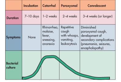
17 isolated from immunocompetent individuals and has been hypothesized to have undergone zoonotic transmission (144, 183). Pathogenesis of a B. pertussis infection The classical presentation of whooping cough or pertussis in infants is traditionally classified into three distinct stages known as the catarrhal, paroxysmal, and convalescent phases (30). After an incubation period of 7-10 days the patient will enter into the catarrhal phase (Fig.2). This period lasts for 1-2 weeks and is associated with mild symptoms similar to the common cold, including rhinorrhea and a mild cough which gradually increases in intensity and frequency as the disease progresses. As the catarrhal stage concludes and paroxysmal phase begins, the cough continues to increase in severity until it is characterized by coughing fits associated with a distinctive whooping sound as the patient makes massive inspirations after the coughing episodes. Symptoms associated with this stage in addition to the paroxysmal cough include vomiting, cyanosis, and leukocytosis. This phase usually persists 1-6 weeks but has been documented to persist up to 10 weeks. Diagnosis of whooping cough generally does not occur until the paroxysmal cough is present, but by this time the bacterial load is decreasing and confirmation via culture methods is often elusive. Upon diagnosis, antibiotics are often prescribed but generally these do not affect the severity or the length of the symptoms due to the fact that pertussis is a toxinmediated disease and by the time of diagnosis the bacterial numbers are already in decline. As the cough diminishes, the patients progress to the convalescent 8
18 Fig. 2. Clinical presentation of B. pertussis disease. B. pertussis infection is initiated by the inhalation of the bacteria, traditionally in aerosol droplets. After an incubation period of 7-10 days the subject enters the catarrhal phase. Symptoms resembling the common cold are generally associated with this stage with no overt signs of serious complications. As the infection enters its second week the paroxysmal stage begins and can last from 2-6 weeks. At this point in the disease the characteristic whooping cough is prominent along with frequent vomiting. The convalescent stage begins as the paroxysmal cough subsides and persists for several weeks. The general weakness of the individual and the immune system often leave the patient susceptible to complications such as a secondary pneumonia. It is important to note that symptom severity does not correlate with bacterial load. This is a common theme for pathogens whose sequela are mediated by toxin accumulation. 9
19 10
20 phase which is generally a time for recovery. However, it is common for complications to develop in this stage due to the general weakness of the individual after struggling with the paroxysmal cough and vomiting. Pneumonia, seizures and encephalopathy are common life threatening complications associated with the phase, as well as increased susceptibility to secondary infections (116). In contrast to the severe presentation of pertussis in infants, adults and adolescents are often asymptomatic or display only mild disease without the characteristic whooping cough (49, 107, 108). The BvgAS Two Component System Bacteria utilize a wide variety of mechanisms to sense their environmental conditions and to respond to their ever changing surroundings. One of the most common mechanisms used to sense these environmental alterations is the two component signal transduction system. These systems are generally composed of a membrane-spanning sensor kinase which processes extracellular stimuli through a phosphorylation cascade or phospho-relay to a response regulator. The sensor protein typically auto-phosphorylates at a conserved histidine residue which is then transferred to an aspartate residue either within the sensor or on the response regulator which alters its DNA binding activity (70). Bordetella control the expression of most of their known virulence genes through an elegant two component system known as the BvgAS signal transduction system. The bvgas locus encodes for a membrane spanning sensor, BvgS, and a DNA binding response regulator, BvgA (37). This locus is 96% conserved between the 11
21 three classical species at the nucleotide level and is functionally interchangeable between the species (114). The BvgAS system belongs to a class of two component systems that varies from the normal paradigm. The phosphorylation cascade undertaken by these proteins is not the traditional two step communication between the sensor and the response regulator. BvgAS signal transduction involves a more complicated four step process in which the sensor kinase, BvgS, is auto-phosphorylated at a histidine residue (H 729) upon sensing changes in certain environmental stimuli. This phosphate is then transferred to an aspartate (D 1023) within BvgS before being shuttled to a second histidine (H 1172) prior to reaching its final destination on an aspartate on the response regulator, BvgA (172). BvgA is a response regulator which is composed of an N- terminal receiver domain and a C-terminal helix-turn-helix DNA binding domain (8, 82). Upon activation by phosphorylation, BvgA regulates a large array of virulence activated genes (vags) by binding to the promoter region of these genes and either displacing a repressor or by recruiting RNA polymerase for transcriptional initiation (Fig. 3) (14, 15). BvgA can not only function as an activator, but is also capable of down regulating virulence repressed genes (vrgs) (Fig 3). This is mainly accomplished when activated BvgA induces the expression of BvgR which in turn acts as a repressor of the vrgs. These genes must be repressed in order for Bordetella to be fully virulent since strains lacking BvgR have been shown to be incapable of causing disease in a host ( ). 12
22 Fig. 3. The BvgAS two component system regulates genes expression patterns and phases in Bordetella. BvgAS is comprised of the membrane spanning sensor kinase, BvgS, and the response regulator BvgA. Upon stimulation, BvgS undergoes a phosphorylation cascade which ends in the phosphorylation of BvgA. This activated protein is capable of regulating the expression of numerous virulence determinants in Bordetella. In fact, the bacteria exist in different virulence phases based on the phosphorylation state of BvgA including Bvg +, Bvg i, and Bvg - phases. These different states correlate with the maximal expression of various virulence factors which are hypothesized to help the bacteria survive in their given environment. In order to colonize a host, the bacteria must be in Bvg + phase which correlates with the peak expression of most adhesins and toxins in Bordetella. 13
23 14
24 Phase Variation in Bordetella One hallmark of the BvgAS system is its ability to control numerous genes in Bordetella in response to relatively few known signals. This two component system alters the expression pattern of so many genes that Bordetella are considered to exist in various phases based on the activation state of BvgAS. As early as 1931, Leslie and Garner observed that Bordetella possessed the ability to change phases and modulate antigen expression in response to environmental signals (104). They noted three distinct phenotypic phases which were later termed Bvg +, Bvg -, and Bvg i. The Bvg + phase correlates with the phosphorylation of BvgA and is characterized by the expression of numerous adhesins and toxins, such as FHA, pertactin, fimbria, adenylate cyclase, and pertussis toxin in B. pertussis (Fig. 3) (116). It is widely accepted that Bordetella must be in Bvg + phase to be virulent and therefore it is the most studied phenotypic state of the organism (126). Under conditions where BvgAS is not active, either by mutation, low temperature growth conditions, or the presence of modulators such as nicotinic acid or magnesium sulfate, the bacteria enter into the avirulent Bvg - phase (Fig. 3) (126). This phase is associated with the lack of expression of traditional virulence factors, but also with expression of motility genes in B. bronchiseptica as well as sets of genes which are hypothesized to help the bacteria survive difficult growth conditions (2, 3). It was once thought that Bordetella existed in these two phases and there was no intermediate or transition state. However, recent studies have revealed that the BvgAS system 15
25 does not function merely as a biphasic switch, but more by a gradient or rheostat mechanism to control the gene expression profile in response to subtle changes in environmental conditions (50, 164). This transitional or Bvg i phase is not only characterized by intermediate expression levels of several virulence factors, but also by maximal expression of several unique virulence factors including the adhesin BipA (164). To date, there has been no distinct role established for the Bvg i phase but it is postulated to be required for aerosol transmission of Bordetella between hosts. Pathogenesis Determinants of Bordetella Virulence Factors. Like most bacterial species, Bordetella produce two main classes of virulence factors, toxins and adhesins. These factors exist in a variety of forms and are not limited to traditional protein components. An intricate interplay of these elements has been shown to be necessary for efficient colonization of the mammalian respiratory tract. Previous studies on Bordetella have shown that various virulence factors are necessary for attachment, invasion, as well as immune evasion or suppression. High immunogenicity is also a common characteristic of many of these antigens as evidenced by the inclusion some of the factors in vaccinations which protect from Bordetella infection. Adhesins. The following is a succinct discussion of some of the wellcharacterized proteins necessary for the efficient attachment and pathogenesis of Bordetella. 16
26 Filamentous hemagglutinin Filamentous hemagglutinin (FHA) is a strongly activated BvgAS regulated protein with its maximal production occurring under Bvg + and Bvg i phase conditions (152). This hairpin-shaped protein is one of most immunogenic virulence factors of B. bronchiseptica and B. pertussis (146). FHA is a 220 kda protein that exists both as a surface attached component, as well as a portion that is secreted into the extracellular environment (39, 40). Abundant research performed in vitro has determined that FHA is an adhesin which has four functional domains that confer its binding ability to both epithelial and immune cells. One of these domains consists of an Arginine-Glycine-Aspartic Acid (RGD) motif which has been shown to mediate adherence to complement receptor 3 (CR3) and leukocyte response integrin/integrin-associated protein found on macrophage/monocytes and leukocytes respectively (86, 145, 154). This RGD domain has also been shown to stimulate binding to bronchial epithelial cells through interactions with very late antigen 5 which can lead to NF-κB signaling and leukocyte accumulation (87-89). A second motif present on this protein is a carbohydrate recognition domain which initiates attachment to macrophages and to the ciliated epithelial cells of the respiratory tract (140). FHA also possesses a heparin binding domain which promotes hemagglutination (122). A final adhesive property of FHA is attributed to a CR3 recognition domain for which a function has yet to be elucidated. Other non-adhesive properties of FHA include its ability inhibit CD4 + T cell proliferation and induce apoptosis in these cells which 17
27 interferes with the capability to mount an adaptive immune response to clear the pathogen (13). Additionally, FHA has been shown to down regulate the innate immune response mounted against the pathogen by dendritic cells and macrophages by suppressing interleukin (IL)-12 production through the induction of IL-10 (118, 120). Research for a function of FHA in vivo has determined that the protein is required for efficient colonization of the trachea in B. bronchiseptica infected rodents (38). However, the role for FHA in B. pertussis infection has yet to be clearly defined due to conflicting reports. McGuirk et al. have shown that a B. pertussis strain harboring a deletion of FHA is defective in its ability to colonize the lungs of infected mice (119). However, Goodwin and Weiss have reported that there is no difference in colonization in mice infected with either the wild type or a strain lacking FHA (179). One possible explanation for these conflicting accounts could be that mice are not the natural host for B. pertussis and thus fail to elucidate a phenotype due to the fact that B. pertussis FHA may be specifically adapted to the human respiratory tract. While B. bronchiseptica and B. pertussis are closely related, genomic comparison of the genes encoding for FHA between the two species does reveal some differences. Conflicting reports have been published as to whether or not FHA of B. bronchiseptica and B. pertussis are functionally interchangeable in vivo. However, it appears FHA is highly adapted to the targeted host of the bacteria and its precise contribution to colonization of heterologous organisms has yet to be definitively determined. 18
28 Fimbriae Many Gram negative pathogens, such as Escherichia coli and Salmonella spp., express filamentous polymeric proteins on their cell surface called fimbriae (Fim) (53, 54). The Bordetella species are capable of expressing four fimbriae, Fim2, Fim3, FimX and FimN (96, 105, 129, 148). These proteins are expressed at varying levels with Fim2 and Fim3 being highly induced under Bvg + phase conditions where as FimX and FimN are produced at much lower levels (96, 148). It has been difficult to ascertain a definitive function for each of the fim genes since they are hypothesized to have overlapping functions between themselves, as well as with other proteins in Bordetella, such as FHA. However, studies have shown that these proteins are capable of mediating adherence to respiratory epithelial cells and host monocytes (75, 76). In vivo, research has implicated Fim as an adhesin necessary for efficient tracheal colonization (117). In addition to these attachment roles, the Fim proteins have been known to down regulate the inflammatory response of the host and protect Bordetella from killing by alveolar macrophages (116). The adaptive immune response recognizes and elicits a significant antibody response to the Fim proteins, especially Fim2/3. These proteins have been shown to induce both an immunoglobulin (Ig) M and IgG2a response in a rat model of infection (117). This major stimulation of the host adaptive response has led to the inclusion of Fim2/3 in currently available acellular vaccines. In fact, addition of Fim2/3 to the acellular vaccine has led to significant enhancement of its overall efficacy (135). 19
29 Pertactin Bordetella express numerous surface associated proteins related to the autotransporter secretion system including the highly Bvg-regulated protein, pertactin (Prn) (78). These autotransporters usually are composed of an N- terminal domain which elicits the effector function of the protein and a C-terminal β-barrel which aids in the transport of the N-terminal domain across the target membrane. Prn is an approximately 69 kda protein which has an RGD motif in the N-terminal effector domain of this protein which has led to the hypothesis that Prn is involved in attachment (27, 58). Purified Prn has been shown to impart binding of CHO cells to tissue culture plates, as well as increasing the attachment of prn expressing Salmonella to target cell lines (59). However, Bordetella strains harboring a deletion of prn show no significant difference in their ability to attach to cell lines in vitro or colonize the mouse respiratory tract (149). Although no pathogenic phenotype has been established for Prn, it is important to note that several studies have documented the necessity of anti-prn antibodies in mediating protection against Bordetella (31, 165). This has led to the inclusion of Prn in many currently available acellular vaccines and has increased their protective efficacy over that of vaccines containing FHA and pertussis toxin alone (28, 72). Toxins. In addition to possessing multiple adhesins, Bordetella produces numerous toxins which contribute to the virulence of the organism. The following 20
30 is a summary of the toxins secreted by the genus and their function in bacterial pathogenesis. Adenylate cyclase CyaA is a toxin secreted by Bordetella which functions as both a calmodulin-sensitive adenylate cyclase and hemolysin. This protein belongs to the repeats-in-toxin (RTX) family of calcium-dependent pore forming toxins (147). The N-terminal domain of CyaA confers the catalytic activity of the toxin while the C-terminal domain is responsible for translocation into the target cell membrane as well as the hemolytic properties of CyaA (64, 79). Unlike other hemolysins, such as HlyA of E. coli, CyaA is not secreted in large quantities into the extracellular milieu (180). In fact, it has been demonstrated that CyaA must be associated with the bacterial cell surface to be functional. This localization with the cell surface has been shown to be dependent on FHA in that a deletion of FHA causes the majority of CyaA to be released into the surrounding environment; however, the mechanism of this interaction has yet to be determined (188). CD11b, a eukaryotic cell surface glycoprotein expressed by various immune cells including, natural killer cells, macrophages, and neutrophils, has been determined to be the receptor for CyaA (69). Upon interaction with CD11b, CyaA is internalized into the mammalian cell where binding with calmodulin activates the adenylate cyclase properties of the toxin, catalyzing the conversion of cellular ATP to cyclic AMP (camp) (182). This leads to the accumulation of camp, which exceeds normal physiological conditions and 21
31 interferes with normal cellular signaling or processes (24, 33). Overproduction of camp inside many cells of the immune system leads to a reduction of their ability to take up and kill bacteria. More specifically, purified CyaA has been shown to inhibit super oxide production, chemotaxis of monocytes and neutrophils, and induce apoptosis in macrophages (33). In addition to these effects, CyaA is capable of suppressing IL-12 and tumor necrosis factor (TNF)α production from bone marrow derived dendritic cells suggesting an anti-inflammatory role for the toxin (159). In accordance with these in vitro studies, infection of infant mice with a strain incapable of expressing CyaA displayed a defect in causing lethal infection (68, 71). These studies demonstrate that CyaA plays an anti-phagocytic and anti-inflammatory role in Bordetella respiratory infections. While CyaA is not a component on the current acellular vaccine, anti- CyaA antibodies have been found in children who have developed a B. pertussis infection (32). In fact, anti-cyaa antibodies are capable of promoting phagocytosis of B. pertussis by neutrophils implying a function for these antibodies in natural infection leading to suggestions that inclusion of CyaA in acellular vaccines may induce a more complete protection (73, 116). Dermonecrotic toxin Dermonecrotic toxin (DNT) was one of the first virulence factors to be discovered for B. pertussis. This heat-labile A-B toxin is composed of a 54 amino acid N-terminal receptor binding domain and a C-terminal enzymatic domain composed of 300 amino acids (115). Although the receptor for DNT has yet to be 22
32 identified, it is know that DNT is internalized through dynamin-dependent endocytosis and is activated via proteolytic nicking by eukaryotic proteases as it enter the cell (115). This activated form of DNT is thought to act on the small GTP-binding protein Rho which leads to a signaling cascade that alters cytoskeletal arrangement, DNA replication, and cell movement (81). Currently a defined role for DNT in Bordetella pathogenesis is unknown, but it has been linked to necrotic lesions in experimentally infected animals and turbinate atrophy in pigs colonized by B. bronchiseptica (111, 150). Tracheal Cytotoxin Unlike most toxins, which are protein based, tracheal cytotoxin (TCT) is composed of a disaccharide-tetrapeptide monomer of the peptidoglycan cell wall produced by all Gram negative bacteria (35). In contrast to other Gram negative species which recycle this peptidoglycan fragment, Bordetella release this monomer into the surrounding environment (92). This secretion of TCT is due to the lack of a functional AmpG homologue in Bordetella species which is responsible for the recycling of this peptidoglycan component in most bacteria (35, 93, 151). TCT is capable of causing cilia destruction, cell blebbing, and damaging cell mitochondria (181). This toxin targets only ciliated cells and appears to leave non-ciliated cells undamaged (65). In a tissue culture model, the cytopathic effects of TCT are mediated by its ability to induce IL-1α which increases the production of nitric oxide (NO) and in turn destroys the cilia in the surrounding area (60, 77). In vivo, it is hypothesized that TCT stimulates IL-1α 23
33 production and NO discharge by non-ciliated mucus secreting cells resulting in cilia destruction on the surrounding cells (60). Pertussis Toxin Unlike the aforementioned toxins produced by all Bordetella species which infect mammals, pertussis toxin (PT) is expressed only by the human pathogen B. pertussis. Although both B. bronchiseptica and B. parapertussis harbor the genes necessary to synthesize pertussis toxin, because of promoter mutations, the loci transcriptionally inactive (4). PT is an ADP-ribosylating toxin composed of six polypeptides, S1-S5, encoded by the genes ptxa-ptxe (106, 133). This AB toxin functions by the B subunits, S2 to S5, forming a ring-like structure which binds to the eukaryotic cell membrane and causes the cell to intake PT via a cytochalasin D independent uptake pathway (168, 185, 186). Once inside the eukaryotic cell, ATP binds to the B subunits causing a release of the enzymatically active A subunit, S1 (98). Upon entry into the eukaryotic cytosol, the S1 subunit transfers ADP ribose to Guanine (G) binding proteins (99). This ADP ribosylation of the cell G proteins causes a disruption of cell signaling and ion channel function (83, 157). Recent studies have shown that PT is involved in down-regulating the immune response to B. pertussis by inhibiting chemotaxis, decreasing the production of reactive oxygen species, and altering lysosome fusion in human neutrophils and macrophages (17, 121). Studies have also shown that strains lacking PT have an increased anti-bordetella antibody response suggesting that the toxin is involved in suppressing or evading the 24
34 adaptive immune response (25). In addition to its role as an immunomodulatory component, PT has been shown to act as an adhesin to the ciliated cells of the mammalian respiratory tract as well as to macrophages (171). In light of this dual role in pathogenesis PT has been proposed to be the major virulence factor responsible for the typical disease symptoms associated with whooping cough and is included in currently available acellular vaccines. Lipopolysaccharide Similar to other Gram negative species, Bordetellae produce endotoxin or lipopolysaccharide (LPS) as a component of their outer membrane. LPS is known to be pyrogenic, toxic, and capable of inducing TNF expression in macrophages (5). Bordetella LPS is composed of three distinct components, band A, band B, and the O antigen. The band A and B portions of LPS are named due to their different mobilities on sodium dodecyl sulfate polyacrylamide gels, but are similar in composition (138). The lower molecular weight band B is comprised of a lipid A molecule which is covalently linked to a branched oligosaccharide core. The higher molecular weight band A consists of band B plus an additional trisaccharide (26). B. bronchiseptica and B. parapertussis also contain an O antigen made of a repeating sugar polymers linked to band A and B (51). The loci wlb and wbm encode for the enzymes necessary for the synthesis of LPS in Bordetella. However, B. pertussis does not contain an intact wbm locus and therefore does not produce the enzymes necessary to construct a functional O antigen (22, 142). A definitive role in pathogenesis has yet to be determined 25
35 for LPS among Bordetella species, but strains deficient in LPS production have a defect in colonization of the murine respiratory tract (74). Recent studies have also suggested the LPS is involved in resistance to complement mediated killing of B. bronchiseptica and protects B. pertussis from the antimicrobial agent surfactant protein A in the mouse lung (155). Biofilm development When studied in laboratory conditions, bacteria are traditionally grown in liquid culture and exist mainly as free swimming individual cells. However, recent research has demonstrated that many bacteria do not always exist in this planktonic state, but are often attached to a surface or clustered together in large conglomerates (62). These microbial communities have not only been observed in the environment, but are also routinely found in the host organism. The first documented observation of these attached bacterial groups occurred in 1933 by Henrici when he noticed that a significant portion of his cultures grew attached to the walls of his culture system and not in the liquid media (189). As technology and research progressed, many studies have been performed using microscopy and transcriptome analysis to determine that the microbial communities adhered to a surface are distinctly different from their free swimming counterparts (103). The structures eventually came to be known as biofilms and are defined as a community of organisms attached to a surface and encased in a self produced polymeric matrix. This matrix can be composed of exopolysaccharides, various proteins, as well as extracellular DNA. 26
36 Biofilms are organized structures that develop in an orderly process which can be divided into various steps or phases. Biofilm formation begins when a free swimming cell comes into contact with a surface and undergoes an initial reversible attachment (Fig. 4). Upon initial adherence, the cell is not committed to form a biofilm and may detach from the surface to rejoin the planktonic population. This temporary surface association is often mediated by flagella initiated contact which allows for the cell pole to interact with the solid media (1). However, favorable conditions can allow the bacteria to progress past this transient attachment step to an irreversible attachment which commits the cell to biofilm formation (Fig. 4). This permanent adherence is associated with the cell initiating a more lateral contact with the surface and the expression of extracellular polymers which are often either not present when the cells exhibit planktonic growth or are present at lower levels than in the biofilm. One example of this is the PGA exopolysaccharide of E. coli which has been shown to be crucial for irreversible attachment, but does not alter the cell s ability to form reversible interactions (1). Studies involving Pseudomonas aeruginosa have demonstrated that as the bacteria make this irreversible attachment, the gene expression profile of the organism changes and genes involved in matrix production are up-regulated, such as exopolysaccharide synthesis genes (46). Once adherence to a surface has been established, the biofilm can increase in size by two mechanisms, clonal expansion and cells migrating together. Translocation is accomplished via twitching motility mediated by the type IV pilus. By utilizing this motility across a surface, bacteria are capable of 27
37 clustering together to construct a growing biofilm (100). A second method of biofilm maturation is clonal expansion of the attached cells to create a monolayer covering a local area. Upon saturation of this monolayer, bacteria then expand vertically and produce what are known as microcolonies (Fig. 4). This cell conglomeration can lead to dramatic three dimensional structures such as towers, water channels, or mushroom like stalks (134). As the biofilm matures in size copious amounts of matrix material is produced to encase the bacteria (18, 19). The final stage of biofilm development is the release of cells from the biofilm into the surrounding environment (Fig. 4). Dispersal from the bacterial biofilm is mediated through the expression of enzymes capable of degrading the surrounding matrix material. For example, upon biofilm maturation, P aeruginosa overexpresses the enzyme alginate lyase which breaks down the exopolysaccharide alginate into short chains allowing for bacterial release from the biofilm (16). Actinobacillus species achieve dispersal through a similar manner by producing the glycosyl hydrolase DspB which cleaves β-1-6 linked N- acetylglucosamine in the surrounding matrix encasing the bacteria (97). The bacteria released from the biofilm by dispersion have a gene expression profile similar to that of planktonic cells and behave in a comparable manner (153). This release of bacteria into the surrounding environment allows for the seeding of new sites of infection and is the last stage of the biofilm cycle. 28
38 Fig.4. Biofilm formation model. Biofilm formation is initiated when planktonic cells come into contact with a sold surface and initiate a reversible attachment. If the bacteria remain adhered to the surface, they commence a change in gene expression resulting in the down regulation of motility genes and an increased production of factors necessary for irreversible attachment. After initial attachment and monolayer formation the biofilm increases in size by either clonal expansion or translocation to form microcolony structures. As the biofilm matures, large amounts of matrix material is produced and the biofilm takes on a three dimensional architecture complete with tower and water channel formation. The final hallmark of biofilm development is the dispersal of bacteria from the structure to seed other areas in the environment. 29

39 30
40 Polysaccharides As discussed above, exopolysaccharides are a critical component of the biofilm matrix. These extracellular polysaccharides contribute in a variety of ways toward biofilm formation, including initial attachment to a surface, intercellular adhesion, and providing a scaffolding for the three dimensional nature of the biofilm (44, 110, 162). Polysaccharides have also been demonstrated to perform functions beyond that of a structural component including protecting the bacteria from antimicrobial peptides, phagocytosis, and antibody mediated clearance (175). The composition of these extracellular polysaccharides varies greatly between bacterial species. For example, E. coli produces at least two polysaccharides which are involved in biofilm formation. The first discovered was colanic acid which is a polymer composed of glucuronate, fructose, glucose, and galactose (163). More recently E. coli has been shown to produce the exopolysaccharide PGA, which is made of N-acetylglucosamine, mannose, glucose, and galactose (177). P. aeruginosa produces three exopolysaccharides Psl, Pel, and alginate which have varying compositions and roles in biofilm formation (23, 91, 156). Although there is great diversity in the exopolysaccharides made by biofilm forming species, many species produce similar structured polysaccharides such as the poly-β-1-6 N-acetylglucosamine (PGA/PIA/PNAG) which is conserved in a number of Gram positive and Gram negative species (41, 110, 136, 177, 187). Staphylococcal species produce one such polysaccharide entitled PIA or PNAG which has been shown to be crucial for biofilm formation and surface adherence. The enzymes necessary for the 31
41 construction of PIA/PNAG are encoded by the icaadbc locus (41, 110, 113). Similar gene loci and polysaccharides have been documented in multiple other species including, pga in E. coli and Actinobacillus, hms in Yersinia, and bps in B. bronchiseptica (136, 139, 177). B. pertussis contains homologues to the bpsabcd locus in B. bronchiseptica and is hypothesized to express a similarly structured Bps polysaccharide. Bps production and its function in B. pertussis pathogenesis is discussed in detail in chapter 2. Traditionally these exopolysaccharides are studied in the context of biofilm formation, yet many have shown to have pathogenic effects exceeding that of forming multicellular communities. The PIA/PNAG of Staphylococcal species has been demonstrated to not only function as an adhesin and biofilm maintenance factor, but also to protect the bacteria from phagocytic uptake, killing by antimicrobial peptides produced by the host, and provide resistance to various antibiotics in vitro (7, 175). An adaptive immune response to PIA/PNAG is capable of providing protection from subsequent lethal challenge demonstrating that this polysaccharide is an important antigen recognized by the immune system (101, 113). It is also interesting to note that clinical isolates are more likely to express PIA/PNAG than Staphylococcus isolated from an environmental source (101, 130). Polysaccharide and Biofilm Regulation Exopolysaccharides have been implicated to function as virulence factors necessary for initial attachment, cell aggregation, biofilm development and 32
42 maintenance, as well as modifying the host immune response to the organism. Given these undeniable roles for polysaccharides in colonization and virulence, it can be anticipated that the expression of these molecules is intricately controlled. One extensively studied example of the regulation of biofilm development and polysaccharide production is the ica locus of S. aureus. Under planktonic growth conditions, transcription of icaadbc occurs at low levels due to the repressor activities of the proteins IcaR, TcaR, and LuxS (34, 95, 184). The divergently transcribed icar gene is located upstream of the ica locus and encodes for a TetR family transcriptional repressor which inhibits transcription unless certain environmental cues, such as ethanol and high salt concentrations, are present (34). In addition to IcaR, the MarR family regulator TcaR also binds to the ica promoter and functions as a weak repressor of transcription (95). A recent report also identified LuxS as a repressor of PNAG synthesis, but the exact mechanism of repression by this quorum sensing system is still being investigated (102). As the local environment changes to favor Staphylococcal biofilm formation, the binding of these repressors to the ica promoter is inhibited and transcription is initiated. In addition to repressors, several other factors which increase the transcription efficacy of this locus have been identified. Under high salt growth conditions, an alternate sigma factor, sigb, is induced which regulates the expression of the DNA binding protein SarA (173). SarA has been found to bind to the ica promoter and lead to an increase in the production of PNAG/PIA (170). Decreased transcription of icaadbc has also been demonstrated in purr deletion strains, however, the mechanism of how PurR functions as an activator 33
43 of this locus has yet to be determined since there are no recognizable binding sites for PurR on the ica promoter (109). As exemplified in Staphylococcus Spp., polysaccharide regulation can often be complex and multifactorial. This intricate paradigm is true of the regulation of biofilm associated exopolysaccharides in other species such as alginate of Pseudomonas, VPS of Vibrio, and PGA of E. coli (11, 66, 143, 176). Similar to these polysaccharides, we have found that Bps is expressed at a higher level in the biofilm and is regulated by a repressor protein, BpsR. This is described in chapter 3 of this manuscript. Biofilms in Human Infections It has been estimated that biofilms are associated with 65-80% of all human bacterial infections and cost billions of dollar every year in their prevention and eradication (112). Biofilms have been linked to numerous infections including heart valve endocarditis, otitis media, tonsillitis, dental carries, chronic lung infections associated with cystic fibrosis and chronic obstructive pulmonary disorder, urinary tract infections, and various gastrointestinal infections (94). Treatment strategies developed to combat most infections are designed to target free swimming metabolically active cells, and are often ineffective against biofilms. Therefore, current research targeting methods to kill, disrupt, or prevent biofilms inside the host are vital to combating this plethora of biofilm related diseases. 34
44 Biofilms and Persistent Infections As mentioned above, biofilms are linked to a variety of diseases in humans, many of which can be chronic in nature. Perhaps the most extensively studied example of this persistence in a host is the colonization of the cystic fibrosis lung by P. aeruginosa. Long term presence of the bacteria in the lung eventually leads to compromised lung function and eventual respiratory failure (131). Immunohistopathological samples of lung tissue removed from cystic fibrosis patients has repeatedly revealed the presence of P. aeruginosa aggregates encased in a matrix-like material (6). Multicellular bacterial communities have also been observed via transmission electron microscopy. In addition, quorum sensing molecules, a hallmark of mature biofilm formation, are often recovered from the sputum of individuals afflicted with cystic fibrosis (158). Taken together, this data suggests that P. aeruginosa displays a biofilm mode of existence in the cystic fibrosis lung which contributes to the chronic establishment of the pathogen. Another example of persistent infections associated with biofilm formation is the prolonged colonization of implanted medical devices, particularly by Staphylococcus. The Staphylococcal species have a unique ability to efficiently adhere to abiotic surfaces, including the metals and plastics used in implanted medical equipment such as catheters, artificial heart valves and joints, as well as others devices (36, 45, 137). Treatment of these infections is often complicated due to the recalcitrance of the biofilm to antibiotics, and the restricted ability of the immune system to access the site because of the abiotic nature of the device 35
45 (52). This resistance to clearance often results in the removal of the implanted device due to damage of host tissue as a result of the infection or impared function of the object due to biofilm accumulation (45). Upon removal of the medical device Staphylococci are frequently observed attached to the apparatus and encased in thick exopolysaccharide matrix suggesting that the biofilm mode of growth has allowed for the persistance of the bacteria on the surface (61). Multiple other pathogens have either been shown or hypothesized to form biofilms inside humans, including Streptococcus pneumoniae, Haemophilus influenzae, Candida albicans, and Escherchia coli (9, 132, 178). It has been postulated that a similar lifecycle could exist for Bordetella given that the bacteria can cause persistent symptoms lasting up to 100 days as well as asymptomatic infections in vaccinated individuals (116). Bordetella Biofilms While the Bordetella biofilm field is still in its infancy compared to some other pathogens like S. aureus and P. aeruginosa, several studies have revealed key components necessary for biofilm formation. Our lab and others have determined that both B. bronchiseptica and B. pertussis are capable of forming biofilms in vitro and that this phenotype is dependent on the BvgAS system (85, 127). Strains grown under modulating conditions or locked in the Bvg - phase are incapable of forming a biofilm and do not progress past the monolayer or reversible attachment phase of development (127). However, when the bacteria are grown under Bvg + or Bvg i phase conditions, Bordetella are capable of 36
46 forming robust mature biofilms on a variety of surfaces (85, 127). This suggests that the BvgAS system regulates one or multiple factors that are crucial for the development of structured biofilms. In accordance with this finding, one study has shown that both FHA and Fim production positively influence biofilm formation (85). However, deletion of cyaa, a Bvg + phase gene, results in an increased ability to form biofilms when compared to the wild-type, suggesting that CyaA interferes with maximal biofilm development (85). As mentioned above, Bordetella species are capable of expressing the Bps polysaccharide that is antigenically similar to poly-β-1-6 N- acetylglucosamine family of polysaccharides. To date the function of Bps has only been examined in animal pathogen B. bronchiseptica. Our laboratory has determined that the enzymes necessary for its synthesis are encoded by the bpsabcd locus (136). Bps was found to be crucial for biofilm formation in vitro on glass surfaces under both static and dynamic growth conditions (136). This phenotype translated to in vivo experiments where a bps strain was found to form less structured biofilms than the wild-type and shown to have a persistence defect in the mouse respiratory tract at extended time points of colonization (160). This suggests that Bps production and biofilm formation is necessary for the long term carriage of B. bronchiseptica in the mouse respiratory tract. While B. pertussis and B. bronchiseptica are closely related and share large similarities in their genomes, it is important to note that B. pertussis has evolved from B. bronchiseptica through genome decay and has lost or inactivated many potential virulence factors. In light of this, B. pertussis has 37
47 retained the bps locus which shares 98% homology to B. bronchiseptica at the nucleotide level. However, retention of these genes does not mean the exact function of the locus is conserved. It is conceivable that Bps could interact with other gene products in B. bronchiseptica that are missing in B. pertussis thereby changing its function or revealing a phenotype not seen in the other species. The function of Bps in B. pertussis pathogenesis is investigated in chapter 2 of this manuscript. 38
48 References 1. Agladze, K., X. Wang, and T. Romeo Spatial periodicity of Escherichia coli K-12 biofilm microstructure initiates during a reversible, polar attachment phase of development and requires the polysaccharide adhesin PGA. J Bacteriol 187: Akerley, B. J., and J. F. Miller Flagellin gene transcription in Bordetella bronchiseptica is regulated by the BvgAS virulence control system. J Bacteriol 175: Akerley, B. J., D. M. Monack, S. Falkow, and J. F. Miller The bvgas locus negatively controls motility and synthesis of flagella in Bordetella bronchiseptica. J Bacteriol 174: Arico, B., and R. Rappuoli Bordetella parapertussis and Bordetella bronchiseptica contain transcriptionally silent pertussis toxin genes. J Bacteriol 169: Ayme, G., M. Caroff, R. Chaby, N. Haeffner-Cavaillon, A. Le Dur, M. Moreau, M. Muset, M. C. Mynard, M. Roumiantzeff, D. Schulz, and L. Szabo Biological activities of fragments derived from Bordetella pertussis endotoxin: isolation of a nontoxic, Shwartzman-negative lipid A possessing high adjuvant properties. Infect Immun 27: Baltimore, R. S., C. D. Christie, and G. J. Smith Immunohistopathologic localization of Pseudomonas aeruginosa in lungs from patients with cystic fibrosis. Implications for the pathogenesis of progressive lung deterioration. Am Rev Respir Dis 140: Begun, J., J. M. Gaiani, H. Rohde, D. Mack, S. B. Calderwood, F. M. Ausubel, and C. D. Sifri Staphylococcal biofilm exopolysaccharide protects against Caenorhabditis elegans immune defenses. PLoS Pathog 3:e Beier, D., and R. Gross Regulation of bacterial virulence by twocomponent systems. Curr Opin Microbiol 9: Beloin, C., A. Roux, and J. M. Ghigo Escherichia coli biofilms. Curr Top Microbiol Immunol 322: Bergfors, E., B. Trollfors, J. Taranger, T. Lagergard, V. Sundh, and G. Zackrisson Parapertussis and pertussis: differences and similarities in incidence, clinical course, and antibody responses. Int J Infect Dis 3: Beyhan, S., K. Bilecen, S. R. Salama, C. Casper-Lindley, and F. H. Yildiz Regulation of rugosity and biofilm formation in Vibrio cholerae: 39
49 comparison of VpsT and VpsR regulons and epistasis analysis of vpst, vpsr, and hapr. J Bacteriol 189: Bjornstad, O. N., and E. T. Harvill Evolution and emergence of Bordetella in humans. Trends Microbiol 13: Boschwitz, J. S., J. W. Batanghari, H. Kedem, and D. A. Relman Bordetella pertussis infection of human monocytes inhibits antigen-dependent CD4 T cell proliferation. J Infect Dis 176: Boucher, P. E., A. E. Maris, M. S. Yang, and S. Stibitz The response regulator BvgA and RNA polymerase alpha subunit C-terminal domain bind simultaneously to different faces of the same segment of promoter DNA. Mol Cell 11: Boucher, P. E., K. Murakami, A. Ishihama, and S. Stibitz Nature of DNA binding and RNA polymerase interaction of the Bordetella pertussis BvgA transcriptional activator at the fha promoter. J Bacteriol 179: Boyd, A., and A. M. Chakrabarty Role of alginate lyase in cell detachment of Pseudomonas aeruginosa. Appl Environ Microbiol 60: Bradford, P. G., and R. P. Rubin Pertussis toxin inhibits chemotactic factor-induced phospholipase C stimulation and lysosomal enzyme secretion in rabbit neutrophils. FEBS Lett 183: Branda, S. S., F. Chu, D. B. Kearns, R. Losick, and R. Kolter A major protein component of the Bacillus subtilis biofilm matrix. Mol Microbiol 59: Branda, S. S., S. Vik, L. Friedman, and R. Kolter Biofilms: the matrix revisited. Trends Microbiol 13: Brinig, M. M., K. B. Register, M. R. Ackermann, and D. A. Relman Genomic features of Bordetella parapertussis clades with distinct host species specificity. Genome Biol 7:R Buonavoglia, C., and V. Martella Canine respiratory viruses. Vet Res 38: Burns, V. C., E. J. Pishko, A. Preston, D. J. Maskell, and E. T. Harvill Role of Bordetella O antigen in respiratory tract infection. Infect Immun 71: Byrd, M. S., I. Sadovskaya, E. Vinogradov, H. Lu, A. B. Sprinkle, S. H. Richardson, L. Ma, B. Ralston, M. R. Parsek, E. M. Anderson, J. S. Lam, 40
50 and D. J. Wozniak Genetic and biochemical analyses of the Pseudomonas aeruginosa Psl exopolysaccharide reveal overlapping roles for polysaccharide synthesis enzymes in Psl and LPS production. Mol Microbiol 73: Cahill, E. S., D. T. O'Hagan, L. Illum, and K. Redhead Mice are protected against Bordetella pertussis infection by intra-nasal immunization with filamentous haemagglutinin. FEMS Microbiol Lett 107: Carbonetti, N. H Immunomodulation in the pathogenesis of Bordetella pertussis infection and disease. Curr Opin Pharmacol 7: Caroff, M., R. Chaby, D. Karibian, J. Perry, C. Deprun, and L. Szabo Variations in the carbohydrate regions of Bordetella pertussis lipopolysaccharides: electrophoretic, serological, and structural features. J Bacteriol 172: Charles, I. G., G. Dougan, D. Pickard, S. Chatfield, M. Smith, P. Novotny, P. Morrissey, and N. F. Fairweather Molecular cloning and characterization of protective outer membrane protein P.69 from Bordetella pertussis. Proc Natl Acad Sci U S A 86: Cherry, J. D Comparative efficacy of acellular pertussis vaccines: an analysis of recent trials. Pediatr Infect Dis J 16:S Cherry, J. D Epidemiology of pertussis. Pediatr Infect Dis J 25: Cherry, J. D Pertussis in the preantibiotic and prevaccine era, with emphasis on adult pertussis. Clin Infect Dis 28 Suppl 2:S Cherry, J. D., J. Gornbein, U. Heininger, and K. Stehr A search for serologic correlates of immunity to Bordetella pertussis cough illnesses. Vaccine 16: Cherry, J. D., D. X. Xing, P. Newland, K. Patel, U. Heininger, and M. J. Corbel Determination of serum antibody to Bordetella pertussis adenylate cyclase toxin in vaccinated and unvaccinated children and in children and adults with pertussis. Clin Infect Dis 38: Confer, D. L., and J. W. Eaton Phagocyte impotence caused by an invasive bacterial adenylate cyclase. Science 217: Conlon, K. M., H. Humphreys, and J. P. O'Gara icar encodes a transcriptional repressor involved in environmental regulation of ica operon expression and biofilm formation in Staphylococcus epidermidis. J Bacteriol 184:
51 35. Cookson, B. T., A. N. Tyler, and W. E. Goldman Primary structure of the peptidoglycan-derived tracheal cytotoxin of Bordetella pertussis. Biochemistry 28: Costerton, J. W., L. Montanaro, and C. R. Arciola Biofilm in implant infections: its production and regulation. Int J Artif Organs 28: Cotter, P. A., and A. M. Jones Phosphorelay control of virulence gene expression in Bordetella. Trends Microbiol 11: Cotter, P. A., M. H. Yuk, S. Mattoo, B. J. Akerley, J. Boschwitz, D. A. Relman, and J. F. Miller Filamentous hemagglutinin of Bordetella bronchiseptica is required for efficient establishment of tracheal colonization. Infect Immun 66: Coutte, L., R. Antoine, H. Drobecq, C. Locht, and F. Jacob-Dubuisson Subtilisin-like autotransporter serves as maturation protease in a bacterial secretion pathway. EMBO J 20: Coutte, L., E. Willery, R. Antoine, H. Drobecq, C. Locht, and F. Jacob- Dubuisson Surface anchoring of bacterial subtilisin important for maturation function. Mol Microbiol 49: Cramton, S. E., C. Gerke, N. F. Schnell, W. W. Nichols, and F. Gotz The intercellular adhesion (ica) locus is present in Staphylococcus aureus and is required for biofilm formation. Infect Immun 67: Crowcroft, N. S., C. Stein, P. Duclos, and M. Birmingham How best to estimate the global burden of pertussis? Lancet Infect Dis 3: Cummings, C. A., M. M. Brinig, P. W. Lepp, S. van de Pas, and D. A. Relman Bordetella species are distinguished by patterns of substantial gene loss and host adaptation. J Bacteriol 186: Danese, P. N., L. A. Pratt, and R. Kolter Exopolysaccharide production is required for development of Escherichia coli K-12 biofilm architecture. J Bacteriol 182: Darouiche, R. O Treatment of infections associated with surgical implants. N Engl J Med 350: Davies, D. G., A. M. Chakrabarty, and G. G. Geesey Exopolysaccharide production in biofilms: substratum activation of alginate gene expression by Pseudomonas aeruginosa. Appl Environ Microbiol 59:
52 47. de Greeff, S. C., F. R. Mooi, A. Westerhof, J. M. Verbakel, M. F. Peeters, C. J. Heuvelman, D. W. Notermans, L. H. Elvers, J. F. Schellekens, and H. E. de Melker. Pertussis disease burden in the household: how to protect young infants. Clin Infect Dis 50: De Jesus-Berrios, Y., S. Gonzalez, and M. Sante Respiratory pathogens in bronchoalveolar lavage in a Puerto Rican population infected with the human immunodeficiency virus. P R Health Sci J 24: De Serres, G., R. Shadmani, B. Duval, N. Boulianne, P. Dery, M. Douville Fradet, L. Rochette, and S. A. Halperin Morbidity of pertussis in adolescents and adults. J Infect Dis 182: Deora, R., H. J. Bootsma, J. F. Miller, and P. A. Cotter Diversity in the Bordetella virulence regulon: transcriptional control of a Bvg-intermediate phase gene. Mol Microbiol 40: Di Fabio, J. L., M. Caroff, D. Karibian, J. C. Richards, and M. B. Perry Characterization of the common antigenic lipopolysaccharide O-chains produced by Bordetella bronchiseptica and Bordetella parapertussis. FEMS Microbiol Lett 76: Donlan, R. M Role of biofilms in antimicrobial resistance. ASAIO J 46:S Duguid, J. P., E. S. Anderson, and I. Campbell Fimbriae and adhesive properties in Salmonellae. J Pathol Bacteriol 92: Duguid, J. P., I. W. Smith, G. Dempster, and P. N. Edmunds Nonflagellar filamentous appendages (fimbriae) and haemagglutinating activity in Bacterium coli. J Pathol Bacteriol 70: Dworkin, M. S., P. S. Sullivan, S. E. Buskin, R. D. Harrington, J. Olliffe, R. D. MacArthur, and C. E. Lopez Bordetella bronchiseptica infection in human immunodeficiency virus-infected patients. Clin Infect Dis 28: Edwards, K. M Overview of pertussis: focus on epidemiology, sources of infection, and long term protection after infant vaccination. Pediatr Infect Dis J 24:S Eldering, G., and P. L. Kendrick Incidence of parapertussis in the Grand Rapids area as indicated by 16 years' experience with diagnostic cultures. Am J Public Health Nations Health 42:
53 58. Emsley, P., G. McDermott, I. G. Charles, N. F. Fairweather, and N. W. Isaacs Crystallographic characterization of pertactin, a membraneassociated protein from Bordetella pertussis. J Mol Biol 235: Everest, P., J. Li, G. Douce, I. Charles, J. De Azavedo, S. Chatfield, G. Dougan, and M. Roberts Role of the Bordetella pertussis P.69/pertactin protein and the P.69/pertactin RGD motif in the adherence to and invasion of mammalian cells. Microbiology 142 ( Pt 11): Flak, T. A., and W. E. Goldman Autotoxicity of nitric oxide in airway disease. Am J Respir Crit Care Med 154:S Gad, G. F., M. A. El-Feky, M. S. El-Rehewy, M. A. Hassan, H. Abolella, and R. M. El-Baky Detection of icaa, icad genes and biofilm production by Staphylococcus aureus and Staphylococcus epidermidis isolated from urinary tract catheterized patients. J Infect Dev Ctries 3: Geesey, G. G., W. T. Richardson, H. G. Yeomans, R. T. Irvin, and J. W. Costerton Microscopic examination of natural sessile bacterial populations from an alpine stream. Can J Microbiol 23: Gilberg, S., E. Njamkepo, I. P. Du Chatelet, H. Partouche, P. Gueirard, C. Ghasarossian, M. Schlumberger, and N. Guiso Evidence of Bordetella pertussis infection in adults presenting with persistent cough in a french area with very high whole-cell vaccine coverage. J Infect Dis 186: Glaser, P., D. Ladant, O. Sezer, F. Pichot, A. Ullmann, and A. Danchin The calmodulin-sensitive adenylate cyclase of Bordetella pertussis: cloning and expression in Escherichia coli. Mol Microbiol 2: Goldman, W. E., D. G. Klapper, and J. B. Baseman Detection, isolation, and analysis of a released Bordetella pertussis product toxic to cultured tracheal cells. Infect Immun 36: Goller, C., X. Wang, Y. Itoh, and T. Romeo The cation-responsive protein NhaR of Escherichia coli activates pgaabcd transcription, required for production of the biofilm adhesin poly-beta-1,6-n-acetyl-d-glucosamine. J Bacteriol 188: Goodnow, R. A Biology of Bordetella bronchiseptica. Microbiol Rev 44: Goodwin, M. S., and A. A. Weiss Adenylate cyclase toxin is critical for colonization and pertussis toxin is critical for lethal infection by Bordetella pertussis in infant mice. Infect Immun 58:
54 69. Gray, M. C., G. M. Donato, F. R. Jones, T. Kim, and E. L. Hewlett Newly secreted adenylate cyclase toxin is responsible for intoxication of target cells by Bordetella pertussis. Mol Microbiol 53: Gross, R., B. Arico, and R. Rappuoli Families of bacterial signaltransducing proteins. Mol Microbiol 3: Gueirard, P., and N. Guiso Virulence of Bordetella bronchiseptica: role of adenylate cyclase-hemolysin. Infect Immun 61: Gustafsson, L., H. O. Hallander, P. Olin, E. Reizenstein, and J. Storsaeter A controlled trial of a two-component acellular, a five-component acellular, and a whole-cell pertussis vaccine. N Engl J Med 334: Harvill, E. T., P. A. Cotter, M. H. Yuk, and J. F. Miller Probing the function of Bordetella bronchiseptica adenylate cyclase toxin by manipulating host immunity. Infect Immun 67: Harvill, E. T., A. Preston, P. A. Cotter, A. G. Allen, D. J. Maskell, and J. F. Miller Multiple roles for Bordetella lipopolysaccharide molecules during respiratory tract infection. Infect Immun 68: Hazenbos, W. L., C. A. Geuijen, B. M. van den Berg, F. R. Mooi, and R. van Furth Bordetella pertussis fimbriae bind to human monocytes via the minor fimbrial subunit FimD. J Infect Dis 171: Hazenbos, W. L., B. M. van den Berg, C. W. Geuijen, F. R. Mooi, and R. van Furth Binding of FimD on Bordetella pertussis to very late antigen-5 on monocytes activates complement receptor type 3 via protein tyrosine kinases. J Immunol 155: Heiss, L. N., S. A. Moser, E. R. Unanue, and W. E. Goldman Interleukin-1 is linked to the respiratory epithelial cytopathology of pertussis. Infect Immun 61: Henderson, I. R., and J. P. Nataro Virulence functions of autotransporter proteins. Infect Immun 69: Hewlett, E. L., V. M. Gordon, J. D. McCaffery, W. M. Sutherland, and M. C. Gray Adenylate cyclase toxin from Bordetella pertussis. Identification and purification of the holotoxin molecule. J Biol Chem 264: Hoppe, J. E Update on respiratory infection caused by Bordetella parapertussis. Pediatr Infect Dis J 18: Horiguchi, Y., T. Senda, N. Sugimoto, J. Katahira, and M. Matsuda Bordetella bronchiseptica dermonecrotizing toxin stimulates assembly of actin 45
55 stress fibers and focal adhesions by modifying the small GTP-binding protein rho. J Cell Sci 108 ( Pt 10): Horvat, A., and R. Gross Molecular characterization of the BvgA response regulator of Bordetella holmesii. Microbiol Res 164: Hoshino, S., S. Kikkawa, K. Takahashi, H. Itoh, Y. Kaziro, H. Kawasaki, K. Suzuki, T. Katada, and M. Ui Identification of sites for alkylation by N- ethylmaleimide and pertussis toxin-catalyzed ADP-ribosylation on GTP-binding proteins. FEBS Lett 276: Hu, J. J., C. Y. Lu, L. Y. Chang, C. H. Huang, C. C. Chou, F. Y. Huang, C. Y. Lee, and L. M. Huang Survey of pertussis in patients with prolonged cough. J Microbiol Immunol Infect 39: Irie, Y., S. Mattoo, and M. H. Yuk The Bvg virulence control system regulates biofilm formation in Bordetella bronchiseptica. J Bacteriol 186: Ishibashi, Y., S. Claus, and D. A. Relman Bordetella pertussis filamentous hemagglutinin interacts with a leukocyte signal transduction complex and stimulates bacterial adherence to monocyte CR3 (CD11b/CD18). J Exp Med 180: Ishibashi, Y., and A. Nishikawa Bordetella pertussis infection of human respiratory epithelial cells up-regulates intercellular adhesion molecule-1 expression: role of filamentous hemagglutinin and pertussis toxin. Microb Pathog 33: Ishibashi, Y., and A. Nishikawa Role of nuclear factor-kappa B in the regulation of intercellular adhesion molecule 1 after infection of human bronchial epithelial cells by Bordetella pertussis. Microb Pathog 35: Ishibashi, Y., D. A. Relman, and A. Nishikawa Invasion of human respiratory epithelial cells by Bordetella pertussis: possible role for a filamentous hemagglutinin Arg-Gly-Asp sequence and alpha5beta1 integrin. Microb Pathog 30: Iwata, S., T. Aoyama, A. Goto, H. Iwai, Y. Sato, H. Akita, Y. Murase, T. Oikawa, T. Iwata, S. Kusano, and et al Mixed outbreak of Bordetella pertussis and Bordetella parapertussis in an apartment house. Dev Biol Stand 73: Jackson, K. D., M. Starkey, S. Kremer, M. R. Parsek, and D. J. Wozniak Identification of psl, a locus encoding a potential exopolysaccharide that is essential for Pseudomonas aeruginosa PAO1 biofilm formation. J Bacteriol 186:
56 92. Jacobs, C., L. J. Huang, E. Bartowsky, S. Normark, and J. T. Park Bacterial cell wall recycling provides cytosolic muropeptides as effectors for betalactamase induction. EMBO J 13: Jacobs, C., B. Joris, M. Jamin, K. Klarsov, J. Van Beeumen, D. Mengin- Lecreulx, J. van Heijenoort, J. T. Park, S. Normark, and J. M. Frere AmpD, essential for both beta-lactamase regulation and cell wall recycling, is a novel cytosolic N-acetylmuramyl-L-alanine amidase. Mol Microbiol 15: Jefferson, K. K What drives bacteria to produce a biofilm? FEMS Microbiol Lett 236: Jefferson, K. K., D. B. Pier, D. A. Goldmann, and G. B. Pier The teicoplanin-associated locus regulator (TcaR) and the intercellular adhesin locus regulator (IcaR) are transcriptional inhibitors of the ica locus in Staphylococcus aureus. J Bacteriol 186: Kania, S. A., S. Rajeev, E. H. Burns, Jr., T. F. Odom, S. M. Holloway, and D. A. Bemis Characterization of fimn, a new Bordetella bronchiseptica major fimbrial subunit gene. Gene 256: Kaplan, J. B., K. Velliyagounder, C. Ragunath, H. Rohde, D. Mack, J. K. Knobloch, and N. Ramasubbu Genes involved in the synthesis and degradation of matrix polysaccharide in Actinobacillus actinomycetemcomitans and Actinobacillus pleuropneumoniae biofilms. J Bacteriol 186: Kaslow, H. R., and D. L. Burns Pertussis toxin and target eukaryotic cells: binding, entry, and activation. FASEB J 6: Katada, T., M. Tamura, and M. Ui The A protomer of islet-activating protein, pertussis toxin, as an active peptide catalyzing ADP-ribosylation of a membrane protein. Arch Biochem Biophys 224: Klausen, M., A. Heydorn, P. Ragas, L. Lambertsen, A. Aaes-Jorgensen, S. Molin, and T. Tolker-Nielsen Biofilm formation by Pseudomonas aeruginosa wild type, flagella and type IV pili mutants. Mol Microbiol 48: Kropec, A., T. Maira-Litran, K. K. Jefferson, M. Grout, S. E. Cramton, F. Gotz, D. A. Goldmann, and G. B. Pier Poly-N-acetylglucosamine production in Staphylococcus aureus is essential for virulence in murine models of systemic infection. Infect Immun 73: Kuehl, R., S. Al-Bataineh, O. Gordon, R. Luginbuehl, M. Otto, M. Textor, and R. Landmann Furanone at subinhibitory concentrations enhances 47
57 staphylococcal biofilm formation by luxs repression. Antimicrob Agents Chemother 53: Lazazzera, B. A Lessons from DNA microarray analysis: the gene expression profile of biofilms. Curr Opin Microbiol 8: Leslie, P. H., and A. D. Gardner The Phases of Haemophilus pertussis. J Hyg (Lond) 31: Livey, I., C. J. Duggleby, and A. Robinson Cloning and nucleotide sequence analysis of the serotype 2 fimbrial subunit gene of Bordetella pertussis. Mol Microbiol 1: Locht, C., and J. M. Keith Pertussis toxin gene: nucleotide sequence and genetic organization. Science 232: Long, S. S., H. W. Lischner, A. Deforest, and J. L. Clark Serologic evidence of subclinical pertussis in immunized children. Pediatr Infect Dis J 9: Long, S. S., C. J. Welkon, and J. L. Clark Widespread silent transmission of pertussis in families: antibody correlates of infection and symptomatology. J Infect Dis 161: Mack, D., A. P. Davies, L. G. Harris, H. Rohde, M. A. Horstkotte, and J. K. Knobloch Microbial interactions in Staphylococcus epidermidis biofilms. Anal Bioanal Chem 387: Mack, D., W. Fischer, A. Krokotsch, K. Leopold, R. Hartmann, H. Egge, and R. Laufs The intercellular adhesin involved in biofilm accumulation of Staphylococcus epidermidis is a linear beta-1,6-linked glucosaminoglycan: purification and structural analysis. J Bacteriol 178: Magyar, T., N. Chanter, A. J. Lax, J. M. Rutter, and G. A. Hall The pathogenesis of turbinate atrophy in pigs caused by Bordetella bronchiseptica. Vet Microbiol 18: Mah, T. F., and G. A. O'Toole Mechanisms of biofilm resistance to antimicrobial agents. Trends Microbiol 9: Maira-Litran, T., A. Kropec, C. Abeygunawardana, J. Joyce, G. Mark, 3rd, D. A. Goldmann, and G. B. Pier Immunochemical properties of the staphylococcal poly-n-acetylglucosamine surface polysaccharide. Infect Immun 70:
58 114. Martinez de Tejada, G., J. F. Miller, and P. A. Cotter Comparative analysis of the virulence control systems of Bordetella pertussis and Bordetella bronchiseptica. Mol Microbiol 22: Matsuzawa, T., A. Fukui, T. Kashimoto, K. Nagao, K. Oka, M. Miyake, and Y. Horiguchi Bordetella dermonecrotic toxin undergoes proteolytic processing to be translocated from a dynamin-related endosome into the cytoplasm in an acidification-independent manner. J Biol Chem 279: Mattoo, S., and J. D. Cherry Molecular pathogenesis, epidemiology, and clinical manifestations of respiratory infections due to Bordetella pertussis and other Bordetella subspecies. Clin Microbiol Rev 18: Mattoo, S., J. F. Miller, and P. A. Cotter Role of Bordetella bronchiseptica fimbriae in tracheal colonization and development of a humoral immune response. Infect Immun 68: McGuirk, P., P. A. Johnson, E. J. Ryan, and K. H. Mills Filamentous hemagglutinin and pertussis toxin from Bordetella pertussis modulate immune responses to unrelated antigens. J Infect Dis 182: McGuirk, P., C. McCann, and K. H. Mills Pathogen-specific T regulatory 1 cells induced in the respiratory tract by a bacterial molecule that stimulates interleukin 10 production by dendritic cells: a novel strategy for evasion of protective T helper type 1 responses by Bordetella pertussis. J Exp Med 195: McGuirk, P., and K. H. Mills Direct anti-inflammatory effect of a bacterial virulence factor: IL-10-dependent suppression of IL-12 production by filamentous hemagglutinin from Bordetella pertussis. Eur J Immunol 30: Meade, B. D., P. D. Kind, J. B. Ewell, P. P. McGrath, and C. R. Manclark In vitro inhibition of murine macrophage migration by Bordetella pertussis lymphocytosis-promoting factor. Infect Immun 45: Menozzi, F. D., C. Gantiez, and C. Locht Interaction of the Bordetella pertussis filamentous hemagglutinin with heparin. FEMS Microbiol Lett 62: Merkel, T. J., C. Barros, and S. Stibitz Characterization of the bvgr locus of Bordetella pertussis. J Bacteriol 180: Merkel, T. J., P. E. Boucher, S. Stibitz, and V. K. Grippe Analysis of bvgr expression in Bordetella pertussis. J Bacteriol 185:
59 125. Merkel, T. J., and S. Stibitz Identification of a locus required for the regulation of bvg-repressed genes in Bordetella pertussis. J Bacteriol 177: Miller, J. F., S. A. Johnson, W. J. Black, D. T. Beattie, J. J. Mekalanos, and S. Falkow Constitutive sensory transduction mutations in the Bordetella pertussis bvgs gene. J Bacteriol 174: Mishra, M., G. Parise, K. D. Jackson, D. J. Wozniak, and R. Deora The BvgAS signal transduction system regulates biofilm development in Bordetella. J Bacteriol 187: Mochizuki, M., A. Yachi, T. Ohshima, A. Ohuchi, and T. Ishida Etiologic study of upper respiratory infections of household dogs. J Vet Med Sci 70: Mooi, F. R., H. G. van der Heide, A. R. ter Avest, K. G. Welinder, I. Livey, B. A. van der Zeijst, and W. Gaastra Characterization of fimbrial subunits from Bordetella species. Microb Pathog 2: Muller, E., S. Takeda, H. Shiro, D. Goldmann, and G. B. Pier Occurrence of capsular polysaccharide/adhesin among clinical isolates of coagulase-negative staphylococci. J Infect Dis 168: Murray, T. S., M. Egan, and B. I. Kazmierczak Pseudomonas aeruginosa chronic colonization in cystic fibrosis patients. Curr Opin Pediatr 19: Nett, J., and D. Andes Candida albicans biofilm development, modeling a host-pathogen interaction. Curr Opin Microbiol 9: Nicosia, A., M. Perugini, C. Franzini, M. C. Casagli, M. G. Borri, G. Antoni, M. Almoni, P. Neri, G. Ratti, and R. Rappuoli Cloning and sequencing of the pertussis toxin genes: operon structure and gene duplication. Proc Natl Acad Sci U S A 83: O'Toole, G., H. B. Kaplan, and R. Kolter Biofilm formation as microbial development. Annu Rev Microbiol 54: Olin, P., F. Rasmussen, L. Gustafsson, H. O. Hallander, and H. Heijbel Randomised controlled trial of two-component, three-component, and fivecomponent acellular pertussis vaccines compared with whole-cell pertussis vaccine. Ad Hoc Group for the Study of Pertussis Vaccines. Lancet 350:
60 136. Parise, G., M. Mishra, Y. Itoh, T. Romeo, and R. Deora Role of a putative polysaccharide locus in Bordetella biofilm development. J Bacteriol 189: Passerini, L., P. T. Phang, F. L. Jackson, K. Lam, J. W. Costerton, and E. G. King Biofilms on right heart flow-directed catheters. Chest 92: Peppler, M. S Two physically and serologically distinct lipopolysaccharide profiles in strains of Bordetella pertussis and their phenotype variants. Infect Immun 43: Perry, R. D., A. G. Bobrov, O. Kirillina, H. A. Jones, L. Pedersen, J. Abney, and J. D. Fetherston Temperature regulation of the hemin storage (Hms+) phenotype of Yersinia pestis is posttranscriptional. J Bacteriol 186: Prasad, S. M., Y. Yin, E. Rodzinski, E. I. Tuomanen, and H. R. Masure Identification of a carbohydrate recognition domain in filamentous hemagglutinin from Bordetella pertussis. Infect Immun 61: Preston, A., J. Parkhill, and D. J. Maskell The bordetellae: lessons from genomics. Nat Rev Microbiol 2: Preston, A., R. Thomas, and D. J. Maskell Mutational analysis of the Bordetella pertussis wlb LPS biosynthesis locus. Microb Pathog 33: Ramsey, D. M., and D. J. Wozniak Understanding the control of Pseudomonas aeruginosa alginate synthesis and the prospects for management of chronic infections in cystic fibrosis. Mol Microbiol 56: Rath, B. A., K. B. Register, J. Wall, D. M. Sokol, and R. B. Van Dyke Persistent Bordetella bronchiseptica pneumonia in an immunocompetent infant and genetic comparison of clinical isolates with kennel cough vaccine strains. Clin Infect Dis 46: Relman, D., E. Tuomanen, S. Falkow, D. T. Golenbock, K. Saukkonen, and S. D. Wright Recognition of a bacterial adhesion by an integrin: macrophage CR3 (alpha M beta 2, CD11b/CD18) binds filamentous hemagglutinin of Bordetella pertussis. Cell 61: Relman, D. A., M. Domenighini, E. Tuomanen, R. Rappuoli, and S. Falkow Filamentous hemagglutinin of Bordetella pertussis: nucleotide sequence and crucial role in adherence. Proc Natl Acad Sci U S A 86: Rhodes, C. R., M. C. Gray, J. M. Watson, T. L. Muratore, S. B. Kim, E. L. Hewlett, and C. M. Grisham Structural consequences of divalent metal 51
61 binding by the adenylyl cyclase toxin of Bordetella pertussis. Arch Biochem Biophys 395: Riboli, B., P. Pedroni, A. Cuzzoni, G. Grandi, and F. de Ferra Expression of Bordetella pertussis fimbrial (fim) genes in Bordetella bronchiseptica: fimx is expressed at a low level and vir-regulated. Microb Pathog 10: Roberts, M., N. F. Fairweather, E. Leininger, D. Pickard, E. L. Hewlett, A. Robinson, C. Hayward, G. Dougan, and I. G. Charles Construction and characterization of Bordetella pertussis mutants lacking the vir-regulated P.69 outer membrane protein. Mol Microbiol 5: Roop, R. M., 2nd, H. P. Veit, R. J. Sinsky, S. P. Veit, E. L. Hewlett, and E. T. Kornegay Virulence factors of Bordetella bronchiseptica associated with the production of infectious atrophic rhinitis and pneumonia in experimentally infected neonatal swine. Infect Immun 55: Rosenthal, R. S., W. Nogami, B. T. Cookson, W. E. Goldman, and W. J. Folkening Major fragment of soluble peptidoglycan released from growing Bordetella pertussis is tracheal cytotoxin. Infect Immun 55: Roy, C. R., and S. Falkow Identification of Bordetella pertussis regulatory sequences required for transcriptional activation of the fhab gene and autoregulation of the bvgas operon. J Bacteriol 173: Sauer, K., A. K. Camper, G. D. Ehrlich, J. W. Costerton, and D. G. Davies Pseudomonas aeruginosa displays multiple phenotypes during development as a biofilm. J Bacteriol 184: Saukkonen, K., C. Cabellos, M. Burroughs, S. Prasad, and E. Tuomanen Integrin-mediated localization of Bordetella pertussis within macrophages: role in pulmonary colonization. J Exp Med 173: Schaeffer, L. M., F. X. McCormack, H. Wu, and A. A. Weiss Bordetella pertussis lipopolysaccharide resists the bactericidal effects of pulmonary surfactant protein A. J Immunol 173: Sherbrock-Cox, V., N. J. Russell, and P. Gacesa The purification and chemical characterisation of the alginate present in extracellular material produced by mucoid strains of Pseudomonas aeruginosa. Carbohydr Res 135: Shinoda, M., T. Katada, and M. Ui Selective coupling of purified alphasubunits of pertussis toxin-substrate GTP-binding proteins to endogenous receptors in rat brain membranes treated with N-ethylmaleimide. Cell Signal 2:
62 158. Singh, P. K., A. L. Schaefer, M. R. Parsek, T. O. Moninger, M. J. Welsh, and E. P. Greenberg Quorum-sensing signals indicate that cystic fibrosis lungs are infected with bacterial biofilms. Nature 407: Skinner, J. A., A. Reissinger, H. Shen, and M. H. Yuk Bordetella type III secretion and adenylate cyclase toxin synergize to drive dendritic cells into a semimature state. J Immunol 173: Sloan, G. P., C. F. Love, N. Sukumar, M. Mishra, and R. Deora The Bordetella Bps polysaccharide is critical for biofilm development in the mouse respiratory tract. J Bacteriol 189: Spilker, T., A. A. Liwienski, and J. J. LiPuma Identification of Bordetella spp. in respiratory specimens from individuals with cystic fibrosis. Clin Microbiol Infect 14: Stapper, A. P., G. Narasimhan, D. E. Ohman, J. Barakat, M. Hentzer, S. Molin, A. Kharazmi, N. Hoiby, and K. Mathee Alginate production affects Pseudomonas aeruginosa biofilm development and architecture, but is not essential for biofilm formation. J Med Microbiol 53: Stevenson, G., K. Andrianopoulos, M. Hobbs, and P. R. Reeves Organization of the Escherichia coli K-12 gene cluster responsible for production of the extracellular polysaccharide colanic acid. J Bacteriol 178: Stockbauer, K. E., B. Fuchslocher, J. F. Miller, and P. A. Cotter Identification and characterization of BipA, a Bordetella Bvg-intermediate phase protein. Mol Microbiol 39: Storsaeter, J., H. O. Hallander, L. Gustafsson, and P. Olin Levels of anti-pertussis antibodies related to protection after household exposure to Bordetella pertussis. Vaccine 16: Strebel, P., J. Nordin, K. Edwards, J. Hunt, J. Besser, S. Burns, G. Amundson, A. Baughman, and W. Wattigney Population-based incidence of pertussis among adolescents and adults, Minnesota, J Infect Dis 183: Sukumar, N., C. F. Love, M. S. Conover, N. D. Kock, P. Dubey, and R. Deora Active and passive immunizations with Bordetella colonization factor a protect mice against respiratory challenge with Bordetella bronchiseptica. Infect Immun 77:
63 168. Tamura, M., K. Nogimori, S. Murai, M. Yajima, K. Ito, T. Katada, M. Ui, and S. Ishii Subunit structure of islet-activating protein, pertussis toxin, in conformity with the A-B model. Biochemistry 21: Tan, T., E. Trindade, and D. Skowronski Epidemiology of pertussis. Pediatr Infect Dis J 24:S Tormo, M. A., M. Marti, J. Valle, A. C. Manna, A. L. Cheung, I. Lasa, and J. R. Penades SarA is an essential positive regulator of Staphylococcus epidermidis biofilm development. J Bacteriol 187: Tuomanen, E., and A. Weiss Characterization of two adhesins of Bordetella pertussis for human ciliated respiratory-epithelial cells. J Infect Dis 152: Uhl, M. A., and J. F. Miller Autophosphorylation and phosphotransfer in the Bordetella pertussis BvgAS signal transduction cascade. Proc Natl Acad Sci U S A 91: Valle, J., A. Toledo-Arana, C. Berasain, J. M. Ghigo, B. Amorena, J. R. Penades, and I. Lasa SarA and not sigmab is essential for biofilm development by Staphylococcus aureus. Mol Microbiol 48: Vincent, J. M., J. D. Cherry, W. F. Nauschuetz, A. Lipton, C. M. Ono, C. N. Costello, L. K. Sakaguchi, G. Hsue, L. A. Jackson, R. Tachdjian, P. A. Cotter, and J. A. Gornbein Prolonged afebrile nonproductive cough illnesses in American soldiers in Korea: a serological search for causation. Clin Infect Dis 30: Vuong, C., J. M. Voyich, E. R. Fischer, K. R. Braughton, A. R. Whitney, F. R. DeLeo, and M. Otto Polysaccharide intercellular adhesin (PIA) protects Staphylococcus epidermidis against major components of the human innate immune system. Cell Microbiol 6: Wang, X., A. K. Dubey, K. Suzuki, C. S. Baker, P. Babitzke, and T. Romeo CsrA post-transcriptionally represses pgaabcd, responsible for synthesis of a biofilm polysaccharide adhesin of Escherichia coli. Mol Microbiol 56: Wang, X., J. F. Preston, 3rd, and T. Romeo The pgaabcd locus of Escherichia coli promotes the synthesis of a polysaccharide adhesin required for biofilm formation. J Bacteriol 186: Weimer, K. E., C. E. Armbruster, R. A. Juneau, W. Hong, B. Pang, and W. E. Swords. Coinfection with Haemophilus influenzae Promotes Pneumococcal 54
64 Biofilm Formation during Experimental Otitis Media and Impedes the Progression of Pneumococcal Disease. J Infect Dis Weiss, A. A., and M. S. Goodwin Lethal infection by Bordetella pertussis mutants in the infant mouse model. Infect Immun 57: Welch, R. A Pore-forming cytolysins of gram-negative bacteria. Mol Microbiol 5: Wilson, R., R. Read, M. Thomas, A. Rutman, K. Harrison, V. Lund, B. Cookson, W. Goldman, H. Lambert, and P. Cole Effects of Bordetella pertussis infection on human respiratory epithelium in vivo and in vitro. Infect Immun 59: Wolff, J., G. H. Cook, A. R. Goldhammer, and S. A. Berkowitz Calmodulin activates prokaryotic adenylate cyclase. Proc Natl Acad Sci U S A 77: Woolfrey, B. F., and J. A. Moody Human infections associated with Bordetella bronchiseptica. Clin Microbiol Rev 4: Xu, L., H. Li, C. Vuong, V. Vadyvaloo, J. Wang, Y. Yao, M. Otto, and Q. Gao Role of the luxs quorum-sensing system in biofilm formation and virulence of Staphylococcus epidermidis. Infect Immun 74: Xu, Y., and J. T. Barbieri Pertussis toxin-catalyzed ADP-ribosylation of Gi-2 and Gi-3 in CHO cells is modulated by inhibitors of intracellular trafficking. Infect Immun 64: Xu, Y., and J. T. Barbieri Pertussis toxin-mediated ADP-ribosylation of target proteins in Chinese hamster ovary cells involves a vesicle trafficking mechanism. Infect Immun 63: Yildiz, F. H., and G. K. Schoolnik Vibrio cholerae O1 El Tor: identification of a gene cluster required for the rugose colony type, exopolysaccharide production, chlorine resistance, and biofilm formation. Proc Natl Acad Sci U S A 96: Zaretzky, F. R., M. C. Gray, and E. L. Hewlett Mechanism of association of adenylate cyclase toxin with the surface of Bordetella pertussis: a role for toxin-filamentous haemagglutinin interaction. Mol Microbiol 45: Zobell, C. E The Effect of Solid Surfaces upon Bacterial Activity. J Bacteriol 46:
65 Chapter 2: The Bps Polysaccharide of Bordetella pertussis Promotes Colonization and Biofilm Formation in the Nose by Functioning as an Adhesin The following manuscript has been published in Molecular Microbiology, volume 77, issue 6, pages , September 2010, and is being reprinted with permission from the journal. Any formatting differences are due to the requirements of the journal. The majority of the experiments were performed by Matt Conover with Neelima Sukumar and Cheraton Love helping with mouse experiment and Gina Sloan contributing to figure 4. 56
66 The Bps polysaccharide of Bordetella pertussis promotes colonization and biofilm formation in the nose by functioning as an adhesin Matt S. Conover 1, Gina Parise Sloan 1,2, Cheraton F. Love 2, Neelima Sukumar 2 and Rajendar Deora 1,2*. 1 Program in Molecular Genetics, 2 Department of Microbiology and Immunology, Wake Forest University Health Sciences, Medical Center Blvd., Winston-Salem, NC Correspondent Footnote: Rajendar Deora Department of Microbiology and Immunology Wake Forest University Health Sciences Medical Center Blvd., Gray 5086 Winston-Salem, NC (336) , (336) (Fax), Address: rdeora@wfubmc.edu 57
67 Summary Many respiratory pathogens establish persistent infection or a carrier state in the human nasopharynx without overt disease symptoms but the presence of these in the lungs usually results in disease. Although the anatomy and microenvironments between nasopharynx and lungs are different, a virulence factor with an organspecific function in the colonization of the nasopharynx is unknown. In contrast to the severity of pertussis and mortality in non vaccinated young children, Bordetella pertussis results in milder and prolonged cough in vaccinated adolescents and adults. Individuals harboring bacteria in the nasopharynx serve as reservoirs for intra-familial and nosocomial transmission. We show that the Bps polysaccharide of B. pertussis is critical for initial colonization of the mouse nose and the trachea but not of the lungs. Our data reveal a biofilm lifestyle for B. pertussis in the nose and the requirement of Bps in this developmental process. Bps functions as an adhesin by promoting adherence of B. pertussis and E. coli to human nasal but not to human lung epithelia. Patient serum specifically recognized Bps suggesting its expression during natural human infections. We describe the first bacterial factor that exhibits a differential role in colonization and adherence between the nasopharynx and the lungs. 58
68 Introduction Bacterial pathogens utilize numerous strategies to initiate intimate association with their host organisms. These interactions can be beneficial, neutral or harmful to their hosts. Association with the respiratory mucosa is a major step that leads to successful colonization, multiplication and transmission. While studies of bacterial virulence factors have provided significant insights into the development of the disease state, little is known about mechanisms that allow bacteria to utilize the same hosts as reservoirs without causing disease and subsequent death of the host. Among the respiratory tract organs, the human nasopharyngeal cavity serves as the primary site for the development of persistent infections for a myriad of bacterial pathogens, including the Gram negative bacterium Bordetella pertussis (Hava et al., 2003, Mattoo & Cherry, 2005, Cole et al., 2001). The yearly number of pertussis cases worldwide has been estimated to be in the range of million per year (Tan et al., 2005). While B. pertussis causes typical acute and life-threatening disease whooping cough in very young unvaccinated children (Carbonetti, 2007, Cherry, 2005), it can cause prolonged cough with milder symptoms in previously vaccinated adolescents and adults (Strebel et al., 2001, Wilson, 2006). The most notable shift in incidence of pertussis cases in many developed countries has been in individuals 15 or older and is estimated to exceed more than one million a year (Strebel et al., 2001). Family members including siblings and parents have been found to serve as sources of infection for unvaccinated young children (Raymond et al., 2007). 59
69 Despite the well-documented role of nasopharyngeal colonization in the development of bacterial disease and the establishment of persistent infections or the carrier state, the identity and the role of bacterial factors that promote nasobacterial interactions are poorly understood (Hava et al., 2003, Foster, 2004). While it has been argued that differences in anatomical features and microenvironment between the nasopharynx and lungs necessitate a specific nasopharyngeal colonization factor (Hava et al., 2003), the existence of such a factor is not known. In this report, we have identified one such bacterial factor, the Bps polysaccharide of B. pertussis. The Bordetella bps locus is critical for the synthesis of a polysaccharide that is similar to the poly-β-1,6-n-acetylglucosamine polysaccharides (PGA/PIA/PNAG) synthesized by multiple pathogens and other bacteria (Vuong et al., 2004b, Kropec et al., 2005, Choi et al., 2009, Wang et al., 2004, Izano et al., 2007b, Izano et al., 2007a, Hinnebusch et al., 1996, Parise et al., 2007). We show that Bps is essential for colonization of the mouse nose and the trachea. Strikingly, we found that it did not appear to be critical for early lung colonization. Consistent with these in vivo data, lack of Bps led to a deficiency in binding to the human nasal epithelial cells but not to the human lung epithelial cells. We also examined the ability of B. pertussis and the role of Bps in mediating the formation of biofilms, a developmental mode that is frequently associated with prolonged bacterial survival in hosts (Parsek & Singh, 2003, Hall-Stoodley & Stoodley, 2009). Data presented herein provide the first evidence of a biofilm-lifestyle for B. pertussis in the mammalian nose. We conclude that by functioning as an adhesin to the nasalepithelial cells, Bps promotes the formation of biofilms at this site. Sera from patients 60
70 that were culture positive or linked to a culture-positive individual recognized Bps, suggesting its expression during natural infection. These results highlight the importance of Bps in B. pertussis pathogenesis during human infections. We believe that Bps is the first bacterial factor that is essential for initial colonization of the nasal cavity but not of the lungs. 61
71 Results The B. pertussis bps locus is required for the production of the Bps polysaccharide. The B. pertussis bpsabcd locus is homologous to bacterial loci involved in the synthesis of poly-β-1,6-n-acetylglucosamine polysaccharides (Vuong et al., 2004b, Kropec et al., 2005, Choi et al., 2009, Wang et al., 2004, Izano et al., 2007b, Izano et al., 2007a, Hinnebusch et al., 1996, Parise et al., 2007). Previously, we have shown that B. pertussis expresses a polysaccharide that is recognized by an antibody raised to a conjugate of deacetylated PNAG from S. aureus (Parise et al., 2007). To determine the genetic basis of the synthesis of this polysaccharide, we generated the bps strain, harboring a non-polar in-frame deletion of the bps locus (see Experimental Procedures). Since we have not yet been able to successfully raise antibodies against Bps, we utilized the anti-dpnag antibody (Maira-Litran et al., 2005) in immunoblot assays to compare the mutant strain to Bp 536 for the production of a PNAG-like material. As shown in Fig. 1A, deletion of the bps locus resulted in the production of very low levels of the immuno-reactive material. Transformation of the bps strain with a broad host range plasmid containing the entire bps locus ( bps comp ) resulted in both the restoration and hyperexpression of the immuno-reactive material. As expected, the bps strain containing the vector plasmid alone ( bps vec ) produced little of the PNAG-like material. The residual immuno-reactive material that is observed in the bps and the bps vec strains probably represents some other material that is weakly cross-reactive with the dpnag antibody. These results confirm the involvement of the bps locus in the synthesis of a polysaccharide that is antigenically similar to S. aureus dpnag. 62
72 The bps locus is critical for B. pertussis biofilm development. We and others have previously shown that B. pertussis forms biofilms on abiotic surfaces (Serra et al., 2007, Mishra et al., 2005). No information is available on the factors that promote B. pertussis biofilm development. Based on the demonstrated role of bps-like loci in the growth and maturation of bacterial biofilms (Wang et al., 2004, Maira-Litran et al., 2005, Cramton et al., 1999, Vuong et al., 2004a, Hinnebusch et al., 1996, Choi et al., 2009, Parise et al., 2007), we utilized three independent assays to determine the contribution of this locus in B. pertussis biofilm formation on different abiotic surfaces (glass and plastic) and under different conditions (static vs. continuous flow). Crystal violet assay. Bp 536 does not grow well in 96 well microtitre plates under shaking or static conditions (Mishra et al., 2005). In order to utilize the commonly used crystal violet microtitre plate assay (O'Toole & Kolter, 1998) to quantitate the biofilm biomass of B. pertussis, we cultured these strains in twelve well tissue culture plates and examined the adherence to the polystyrene surfaces in a time-dependent manner. Compared to Bp 536, the bps strain was defective in its ability to form biofilms at 24h (Fig. 1B). Even after 72h of growth in the culture plates, the biofilm biomass of the bps strain remained only slightly above the background level of staining. The reduction in biofilm biomass of the bps strain is not due to slower growth, since this strain grows similar to Bp 536 under both shaking and static conditions (data not shown). 63
73 We also examined the bps comp and the bps vec strains in the CV assay. These strains were cultured in the presence of chloramphenicol, since growth without this antibiotic caused plasmid loss in greater than 50% of the bacterial cells by 24h (data not shown). Surprisingly, we found that presence of chloramphenicol led to a slower growth of both these strains under shaking conditions (Fig. S1). When grown statically in tissues culture plates, these strains grew even slower (data not shown). Therefore, to demonstrate complementation of the biofilm defect observed in the bps strain, we directly compared biofilms formed by the bps vec and the bps comp strains. On culturing for extended periods (96 and 144h), while the biofilm biomass formed by the bps vec strain did not increase significantly, a four fold increase was observed for the bps comp strain (Fig. 1C), suggesting that complementation of the bps mutant restores biofilm formation in the 12 well plates. Note that the reduction in biofilm biomass of the bps vec strain is not due to slower growth, since this strain grows similar to bps comp under both shaking and static conditions (Fig. S1 and data not shown). Microscopic analyses of B. pertussis biofilms on abiotic surfaces. Scanning electron microscopy (SEM) was used to analyze the impact of Bps on B. pertussis biofilm architecture. Bacterial strains were cultured on glass coverslips and the kinetics of biofilms formed at the air liquid interface was examined. After 24h of growth, both Bp 536 and the bps strains were present as scattered single cells (Figs. 2A and 2B, respectively). At 96h, while Bp 536 displayed a complex multilayered three dimensional structure that was characteristic of bacterial biofilms (Fig. 64
74 2C), the mutant strain continued to remain as single cells with large regions of the coverslip remaining unoccupied (Fig. 2D). As expected, SEM of biofilms formed by the bps comp strain resulted in the restoration of the multicellular appearance of the bacterial cells as observed for the wild type strain (Fig. 2E), while the bps vec strain was observed on the coverslips as single cells (data not shown). To further examine how Bps influences the biofilm architecture, biofilms formed in flow cells under constant medium flow were examined. The advantage of a flow cell system is that it allows for the continuous analyses of live fully hydrated biofilms grown on glass coverslips by confocal microscopy (Heydorn et al., 2000). After injection of equal numbers of either Bp 536 or the bps cells harboring the GFP plasmid pgb5p1, flow cells were incubated for 12h to allow efficient adherence to the glass cover slips. The flow of the growth medium was then resumed and the dynamics of biofilm development was visualized by confocal microscopy over a period of 72h. After 24h of flow, there were no significant differences between Bp 536 and the bps strains in either the biofilm thickness or the confluence of the cells (Fig. 3). At this time point, both these strains existed in the form of a monolayer across the cover slip with no evidence of a structured biofilm. At later time points (48 and 72h after the initiation of the flow), Bp 536 continued to increase in thickness and density resulting in a highly structured biofilm. In contrast, the bps strain displayed a severe defect in biofilm formation. Notably, after 72h of continuous flow, there were very few cells of the mutant strain that remained adherent to the glass slide (Fig. 3, compare top right panel with the bottom right panel). This demonstrates that while 65
75 the bps strain is still able to attach to the glass surface at early time points, it may be a transient interaction that prevents stable biofilm formation. Collectively, these results suggest that B. pertussis Bps functions to promote the complex architecture and stability of the biofilm. B. pertussis exists as a community in the mouse nose. Despite widespread and very efficient vaccination, B. pertussis is found to colonize the nasopharynx of older children and adults (Strebel et al., 2001, Wilson, 2006). We hypothesized that the survival and persistence of B. pertussis in the human nasopharynx is due to the formation of biofilms. To investigate this form of existence in an experimental mouse model of infection (Carbonetti et al., 2005, Kirimanjeswara et al., 2005), nasal septa from mice inoculated intranasally with PBS, Bp 536 or with the bps strain were probed for B. pertussis and the respiratory epithelium followed by visualization by CSLM (See Experimental Procedures). As shown in Fig. 4A, Bp 536 existed as distinct clusters of cells resembling towers or pillars on the apical surface of the epithelium, which are similar to bacterial biofilms formed on abiotic surfaces (Kuchma & O'Toole, 2000). Although the majority of the epithelium stained positive for B. pertussis, the height and the distribution of the tower-like structures varied considerably suggesting the formation of focal biofilms. Sera from PBSinfected mice or the secondary antibody alone did not cross-react with nasal tissues (data not shown), confirming the identity of the biofilms as Bordetella. Nasal septa from mice infected with PBS or with the bps strain displayed reactivity to actin only (Fig. 4A). 66
76 Independent evidence of nasal biofilms was obtained by SEM. Nasal septa harvested from Bp 536-infected mice revealed adherent micro-colony structures consistent with bacterial forms which were encased in a matrix-like material (Fig. 4B, top right and bottom left). Similar to that observed with CSLM, these bacterial clusters were interspersed. On some regions of the nasal septum, the bacterial microcolonies were separated, while at other regions confluent layers of bacteria obscured the underlying cilia. The inability to visualize cilia in the B. pertussisinfected mice is probably due to dense coverage by the bacterial cells and the associated matrix material. Consistent with this, during infection of organ cultures of human nasal turbinates, Bp 536 obscured cilia (Soane et al., 2000). Another possibility is that Bordetella-specific factors like tracheal cytotoxin and pertussis toxin (PT) are resulting in the senescence of the ciliated cells (Heiss et al., 1993). Micro or macro colony like structures were not observed on nasal septa harvested from mice inoculated either with PBS (Fig. 4B, top left panel) or infected with the bps strain (Fig. 4B, bottom right panel). The nasal epithelia harvested from these animals had a tissue surface mainly composed of ciliated cells. The lack of damage to the host epithelium in bps-infected animals probably reflects the failure to detect any bacteria associated with the nasal tissue. Production of Bps by B. pertussis during mouse infection. A defining characteristic of mature bacterial biofilms is the presence of an extracellular matrix (Branda et al., 2005, Parsek & Singh, 2003). We reasoned that if the observed micro and macro-colonies of B. pertussis were truly in the form of 67
77 biofilms, then Bps will be produced within these biofilms. We stained infected mouse nasal septa for B. pertussis and Bps. Our results demonstrate that the Bps stain (red) co-localized with the majority of B. pertussis biofilm cells (green) (Fig. 4C). The intensity of the Bps stain was higher in areas of the nasal septum where the bacterial cells were present in the form of microcolonies or in a community-like structure. In contrast, the intervening areas corresponding to diffused cells showed very little to no staining. Most importantly, the detection of an extruded polysaccharide along with the observation of distinct architectural features of the bacterial cells further confirmed the biofilm nature of B. pertussis in the nasal cavity. Tissues from mice inoculated with PBS (Fig. 4C, right panel) or the bps strain displayed little cross reactivity to Bps (and data not shown). Sera from PBSinoculated mice, heterologous goat sera or the secondary antibody alone did not cross-react with nasal tissues (data not shown). Taken together, data from Fig. 4 confirm the biofilm nature of B. pertussis in the mouse respiratory tract. Role of the B. pertussis bps locus in colonization of the mouse respiratory tract. Based on the essential requirement of Bps in the development of nasal biofilms, we hypothesized that Bps will contribute to the colonization of the mouse nares. By utilizing the intranasal inoculation protocol that leads to the seeding of the entire respiratory tract (Carbonetti et al., 2005, Kirimanjeswara et al., 2005), we also assessed the colonization of the trachea and the lungs along with that of the nasal cavity. Compared to Bp 536, the bps strain was obtained from the nose and the trachea at lower numbers for all time-points examined (Fig. 5). Even as early as six 68
78 hours post-inoculation, the numbers of the bps strain in the nose and the trachea were either at or below the lower limit of detection. While there was a slight increase in the cfus of this strain at 24h and 3 days in the trachea, it was rapidly cleared from the nose at these time-points. Although for the first three days there was a statistically significant reduction in the numbers of bacteria harvested from the lungs of mice infected with the bps strain, greater than 10 4 cfus of the mutant strain were still detected in the lungs. Strikingly, at seven days post-inoculation, the mutant strain colonized the lungs at numbers that were similar to that of the wild type strain (Fig. 5). Overall these data suggest that while B. pertussis Bps is crucial for initial colonization of the mouse nose and trachea, it is not essential in early stages of lung colonization. Bps functions as a nasal adhesin. One mechanism by which Bps may lead to initial colonization and subsequently promote biofilm formation in the nose is by mediating attachment of B. pertussis to the nares. On the basis of the mouse colonization data, we further hypothesized that while Bps will promote attachment of B. pertussis to nasal epithelial cells, it will have a minor role in adherence to lung epithelial cells. Adherence assays were carried out with immortalized epithelial cell-lines originating from the human nasal septum (RPMI 2650) and human lungs (A549). The nasal cell line RPMI 2650 has several characteristics that are closely related to those of the normal human nasal epithelium and has been used in a number of model systems including binding of bacteria to epithelial cells (Merkle et al., 1998). The bps mutant adhered at 69
79 significantly lower numbers to RPMI 2650 than did Bp 536 (Fig. 6A). In contrast, there were no statistically significant differences in the adherence of either of these strains to A549 or to the rat lung epithelial cell line, L2 (Fig. 6A). Both L2 and A549 have previously been used to study Bordetella attachment to epithelial cells and also for other aspects of pathogenesis (Kuwae et al., 2003, Cotter et al., 1998, Lamberti et al., 2009). These data indicate that while Bps is essential for promoting attachment to nasal epithelial cells, it is not required for binding to the lung epithelial cells. It is also apparent from Fig. 6A, that both Bp 536 and the bps mutant exhibited a higher degree of adherence to the human epithelial cell lines than to L2 cells. This is consistent with the demonstrated tropism in adherence of Bordetella spp. to respiratory cells. It has been previously shown that while B. pertussis and B. parapertussis adhere better to cell originating from humans, B. bronchiseptica demonstrates preferential adherence to nonhuman cells (Tuomanen et al., 1983). If Bps has a specific role in attachment to the nasal epithelial cell line, then it should be possible to block adherence of Bp 536 to RPMI 2650 using the anti-dpnag antibody that specifically recognizes Bps (Fig. 1A). Pre-incubation of Bp 536 with this antibody resulted in an inhibition in the attachment of B. pertussis to RPMI 2650 cells (Fig. 6B) whereas the isotype antibody control or non-specific sera did not have any significant effect (Fig. 6C, and data not shown). The somewhat modest reduction in the attachment (40-50%) of Bp 536 to RPMI 2650, despite an increase in the antibody concentration from 1% to 10%, is probably because of the inability of the heterologous antibody raised against S. aureus dpnag to completely inhibit attachment. In contrast to that observed for RPMI 2650, prior incubation of Bp
80 with the anti-dpnag antibody or the goat IgG control failed to significantly block the adherence to A549 (Fig. 6D). To further define the role of Bps as a nasal adhesin, we conducted adherence assays with nasal septa extracted from naïve mice. Compared to Bp 536, which attached at an efficiency approximating 70% of the inoculum, only 22% of the mutant bacteria bound to the nasal septum. The presence of the plasmid containing the bps locus ameliorated the attachment defect of the bps strain, whereas the presence of the vector plasmid had no significant effect (Fig. 7). Ectopic expression of Bps in E. coli confers the ability to attach to nasal epithelia. We also expressed Bps in an E. coli strain that lacks the pga locus, which is a homolog of the bps locus. Expression of Bps by the pga strain led to a significantly higher level of attachment to RPMI 2650 compared to that containing the expression vector alone (Fig. 8). However, there were no significant differences between these two E. coli strains in adherence to A549 cells (Fig. 8). These data clearly demonstrate that while Bps directly mediated adherence to nasal epithelial cells, its expression alone was not sufficient to increase the binding of E. coli to lung epithelial cells. Bps binds to human nasal epithelial cells. To demonstrate direct binding of Bps to the nasal epithelial cells, we incubated RPMI 2650 with Bps followed by detection of bound Bps by confocal microscopy. We observed that Bps bound to the surface of RPMI 2650 cells in a predominantly 71
81 punctuate manner, whereas a mock-purified preparation did not show any binding. Consistent with the above results which showed that lack of Bps from B. pertussis or heterologous expression of Bps in E. coli had no effect on adherence to the lung epithelial cell line, incubation of A549 cells with the Bps preparation revealed no significant binding (Fig. 9). These results further substantiate a cell-specific interaction between Bps and nasal epithelia. In combination, results from the above experiments strongly suggest that Bps imparts a nasal epithelial adhesive function to B. pertussis. Expression of Bps during natural human infections. The experiments presented above clearly demonstrate that Bps is expressed in the mouse nasal cavity and promotes colonization of the nose and the trachea. B. pertussis is a strict human pathogen with no other reservoir and mice are not natural hosts for B. pertussis. To determine the expression of Bps in human hosts, we screened sera obtained from people either culture positive or epidemiologically linked to a culture positive contact for antibody reactivity to Bps. Immunoblot analyses using the Bps preparation revealed that ten of eleven human serum samples tested, reacted with a high molecular weight material that barely entered the resolving gel during extended electrophoresis (representative results from three individuals are shown in Fig. 10). The anti-dpnag antibody also recognized a similarly sized band (Fig. 10). Lack of detection of this material from mock purified preparation (Fig. 10, mock lanes) by either the human sera or the anti-dpnag antibody further confirms the identity of this band as Bps. We also examined normal 72
82 human sera for reactivity against Bps and against B. pertussis antigens in an immunoblot assay. While five of these sera displayed high cross-reactivity against multiple B. pertussis antigens and Bps, three were found to have very low reactivity against pertussis antigens and correspondingly did not react with Bps (data not shown). Our results are similar to a recently published report where some of the sera from normal donors recognized B. pertussis antigens (Brickman et al., 2008). Presently, it is not possible to completely exclude that the observed recognition of Bps was because of cross-reactivity from similar polysaccharides from other bacteria that may have infected these individuals. Nonetheless, the detection of Bps by human sera provides reasonable evidence that Bps is expressed during human infections. 73
83 Discussion A detailed knowledge of the adherence mechanisms to host surfaces is a prerequisite towards an advanced understanding of bacterial colonization and ultimately the establishment of persistent infections or the carrier state. The human nasopharyngeal cavity serves as a major reservoir for many bacterial pathogens (Murphy et al., 2009, Foster, 2004). Although colonization of the nasopharynx by these bacteria mainly leads to asymptomatic infections, it is often a major risk factor for the pathogenesis of infections. We are studying the members of the Bordetella species with a goal towards understanding the role of bacterial factors in promoting respiratory tract survival. Several recent studies have documented that B. pertussis can persist in the nasopharynx of children and adults (Strebel et al., 2001, Wilson, 2006). Adults and adolescents harboring B. pertussis in the nasopharynx are responsible for familial transmission to infants and young children, in whom the disease is severe and sometimes lethal (Raymond et al., 2007). In human subjects and organ cultures of human tissues (Soane et al., 2000), B. pertussis is found to mainly colonize the ciliated epithelial cells of the nares. In rodent infections, while many B. pertussis virulence factors are essential for colonization of the lower respiratory tract, none of these factors have been demonstrated to have a marked impact on colonization of the nose (Carbonetti et al., 2005, Harvill et al., 1999, Alonso et al., 2002, Geuijen et al., 1997). In this report, we show that the B. pertussis Bps polysaccharide is essential for colonization of the mouse nose and the trachea. In contrast, Bps plays a nonessential role in lung colonization. It is generally believed that colonization of the 74
84 nose and the trachea is a prerequisite for both initiation and perpetuation of infection in the lungs. Thus our finding of that Bps is not required for initial colonization of the lungs compared to that of the nose and the trachea is quite unusual and has not been observed in case of respiratory pathogens. For bacterial pathogens that are frequent colonizers of the nose, while many factors have been shown to be critical for nasal colonization, their role in promoting lung colonization or attachment to the lung epithelia is not known (Cole et al., 2001, Weidenmaier et al., 2004). In case of S. aureus, while multiple surface-exposed factors like Wta (wall teichoic acid), clumping factor B, and iron regulated surface protein A are critical for colonization of rodent nasopharynx, their role in lung colonization has not been examined (Corrigan et al., 2009, Schaffer et al., 2006). Similarly, while type IV pili of Moraxella catarrhalis has been shown to promote colonization of the chinchilla nasopharynx, its role in lung colonization is not known (Luke et al., 2007). In instances where such a differential role has been experimentally determined, these factors have also been found to be essential for efficient lung colonization and adherence. Mutants lacking pneumococcal surface protein A and pneumolysin were found to be deficient in colonization of both the nasopharynx and the lungs (Ogunniyi et al., 2007). Similarly, a S. aureus mutant defective in the production of Wta attached less efficiently to both nasal and lung epithelial cells (Weidenmaier et al., 2004). Thus, we believe that Bps represents the first bacterial factor that is essential for promoting efficient early stage colonization of the nose but not the lungs. Bps is a surface polysaccharide of B. pertussis and one mechanism by which it may confer the observed cellular tropism between nose and the lungs is by promoting 75
85 differential adherence to the respective respiratory epithelia. The bps mutant, when compared to Bp 536, was found to be highly attenuated in attachment to the human nasal epithelial cell line, RPMI In contrast, there were no significant differences in the binding of these strains to lung epithelial cell lines of rat and human origin. Adherence assays conducted with nasal explants further strengthen the adhesive function of Bps for the nasal epithelial cells. In the present study, we further demonstrate a direct interaction between Bps and nasal epithelial cells, since Bps bound to the nasal but not to the lung epithelial cell line. One determinant of the observed tropism in epithelial cell adherence could be that nasal epithelial cells exclusively express a Bps-binding surface receptor that is not expressed on lung epithelial cells. We are currently exploring the presence of a Bps-specific nasal receptor. Identification of such a mammalian receptor will enable a better understanding of the interactions between B. pertussis and the nasopharynx. Despite the unequivocal importance of chronic bacterial infections in disease pathogenesis, there is a dearth of knowledge on the mechanisms that lead to their establishment and maintenance. Our data provide the first potential mechanistic explanation for the continued presence of B. pertussis in the human nasopharynx. We show that in an experimental infection setting, B. pertussis exists in the form of biofilms in the mouse nose. The in vivo biofilm state has been defined by us and others as a surface-adherent community of bacteria that are covered by a matrix material and are resistance to host clearance (Sloan et al., 2007, Parsek & Singh, 2003, Hall-Stoodley & Stoodley, 2009). We have found clear evidence by two independent techniques of the presence of Bordetella communities attached to the 76
86 nasal epithelium. We also show that these biofilm-like structures are characterized by the production of the Bps polysaccharide, which co-localized with the bacterial cells. Consistent with the essential role of Bps in attachment to the nasal septum, the bps mutant failed to display any evidence of biofilms in the nose. The production of the biofilm matrix consisting of Bps and the development of sporadic microcolonies in the form of focal biofilms may allow Bordetella to develop specific areas of residence in the nasopharynx thereby allowing for increased survival. In the intranasal mouse model of infection utilized here, the Bp 536 strain of B. pertussis is not completely cleared from the nose until a month following inoculation. Additionally, the biofilm-borne bacteria possess physicochemical properties distinct from those of the planktonic cells. When in the biofilm mode, bacteria are protected from host defenses and become resistant to multiple antimicrobial agents, e.g., antibiotics, reactive oxygen species, and detergents (Anderson & O'Toole, 2008, Lewis, 2007). Thus, we propose that the biofilm-like attributes observed for B. pertussis in the mouse nose afford resistance to clearance by host immune mechanisms. We have previously examined the function of Bps in biofilm development and respiratory tract colonization of B. bronchiseptica, an animal pathogen and an evolutionary progenitor of B. pertussis (Parise et al., 2007, Sloan et al., 2007). Comparison of the results from these studies with that of the current study, suggests that with respect to biofilm formation on abiotic surfaces, Bps performs similar functions. In both these species, Bps is not essential at the initial stages of biofilm formation but is required for the stability and the maintenance of the complex 77
87 architecture of biofilms. However, with respect to host colonization and biofilm development in the nose, Bps appears to function in a mechanistically different manner. In contrast to the presently observed role in early nasal colonization of B. pertussis, Bps promotes persistent colonization of B. bronchiseptica in the mouse nose. B. pertussis has evolved from B. bronchiseptica through genome decay resulting in a loss of nearly 1 Mb of genome and inactivation of a large number of ORFs (Parkhill et al., 2003). We hypothesize that if Bps has a role in early respiratory colonization and cell attachment of B. bronchiseptica, we were unable to resolve this in our assays because of the presence of multiple or redundant adhesins. Consistent with this hypothesis, it has been shown that FHA of B. bronchiseptica plays a role in the colonization of non-ciliated respiratory epithelia (Irie & Yuk, 2007). In addition, we have previously shown that two outer membrane proteins of B. bronchiseptica, BipA and BcfA, have a combinatorial role in colonization of the rat trachea (Sukumar et al., 2007). The ability of B. pertussis to form biofilms in mice implicates a role for this mode of existence during human infections. We hypothesize that biofilm formation in the human nasopharynx allows B. pertussis to escape immune defenses and ultimately serve as reservoirs for transmission of the organism to unvaccinated infants and children (Tan et al., 2005). A principal impediment towards the development of improved vaccines for B. pertussis and other bacterial pathogens is a gap in our understanding of factors that promote colonization in the nasopharynx. A potential reduction in nasal colonization will disrupt the cycle of transmission and thereby the incidence of infection and disease. For B. pertussis, while the current pertussis 78
88 vaccines have been remarkably successful in the prevention of the severe disease, these do not prevent colonization of the nasopharynx. The finding in the present study that Bps is expressed during human infection leads us to believe that therapeutic interventions aimed at targeting or inhibiting Bps synthesis will eliminate B. pertussis from asymptomatic carriers and will have profound impact on the familial and adult transmission to infants and young children. In conclusion, we have identified a novel function for the Bps polysaccharide in adherence to the nasopharynx, a conserved site of chronic residence for a multitude of bacterial pathogens like Streptococcus pneumoniae, S. aureus and Haemophilus influenzae (Murphy et al., 2009, Foster, 2004). The production of Bps-like polysaccharides and a functional role for this family of polysaccharides in biofilm development is conserved in a number of bacterial pathogens unrelated to Bordetella spp., (Wang et al., 2004, Maira-Litran et al., 2005, Cramton et al., 1999, Vuong et al., 2004a, Hinnebusch et al., 1996, Choi et al., 2009, Parise et al., 2007, Sloan et al., 2007) thus suggesting not only a common mechanism of pathogenesis but potentially the development of widely applicable preventive or prophylactic measures. 79
89 Experimental procedures Bacterial strains and growth conditions. Bp 536 was utilized as the parental wild type strain of B. pertussis (Mishra et al., 2005). The E. coli strain TRXWMG1655 containing a deletion in the pga locus (Wang et al., 2004) was used as the surrogate host for attachment assays. B. pertussis strains were maintained on Bordet-Gengou agar (BG) supplemented with 7.5% defibrinated sheep blood. Liquid cultures were grown in Stainer-Scholte (SS) broth with supplement and heptakis (2,6-di-O-methyl-β-cyclodextrin) as described (Parise et al., 2007). E. coli strains were grown in Luria-Bertani medium. As necessary, the various growth media were supplemented with the appropriate antibiotics, chloramphenicol (10µg ml -1 ), kanamycin (25µg ml -1 ) and streptomycin (50µg ml -1 ). Construction of the bps strain. An in-frame non-polar deletion of the entire bpsabcd locus (ORFs; Bp1941- Bp1944) was constructed using allelic exchange as previously described by utilizing the plasmid pgp8 (Parise et al., 2007, Sukumar et al., 2007). This plasmid was constructed by ligating upstream and downstream regions flanking 5 to the bpsa and 3 to the bpsd ORFs for allelic exchange. The upstream region was amplified using primers GP1 (ctagtctagaggcgaaattataccgcgtt) and GP2 (cccaagcttccccgccaccagcagccgagt) while the downstream region was amplified using primers GP3 (cccaagcttcagcggcaacccgacggacgcat) and GP4 (cggggtaccgggcgcggctgctgctgcagg). Allelic exchange utilizing the resultant 80
90 plasmid led to an in-frame deletion of the entire bps locus except the first 20 codons of the bpsa ORF and the last 20 codons of the bpsd ORF. The plasmid pgp8 was transformed into the E. coli strain SM10λpir and mobilized into Bp 536. Exoconjugates were selected on BG agar containing chloramphenicol and streptomycin. Colonies that underwent second recombination events were selected on BG agar containing 7.5% sucrose as described previously (Sukumar et al., 2007). The genotype of the bps strain was confirmed by PCR and DNA sequencing of the PCR product. Genetic complementation of the bpsabcd locus in B. pertussis and E. coli. The plasmid pmm11(parise et al., 2007) containing the entire bps locus was utilized for complementation. Cognate strains containing the parent vector plasmid pbbr1mcs were used as negative controls. Construction of GFP strains. The previously described GFP plasmid pgb5p1 (Weingart et al., 1999) was transformed into either Bp 536 or the bps strains by electroporation and the recombinants were selected on BG agar containing kanamycin. Randomly picked colonies containing pgb5p1 were grown in SS broth with kanamycin and were analyzed for GFP expression utilizing a Nikon Eclipse TE300 inverted microscope. One of the GFP-expressing clones corresponding to each of the strains was chosen for experimental analysis. Comparison of the GFP-expressing strains with the 81
91 respective parental strains not containing the plasmids revealed no differences in growth in batch cultures or colony morphology on BG agar containing blood. Detection of Bps by Immunoblot Crude exopolysaccharide extracts were prepared using a previously described method for purification of PNAG in Staphylococcus species (Kropec et al., 2005). Approximately, 5x10 9 cells of different strains grown for three-four days at 37 o C in broth culture were harvested by centrifugation, resuspended in 100 µl of 0.5M EDTA and boiled for 5 min at 100 o C. Cells were removed by centrifugation and the supernatant was treated with 1mg ml -1 of pronase for 3h at 37 o C. At the end of the incubation period, samples were heated to 85 o C for 15 min to inactivate the pronase. 5µl of the extract was spotted on a nitrocellulose membrane and allowed to dry overnight. The membrane was blocked with 5% nonfat milk and probed with a 1:5000 dilution of a goat antibody raised against S. aureus dpnag conjugated to diphtheria toxoid (Maria-Litran 2005). A secondary mouse anti-goat IgG antibody conjugated to horseradish peroxidase (Pierce) was used at a concentration of 1:20,000 for detection in conjunction with the Amersham ECL (enhanced chemiluminescence) Western blotting system. Crystal Violet Assay B. pertussis strains were inoculated at an OD 600 of 0.1 into 12 well tissue culture plates containing 1.5ml of supplemented SS broth and heptakis and incubated at 37 o C under static conditions. At each time point, the nonattached and loosely 82
92 adherent bacteria were removed by discarding the media and the wells were vigorously washed three times with water. Adherent cells were then stained with a 0.1% solution of crystal violet (CV) incubated at room temperature for 30 min and the washing process was repeated. The CV staining the cells was solubilized with 95% ethanol. Biofilm formation was quantitated by measuring the OD 540 for each well by transferring 100 µl of the solubilized CV stain to a fresh polystyrene microtiter dish. Scanning Electron Microscopy B. pertussis strains were inoculated at an OD 600 of 0.1 into the wells of 12 well tissue culture plates, each containing 1.5ml of supplemented SS broth and heptakis. A sterilized glass coverslip was suspended vertically against the walls of each well and incubated at 37 o C. After incubating for the designated time points, the coverslips were removed, washed gently with sterile PBS and fixed with 2.5% glutaraldehyde for SEM processing as previously described (Parise et al., 2007, Swords et al., 2004). A similar procedure was used for visualizing biofilms formed in vivo. Nasal septa were harvested from mice infected with PBS-inoculated or Bordetella-infected animals, washed with PBS and processed for SEM. Continuous flow confocal microscopy. Three-chambered flow cells were obtained as sterile units from Stovall. 500µl suspensions of B. pertussis strains at an OD 600 of 0.5 were inoculated into the 83
93 chambers using sterile 25 5/8 gauge needle. Cells were allowed to attach to the chamber without flow for 12h at 37 C. After attachment, the chamber was inverted and medium flow (SS broth containing 25 µg ml -1 of kanamycin) was initiated at a rate of 0.5 ml min -1. Biofilms were observed every 24h using a Ziess LSM 510 confocal scanning laser microscope as described (Parise et al., 2007). Animal Colonization. Six to eight week old female C57BL/6 mice obtained from Jackson laboratory were used for all experiments. Mice were infected intranasally with 5x10 5 cfus of Bp 536 or the bps mutant suspended in a 50µl droplet of PBS. At designated time points, mice were sacrificed and nasal septum, trachea, and two lobes of the lungs were harvested, homogenized and plated on BG agar containing 7.5% blood and streptomycin for colony counts. All animal procedures were conducted according to protocols approved by University Committee on the Care and Use of Animals at the Wake Forest University Health Sciences. Statistical significance was determined by the unpaired two tailed student s t- test and data were determined to be significant if p<.05. Confocal Microscopy. Nasal septa were harvested and placed immediately in 10% neutral buffered formalin for fixation overnight. Formalin was removed and the nasal septa were washed twice with PBS. Tissues were blocked for 30 min with 10% normal donkey serum (NDS), incubated with 1:200 dilution of rat anti-bordetella serum at room 84
94 temperature for 2h with shaking followed by extensive washing with PBS. The goat anti-rat IgG antibody conjugated to Alexa-flour 488 was added for 2h with shaking. The tissues were then washed with PBS as before and fixed with formalin for 30 min to prevent antibody separation. After removal of formalin, tissues were washed briefly with PBS to remove any traces of the fixative followed by the addition of 0.1% Triton X-100 for 5 min to permeabilize the tissues. A 1:40 dilution of Alexa fluor 633 labeled phalloidin, which binds to cellular actin was added, incubated for 1h with shaking followed by washing with PBS. The tissues were then mounted in ProLong Gold anti-fade reagent in four-chambered cover glass. Samples were viewed using Zeiss LSM 510 confocal scanning laser microscope. A similar staining procedure was used for observing in vivo Bps production except that a 1:500 dilution of the anti-dpnag antibody and an anti-goat IgG conjugated to Alexa-fluor 633 was utilized. Attachment to immortalized cell lines and explants. A549, L2 and RPMI 2650 cells were grown in DMEM supplemented with 10% FBS and 4mM L-glutamine at 37 o C under 5% CO 2. A549 cells were originally isolated from a 58 year old cancer patient in 1972 and express numerous morphological characteristics similar to type II pneumocytes including lamellar body formation as well as expressing various cytokeratins and phospholipid markers traditionally expressed by lung epithelial cells (Nardone & Andrews, 1979). L2 cells are immortalized rat lung epithelial cells which also have shown numerous type II pneumocyte phospholipid markers and express copious amounts of surfactant 85
95 (Douglas et al., 1983). RPMI 2650 cells were originally isolated from a human with squamous cell carcinoma of the nasal septum. These cells closely represent the human nasal epithelia in terms of their karyotype, cytokeratin expression profile, and their ability to excrete a mucin like material on the cell surfaces (Salib et al., 2005). Approximately, 2x10 5 cells were seeded in 24 well culture plates containing media and incubated overnight at 37 o C under 5% CO 2. The following day, 2x10 6 B. pertussis cells were added to the wells, centrifuged at 900 rpm for 5 min in order to facilitate contact between bacteria and epithelial cells. The plates were then incubated at 37 o C for 15 min to allow bacterial attachment. The media was removed and the wells were washed four times with sterile PBS to remove any nonattached bacteria. The eukaryotic cells were then lysed with 0.05% saponin and the mixture was plated on BG-agar containing 10% blood and streptomycin for enumeration of attached bacteria. Note that saponin did not have any significant effect on the survival of any of the B. pertussis strains used in this study (data not shown). Nasal septa were aseptically removed from naïve C57/BL6 mice and immediately placed in a 1.5ml tube containing 5x10 5 of B. pertussis cells in 1ml of DMEM followed by incubation at 37 o C for 1.5h with gentle rocking. After incubation, the medium was removed, tissues were washed twice with sterile PBS to remove any nonattached bacteria followed by brief centrifugation at room temperature. 200µl of ice-cold PBS was then added to the tissues and homogenized. Serial dilutions were then plated to determine the numbers of bacteria attached to each tissue. 86
96 Inhibition of bacterial attachment. The anti-dpnag, purified goat IgG and NDS were heat-inactivated at 55 o C for 15 min. 2x10 6 B. pertussis cells were incubated under gentle rocking at 37 o C for 30 min with the designated concentrations of the sera. The B. pertussis cells were then added to either RPMI 2650 or A549 cells and the attachment assay was performed as described above. Bps purification An exopolysaccharide preparation enriched in Bps or a mock preparation was purified from the bps comp or the bps vec strains, respectively. Cells were grown in 10 liters of supplemented SS media with aeration at 37 o C to 1 O.D. and harvested at 4 o C. The cell pellet was resuspended in 30 ml of 0.5M EDTA, boiled for 5 min and centrifuged at 4 o C to remove cell debris. The supernatant was extracted with phenol:chloroform to remove proteins. The aqueous phase was extracted with an equal volume of chloroform, precipitated using 100% ethanol overnight at 4 o C and centrifugation. The pellet was washed with 70% ethanol and resuspended in 50ml of water. The solution was then treated with DNase I and RNase for 2h at 37 o C to remove nucleic acids, followed by protease treatment to degrade any remaining proteins. A final phenol:chloroform extraction and ethanol precipitation were performed before the remaining solution was dialyzed against dh 2 O, lyophilized and stored at -80 o C. Neutral-sugar content was determined using the phenol sulfuric acid assay (Wang et al., 2004). For assays utilizing the Bps and mock preparations, the samples were standardized by neutral-sugar content. Additionally, the two 87
97 preparations contained similar amounts of LPS as determined by the Limulus Amebocyte Lysate assay (Cape Cod Inc.). Bps binding to epithelial cell lines. Approximately 10 5 epithelial cells grown as above were added to tissue culture plates containing sterile small circular glass coverslips and cultured for two days. The coverslips containing the adhered cells were washed in PBS, transferred in fresh 35mm dishes and treated with 1% formalin for 10 min to prevent antigen internalization followed by brief washing with PBS. Next cells were incubated with either purified Bps or the mock purified preparation for 1h at 37 o C with 1% BSA. The coverslips were then washed with PBS and treated with a 1:500 dilution of the antidpnag antibody containing 1% BSA for 1h at 37 o C. The coverslips were washed in PBS, incubated with an anti-goat Alexa-fluor 488 antibody at a dilution of 1:200 for 1h at 37 o C, washed again in PBS and then fixed with 10% formalin for 30 min to prevent antigen/antibody separation. After a brief PBS wash the samples were permeabilized with 0.1% Triton X-100 for 5 min. Following another brief PBS wash, cells were counter stained with a 1:40 dilution of rhodamine labeled phalloidin for 1h. Subsequent to a final PBS wash, the coverslips were mounted in ProLong anti-fade gold reagent and viewed using a Zeiss LSM 510 confocal scanning laser microscope. 88
98 SDS-PAGE and immunoblot analysis. Purified Bps or the mock purified material was run on a 12% SDS-polyacrylamide gel (5% stacking) and transferred to a nitrocellulose membrane. The membranes were then probed with either the anti-dpnag antibody as described (Parise et al., 2007) or various human sera. The human sera used were from individuals who were culture positive for B. pertussis or epidemiologically linked to a culture positive patient. A 1:1000 dilution of various human sera was used. A goat anti-human IgG conjugated to HRP (1:2000) was used as a secondary antibody. Statistical Analysis All statistics were performed using the student s t-test and were determined to be significant if p<
99 Acknowledgements. We are grateful to Dr. Gary N. Sanden and Dr. Lucia Pawloski for the human sera, Dr. Gerry B. Pier for the anti-dpnag antibody and Dr. Allison Weiss for the GFP-expressing plasmid. We thank Dr. Manish Bharadwaj for the preparation of the Bps polysaccharide. We also acknowledge the generous help of Ken Grant and Dr. Purnima Dubey with microscopy and tissue culture, respectively. Research in the laboratory of R.D. is supported by funds from the NIH (grant no. 1R01AI075081), National Research Initiative Grant no from the USDA National Institute of Food and Agriculture, Microbial Functional Genomics Program and the American Heart Association. M.C. is supported by a NIH predoctoral training grant, T32 AI
100 Figure Legends Fig. 1. Critical role of the bps locus in Bps synthesis and formation of biofilms on abiotic surfaces. (A) Boiled EDTA surface extracts were treated with pronase and spotted onto a nitrocellulose membrane followed by detection using the anti-dpnag antibody as described previously (Parise et al., 2007) and in the experimental procedures. (B) and (C), Crystal violet assays were performed on surface-attached cells grown statically in 12 well tissue culture plates. The OD 540 of solubilized crystal violet from surface-attached cells is shown on the y axis. Note that the time-course of biofilm formation was extended in C because of the slower growth of these strains in the presence of chloramphenicol. Each data point is the average of three wells and error bars indicate the standard deviation. Representative data from one of at least three independent experiments are shown. Asterisks designate a P<.05 (students t-test). 91
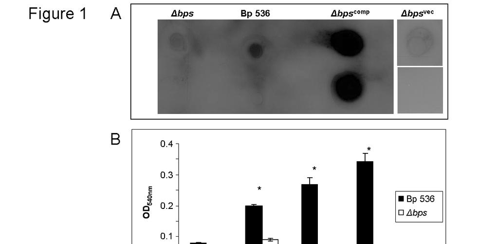
101 92
102 Fig. 2. SEM analysis of the role of Bps in B. pertussis biofilm formation. SEM of biofilms formed at the air-liquid interface on glass coverslips. Bp 536 (left panels) grown for 24h (A) and 96h (C); bps mutant grown for 24h (B) and 96h (D); bps comp strain grown for 96h (E). Bar, 10µm. 93
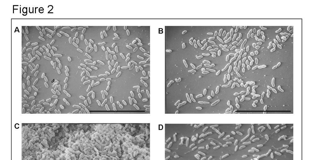
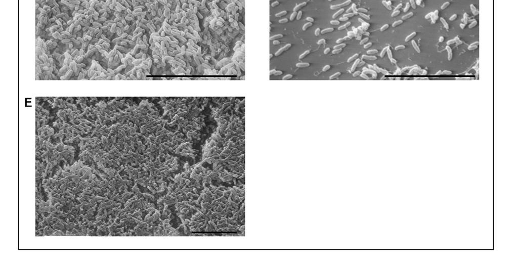
103 94
104 Fig. 3. Bps is crucial for biofilm development under flow conditions. Confocal scanning laser micrographs of biofilms formed in flow cells by Bp 536 (top panels) and the bps mutant (bottom panels). Strains were inoculated directly in the flow cell and visualized in situ every 24h. For each micrograph, the middle panel represents the x-y plane, and the adjacent top and side panels represent the x-z and y-z planes, respectively. For each strain, images were taken from at least eight areas and the experiment was repeated three times. A representative CSLM image for each sample is shown. Bar, 50µm. 95
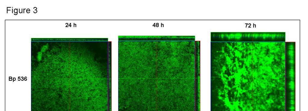
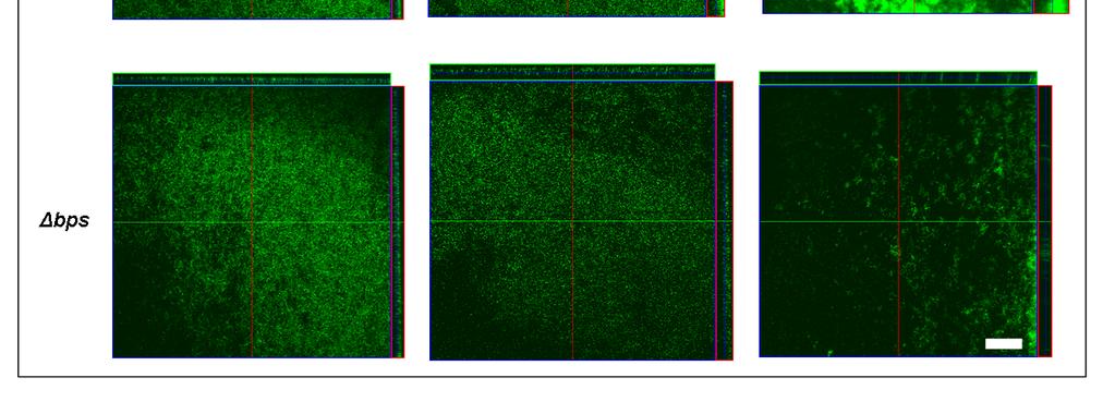
105 96
106 Fig. 4. B. pertussis forms biofilms in the mouse nose. (A) CSLM of biofilms formed within the murine nasal cavity by B. pertussis. C57BL/6 mice were inoculated with PBS, Bp 536, or the bps strain. Seven days postinoculation, nasal septa were harvested, immediately fixed, and probed with rat anti- Bordetella serum followed by a secondary anti-rat antibody conjugated to Alexa Fluor 488 (which stains bacteria green). To determine the localization of the host epithelium, specimens were stained for F-actin using phalloidin conjugated to Alexa Fluor 633 (which stains the epithelium red) and visualized with CSLM. Each micrograph represents an x-z reconstruction. For each specimen, images were obtained from at least five areas of the nasal septum and from at least three independent animals. Bar, 10µm. (B) SEM of B. pertussis biofilm formation on nasal septa. Specimens were collected from animals 7 days post-inoculation with PBS (top left), Bp 536 (top right and bottom left), bps mutant (bottom right), directly fixed, and processed for SEM. Scale bars, 10 µm. (C) Bps co-localizes with nasal biofilms. Nasal septa were harvested from C57BL/6 mice inoculated with PBS or Bp 536. Samples were collected 7 days postinoculation and were stained for Bordetella (green) as described in the legend to Fig. 4A. To detect Bps production (red), specimens were stained using goat anti-dpnag, followed by anti-goat conjugated to Alexa Fluor 633. Yellow staining indicates colocalization of B. pertussis and Bps. Micrographs are x-z reconstructions and are representative of at least three independently harvested tissues. 97
107 98
108 Fig. 5. Bps is essential for nasal colonization Colonization of the mouse respiratory tract by Bp 536 and the bps mutant. Groups of five 6-week-old C57BL/6 mice were intranasally inoculated with 50 µl containing 5 x 10 5 cfus of either the Bp 536 or bps strain. At the designated time points, mice were sacrificed and the nasal septum, trachea, and lungs removed, homogenized and plated for enumeration of the resident bacteria. Statistically significant differences were observed at all time points and tissues except at 7 days in the lungs. Error bars represent standard deviation. *P<0.05, students t-test. 99
109 100
110 Fig. 6. Bps functions as a nasal adhesin (A) Attachment assays were conducted with L2 (rat lung), A549 (human lung) and RPMI 2650 (human nasal septum) epithelial cell lines. Bp 536 or the bps strain was incubated at a multiplicity of infection of 10. Results are expressed as adherent cfus. Each data point is the average for six wells, and error bars indicate the standard deviation. Representative data from one of at least five independent experiments are shown. Asterisks designate a P<0.05 (students t-test). (B and C) RPMI 2650 cells were used for attachment assays. Bp 536 was preincubated with various concentrations of the anti-dpnag antibody (B) or the goat IgG antibody (C). Each data point is the average for three wells, and error bars indicate the standard deviation. Representative data from one of at least three independent experiments are shown. Asterisks designate a P<0.05 (students t-test). (D) Attachment assays were conducted with A549 cell line as above except that Bp 536 was pre-incubated with various concentrations of the anti-dpnag antibody or goat IgG control. Each data point is the average for three wells, and error bars indicate the standard deviation. Representative data from one of at least three independent experiments are shown. 101
111 102
112 Fig. 7. Attachment assays with nasal explants. Similar sized mouse nasal septa were excised from naïve mice and incubated with the indicated strains as described in experimental procedures. Results are expressed as adherent cfu from 5-6 nasal septa. Error bars represent standard deviation. *P<0.05, students t-test. 103
113 104
114 Fig. 8. Ectopic expression of Bps confers adherence. Attachment assays were conducted with the pga strain of E. coli containing the empty pbbr1mcs vector or the pmm11 plasmid that expresses Bps. Each data point is the average for six wells, and error bars indicate the standard deviation. Representative data from one of at least five independent experiments are shown. *P<0.05, students t-test. 105
115 106
116 Fig. 9. The Bps polysaccharide binds to RPMI 2650 but not to A549 cells. Epithelial cells adherent to glass coverslips were treated with purified Bps preparation from the bps comp strain or a mock-purified preparation from the bps vec strain. Bound Bps was detected by anti-dpnag antibody followed by secondary antibody coupled to Alexa-fluor 488. Epithelial cells were counter stained with rhodamine labeled phalloidin. Yellow staining denotes co-localization of Bps with the RPMI 2650 cells (indicated by arrows). 107
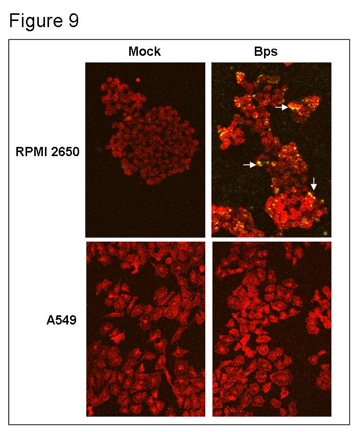
117 108
118 Fig. 10. Bps is expressed during human infections Immunoblot analysis of Bps reactivity with the human sera. Bps or the mock-purified preparation were separated by SDS-PAGE and after transfer onto nitrocellulose were probed with either anti-dpnag antibody or human serum. The separation of the stacking and the resolving gel is indicated by the arrow on the left. 109
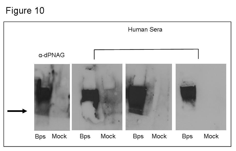
119 110
Bordetella bronchiseptica: A Candidate Mucosal Vaccine Vector
 University of Tennessee, Knoxville Trace: Tennessee Research and Creative Exchange Doctoral Dissertations Graduate School 5-2002 Bordetella bronchiseptica: A Candidate Mucosal Vaccine Vector Sreekumari
Consequences of Antimicrobial Resistant Bacteria. Antimicrobial Resistance. Molecular Genetics of Antimicrobial Resistance. Topics to be Covered
 Antimicrobial Resistance Consequences of Antimicrobial Resistant Bacteria Change in the approach to the administration of empiric antimicrobial therapy Increased number of hospitalizations Increased length
Antimicrobial Resistance
 Antimicrobial Resistance Consequences of Antimicrobial Resistant Bacteria Change in the approach to the administration of empiric antimicrobial therapy Increased number of hospitalizations Increased length
Antimicrobial Resistance Acquisition of Foreign DNA
 Antimicrobial Resistance Acquisition of Foreign DNA Levy, Scientific American Horizontal gene transfer is common, even between Gram positive and negative bacteria Plasmid - transfer of single or multiple
MID 23. Antimicrobial Resistance. Consequences of Antimicrobial Resistant Bacteria. Molecular Genetics of Antimicrobial Resistance
 Antimicrobial Resistance Molecular Genetics of Antimicrobial Resistance Micro evolutionary change - point mutations Beta-lactamase mutation extends spectrum of the enzyme rpob gene (RNA polymerase) mutation
Phenotypic modulation of the Bvg+ phase is not required for pathogenesis and. transmission of Bordetella bronchiseptica in swine
 IAI Accepts, published online ahead of print on 12 December 2011 Infect. Immun. doi:10.1128/iai.06016-11 Copyright 2011, American Society for Microbiology and/or the Listed Authors/Institutions. All Rights
Antimicrobial Resistance
 Antimicrobial Resistance Consequences of Antimicrobial Resistant Bacteria Change in the approach to the administration of Change in the approach to the administration of empiric antimicrobial therapy Increased
WHY IS THIS IMPORTANT?
 CHAPTER 20 ANTIBIOTIC RESISTANCE WHY IS THIS IMPORTANT? The most important problem associated with infectious disease today is the rapid development of resistance to antibiotics It will force us to change
1. Division of Bacterial, Parasitic, and Allergenic Products, Center for Biologics Evaluation and
 JB Accepted Manuscript Posted Online 30 July 2018 J. Bacteriol. doi:10.1128/jb.00175-18 This is a work of the U.S. Government and is not subject to copyright protection in the United States. Foreign copyrights
THE COST OF COMPANIONSHIP
 THE COST OF COMPANIONSHIP Jared Gillingham and Robert Burlage Concordia University School of Pharmacy Mequon, WI Synopsis: Infectious diseases are always a concern, but when you are a person in an at-risk
Methicillin-Resistant Staphylococcus aureus
 Methicillin-Resistant Staphylococcus aureus By Karla Givens Means of Transmission and Usual Reservoirs Staphylococcus aureus is part of normal flora and can be found on the skin and in the noses of one
Burton's Microbiology for the Health Sciences. Chapter 9. Controlling Microbial Growth in Vivo Using Antimicrobial Agents
 Burton's Microbiology for the Health Sciences Chapter 9. Controlling Microbial Growth in Vivo Using Antimicrobial Agents Chapter 9 Outline Introduction Characteristics of an Ideal Antimicrobial Agent How
Overview. There are commonly found arrangements of bacteria based on their division. Spheres, Rods, Spirals
 Bacteria Overview Bacteria live almost everywhere. Most are microscopic ranging from 0.5 5 m in size, and unicellular. They have a variety of shapes when viewed under a microscope, most commonly: Spheres,
Microarray and Functional Analysis of Growth Phase-Dependent Gene Regulation in Bordetella bronchiseptica
 INFECTION AND IMMUNITY, Oct. 2009, p. 4221 4231 Vol. 77, No. 10 0019-9567/09/$08.00 0 doi:10.1128/iai.00136-09 Copyright 2009, American Society for Microbiology. All Rights Reserved. Microarray and Functional
Mastitis cows and immunization
 In Spain, the antibiotherapy against mastitis moves 12,000,000 with an interannual growth of 10.2%. Only 4 of these millions are drying antibiotherapy. Conclusion: farmers spend a lot of money on mastitis
Boosting Bacterial Metabolism to Combat Antibiotic Resistance
 Boosting Bacterial Metabolism to Combat Antibiotic Resistance The MIT Faculty has made this article openly available. Please share how this access benefits you. Your story matters. Citation As Published
Inhibiting Microbial Growth in vivo. CLS 212: Medical Microbiology Zeina Alkudmani
 Inhibiting Microbial Growth in vivo CLS 212: Medical Microbiology Zeina Alkudmani Chemotherapy Definitions The use of any chemical (drug) to treat any disease or condition. Chemotherapeutic Agent Any drug
Staphylococcus aureus
 Staphylococcus aureus Significant human pathogen. SSTI Biomaterial related infections Osteomyelitis Endocarditis Toxin mediated diseases TSST Staphylococcal enterotoxins Quintessential Pathogen? Nizet
Significant human pathogen. SSTI Biomaterial related infections Osteomyelitis Endocarditis Toxin mediated diseases TSST Staphylococcal enterotoxins
 Staphylococcus aureus Significant human pathogen. SSTI Biomaterial related infections Osteomyelitis Endocarditis Toxin mediated diseases TSST Staphylococcal enterotoxins Quintessential Pathogen? Nizet
Vaccines for Cats. 2. Feline viral rhinotracheitis, FVR caused by FVR virus, also known as herpes virus type 1, FHV-1
 Vaccines for Cats Recent advances in veterinary medical science have resulted in an increase in the number and type of vaccines that are available for use in cats, and improvements are continuously being
National Research Center
 National Research Center Update of immunodiagnosis of cystic echinococcosis cysts Global distribution of zoonotic strains of Echinococcus granulosus (Adapted from Eckert and Deplazes, 2004) Echinococcus
Selective toxicity. Antimicrobial Drugs. Alexander Fleming 10/17/2016
 Selective toxicity Antimicrobial Drugs Chapter 20 BIO 220 Drugs must work inside the host and harm the infective pathogens, but not the host Antibiotics are compounds produced by fungi or bacteria that
Electron Microscopic Observations on Ciliated Epithelium of Tracheal Organ Cultures Infected with Bordetella bronchiseptica
 Microbiol. Immunol. Vol. 33 (2), 111-121, 1989 Electron Microscopic Observations on Ciliated Epithelium of Tracheal Organ Cultures Infected with Bordetella bronchiseptica Kachiko SEKIYA,*,1 Yutaka FUTAESAKU,2
Role of Antibodies in Immunity to Bordetella Infections
 INFECTION AND IMMUNITY, Apr. 2003, p. 1719 1724 Vol. 71, No. 4 0019-9567/03/$08.00 0 DOI: 10.1128/IAI.71.4.1719 1724.2003 Copyright 2003, American Society for Microbiology. All Rights Reserved. Role of
Amoxicillin trihydrate. Amoxicillin trihydrate. Amoxicillin trihydrate. Amoxicillin trihydrate. Amoxicillin trihydrate. Amoxicillin trihydrate
 Annex I List of the names, pharmaceutical form, strength of the veterinary medicinal product, animal species, route of administration, applicant in the Member States Member State EU/EEA Applicant Name
Antibiotics & Resistance
 What are antibiotics? Antibiotics & esistance Antibiotics are molecules that stop bacteria from growing or kill them Antibiotics, agents against life - either natural or synthetic chemicals - designed
Dier & Kruid Prof. Dr. J. Fink-Gremmels DVM, PhD, Dip ECVPT
 Dier & Kruid 03-06-2015 Prof. Dr. J. Fink-Gremmels DVM, PhD, Dip ECVPT J.Fink@uu.nl Antibiotics secondary metabolites produced under conditions of stress Fungi imperfecti (Penicillium Fusarium) Streptomyces
Q1. (a) Clostridium difficile is a bacterium that is present in the gut of up to 3% of healthy adults and 66% of healthy infants.
 Clostridium difficile is a bacterium that is present in the gut of up to 3% of healthy adults and 66% of healthy infants.") Q1. (a) Clostridium difficile is a bacterium that is present in the gut of up to 3% of healthy adults and 66% of healthy infants. C. difficile rarely causes problems, either in healthy adults or in infants.
Q1. (a) Clostridium difficile is a bacterium that is present in the gut of up to 3% of healthy adults and 66% of healthy infants. C. difficile rarely causes problems, either in healthy adults or in infants.
VACCINE-INDUCED-IMMUNITY-MEDIATED COMPETITION BETWEEN ENDEMIC BORDETELLAE AND HOST IMMUNITY AGAINST THEM
 The Pennsylvania State University The Graduate School College of Agricultural Sciences VACCINE-INDUCED-IMMUNITY-MEDIATED COMPETITION BETWEEN ENDEMIC BORDETELLAE AND HOST IMMUNITY AGAINST THEM A Dissertation
Mechanism of antibiotic resistance
 Mechanism of antibiotic resistance Dr.Siriwoot Sookkhee Ph.D (Biopharmaceutics) Department of Microbiology Faculty of Medicine, Chiang Mai University Antibiotic resistance Cross-resistance : resistance
TEAT DIP- POST DIP- PRE DIP- STRIPING
 TEAT DIP- POST DIP- PRE DIP- STRIPING KRISHIMATE AGRO AND DAIRY PVT LTD NO.1176, 1ST CROSS, 12TH B MAIN, H A L 2ND STAGE, INDIRANAGAR BANGALORE-560008, INDIA Email: sales@srisaiagro.com Www.srisaiagro.com
Cell Wall Inhibitors. Assistant Professor Naza M. Ali. Lec 3 7 Nov 2017
 Cell Wall Inhibitors Assistant Professor Naza M. Ali Lec 3 7 Nov 2017 Cell wall The cell wall is a rigid outer layer, it completely surrounds the cytoplasmic membrane, maintaining the shape of the cell
The Bvg Virulence Control System Regulates Biofilm Formation in Bordetella bronchiseptica
 JOURNAL OF BACTERIOLOGY, Sept. 2004, p. 5692 5698 Vol. 186, No. 17 0021-9193/04/$08.00 0 DOI: 10.1128/JB.186.17.5692 5698.2004 Copyright 2004, American Society for Microbiology. All Rights Reserved. The
Challenges Emerging resistance Fewer new drugs MRSA and other resistant pathogens are major problems
 Micro 301 Antimicrobial Drugs 11/7/12 Significance of antimicrobial drugs Challenges Emerging resistance Fewer new drugs MRSA and other resistant pathogens are major problems Definitions Antibiotic Selective
Investigation of the molecular biology and contribution to virulence of Bordetella bronchiseptica urease
 University of Wollongong Research Online University of Wollongong Thesis Collection University of Wollongong Thesis Collections 1999 Investigation of the molecular biology and contribution to virulence
Medical bacteriology Lecture 8. Streptococcal Diseases
 Medical bacteriology Lecture 8 Streptococcal Diseases Streptococcus agalactiae Beat haemolytic Lancifield group B Regularly resides in human vagina, pharynx and large inine Can be transferred to infant
Mechanisms and Pathways of AMR in the environment
 FMM/RAS/298: Strengthening capacities, policies and national action plans on prudent and responsible use of antimicrobials in fisheries Final Workshop in cooperation with AVA Singapore and INFOFISH 12-14
Mastitis: Background, Management and Control
 New York State Cattle Health Assurance Program Mastitis Module Mastitis: Background, Management and Control Introduction Mastitis remains one of the most costly diseases of dairy cattle in the US despite
Int.J.Curr.Microbiol.App.Sci (2018) 7(8):
 7(8):") International Journal of Current Microbiology and Applied Sciences ISSN: 2319-7706 Volume 7 Number 08 (2018) Journal homepage: http://www.ijcmas.com Original Research Article https://doi.org/10.20546/ijcmas.2018.708.378
International Journal of Current Microbiology and Applied Sciences ISSN: 2319-7706 Volume 7 Number 08 (2018) Journal homepage: http://www.ijcmas.com Original Research Article https://doi.org/10.20546/ijcmas.2018.708.378
Antibiotics. Antimicrobial Drugs. Alexander Fleming 10/18/2017
 Antibiotics Antimicrobial Drugs Chapter 20 BIO 220 Antibiotics are compounds produced by fungi or bacteria that inhibit or kill competing microbial species Antimicrobial drugs must display selective toxicity,
مادة االدوية المرحلة الثالثة م. غدير حاتم محمد
 م. مادة االدوية المرحلة الثالثة م. غدير حاتم محمد 2017-2016 ANTIMICROBIAL DRUGS Antimicrobial drugs Lecture 1 Antimicrobial Drugs Chemotherapy: The use of drugs to treat a disease. Antimicrobial drugs:
CHAPTER 1 INTRODUCTION
 1 CHAPTER 1 INTRODUCTION The Staphylococci are a group of Gram-positive bacteria, 14 species are known to cause human infections but the vast majority of infections are caused by only three of them. They
SUMMARY OF PRODUCT CHARACTERISTICS
 SUMMARY OF PRODUCT CHARACTERISTICS 1. NAME OF THE VETERINARY MEDICINAL PRODUCT Oxycare 20 %w/v LA Solution for Injection 2. QUALITATIVE AND QUANTITATIVE COMPOSITION Active Substance: Oxytetracycline (Equivalent
Growth Phase- and Nutrient Limitation-Associated Transcript Abundance Regulation in Bordetella pertussis
 INFECTION AND IMMUNITY, Oct. 2006, p. 5537 5548 Vol. 74, No. 10 0019-9567/06/$08.00 0 doi:10.1128/iai.00781-06 Copyright 2006, American Society for Microbiology. All Rights Reserved. Growth Phase- and
CHAPTER 18 THE COCCI OF MEDICAL IMPORTANCE. Learning Objectives
 CHAPTER 18 THE COCCI OF MEDICAL IMPORTANCE Gram-positive and gram-negative cocci that cause infection are presented. The difference between commensal and pathogenic strains is explained, because many of
The Pennsylvania State University. The Graduate School. College of Agricultural Science UNDERSTANDING HOW VACCINATION AND PARTICULAR VIRULENCE
 The Pennsylvania State University The Graduate School College of Agricultural Science UNDERSTANDING HOW VACCINATION AND PARTICULAR VIRULENCE FACTORS CONTRIBUTE TO BORDETELLA TRANSMISSION A Dissertation
CONFLICT OF INTEREST ANTIMICROBIAL LOCK SOLUTIONS INCREASE BACTEREMIA
 CONFLICT OF INTEREST ANTIMICROBIAL LOCK SOLUTIONS INCREASE BACTEREMIA NONE Vandana Dua Niyyar, MD Associate Professor of Medicine, Division of Nephrology, Emory University. OBJECTIVES Role of biofilm in
Antibiotic Resistance in Bacteria
 Antibiotic Resistance in Bacteria Electron Micrograph of E. Coli Diseases Caused by Bacteria 1928 1 2 Fleming 3 discovers penicillin the first antibiotic. Some Clinically Important Antibiotics Antibiotic
ANTIBIOTICS USED FOR RESISTACE BACTERIA. 1. Vancomicin
 ANTIBIOTICS USED FOR RESISTACE BACTERIA 1. Vancomicin Vancomycin is used to treat infections caused by bacteria. It belongs to the family of medicines called antibiotics. Vancomycin works by killing bacteria
Burn Infection & Laboratory Diagnosis
 Burn Infection & Laboratory Diagnosis Introduction Burns are one the most common forms of trauma. 2 million fires each years 1.2 million people with burn injuries 100000 hospitalization 5000 patients die
Antimicrobial use in poultry: Emerging public health problem
 Antimicrobial use in poultry: Emerging public health problem Eric S. Mitema, BVM, MS, PhD CPD- Diagnosis and Treatment of Poultry Diseases FVM, CAVS, 6 th. August, 2014 AMR cont Antibiotics - Natural or
CLADISTICS Student Packet SUMMARY Phylogeny Phylogenetic trees/cladograms
 CLADISTICS Student Packet SUMMARY PHYLOGENETIC TREES AND CLADOGRAMS ARE MODELS OF EVOLUTIONARY HISTORY THAT CAN BE TESTED Phylogeny is the history of descent of organisms from their common ancestor. Phylogenetic
Probing the Function of Bordetella bronchiseptica Adenylate Cyclase Toxin by Manipulating Host Immunity
 INFECTION AND IMMUNITY, Mar. 1999, p. 1493 1500 Vol. 67, No. 3 0019-9567/99/$04.00 0 Copyright 1999, American Society for Microbiology. All Rights Reserved. Probing the Function of Bordetella bronchiseptica
ESBL Producers An Increasing Problem: An Overview Of An Underrated Threat
 ESBL Producers An Increasing Problem: An Overview Of An Underrated Threat Hicham Ezzat Professor of Microbiology and Immunology Cairo University Introduction 1 Since the 1980s there have been dramatic
Redefining Infection Management. Proven Clinical Outcomes
 Proven Clinical Outcomes Proof of Bacteria-Binding1 In the first 30 seconds, 1 square centimeter of Cutimed Sorbact binds wound bacteria - after 2 hours, the amount of bacteria bound are more than would
12 TIPS HOW TO TREAT BACTERIAL INFECTION WITHOUT ANTIBIOTICS
 PDF STD FACTS - BACTERIAL VAGINOSIS 12 TIPS HOW TO TREAT BACTERIAL INFECTION WITHOUT ANTIBIOTICS 1 / 6 2 / 6 3 / 6 bacterial infection close pdf Bacterial vaginosis (BV) is a condition that happens when
Impact of Antimicrobial Resistance on Human Health. Robert Cunney HSE HCAI/AMR Programme and Temple Street Children s University Hospital
 Impact of Antimicrobial Resistance on Human Health Robert Cunney HSE HCAI/AMR Programme and Temple Street Children s University Hospital AMR in Foodchain Conference, UCD, Dec 2014 Sir Patrick Dun s Hospital
Microbiology ( Bacteriology) sheet # 7
 sheet # 7") Microbiology ( Bacteriology) sheet # 7 Revision of last lecture : Each type of antimicrobial drug normally targets a specific structure or component of the bacterial cell eg:( cell wall, cell membrane,
Microbiology ( Bacteriology) sheet # 7 Revision of last lecture : Each type of antimicrobial drug normally targets a specific structure or component of the bacterial cell eg:( cell wall, cell membrane,
No-leaching. No-resistance. No-toxicity. >99.999% Introducing BIOGUARD. Best-in-class dressings for your infection control program
 Introducing BIOGUARD No-leaching. >99.999% No-resistance. No-toxicity. Just cost-efficient, broad-spectrum, rapid effectiveness you can rely on. Best-in-class dressings for your infection control program
Evolution of the Bordetella autotransporter Pertactin: identifications of regions subject to positive selection
 Evolution of the Bordetella autotransporter Pertactin: identifications of regions subject to positive selection Marcel Hijnen 1,2, Dimitri Diavatopoulos 1,2 and Frits R. Mooi 1,2 Both authors contributed
ANNEX I SUMMARY OF PRODUCT CHARACTERISTICS
 ANNEX I SUMMARY OF PRODUCT CHARACTERISTICS 1 1. NAME OF THE VETERINARY MEDICINAL PRODUCT CYTOPOINT 10 mg solution for injection for dogs CYTOPOINT 20 mg solution for injection for dogs CYTOPOINT 30 mg
Medical Bacteriology- Lecture 14. Gram negative coccobacilli. Zoonosis. Brucella. Yersinia. Francesiella
 Medical Bacteriology- Lecture 14 Gram negative coccobacilli Zoonosis Brucella Yersinia Francesiella 1 Zoonosis: A disease, primarily of animals, which is transmitted to humans as a result of direct or
Caused by microorganisms (usually bacteria) that invade the udder, multiply, and produce toxins that are harmful to the mammary gland
 that invade the udder, multiply, and produce toxins that are harmful to the mammary gland") MASTITIS PA R T 1 MASTITIS Mast = breast; itis = inflammation Inflammation of the mammary gland Caused by microorganisms (usually bacteria) that invade the udder, multiply, and produce toxins that are
MASTITIS PA R T 1 MASTITIS Mast = breast; itis = inflammation Inflammation of the mammary gland Caused by microorganisms (usually bacteria) that invade the udder, multiply, and produce toxins that are
2008 FELINE HEALTH GRANT AWARDS 10 projects funded for a total of $135,860
 2008 FELINE HEALTH GRANT AWARDS 10 projects funded for a total of $135,860 The Winn Feline Foundation receives proposals from veterinary researchers around the world who are interested in improving feline
Introduction to Chemotherapeutic Agents. Munir Gharaibeh MD, PhD, MHPE School of Medicine, The university of Jordan November 2018
 Introduction to Chemotherapeutic Agents Munir Gharaibeh MD, PhD, MHPE School of Medicine, The university of Jordan November 2018 Antimicrobial Agents Substances that kill bacteria without harming the host.
Chapter concepts: What are antibiotics, the different types, and how do they work? Antibiotics
 Chapter concepts: Antibiotics What are antibiotics, the different types, and how do they work? How do we decided on the most appropriate antibiotic treatment? What are some of the ways that bacteria are
MILK COMPOSITIONAL CHANGES DURING MASTITIS
 MASTITIS PA R T 2 MILK COMPOSITIONAL CHANGES DURING MASTITIS Increased SCC Na Cl Whey protein (e.g. serum albumin, Ig, lactoferrin) Decreased Production α-lactalbumin & Lactose Casein K MILK LOSS LACTOFERRIN
Chemotherapeutic Agents
 Chemotherapeutic Agents The cell is the basic structure of all living organisms. The cell membrane features specifi c receptor sites that allow interaction with various chemicals, histocompatibility proteins
10/15/08. Activity of an Antibiotic. Affinity for target. Permeability properties (ability to get to the target)
") Beta-lactam antibiotics Penicillins Target - Cell wall - interfere with cross linking Actively growing cells Bind to Penicillin Binding Proteins Enzymes involved in cell wall synthesis Activity of an Antibiotic
Beta-lactam antibiotics Penicillins Target - Cell wall - interfere with cross linking Actively growing cells Bind to Penicillin Binding Proteins Enzymes involved in cell wall synthesis Activity of an Antibiotic
Running title: Contribution of Bordetella Bps to Biofilm Formation and Respiratory Disease in
 IAI Accepted Manuscript Posted Online 30 May 2017 Infect. Immun. doi:10.1128/iai.00261-17 Copyright 2017 American Society for Microbiology. All Rights Reserved. 1 2 The Bordetella Bps Polysaccharide is
Filamentous Hemagglutinin of Bordetella bronchiseptica Is Required for Efficient Establishment of Tracheal Colonization
 INFECTION AND IMMUNITY, Dec. 1998, p. 5921 5929 Vol. 66, No. 12 0019-9567/98/$04.00 0 Copyright 1998, American Society for Microbiology. All Rights Reserved. Filamentous Hemagglutinin of Bordetella bronchiseptica
Evaluation of the hair growth and retention activity of two solutions on human hair explants
 activity of two solutions on human hair explants Study Directed by Dr E. Lati of Laboratoire Bio-EC, Centre de Recherches Biologiques et d Experimentations Cutanees, on behalf of Pangaea Laboratories Ltd.
Name(s): Period: Date:
: Period: Date:") Evolution in Action: Antibiotic Resistance HASPI Medical Biology Lab 21 Background/Introduction Evolution and Natural Selection Evolution is one of the driving factors in biology. It is simply the concept
Evolution in Action: Antibiotic Resistance HASPI Medical Biology Lab 21 Background/Introduction Evolution and Natural Selection Evolution is one of the driving factors in biology. It is simply the concept
An#bio#cs and challenges in the wake of superbugs
 An#bio#cs and challenges in the wake of superbugs www.biochemj.org/bj/330/0581/bj3300581.htm ciss.blog.olemiss.edu Dr. Vassie Ware Bioscience in the 21 st Century November 14, 2014 Who said this and what
Antimicrobials & Resistance
 Antimicrobials & Resistance History 1908, Paul Ehrlich - Arsenic compound Arsphenamine 1929, Alexander Fleming - Discovery of Penicillin 1935, Gerhard Domag - Discovery of the red dye Prontosil (sulfonamide)
A Unique Approach to Managing the Problem of Antibiotic Resistance
 A Unique Approach to Managing the Problem of Antibiotic Resistance By: Heather Storteboom and Sung-Chul Kim Department of Civil and Environmental Engineering Colorado State University A Quick Review The
The Bordetella Bps Polysaccharide Is Critical for Biofilm Development in the Mouse Respiratory Tract
 JOURNAL OF BACTERIOLOGY, Nov. 2007, p. 8270 8276 Vol. 189, No. 22 0021-9193/07/$08.00 0 doi:10.1128/jb.00785-07 Copyright 2007, American Society for Microbiology. All Rights Reserved. The Bordetella Bps
Drd. OBADĂ MIHAI DORU. PhD THESIS ABSTRACT
 UNIVERSITY OF AGRICULTURAL SCIENCES AND VETERINARY MEDICINE ION IONESCU DE LA BRAD IAŞI FACULTY OF VETERINARY MEDICINE SPECIALIZATION MICROBIOLOGY- IMUNOLOGY Drd. OBADĂ MIHAI DORU PhD THESIS ABSTRACT RESEARCHES
THE PENNSYLVANIA STATE UNIVERSITY SCHREYER HONORS COLLEGE DEPARTMENT OF BIOCHEMISTRY AND MOLECULAR BIOLOGY
 THE PENNSYLVANIA STATE UNIVERSITY SCHREYER HONORS COLLEGE DEPARTMENT OF BIOCHEMISTRY AND MOLECULAR BIOLOGY THE ROLE OF FIMBRIAE IN BORDETELLA COLONIZATION MARGARET CURRY DUNAGIN Spring 2010 A thesis submitted
The color and patterning of pigmentation in cats, dogs, mice horses and other mammals results from the interaction of several different genes
 The color and patterning of pigmentation in cats, dogs, mice horses and other mammals results from the interaction of several different genes 1 Gene Interactions: Specific alleles of one gene mask or modify
Protein Synthesis Inhibitors
 Protein Synthesis Inhibitors Assistant Professor Dr. Naza M. Ali 11 Nov 2018 Lec 7 Aminoglycosides Are structurally related two amino sugars attached by glycosidic linkages. They are bactericidal Inhibitors
Cercetări bacteriologice, epidemiologice şi serologice în bruceloza ovină ABSTRACT
 ABSTRACT Thesis entitled BACTERIOLOGICAL, EPIDEMIOLOGICAL AND SEROLOGICAL RESEARCHES IN BRUCELLOSIS OVINE is scientific and practical reasons the following: - Infectious epididymitis in Romania, described
CLPNA Pressure Ulcers ecourse: Module 5.6 Quiz II page 1
 CLPNA Pressure Ulcers ecourse: Module 5.6 Quiz II 1. What are the symptoms of an infected wound? a. Fever b. Edema c. Erythema d. Local pain and tenderness e. Induration of wound edge 2. A person with
Changing Trends and Issues in Canine and Feline Heartworm Infections
 Changing Trends and Issues in Canine and Feline Heartworm Infections Byron L. Blagburn College of Veterinary Medicine Auburn University Canine and feline heartworm diagnostic, treatment and prevention
Venom Research at Natural Toxins Research Center (NTRC)
") Venom Research at Natural Toxins Research Center (NTRC) Dr. John C. Pérez Regents Professor and Director of the NTRC Texas A&M University-Kingsville Snake Venom Research is Important for Numerous Reasons
Venom Research at Natural Toxins Research Center (NTRC) Dr. John C. Pérez Regents Professor and Director of the NTRC Texas A&M University-Kingsville Snake Venom Research is Important for Numerous Reasons
SUMMARY OF PRODUCT CHARACTERISTICS
 SUMMARY OF PRODUCT CHARACTERISTICS 1. NAME OF THE VETERINARY MEDICINAL PRODUCT Enrocare 50 mg/ml Solution for Injection for Cattle, Pigs, Dogs and Cats (UK, IE, FR) Floxadil 50 mg/ml Solution for Injection
SUPPLEMENTARY INFORMATION
 doi:10.1038/nature12234 Supplementary Figure 1. Embryonic naked mole-rat fibroblasts do not undergo ECI. Embryonic naked mole-rat fibroblasts ( EF) were isolated from eight mid-gestation embryos. All the
EXPRESSION OF BACILLUS ANTHRACIS PROTECTIVE ANTIGEN IN VACCINE STRAIN BRUCELLA ABORTUS RB51. Sherry Poff
 EXPRESSION OF BACILLUS ANTHRACIS PROTECTIVE ANTIGEN IN VACCINE STRAIN BRUCELLA ABORTUS RB51 By Sherry Poff Thesis submitted to the Faculty of the Virginia Polytechnic Institute & State University in partial
Multi-drug resistant microorganisms
 Multi-drug resistant microorganisms Arzu TOPELI Director of MICU Hacettepe University Faculty of Medicine, Ankara-Turkey Council Member of WFSICCM Deaths in the US declined by 220 per 100,000 with the
ANTIBIOTIC RESISTANCE. Syed Ziaur Rahman, MD, PhD D/O Pharmacology, JNMC, AMU, Aligarh
 ANTIBIOTIC RESISTANCE Syed Ziaur Rahman, MD, PhD D/O Pharmacology, JNMC, AMU, Aligarh WHY IS THIS IMPORTANT? The most important problem associated with infectious disease today is the rapid development
The genetic basis of breed diversification: signatures of selection in pig breeds
 The genetic basis of breed diversification: signatures of selection in pig breeds Samantha Wilkinson Lu ZH, Megens H-J, Archibald AL, Haley CS, Jackson IJ, Groenen MAM, Crooijmans RP, Ogden R, Wiener P
Was the Spotted Horse an Imaginary Creature? g.org/sciencenow/2011/11/was-the-spotted-horse-an-imagina.html
 Was the Spotted Horse an Imaginary Creature? http://news.sciencema g.org/sciencenow/2011/11/was-the-spotted-horse-an-imagina.html 1 Genotypes of predomestic horses match phenotypes painted in Paleolithic
THE ROYAL COLLEGE OF VETERINARY SURGEONS DIPLOMA EXAMINATION IN VETERINARY DERMATOLOGY. Tuesday 22 August PAPER 1 (3 hours)
") DIPLOMA EXAMINATION IN VETERINARY DERMATOLOGY Tuesday 22 August 2000 PAPER 1 Candidates are required to answer FOUR questions only. 1. What is meant by the term staphylococcal virulence factors. Indicate
DIPLOMA EXAMINATION IN VETERINARY DERMATOLOGY Tuesday 22 August 2000 PAPER 1 Candidates are required to answer FOUR questions only. 1. What is meant by the term staphylococcal virulence factors. Indicate
BvgAS Is Sufficient for Activation of the Bordetella pertussis ptx Locus in Escherichia coli
 JOURNAL OF BACTERIOLOGY, Nov. 1995, p. 6477 6485 Vol. 177, No. 22 0021-9193/95/$04.00 0 Copyright 1995, American Society for Microbiology BvgAS Is Sufficient for Activation of the Bordetella pertussis
HOW XTC IMPROVED MINOXIDIL PENETRATION - 5 WAYS!
 HOW XTC IMPROVED MINOXIDIL PENETRATION - 5 WAYS! What Hinders Minoxidil from Working Well 1. Sebum from sebaceous gland blocks the hair follicle. 2. Minoxidil therefore, cannot penetrate through the sebum
Block Objectives: Basic Infectious Diseases Block
 Course: Intro to Infectious Diseases IID-BASID-01 Virtual Lab: Infectious Disease Laboratory Session 1 Identify etiologic bacterial organisms from clinical case studies based on the following: Discriminating
Brucellosis is a bacterial zoonosis transmitted directly or indirectly to humans from infected animals,
 Definition Brucellosis is a bacterial zoonosis transmitted directly or indirectly to humans from infected animals, predominantly domesticated ruminants and swine. The disease is known colloquially as undulant
Welcome to Pathogen Group 9
 Welcome to Pathogen Group 9 Yersinia pestis Francisella tularensis Borrelia burgdorferi Rickettsia rickettsii Rickettsia prowazekii Acinetobacter baumannii Yersinia pestis: Plague gram negative oval bacillus,
How to stop the snotty noses: Preventing feline upper respiratory infections. Staci Cannon, DVM, MPH, DACVPM, DABVP (Shelter Medicine Practice)
") How to stop the snotty noses: Preventing feline upper respiratory infections Staci Cannon, DVM, MPH, DACVPM, DABVP (Shelter Medicine Practice) Why is URI so hard to control? Multiple pathogens Chronic
How to stop the snotty noses: Preventing feline upper respiratory infections Staci Cannon, DVM, MPH, DACVPM, DABVP (Shelter Medicine Practice) Why is URI so hard to control? Multiple pathogens Chronic
Why Don t These Drugs Work Anymore? Biosciences in the 21 st Century Dr. Amber Rice October 28, 2013
 Why Don t These Drugs Work Anymore? Biosciences in the 21 st Century Dr. Amber Rice October 28, 2013 Outline Drug resistance: a case study Evolution: the basics How does resistance evolve? Examples of