Host-parasite relationships of Haemoproteus sacharovi Novy and MacNeal, 1904 (Protozoa:Sporozoa)
|
|
|
- Agnes Chapman
- 5 years ago
- Views:
Transcription

1 Retrospective Theses and Dissertations Iowa State University Capstones, Theses and Dissertations 1960 Host-parasite relationships of Haemoproteus sacharovi Novy and MacNeal, 1904 (Protozoa:Sporozoa) John Neville Farmer Iowa State University Follow this and additional works at: Part of the Zoology Commons Recommended Citation Farmer, John Neville, "Host-parasite relationships of Haemoproteus sacharovi Novy and MacNeal, 1904 (Protozoa:Sporozoa) " (1960). Retrospective Theses and Dissertations This Dissertation is brought to you for free and open access by the Iowa State University Capstones, Theses and Dissertations at Iowa State University Digital Repository. It has been accepted for inclusion in Retrospective Theses and Dissertations by an authorized administrator of Iowa State University Digital Repository. For more information, please contact
2 This dissertation has been microfilmed exactly as received Mic FARMER, John Neville. HOST-PAiiASiTE RELATIONSHIPS OF HAEMOFROTEUS SACHARG VÏ NO VY AND -IACN4AL, 1904 (PROTOZOA: SPORGZOA). Iowa State University of Scitince and Technology Ph.D,, I960 Zoology University Microfilms, Inc., Ann Arbor, Michigan
3 HOST-PARASITE RELATIONSHIPS OP HAEMOPEOTEUS SACHAROVI NOVY AND MACNEA1, 1904 (PROTOZOA: SPOROZOA) by John Neville Farmer A Dissertation Submitted to the Graduate Faculty in Partial Fulfillment of The Requirements for the Degree of DOCTOR OF PHILOSOPHY Major Subject: Parasitology Approved: Signature was redacted for privacy. Chairman Signature was redacted for privacy. Co-chairman Signature was redacted for privacy. Head of Major Department Signature was redacted for privacy. Dean of Graduate College Iowa State University Of Science and Technology Ames, Iowa I960
4 ii TABLE OP CONTENTS Page I. INTRODUCTION 1 A. Nature of the Problem 1 B. Protozoan Genera Frequently Reported from Avian Blood 3 II. REVIEW OF LITERATURE 6 A. Early History of Bird Malaria 6 B. The Genus Haemoproteus 8 C. Haemoproteus saoharovi Novy and MacEsal, III. MATERIALS AND METHODS 18 A. Source of Avian Hosts 18 B. Collection and Examination of Blood Samples 19 C. Description of Experiments 20 D. Histological Techniques 21 IV. RESULTS 23 A. Incidence of Avian Haemosporidian Parasites in Central Iowa 23 B. Infections of Haemoproteus saoharovi in Mourning Doves 31 C. Haemoproteus saoharovi Infections in Pigeons 53 D. Attempted Transmission of Haemoproteus saoharovi 73 V. DISCUSSION 93 VI. SUMMARY AND CONCLUSIONS 102 VII. LITERATURE CITED 106 VIII. ACKNOWLEDGEMENTS 115 IX. PLATES 116
5 1 I. INTRODUCTION A. Nature of the Problem Becker and co-workers (1956, 1957) described naturally occurring Plasmodium and Haemoproteus infections in the common pigeon. Birds harboring these infections were obtained from a pigeon colony at Gilbert, Iowa. The occurrence of Haemoproteus saoharovi Novy and MacNeal, in a number of these pigeons afforded an excellent opportunity to investigate the habits of this relatively unknown parasite. The birds in this colony are kept in chicken-wire cages that do not hinder, to any great extent, the passage of free-flying insects, numerous genera of which are undoubtedly attracted to the area by the presence of the pigeons. The susceptibility of the birds to H. saoharovi and the fact that they are readily available to a number of invertebrates which might serve as hosts, make an avian colony such as this suitable for studies on the host-parasite relationships of H. saoharovi. Assuming that H. saoharovi follows a developmental course similar to that of Haemoproteus columbae Kruse and H. lophortyx O'Roke, the following diagram might well represent its developmental pattern: AVIAN HOST 1. Blood cells-< 2. Tissue cells \ / 3. INVERTEBRATE EOS!
6 2 Three probable sites of parasitic development are suggested, namely: 1. gametoeyte development within erythrocytes, 2. exoerythrocytic development and schizogony within tissue cells, and 3. sexual development and sporogony in the invertebrate host. While studying blood films made from pigeons of the colony at Gilbert, the occurrence of H. saoharovi during the summer months and its subsequent disappearance during the winter, raised the question as to the mode of its transmission. Is the yearly appearance of this organism associated with a relapse phenomenon similar to that exhibited by Leucocytozoon infections, or is a natural reservoir host involved? Should the latter relationship be true, the preceding diagram would have to be modified as follows: AVIAN HOST Blood cells «Tissue cells X /.INVERTEBRATE HOST X X Blood cells * Tissue cells RESERVOIR HOST The role of the invertebrate host is emphasized by such a relationship. Although considerable information may be accumulated concerning the developmental stages of H. saoharovi, discovery of the invertebrate host(s) responsible
7 3 for its transmission would permit verification of these data, by enabling bird-to-bird transfer to be carried on in the laboratory. This, in turn, would allow thorough investigation of the various stages involved in the development of the parasite. The following studies constitute an attempt to clarify host-parasite relationships of H. saoharovi and concern themselves, in part, with attempts to discover the invertebrate host or hosts responsible, in nature, for transmission of this sporozoan. B. Protozoan Genera Frequently Reported from Avian Blood A number of protozoan genera, namely Trypanosoma Gruby, Plasmodium Marchiafava and Celli, Haemoproteus Kruse and Leucocytozoon Danilewsky, have been described from birds. Recent investigations of avian blood parasites have concerned themselves primarily with these genera. All trypanosomes reported from birds belong to the genus Trypanosoma. These flagellated organisms are extracellular in birds and have been reported only from the peripheral blood. All other avian protozoan blood parasites belong to the class Sporozoa whose important genera include Plasmodium. Haemoproteus and Leucocytozoon. Pigment-producing parasites are included among members
8 4 of the germs Plasmodium. These are parasites of reptiles, birds and mammals. Both asexual stages and gametocytes may be demonstrated within erythrocytes where schizogony occurs. The presence of trophozoites in the circulating blood permits the transmission of the infection by blood transfusions. Schizogony probably also occurs within endothelial cells of the vertebrate host, where exoerythrocytic phases occur. Sexual processes, including fertilization and development of a motile zygote (ookinete), take place within mosquitoes which serve as arthropod hosts. The genus Leucocytozoon is characterized by gametocytes which are demonstrable in the peripheral blood of birds, the only vertebrates harboring this genus. As schizogony does not occur within erythrocytes, transmission of infection requires another host. Asexual development occurs within vertebrate hosts and sexual reproduction, so far as is known, occurs within blackflies, Simulium spp. Members of the genus Haemoproteus resemble those of Leucocytozoon in that gametocytes are demonstrable in the peripheral blood. The two genera differ, however, since in Haemoproteus«the gametocytes are restricted entirely to erythrocytes. As in Leucocytozoon. schizogony does not occur within blood cells, but takes place within endothelial cells of lungs and other organs. An invertebrate host is required for transmission of infection. It is generally
9 5 accepted that the processes of fertilization and ookinete development are limited to the invertebrate hosts of the family Hippoboscidae (louse flies). Recent authors, however, (Baker, 1957; Faliis and Wood, 1957; Hanson et al., 1957; Huff, 1932, 1942) question louse flies as the sole disseminators of Haemoproteus in nature.
10 6 II. HEVIEW OP LITERATURE A. Early History of Bird Malaria Danilewsky (1885a) is given credit for the initial discovery of protozoa in the blood of birds. In this work, he described three types of protozoans. One, ein 'Blutwiirmchen 1 im Plasma freischwimmend," he considered to be closely related to Haemogregarina previously described by him (Danilewsky, 1885b). A second type he recognized as belonging to the genus Trypanosoma» A third he referred to as a haemocytozoon which, after "Exkapsulation," also became free-swimming in the blood. Within erythrocytes he saw clear, uncolored, transparent "Vakuolen" of various shapes and sizes containing strongly light-refractile, glossy-black particles. He commented that these "Pseudovakuolen" were very common in certain species of birds. He described them as ring-like structures lying alongside the nucleus of the erythrocytes. The more developed forms took on a spherical shape, altering the outline of the red blood corpuscles, which at the same time became more and more distorted. From this last description, it is evident that he was dealing with a species of bird malaria. Danilewsky's observations were timely, in view of the fact that Laveran (1880) had described somewhat similar organisms in man. The similarity between these two types
11 7 of infection was soon realized, as evidenced by the subsequent work of Grassi and Feletti (1890). These investigators placed the intracorpuscular parasites of birds in the same genus as those described from man and established the genus Laverania to include the parasites previously described in birds by Danilewsky, as well as those parasites of man reported by Laveran. The generic name Haemoproteus, however, established by Kruse (1890), which also included the avian forms described by Danilewsky, has priority, since it appeared shortly before the work of Grassi and Peletti (1890). Laveran (1890) confirmed Danilewsky's descriptions of blood parasites from avian hosts and envisioned birds as convenient laboratory hosts through which the mysteries of human malaria might be studied. The first successful transmission of avian malarial parasites from bird to bird by blood inoculations was reported by Celli and San Pelice (1891), who worked with Plasmodium. Attempts by earlier workers to do this had been unsuccessful probably because they were dealing with Haemoproteus rather than Plasmodium. Celli and San Felice (1891) believed that the malarial parasites of man and of birds, although similar, were not identical. The exflagellation of microgametocytes and the union of gametes in blood drawn from a crow infected with
12 8 Haemoproteiis was described by MacCallum (1897, 1898a). In 1898b, he reported an analogous process in what is now known as Plasmodium falciparum Welch. Aspects of the sexual phase of both human and avian malaria were thus demonstrated. Furthermore, encouraged by these observations of MacCallum, Ross (1898) demonstrated the nature of malarial transmission, utilizing Gulex mosquitoes in transferring Plasmodium to sparrows. Without these facts, stemming for the most part from studies of avian malaria, it is probable that our present knowledge of human malaria would not have advanced as rapidly as it has. B. The Genus Haemoproteus The circumstances concerning the first use of the generic name Haemoproteus have already been discussed. As is often the case, old terminology sometimes persists in current literature. Gametocytes of most species belonging to this genus are still referred to as "halteridia." This practice stems from Labbe (1894) who incorrectly employed the generic designation Halteridium for the same parasites. Minchin (1912) observed that the Haemoproteus parasitizing different species of birds varied in size and appearance and concluded that there were many species of the genus. Subsequent investigations proved him correct, as evidenced
13 9 by We jay on (1926) who listed 302 kinds of birds from which the genua Haemoproteus had been reported. A checklist and host-index of the genus Haemoproteus was published by Coatney (1936) who included 45 species of Haemoproteus, most of which were described from birds. A more recent checklist and host-index of the blood protozoa from birds of North America by Herman (1944) included 17 species of Haemoproteus. Fifty-five genera of birds from which Haemoproteus have been described were also included. Recently, Levine and Kantor (1959) published a checklist of blood parasites of birds of the order Columbiformes in which eight species of Haemoproteus are recorded. It is apparent that members of this genus are among the most common malarial parasites of birds. Information concerning their host-parasite relationships, however, is sparse. This lack of information is undoubtedly due to the difficulties involved in maintaining laboratory strains. Bird-to-bird transfer of the parasite demands a suitable invertebrate host. Investigations concerning host-parasite relationships become complicated when the vector is unknown. Life histories are known for very few avian species of Haemoproteus. Sergent and Sergent (1906) and later Aragao (1907, 1908) proved that Haemoproteus columbae Kruse is normally transmitted from pigeon to pigeon by the bite of the hippoboscid, Lynchia maura Speiser. Since Sergent and
 confirmed the role of this fly in the transmission of H. columbae from pigeon to pigeon.")
14 10 Sergent (1906), Aragao (1907, 1908), Mezinescu (1909) and Gronder (1915) were unable to follow the parasite's development beyond ookinete formation in the fly, these investigators concluded, erroneously, that the ookinete itself was inoculated into the pigeon. Adie (1915, 1924) confirmed the role of this fly in the transmission of H. columbae from pigeon to pigeon. In so doing, she was the first to completely describe the life cycle of this parasite in the fly, Lynchia maura. Other hippoboscids that have been incriminated in the transmission of H. columbae are Lynchia capensis Speiser, by Gonder (1915), and Lynchia livideolor Aragao, L. brunea Aragao, and Microlynchia pusilla Lutz, by Aragao (1916). Bequaert (1953, p. 138), however, in his extensive monograph concerning the Hippoboscidae, states : "The name Lynchia maura, L. lividcolor and L. capensis, sometimes cited also among the vectors of pigeon malaria, are all synonyms of Pseudolynchia canariensis. As for Lynchia brunea mentioned by AragSo ( ) as one of the vectors, it was based upon a misidentification of P. canariensis, the true Pseudolynchia brunnea (Latreille) having nothing to do with the transmission of the disease; moreover Aragao synonomized his L. brunea with L. maura in a later paper (1927, p. 827). At one time Aragao (1916, p. 355) included Microlynchia pusilla (Speiser) among the bird-flies transmitting H. columbae in Brazil; unfortunately he never described his experiments with this fly. h In accordance with this apparent invertebrate host specificity, Kartman (194-9), studying Haemoproteus infections of Hawaiian pigeons, reported finding oocysts of
15 11 H. columbae on the midgut of P. canariensis. However, recent studies in England by Baker (1957) indicate a species of Omithornyia Latreille to be a vector of H. columbae in wood pigeons, Columba palumbus Linnaeus. California quail, Lophortyx californica Shaw, may contract a severe malaria-like disease caused by Haemoproteus lophortyx O'Roke. O'Roke (1930) injected a young quail with the macerated salivary glands and with part of the gut of an infected hippoboscid, Lynchia hirsuta Ferris, taken from a wild quail infected with H. lophortyx. After a period of 27 days, gametocytes of H. lophortyx were observed in the blood of the young bird. He also described sporogonic stages (ookinetes, oocysts and sporozoites) in some wild Lynchia hirsuta collected from infected quail. Herman and Bischoff (194-9) described sporozoites in the salivary glands and body cavity of Stilbometopa impressa (Bigot). This material, including the salivary glands, was inoculated into a young quail. Twenty-one days after injection, parasites were observed in the blood. Recently, Tarshis (1955) has demonstrated that H. lophortyx may be transmitted to quail by the bite of infected S. impressa. Laboratory-reared Pseudolynchia maura Bequaert were used by Huff (1931, 1932) to transmit Haemoproteus saoharovi and Haemoproteus maccallumi Novy and MacNeal from the mourning dove to domestic pigeons. He doubted, however, that
16 12 this pigeon louse fly was responsible for the transmission of these parasites in nature. The possibility that invertebrate hosts other than hippoboscids are involved in the transmission and life history of H. columbae has also been investigated. Formation of ookinetes in the gut of mosquitoes and in a mite was described by Aragâo (1916). Roller (1920) noted ookinete development in the gut of the bed bug, Qimex lectularius Linnaeus. Kartman (194-9), on the other hand, failed to observe ookinete formation in the gut of Oulex quinquefasciatus Say and Aedes albopictus (Skuse) mosquitoes that had fed on infected pigeons. More recently, species of Culicoides (Ceratopogonidae) have been suggested as suitable intermediate hosts for Haemoproteus nettionis (Johnson and Cleland) of ducks. Fallis and Wood (1957), investigating H. nettionis infections in domestic ducks in Algonquin Park, Ontario, Canada, observed an abundance of black flies, biting midges and mosquitoes. These blood-sucking insects were collected from caged ducks and from their immediate surroundings. Clean ducks were inoculated with suspensions of these insects after comminution of the latter in blood. H. nettionis infections developed in ducks injected with the specimens of Culicoides. However, the insects employed in these experiments were not specifically identified. Further
17 13 investigations may show that H. nettionis is transmissible by the bite of Culicoides. C. Haemoproteus saoharovi Novy and MacNeal, 1904 Of the few reports dealing with Haemoproteus saoharovi most are concerned solely with its occurrence in nature. The initial description of the parasite was by Novy and MacNeal (1904) who obtained specimens from the blood of the mourning dove, Zenaidura macroura (Linnaeus). They published their findings in three separate journals. The following description appeared twice in 1904, and again in 1905: "Haemoproteus saoharovi, n.sp. This species, probably first observed by Sacharoff, who regarded it as a "leucocytozoon," is related to that of Danilewsky. Pound in young mourning doves and elsewhere. Invasion begins with an infection of very young erythroblasts. As the parasite grows, it pushes the nucleus to the periphery, where it is seen in the adult form on the outer edge as a cap, which is but a trifle larger than the nucleus of a red blood cell. The parasite is spherical, male and female forms common, latter predominate; blepharoplast distinct, adjoining or over the nucleus. Microgamete formation common. Infection not transferable by the blood." (p. 933) Although the description is somewhat abbreviated, it is adequate for enabling one to recognize the species. H. saoharovi has been reported from the mourning dove Zenaidura macroura only one other time, namely, by Herms et al. (1939), who described the infection as occurring in the blood of one of four doves examined in California.
18 14- Most of the literature concerning H. saoharovi, however, deals with its occurrence in the eastern mourning dove, Zenaidura macroura Carolinensis (Linnaeus). Huff (1931, 1932) reported the successful transmission of H. saoharovi from this host to domestic pigeons, using laboratory-reared Pseudolynchia maura Bequaert. Huff's source of H. saoharovi in these studies was from four naturallyinfected doves. Since only one of these, however, had a single infection, it alone was used in the transmission experiments. After the flies had been allowed to remain on this dove for two to eight days, they were placed upon laboratory-reared pigeons. Thirteen days after the first flies had been transferred, gametocytes resembling those of H. saoharovi appeared in the blood of one of the pigeons. This particular infection persisted up to the time the bird was sacrificed, a period of three months. Coatney and Roudabush (1937), while studying the incidence of blood parasites in Nebraska birds, found H. saoharovi in two mourning doves. Similar organisms were described by Coatney and West (1938) who found, in examining the blood of 13 doves over periods of from one to 66 days, that all 13 were infected. In the same year, Herman (1938) reported the blood of six of 86 mourning doves that he examined on Cape Cod, Massachusetts to be positive for H. saoharovi. Huff (1939) examined blood smears of 188 doves
19 15 trapped for banding in various regions of the United States. Of these birds, he found 51 to be infected with H. saoharovi and 34 others to have both H. saoharovi and H. maccallumi. In Nebraska, Coatney and West (1940) reported H. saoharovi from 11 of 20 nestling doves. They offered this as evidence that this parasite was acquired in the North and not necessarily after migration. The natural vector was not found, however. Wetmore (1941), although primarily concerned with the Leucocytozoon species that she observed, described H. saoharovi from two mourning doves. She noted that gametocytes of this parasite disappeared from the blood for days at a time. In Texas, Couch (1952) observed H. saoharovi in 58 of 213 mourning doves ; 11 of these occurring as single infections. In Illinois, Levine et al. (1952) reported 103 of 206 mourning doves to be infected with H. saoharovi. Similar organisms were found to parasitize blood cells of 58.2% of 392 immature doves collected in Illinois by Hanson et al. (1957). These investigators also reported an incidence of 43.1$ in 72 adult birds. The possibilities of a natural vector being responsible for transmission were discussed and an extensive survey of the ectoparasites of these birds was undertaken. The vector was not discovered, however. Other vertebrate hosts recorded as being infected with H. saoharovi include the Western mourning dove, Zenaidura
20 16 macroura marginella (Woodhouse). Wood and Herman (194-3) reported H. saoharovi in 11 of 27 of these doves taken in Arizona and California. They also observed similar organisms in the blood of the Western white winged dove, Zenaida (=Melopelia) asiatica mearnsi (Ridgway). A species of Leucocytozoon was described from the blood of the European turtle dove, Streptopelia turtur (Linnaeus), by Franchini (1924). His descriptions and figures of this organism, however, resemble H. saoharovi rather than a leucocytozoon. Another important host reported for this organism is the common pigeon, Columba livia. Although Huff (1931) was the first to transmit H. saoharovi to pigeons experimentally, Coatney and West (1938, 194-0) were the first to describe natural infections in the common pigeon. These investigators initially observed natural infection of H. saoharovi in an adult pigeon and two squabs. Further study uncovered six infections in 17 adult pigeons and five infections in 33 squabs that were examined. The natural occurrence of H. saoharovi in the common pigeon was not reported again until Becker et al. (1956) described its presence in pigeon squabs reared in a colony at Gilbert, Iowa. Reference was made to abnormally enlarged spleens and to granular gizzards observed in a number of sacrificed birds. Some of the blood smears made from these
21 17 particular birds were diagnosed as positive for H. saoharovi. This pigeon colony was the source for another report by Becker et al» (1957), who examined 114 stained blood films made from pigeons ranging in age from two to eight weeks. Blood samples were taken from the birds «,t various times during the summer of 1956, and it was shown that two squabs harbored patent H. saoharovi infections. This summary of investigations concerning H. saoharovi indicates that this parasite enjoys a relatively high natural incidence among columbiform birds and a fairly wide geographical distribution. On the other hand, it emphasizes the lack of information concerning the biology of the organism. Although its morphology has been described by Huff (1932) and by Coatney and West (1940), no further stages of its life cycle have been clearly defined.
22 18 III. MATERIALS AND METHODS A. Source of Avian Hosts Since this investigation deals primarily with the hostparasite relationships of Haemoproteus saoharovi, special efforts were undertaken to examine the blood of birds belonging to the avian family Coluabidae, certain members of which are recognized hosts for this species. Almost all the common pigeons examined were from a colony at Gilbert, Iowa. Other free or so-called barn pigeons examined were trapped or shot in the vicinity of Ames, Iowa. Drop-door traps operated by trip-wires and baited with cracked corn, and funnel traps similarly baited, were utilized to capture mourning doves. Doves caught in these traps were kept in an animal room. Attempts to maintain several young nestling doves in the laboratory were unsuccessful. Consequently, blood smears from very young birds were taken at the nest. Blood smears were also prepared from other birds such as the redwing [Agelaius phoeniceus (Linnaeus)], domestic duck [Anas platyrhynchus Linnaeus], great horned owl [Bubo virgin!anus (Gmelin)], common nighthawk [Chordeiles minor (iorster)], blue jay [Cyanocitta cristata (Linnaeus)], catbird [Dumetella carolinensis (Linnaeus)], bronzed grackle [Quiscalus quiscula versicolor Vieillot], ringed turtle dove
23 19 [Streptopelia risoria (Linnaeus)], starling [Sturnus vulgaris Linnaeus], and robin [Turdus migrâtorius Linnaeus]. B. Collection and Examination of Blood Samples Blood samples were obtained from living birds by puncturing a toe with the blade of a scalpel. Obtaining blood from nestling birds proved to be more difficult. If unsatisfactory smears resulted due to insufficient blood, the tip of a claw was cut off with a pair of scissors. Blood was easily obtained using this method, but bleeding generally persisted. The toe puncture method was preferred. In examining dead birds, samples of blood were obtained, if possible, from the heart. If not, tissue smears of the liver, lungs or kidney were made. Since direct microscopic examination of stained blood films does not take into consideration subpatent or latent infections, isodiagnosis was sometimes used. This procedure, used extensively by Sergent (1920) to uncover subpatsnt Plasmodium infections, involves transfusing previously uninfected birds with the blood of "suspect" birds. Preliminary examination of an entire smear was made under the low power of a Bausch and Lomb binocular microscope equipped with 10X oculars. If, during this examination, erythrocytes or leucocytes were suspected of infection, the area in question was inspected under oil
24 20 immersion. Blood films, even when apparently negative, were examined under oil immersion for a period of five minutes. During this time, care was taken not to re-examine the same fields, thus permitting the examination of approximately 250 to 300 different fields. All identifications of parasites were confirmed by using oil immersions. The measurements of parasites and blood cells recorded during this investigation were obtained with the aid of a calibrated ocular micrometer. Line drawings were prepared with the aid of the camera-lucida or micro-projector. C. Description of Experiments Materials and methods employed in rearing and maintaining insect colonies used for this study will be included in a later section concerning attempted transmission of H. saoharovi. It should be mentioned, however, that transfer of H. sacharovi from infected birds to pigeons and to mourning doves was attempted in three ways. Uninfected insects were allowed to feed on a bird known to harbor a patent infection. After a number of days, allowing for the possibility of any sporogonic development within the invertebrate host, these insects were allowed to take a blood meal from an uninfected pigeon. Another attempted method of transfer consisted of inoculating uninfected birds with insect salivary glands,
25 21 comminuted in physiological saline. These insects had previously fed on pigeons or doves lmovra. to harbor H. saoharovi. A third method used was the inoculation of uninfected pigeons and doves with comminuted tissues removed from sacrificed pigeons and doves known to have patent infections of H. sacharovi. The tissues used were brain, liver, lungs, spleen and gizzard. Inoculations were made intravenously, using syringes of 2 cc capacity and equipped with 23-gauge needles. In every case, after exposure or inoculation, the blood of the experimental birds was examined for any sign of parasitic development. The pigeons used for both isodiagnosis and transmission attempts were reared from eggs and maintained in a screened brooder house. Accidental infection was unlikely, but, as a precautionary measure, blood films of all experimental birds were always made before the experiment was undertaken. D. Histological Techniques After fixation in absolute methyl alcohol, blood films and tissue impressions were stained in diluted G-iemsa (1:40). A staining time of 40 minutes duration was found to be very satisfactory. Staining techniques employed for sections of avian tissue included Mallory's triple connective stain, Heidenhain's "Azan" triple stain, the Peulgen technique
26 22 with fast green counterstain, and Delafield's haemotoxylin counterstained with eosin. Fixation of avian tissues was usually accomplished with Bouin's, although 10$ formalin, Zenker's and A.P.A. were also used. Insects to be sectioned were always fixed in ale oholic Bouin's. With muscular tissue such as the gizzard, in which infiltration of paraffin would be expected to be difficult, the dioxan method was used. Even this technique was not found to be entirely satisfactory for obtaining unwrinkled sections. Soaking the block in a mixture of glycerine and 95<$> ethanol for a period of several hours proved helpful in obtaining smooth sections. Such glycerine-alcohol treated blocks permitted sections of 8 to 10M to be obtained with little difficulty.
27 23 IV. RESULTS A. Incidence of Avian Haemosporidian Parasites in Central Iowa The following observations on the protozoan genera Haemoproteus, Leucocytozoon, Plasmodium and Trypanosoma are based, for the most part, on material collected at Ames, Boone, and Gilbert, Iowa, from 1957 to 1959* During this period, 1,006 blood smears from 568 birds were examined, with 99 or 17.25$ of these birds being found to harbor blood parasites. The study included 13 species of birds, of which six species were infected. As recorded in Table 1, four species harbored Plasmodium, four species were infected with Leucocytozoon and five species with Haemoproteus. Honhaemosporidian organisms, i.e., trypanosomes and microfilariae, were observed in two species of birds. 1. Plasmodium infections Plasmodium circumflexum Kikuth was diagnosed from a nestling redwing (Agelaius phoeniceus) having 28$ of its erythrocytes parasitized. In August, 1959, a three-week-old pigeon (Oolumba livia) belonging to the pigeon colony at Gilbert, Iowa, was found to harbor a heavy infection of P. relictum Grassi and Peletti. The natural occurrence of P. relictum had been described by Becker and co-workers (1956, 1957) from pigeons
28 24 Tablé 1«OoeUx-x-eiiC-e Ox iutx-a- âuu ex ux"acellula.x' uluou. parasites in birds examined from Story and Boone counties, Iowa, during the period Agelaius phoeniceus Redwing Anas Tolatyrhynchus Domestic duck 6 Bubo virgin!anus Great horned owl 25 Ohordeiles minor Common nighthawk 2 Golumba livia Commonpigeon 451 Cyanocitta cristata Blue jay 6 Dumetella carolinensis Catbird 2 Pi P. -ti P» 0) A A A o 0} m m u a *H. «Ë 81 o m a 3 0)1 0 cd bj 3 M ctsi H S <D Hi Ml A P4 EH <d o m xi ti s-ti <D I -H h -P O k H-H O h CD CO 42 <D OH -P <H H-H 0«H 9 S<H EH O H ) so cd m -d += r& <1> a u -p <v -H O O.Û (0 k <H 0) <H 9 ft OH Quiscalus quiscula versicolor Bronze grackle 16 5 *1.25 Streptopelia risoria Ringed turtle dove 5 0 Sturnus vulgaris Starling 7 0 Turdus migratorius Robin 3 0 Zenaidura macroura Mourning dove j6 1_
29 25 maintained in the same location. Unfortunately, subinoculations were not made in 1959, so that its particular characteristics could not de compared with those from Becker 1 s strain, named 1-B by Huff et al. (1959). Examination of the blood of a single juvenile grackle (Quiscalus quiscula versicolor) revealed a heavy infection with a species of Plasmodium, resembling P. relictum. While re-examining blood films acquired from this bird, it was observed that pigment granules within gametocytes were often elongate and coarse. Also, in the case of mature gametocytes, the host-cell nucleus was sometimes extruded. According to Hewitt (1940), these characters indicate an infection with P. cathemerium Hartman. An adult mourning dove (Zenaidura macroura carolinensis), captured August, 1957, was initially believed to harbor an infection of Haemoproteus. This bird, however, was included with six other doves in isodiagnostic experiments, which resulted in uncovering a subpatent Plasmodium infection in this particular dove. A transfused pigeon* recipient on August 28, 1957 of 0.6 ml. of blood from this dove, developed a patent Plasmodium infection on September 1, The pigeon died September 29, 1957, at which time a parasitemia of 38.6$ was recorded from a blood smear obtained at necropsy. Tissue impressions of the liver, lungs, kidneys, spleen and brain
30 26 revealed the presence of exo-erythrocytic schizonts in these organs. Farmer (1959) observed similar schizonts in organs of pigeons infected with the 1-B strain of P. relictum. Three other pigeons, transfused with blood from this dove, also developed patent infections. In each instance, however, the recipient bird recovered. 2. Leucocytozoon infections Since Leucocytozoon cannot be transferred by blood inoculations and since all cases of Leucocytozoon, with one exception, observed during this study, were low grade infections, species allocation was difficult. In several instances, however, the parasites observed compared favorably with well-described species. Examination of blood films from great horned owls (Bubo virginianus) revealed three birds harboring Leucocytozoon infections, with one of the birds possessing two distinct species. One species common to all three, is characterized by rounded gametocytes, Its identity has not been established. The gametocytes of the other species of Leucocytozoon are unusually conspicuous, for the host-cells are peculiarly spindle-shaped. The host-cell nucleus is distorted to such an extent that it resembles a dumbbell in appearance. A somewhat incomplete description of L. siemani var. bubonis
31 27 was presented "by Pantham (1926) which he observed in the blood of the owl (Bubo maculosus). Goatney and Roudabush (1937) published a detailed description of this species which they recovered from the great horned owl. Since my specimens of the spindle-shaped Leucocytozoon species found in the great homed owl compare favorably with their description, these sporozoans are considered to be L. ziemani var. bubonis. Hounded gametocytes belonging to a species of Leucocytozoon were observed in one nestling and in five adult mourning doves. Two species of Leucocytozoon have been recorded from the avian order Columbiformes. An unnamed elongate Leucocytozoon was described by Minchin (1910) from the dove (Streptopelia semitorquata). Mathis and Léger (1910) described a rounded form, L. marchouxi, from four of nine doves, Streptopelia tranquebarica (=Turtur humilis). Recently, a detailed review of the literature concerning L. marchouxi was published by Levine (1954), who found this species in five adult, one juvenile and four nestling mourning doves. He considered as L. marchouxi only those strains in the mourning dove having rounded gametocytes. Accordingly, the rounded gametocytes observed in the blood of mourning doves examined during this study are considered to be L. marchouxi. Unidentified Leucocytozoon infections involving species
32 28 forming rounded gametocytes were observed in an adult and juvenile redwing and in three adult and two juvenile grackles. One of the juvenile grackles possessed such a massive infection that one suspects this species to be pathogenic. 3. Haemoproteus infections Infections of Haemoproteus, as with Leucocytozoon, usually are not easily transferred by blood inoculations. Morphology of gametocytes becomes the only characteristic useful in differentiating species. Due to the similarity in the appearance of some gametocytes, identification of species is often not possible. In some cases, however, gametocytes vary sufficiently, permitting species allocation to be undertaken with some reliability. For example, H. sacharovi Itovy and MacNeal of the mourning dove may be recognized by the characteristic appearance of its gametocytes. Other well-described species may be differentiated according to the tendency of the gametocytes either to encircle the erythrocyte nucleus completely or to displace it laterally. Examination of blood smears made from great horned owls revealed one immature and four adults possessing light Haemoproteus infections. Gelli and San Felice (1891) described three species of Haemoproteus from owls, namely, H. aluci, H. bubonis, and
33 29 H. noctuae, "varieties A and C. The investigations of Wolf son (1936), however, show that H= noctuae variety C is really a species of Plasmodium and was named P. oti "by her. The true H. noctuae of Oelli and San Felice, however, is generally recognized as being the true halteridium of the owl. The gametocytes of this species displace the host-cell nucleus laterally, but do not enclose this structure. Gametocytes resembling H. noctuae were harbored in two of the four infected adults. Coatney and Roudabush (1937) described H. noctuae var. nebraskensis from a great horned owl. They described these parasites as being similar in appearance to H. noctuae, but that the gametocytes enclosed the host-cell nucleus. Accordingly, gametocytes observed in one immature and two mature great horned owls are considered to belong to this species. H. sacharovi was observed in the blood of 50 of 414 colonized pigeons. The blood of 37 barn pigeons, however, was parasite-free. H. sacharovi was also recovered from the blood of 22 of 41 mourning doves. Three of these infections were harbored in nestling birds. Twenty-two adults of the 41 mourning doves examined harbored what is considered to be H. maccallumi Novy and MacNeal. Huff (1932) questioned the validity of H. maccallumi being a distinct species, since he was unable to
34 30 recognize any constant morphological differences between H. maccailumi of the dove and H. columbae Erase in the pigeon's blood. Although Huff (1931, 1932) reported transmitting both H. sacharovi and H. maccailumi to the pigeon by using the hippoboscid fly, Pseudolynchia maura Bigot, Coatney (1933) was unable to transmit H. columbae to the dove, using the same species of fly. In view of this, Coatney and Roudabush (1937) treated H. maccailumi as a distinct species. Recently, the author, using the hippoboscid fly, Pseudolynchia canariensis (Macquart), was unable to transmit either H. sacharovi or H. maccailumi to pigeons. In view of this, along with the absence of H. columbae in 4-51 pigeons although local doves were infected with gametocytes morphologically similar to H. columbae, H. maccailumi is considered here to be a distinct species. An unidentified Haemoproteus species similar to that mentioned by Coatney and West (1938) was harbored in two of six blue jays = One of the 16 grackles examined possessed a very light infection with Haemoproteus. Only the macrogametocytes of this species were observed, however. The cytoplasm of these female cells possessed a distinctive vacuole. Coatney and West (1938) described H. quiscalus from the blood of an adult and an immature bronzed grackle. The macrogame tocyte s of this species showed a large, irregular vacuole near the center of the parasite. Since micro-
35 31 gametocytes were not seen, however, the species observed during the present study were not identified. 4. Incidence of Trypanosoma and microfilariae Of the 568 birds examined, one grackle was found to harbor an extremely light infection with a species of Trypanosoma. Infections with microfilariae were noted in a single blue jay and a grackle. The following birds were negative for blood-inhabiting organisms: Six domestic ducks (Anas platyrhynchus), two common nighthawks (Chordeiles minor), two catbirds (Dumetella carolinensis), five ringed turtle doves (Streptopelia risoria), seven starlings (Sturnus vulgaris) and three robins (Turdus migrâtorius). B. Infections of Haemoproteus sacharovi in Mourning Doves 1. Incidence Blood smears were examined from 41 mourning doves, ten of which were nestlings. Of these, 39 were living birds. Blood from two dead birds was obtained from the heart, but in the case of living birds, peripheral blood was examined. Three nestlings were infected with H. sacharovi alone and a fourth squab harbored L, marchouxi. Since none of these birds was able to fly, it is obvious that parasites
36 32 had been acquired while birds were still iii. the nest. Eleven adult doves harbored single haemosporidian infections, four of which were diagnosed as H. sacharovi. Eleven other adult doves were infected with both H. sacharovi and H. maccailumi. A latent Plasmodium infection was discovered in one of these doves as a result of isodiagnostic techniques. H. sacharovi, H, maccailumi. and I. marchouxi were demonstrable in three other doves. A combination of H. maccailumi and L. marchouxi was noted in a single adult dove. Ten other doves examined proved to be parasite-free. These data, summarized in Table 2, indicate that multiplicity of infections is apparently far more common in adult birds than in nestlings. These data do not necessarily incriminate local invertebrate hosts as being responsible for the transmission of H. sacharovi and 1. marchouxi. Since mourning doves are a migratory species, it is possible that the vectors responsible are imported by adult birds. To incriminate either local or migratory invertebrate hosts, evidence concerning the known ecto-parasites of mourning doves must be considered. The possibility of age immunity toward protozoan parasites also must not be overlooked. An analysis of the existing data and inclusion of evidence gathered during the present study concerning local invertebrate hosts will be discussed in a later section.
37 33 Table 2. Haemoproteus, Plasmodium, and Leucocytozoon infections in mourning doves captured near Ames, Iowa, from 1957 to Species Number of parasitized birds Nestlings Adults H. sacharovi H. maccailumi L. marchouxi 1 1 H. sacharovi and ÏÏ. maccailumi H. sacharovi, H. maccailumi and L. marchouxi 0 E. sacharovi, H. maccailumi and P. relictum H. maccailumi and H. marchouxi 0 Number of infected birds Number of birds parasite-free 10 Total number of birds examined
38 34 2. Course of infections in mourning doves a. General nature of infection Avian malarial infections involve the reaction of the parasite to the host as well as the host's reaction to the presence of the parasite, The former relationship, according to Hewitt (1940), is known as the parasitological period; the latter, the clinical period. The parasitological period includes prepatent, patent and subpatent periods. The prepatent period encompasses the length of time between entrance of the parasite into the body until it is demonstrable in the peripheral circulation. The patent period includes that interval during which parasites may be readily observed in the blood. The subpatent period is one in which parasites may be present in the blood but are so few in number that they are usually overlooked using routine techniques. The clinical period, generally involving the protective mechanisms of the host, may also involve several recognizable phases. Hewitt (1940) has indicated these as the periods of incubation, symptoms, convalescence and relapse. Any reappearance of young gametocytes following convalescence, excluding reinfection by the intermediate host, constitutes a relapse. b. Development of gametocytes Since none of the infections observed in doves were laboratory-induced, the course of parasite development within the host can be described only from observations of infections that have
39 35 relapsed. At the onset of a relapse, small, round stages are observed in erythrocytes (Pig. 1). On the following day, the developing parasites are usually elongate, lying adjacent to the nucleus of the host cell (Pig. 2). In Giemsa stained smears, the parasite nucleus stains pink and sometimes contains a maroon stained karysome, By the third day, hypertrophy of parasitized red blood cells is very obvious (Pig. 3). Some macrogametocytes at this stage completely fill the red blood cell. In a majority of invaded erythrocytes, the host cell nucleus is displaced laterally by the developing gametocyte. Where the host cell nuclear displacement is polar, however, the parasite tends to envelop the nucleus rather than to displace it. Sex of gametocytes is easily differentiated at this stage of their development. The cytoplasm of macrogametocytes stains blue with Giemsa, and generally possesses a mottled or vacuolated appearance. Numerous fine granules and several slightly larger, more conspicuous granules are scattered throughout the cytoplasm. The nucleus of the macrogametocyte is ovoid to elongate and stains reddish pink. It is generally located toward one end of the parasite and possesses a circular maroon staining karyosome. This karyosome is invariably situated at the periphery of the nucleus (Pig. 4). Indeed, in many macrogametocytes the location of the karyosome is almost
40 36 completely outside of, but never quite losing contact with, the boundaries of the nucleus. The cytoplasm of microgametocytes stains a rather diffuse pink -fith Giemsa. Vacuol&tion is less apparent in microgametocytes than in macrogametocytes. Eosinophilic granules are present and are more conspicuous, but less numerous, than the granules observed in the female stages. The nucleus of the microgametocytes is considerably larger than the corresponding structure in macrogametocytes, and stains a slightly darker pink than the surrounding cytoplasm. It is located near the center of the parasite and possesses a round, maroon staining karyosome generally located near the center of the nucleus (Fig. 5). The peripheral orientation of the karyosome never appears to approach the extremes exhibited by corresponding structures noted in macrogametocytes. By the fourth day, gametocytes may completely fill the infected erythrocytes. Other than an increase in size, there is no morphological change in microgametocytes. In macrogametocytes, however, the cytoplasm tends to stain a darker blue, with pigment-like granules appearing more distinct. Such granules become increasingly conspicuous day by day as long as macrogametocytes are demonstrable in the blood (Fig. 6). Concerning the growth rate of gametocytes of H,
41 37 sacharovi in the blood of mourning doves and pigeons, Coatney and West (194-0) stated that these parasites are morphologically mature after four days of development. In the present study, smears were made from the blood of two mourning doves at 8 A.M., 4- P.M. and 10 P.M. each day during the course of several relapses. Examination of these slides indicates that many gametocytes mature after three and a half days of development. By the sixth day following the onset of a relapse, however, all gametocytes are full size and considered as mature (Fig. 7). c. Length of life of mature gametocytes The continuous observation of the course of relapsed infections in six doves permitted estimation of the length of time that gametocytes may survive in the blood. Acton and Knowles (1914) studied this in H. columbae infections of the pigeon and concluded that gametes could live for "considerable periods" in the peripheral blood. Coatney (1933) presented data indicating that gametocytes of H. columbae were removed from the blood of pigeons within 28 days following a relapse or could remain in the blood for as long as 68 days. During the course of H. sacharovi infections in six doves, the data in Graph 1 indicate that gametocytes may frequently disappear from the blood. The shortest time required for the disappearance of gametocytes from the blood following a relapse was seven days. The maximum length of
42 Graph 1. Relapse phenomena exhibited by mourning doves examined daily for 130 days.
43 13- (CONTROL) MATURE GAMETOCYTES IMMATURE GAMETOCYTES BIRD SACRIFICED J < 8 o Lu h- 7 z UJ - 4 UJ > o k (CONTROL)»» I I I I I I L I NOVEMBER DECEMBER 1958 T.I i i i l I l i i i i i i I i I I II JANUARY FEBRUARY MARCH 1959
44 40 time that gametocytes remained in the blood following a relapse was 33 days, with an average duration of 15.8 days. d. The sex ratio of gametocytes Examination of smears made from the blood of naturally infected mourning doves indicate that macrogametocytes are far more frequently seen (Table 3). Coatney and West (1940) reported a ratio of one male to 6.71 female gametocytes in the blood of mourning doves infected with Et. sacharovi. During the present investigation, it appeared that in some doves the ratio was even lower than this. Initially, it was surmised that the presence of microgametocytes was being overlooked, due to their staining characteristics. The cytoplasm of the male sexual stages stains a diffuse pink compared to the blue coloration of macrogametocyte cytoplasm. To evaluate the actual sex ratio of gametocytes, however, a series of blood films made from Doves #4, #7, #8 and #11 were carefully examined under oil. 10,000 erythrocytes were examined on each slide, with each gametocyte encountered recorded as either a male or female. Since the infections were at extremely low levels (ranging from one parasitized cell to eight parasitized cells per 10,000 red blood cells), enumeration of gametocytes was terminated after 200 had been observed. Ten slides from Dove #4, and nine each from Doves #7, #8 and #11 were examined with microgametocytes being observed only in the blood of #4 and #8. A total of 19
45 41 Table 3. Distribution of micro- and macrogametocytes in the blood of Doves #4, #7» #8 and #11. Identification Number of red Number of gametocytes number of dove blood cells Male Female 4 100, , , , Totals 370, microgametocytes and 181 macrogametocytes were noted, with the overall ratio indicating one male to 9.52 females. In some cases, microgametocytes were not demonstrable in the peripheral circulation for several days. Either they are produced in such low numbers that they are overlooked, or they may be more susceptible to the defensive mechanisms within the host. When they were observed, however, they were always sparse. This fact suggests that under normal conditions fewer microgametocytes are produced than macrogametocytes.
46 42 e. Hypertrophy of parasitized erythrocytes The most striking characteristic of gametocytes of H. sacharovi is their size. Although several investigators have commented on the size of these organisms, none have specified their actual dimensions. According to Huff (1931)» gametocytes of H. sacharovi completely fill infected red blood cells and enlarge them to a size 1.3 times as wide and 1.4 times as long as a normal red blood cell. Illustrations comparing normal and parasitized erythrocytes were published by Huff (1932) but measurements were not included. Coatney and Roudabush (1937) also did not publish any dimensions in their descriptions of H. sacharovi in the mourning dove. In the studies here presented, it was noted that parasitized cells are greatly distorted and enlarged by the presence of gametocytes. Fully-developed gametocytes often fill the host cell completely, although some apparently mature gametocytes are contained within erythrocytes whose dimensions are greater than those of the invading organism. Hypertrophy of erythrocytes is noticeable even in early developmental stages of the parasite. Accordingly, measurements were made to determine the size of uninfected red blood cells, infected erythrocytes and gametocytes. Films made from blood obtained from Doves #4, #7, #8 and #11 were examined. The dimensions of randomly selected red blood cells were recorded from each of the doves, 25 from each
47 43 bird. These data are recorded in Table 4 and indicate that uninfected erythrocytes average 12.94A in length and 5.95^ in width. A total of 19 microgametocytes was measured, 14 from Dove #4 and five from Dove #8. Their mean length was 16.06A, and their mean width, 6.42A. The host cells for these microgametocytes averaged 16.92M. in length and 7.55-^ in width. The dimensions of 181 macrogametocytes were also recorded, 72 from Dove #4, 54 from Dove #7, 18 from Dove #8 and 37 from Dove #11. These data indicate 14.18A and 6.74^ as their mean length and width, respectively. The dimensions of the host cells for these female forms were a mean length of 15.77A and a mean width of These data show conclusively that parasitized cells are larger than uninfected ones. They also show that microgametocytes are longer but a trifle narrower than macrogametocytes. This difference may also be demonstrated by comparing dimensions of host cells for each sex. f. Site of exoerythrocytic development Attempts were made to locate the site of exoerythrocytic development of H. sacharovi in the mourning dove. Since all mourning doves used in this study had acquired their infections naturally, there is the possibility that they may have acquired other infections as well. In an attempt to examine birds infected only with H. sacharovi, eight doves were selected which appeared to possess only single infections.
48 44 Table 4, The mean size of uninfected erythrocytes, infected erythrocytes and gametocytes in the blood of Doves #4, #7, #8 and #11. Uninfected Infected Infected erythrocytes erythrocytes gametocytes (size in microns) (size in microns) (size in microns) Male Female Male Female Length Width Number counted Isodiagnostic techniques were used to eliminate the possibility of latent Plasmodium infections. One dove proved to be positive for Plasmodium and was withdrawn from observation. The blood of the remaining seven doves was carefully examined every other day for a period of 60 days. Three of these birds harbored infections of L. marchouxi which had not been detected previously. The other four birds apparently harbored only H. sacharovi. Since one of them (Dove #3) possessed developing gametocytes, it was sacrificed immediately. The other birds were again placed under observation. Pieces of the liver, lungs, kidney, spleen, gizzard, heart and brain were removed from the infected bird. At
49 45 necropsy, the only abnormality observed was in the spleen. This organ appeared to be swollen and was a mottled purplishblack. It was very fragile and ruptured easily when removed from the bird. The tissues listed were sectioned, stained and examined. No areas of asexual development were demonstrable in any of them. Pigment granules, however, were present in abundance in the spleen, but only to a very limited degree in the liver. Sections of the lung were examined with care, for it is known that the asexual processes of H. columbae occur within the capillaries of this organ. Some localized areas of inflammation were observed, but nothing which indicated the lung as the site for developing asexual stages of H. sacharovi. Dove #6 was sacrificed after being under observation for 120 days. Only H. sacharovi had been diagnosed from its blood during this period. At death, only mature gametocytes were present in the circulation. Tissues were removed, sectioned and examined. The spleen was swollen, fragile and densely purple-black in color. However, no areas of exoerythrocytic development were found. Two doves (#7 and #12) were used in an experiment concerning relapse and were sacrificed on the dates indicated in Graph 1. Both were sacrificed immediately following the onset of a relapse. Pieces of the liver, lung, kidney,
50 46 spleen, heart, gizzard and "brain were removed. As in previous "birds, the spleens were swollen and purplish-black. Examination of sectioned material revealed nothing new concerning the site of exoerythrocytic schizogony of H. sacharovi. 3. Relapse phenomena a. Experimental evidence of relapse In Haemoproteus infections, relapse can be recognized only by the appearance of gametocytes in the peripheral circulation, since exoerythrocytic development occurs elsewhere. For example, the asexual developmental processes of H. columbae in the pigeon is known to take place within capillaries of the lung. An extensive experiment concerning relapse was begun in November, Eight doves were used, seven of which were known to harbor, or to have harbored, H. sacharovi infections at the time the experiment was started. Of these eight birds, two (Doves #1 and #13) were designated as controls. H. sacharovi had been diagnosed from Dove #1 at the time of its capture in June, The infection was apparently a terminal one, since gametocytes had not been demonstrable in the blood for at least six months preceding the start of this experiment. Dove #13 had been under observation since August, 1957 and was considered free from haemosporidian parasites of any kind. All the birds were kept
51 47 in the same cage for the duration of the experiment. As a precautionary measure, to exclude the possibility of reinfection, the birds were examined periodically for the presence of ectoparasites, as were other birds maintained in the same animal room but not involved in the test. Details of the relapse phenomena as shown by six of these infected doves are indicated below, as well as by Graph 1. Dove #1$ During the 130 days that it was under daily observation, its blood remained parasite-free. Although having been infected with H. sacharovi at one time, this bird had apparently been successful in throwing off the infection. Dove #4: During the period of 130 days that this bird was under observation, the infection relapsed six times with five latent periods being recognizable. From November 1 to 19, 1958, parasites were not detected in its blood. The first relapse occurred November 20th - 24th, with fully developed gametocytes being observed November 23rd. Mature gametocytes were present in the blood for a period of twelve days. The sexual stages then disappeared for a period of 18 days, followed by the onset of a second relapse December 23rd - 25th. Fully developed gametocytes were noted December 26th and were demonstrable in the blood for 20 days thereafter. During this period, the appearance of
52 48 young gametocytes was observed December 31st - January 1st, 1959, and again January 8th - 10th. Although they do not follow periods of latency, the reappearance of young gametocytes in this way constitute the third and fourth relapses of infection. After January 15th, the infection appeared to have reached such a low level that only occasional gametocytes could be detected in the blood* with the infection becoming latent January 23rd. A fifth relapse occurred January 31st - February 2nd, with fully-developed gametocytes being demonstrable until February 7th. A latent period of nine days was interrupted by a sixth relapse February 17th - 21st, with mature gametocytes being observed daily until February 28th, at which time the infection became subpatent. Dove #7: During the 81 days that this bird remained under observation, parasites completely disappeared from the blood five times. Fully-developed gametocytes were noted in the blood from the time that the experiment was begun and remained present until November 6th, A latent period, lasting 11 days, ended on November 18th, with the appearance of ring-like stages in the blood. Developing stages were conspicuous in the blood until November 24th, with fullydeveloped gametocytes being detected November 21st and remaining demonstrable for the following 20 days. A single, degenerate macrogametocyte was noted December 12th and
53 49 another on December 15th. There followed a period of nine days during which the blood remained parasite-free. The reappearance of young gametocytes was observed December 25th - 30th, with fully-developed gametocytes first being noted December 27th and remaining demonstrable in the peripheral circulation for the following 16 days. A latent period of only four days was followed by a third relapse January 17th - 20th, 1959, at which time the bird was sacrificed. Dove #9: During the 130-day examination period, only one relapse occurred. A single fully-developed g&metocyte was observed November 4th, 1958, after which the peripheral circulation remained free from parasites for 44 days. The relapse occurred December 19th - 21st, with fully-developed organisms being detected daily December 21st through December 27th. Single gametocytes were again observed December 30th, January 1st, 1959 and January 3rd. The blood remained parasite-free throughout the remainder of the experiment. Dove #11: During the course of infection, six relapses were observed, with parasites completely disappearing from the blood two times, both for intervals of 15 days. Mature gametocytes were noted in the blood at the start of the experiment and remained present until November 6th, A latent period of 15 days duration was interrupted by the first relapse November 22nd - 24th. Fully-developed
54 50 gametocytes could be detected in the blood for the next 39 days. During this span, two relapses were observed; one December 3rd - 6th, the other December 26th - 30th. A latent period, 15 days in length, ended with the appearance of young gametocytes January 18th - 20th, 1959, constituting the fourth relapse of this particular course of infection. Mature sexual forms remained demonstrable in the peripheral circulation for the remainder of the experiment, a period of 48 days. Two more relapses occurred, February 18th - 21st and March 4th - 7th. Dove #12: The course of infection included two relapses separated by a latent period of 66 days. Gametocytes did not appear in the blood until December 18th, Ring stages were observed on this date only, with development of gametocytes continuing until December 21st. Fully-developed gametocytes were first detected December 20th and remained until December 28th. A latent period of 66 days ended with the re-appearance of young gametocytes March 7th - 8th, at which time the bird was sacrificed. Dove #13: The blood of this bird, used as a control, remained parasite-free throughout the course of the experiment. From the data presented above, it is apparent that relapses of infection of H. sacharovi in mourning doves are of common occurrence. Furthermore, it emphasizes the fact
55 51 that birds which appear to be parasite-free when first examined may, nonetheless, be subject to relapse even though maintained under conditions whereby reinfection by possible intermediate hosts is unlikely. Since the birds used in these experiments were all maintained under the same environmental conditions, it would appear that relapse is not necessarily influenced by extrinsic factors but by the variability in physiological processes of individual hosts. Further aspects of relapse phenomena are considered below. b. Periodicity of relapse The data in Graph 1 suggest a lack of periodicity of any type in successive relapses of E. sacharovi. For example, Dove #4- relapsed six times at intervals varying from nine to 33 days. Dove #7 underwent three relapses at intervals of 23 to 37 days, while in Dove #8, a second relapse followed the first after an interval of 4-5 days. Dove #11 underwent six relapses at intervals ranging from 11 to 33 days. Two relapses were recorded in Dove #12, 78 days apart. These data agree with the findings of Coatney (1933), who, although investigating a different species, (H. columbae), recorded similar results concerning the lack of periodicity of relapse. c. Frequency of relapse From the data presented in Graph 1, it is apparent that the frequency of relapse varies
56 52 from bird to bird. Thus, in the space of 150 days, Doves #4 and #11 each relapsed six times while Dove #7 relapsed three times in 111 days. Doves #8 and #12 each relapsed twice during 150 days, while in Dove #9, young gametocytes reappeared only once. These birds were maintained under uniform conditions of light, food, water and temperature, yet relapses occurred at varying intervals. Similar findings were reported by Ben- Hare 1 (1923) in her studies concerning the mechanism of relapse in bird malaria. She stated that some birds relapsed at varying intervals, although environmental conditions had not been altered in any way. Moreover, she was able to provoke relapse in some cases, using ultra-violet radiation and injections of adrenalin. Coatney (1933) also noted a wide variation in the frequency of relapse in H. columbae infections. He stated, however, that there appeared to be some correlation between intensity of initial infections and the frequency of relapse. In the present study, since no experimental infections were made which involved birds known to be parasite-free, no information can be given relative to this aspect of the problem.
57 53 G. Haemoproteus sacharovi Infections in Pigeons 1. Incidence of H. sacharovi in pigeons A three-year survey of blood parasites in pigeons in the vicinity of Gilbert, Iowa, was conducted during 1957 to 1959» In 1957, blood films of 99 birds were examined; during 1958, 148 were studied, and in 1959, 167 birds were examined. Records of all birds, concerning age, pen number and description, were provided by their owner, Dr. W. P. Hollander. These data permitted accurate re-examination of each bird when deemed necessary. Of these 414 pigeons, 51 (12.3$) harbored haemosporidians. One infection was diagnosed as Plasmodium relictum, the remaining 50 as H. sacharovi. In addition, the blood of 37 feral pigeons, shot or captured during the winter months of , was examined and found to be parasite-free. Thus, all H. sacharovi infections diagnosed from pigeons were from birds belonging to the colony at Gilbert. Results of blood examinations, as indicated in Graph 2, reveal an apparent relationship between the age of birds and their susceptibility to H. sacharovi infections. Of the 99 birds examined in 1957, 18 (18.2%) harbored patent H. sacharovi infections. Infections ranging between 15i and 42.9% were limited to birds two to six weeks old.
58 Graph 2. Age of pigeons and their susceptibility to Haemoproteus sacharovi infections.
59 7 0 - CO z o 60 LU O 50 1 I NO INFECTION 40 I 30 z LU U I 0 m L #» I I I 10 I ADULT PIGEONS IN WEEKS
60 56 No infections were observed in older pigeons (see Graph 2). Of the 148 pigeons examined during 1958, 14 (9.4%) possessed H. sacharovi infections. These were limited to birds three to five weeks of age. No infections were demonstrable in birds older than five weeks. Pigeons three to five weeks old showed percentages of infection ranging between 8.5% and 25.8% (see Graph 2). Of the 167 birds examined in 1959> 18 (10.4%) harbored infections of H. sacharovi. These infections were confined to two-to five-week old pigeons; none being observed in older birds. Pigeons two to five weeks old showed percentages of infection ranging between 2.1% and 66.7% (see Graph 2). These data indicate that infections of H. sacharovi are apparently confined to younger birds. Infection levels of the three-year period were highest when birds had attained an age of five weeks. The highest level of infection was shown by 10 of 28 (35.7%) birds of this age. Since Huff (1932) and Coatney and West (1940) report infections of H. sacharovi in adult pigeons, it appears unlikely that an age immunity for this species is involved. Huff (1932), using Pseudolynchia canariensis as an intermediate host in transferring H. sacharovi from a mourning dove to pigeons, reported a prepatent period of 13 days» Since, during the present study, infections were diagnosed
61 57 from birds only two weeks old, the duration of the prepatent period can be no longer than 14 days. Since the incidence of infection is highest in five-week-old birds, it is apparent that three-week-old pigeons are most vulnerable to the bite of the invertebrate host(s) responsible for transmitting H. sacharovi. This suggests that the apparent limitations of H. sacharovi infections to young birds, as observed in the present investigation, is related to the fact that threeweek-old pigeons are quite active and are no longer completely protected by their parents. At this age, they lack their full complement of feathers which probably makes them more susceptible to the attack of blood-sucking invertebrates. They are probably more easily approached than adult birds and, consequently, more likely to be infected with H. sacharovi. The samples of blood taken from pigeons of the Gilbert colony during the three-year survey were examined at weekly intervals during the months of June through October. The results of these weekly surveys are recorded in Graph 3. These data indicate yearly variations in the prevalence of H. sacharovi infections. In 1957, the highest percentage of infected pigeons was recorded during the middle of July. In 1958, the highest percentage was observed in late August, while in 1959, it was recorded during the first two and a
62 Graph. 3. Seasonal incidence of Haemoproteus sacharovi infections in pigeons.
63 (S) Z40- O UJ 235- Q. I PS NO INFECTIONS VI <-o JUNE JULY i r 0" 7' '24' S-r '31 AUGUST '7 1 ' 14' 2 ' '28' ' 4* SEPTEMBER OCTOBER
64 60 half weeks of August. These data also indicate that H. sacharovi infections were not demonstrable in the blood of pigeons until the third week in June. The likelihood of their appearance during the months of July and August on the other hand is very good. Infections were scarce in September, with none being observed after the third week of this month. The appearance of these organisms in pigeons is apparently limited to an interval of approximately 14 weeks in the summer. Ko relapses were observed, however, in H. sacharovi-infected pigeons, examined periodically after the parasites had disappeared from their blood. This suggests the possibility of the presence of a reservoir host, permitting continuation of infections. However, mourning doves, as indicated in an earlier section, are known to harbor these organisms and undoubtedly represent this reservoir host. Also, mourning doves are a migrating species and are not present, to any great extent, during the winter months. Thus, even if the invertebrate host(s) responsible for the transmission of this organism were present during the winter, infections would be unlikely. Either the invertebrate host leaves with the migrating birds, or is an overwintering species, becoming active during the summer months.
65 61 2. General nature of infection a. Development of gametocytes Since none of the infections observed in pigeons were laboratory-induced, the duration of the subpatent period cannot be accurately estimated. However, because infections were diagnosed from twoweek-old birds, this period cannot be longer than 14 days. Pigeons, in whose blood only small, round, ring-like gametocytes were demonstrable, were examined daily for seven days. The development of gametocytes from this initial stage to fully-developed gametocytes was followed in seven pigeons. Three of these birds were considered to have developed patent infections just prior to, or on the same day that the birds were examined. No differences were observed between the developmental rate, staining reaction or morphology of gametocytes in mourning doves and pigeons. b. Sex ratio of gametocytes Examination of blood smears from naturally-infected pigeons indicated that macrogametocytes exceed microgametocytes in number. To estimate the actual ratio between these stages, blood smears of seven infected birds were examined. These pigeons were selected because male and female gametocytes of H. sacharovi were readily distinguishable. The results are summarized in Table 5, and represent numbers of gametocytes present while counting 10,000 red
66 62 Table 5» Distribution of micro- and macrogametocytes in the blood of pigeons infected with H. sacharovi, per red blood cells per bird..bird number Date examined Number Male of gametocytes Female 4 August 14, July 2, August 3, it h h Il II M July 25, July 4, Total blood cells per bird. Coatney and West (1940) reported a ratio of one male to 6.97 females in the blood of pigeons infected with H. sacharovi. The ratio as observed in the present study is one male to 6.19 females. However, many of the gametocytes observed were not considered mature. Consequently, additional data were accumulated by examining blood smears of seven different pigeons infected with H. sacharovi. These individuals were selected because all demonstrable
67 63 gametocytes were considered mature. The results, summarized in Table 6, show a ratio of one male to 10.9 females. These data suggest that as H. sacharovi infections progress, microgametocyte numbers decrease. To substantiate this hypothesis, data were obtained from two naturally-infected pigeons. The blood smears, made every other day from these birds, represent all developmental stages of infection from the appearance of developing stages in the blood until the termination of the infection. The results are indicated in Table 6. Distribution of mature micro- and macrogametocytes in the blood of pigeons infected with H. sacharovi. per 10,000 red blood cells per bird. Number of gametocytes Bird number Date examined Male Female 1 August 14, August 3, it «n h n ii (Baghdad) July 28, (Albino) July 28, July 1, Total
68 64 Table 7 and demonstrate that, as the infections progress, microgametocytes, although demonstrable during early stages of an infection, decrease in number. c. Hypertrophy of parasitized erythrocytes Hypertrophy of parasitized erythrocytes is very evident even during the early development of the parasite. Apparently, invading gametocytes initiate hypertrophy of red blood cells, in many of which the invading parasite occupied less than half the host cell. On the other hand, fully-developed gametocytes often filled the host cell completely. To compare the dimensions of H. sacharovi in the pigeon with those observed in H. sacharovi infected doves, films of blood obtained from five infected pigeons were examined. Measurements were made to determine the size of uninfected red blood cells, infected erythrocytes and gametocytes. The results are summarized in Table 8. These data indicate that micro- and macrogametocytes and their host cells have similar dimensions both in length and width. Comparing these data with those recorded from infected doves, the average size of uninfected erythrocytes in the mourning dove is smaller in all dimensions than in the pigeons. However, the average size of parasitized cells in the dove is greater than in the pigeon. Also, the average length of micro- and macrogametocytes in the dove is greater
69 65 Table 7 Distribution of micro- and macrogametocytes in blood of infected pigeons, per 10,000 red blood cells per bird. Pigeon #32 Day of Number of infection Male gametocytes Female 1 3 Pigeon #33 Number of Sex gametocytes ratio Male Female Sex ratio : : : : : : than similar dimensions of corresponding forms in the pigeon. The average width of male and female stages in the dove, on the other hand, is slightly less than the width of corresponding stages observed in the pigeon. In summary, although slight variations in the size of parasitized red blood cells and gametocytes are evident in both hosts, hypertrophy of erythrocytes invaded by H. sacharovi is very evident. From a morphological standpoint,
70 66 the outstanding feature of this parasite would appear to be its size. Table 8. Mean size of uninfected erythrocytes, infected erythrocytes and gametocytes in the blood of five pigeons harboring H. sacharovi. Infected Uninfected erythrocytes Gametocytes erythrocytes (size in microns) (size in microns) (size in microns) Male Female Male Female Length Width Number counted d. Duration of the patent period Since relapses of H. sacharovi were not demonstrable in infected pigeons, accurate determination of the longevity of mature gametocytes was not possible. However, estimation of the length of patent infections was possible by periodic blood examina 1 tiens. Birds were selected whose blood was observed to be parasite-free prior to, and following, the appearance of gametocytes in their blood. Thus, the maximum length of a patent infection in a pigeon whose blood was parasite-free July 6th, with gametocytes demonstrable July 20th and 24th,
71 67 but not July 31st, would be 24 days. These data are summarized in Graph 4- and indicate the maximum duration of infection ranges between 11 and 27 days. For all three years, the average maximum length of patent infections in 20 pigeons was days. The minimum duration of infection ranges between one and 12 days. For all three years, the average minimum length of patency was 4.4 days. A more accurate method of determining the length of the patent period would be to examine the blood of infected pigeons daily. However, blood slides obtained every other day from pigeons #32 and #33 (Table 7) indicate the maximum length of the patent to be 15 and 15 days, respectively. 5. Gamete formation The phenomenon of exflagellation is, strictly speaking, a process of gamete formation or gametogenesis. The microgametocyte undergoes a metamorphosis, during which the familiar elongate organism observed in erythrocytes emerges from the blood cell, becomes rounded, and eventually produces filamentous projections. These filaments thrash about violently for a brief period, then break away as microgametes. A residual mass may be observed after the microgametes have left. The macrogametocyte, on the other hand, emerges from the host-cell and remains quiescent. In this condition, it apparently awaits penetration by a microgamete. This
72 Graph 4. Duration of patent Haemoproteus sacharovi infections in 20 pigeons examined at Gilbert, Iowa, 1957 to 1959.
73 69 NO INFECTION POSITIVE P-15 P-14.! C-6 e 8-21 D B-5! - 21 B -6 e 8-21» i i i «i» i L SS C H i D 6 e P » i i 1 i i i i A C C A B6-I B P - e A5-I A i JUNE JULY i AUGUST DURATION OF INFECTION (RANGE IN DAYS)
74 70 fertilization process was observed, by MauCallum (1897), in the blood of crows infected with Haemoproteus, and he was the first to interpret it correctly as a fertilization process. An excellent description of the phenomenon as observed in H. lophortyx of quail, is provided by 0'Roke (1930). Although Novy and MacNeal (1904) stated "microgamete formation common" while describing H. sacharovi, no investigator since that time has described this phenomenon in blood withdrawn from either mourning doves or pigeons infected with H. sacharovi. Accordingly, a series of observations was made to obtain this information. By means of a hypodermic syringe, blood was withdrawn from a wing vein of a pigeon infected with H. sacharovi. A drop of this blood, diluted in normal saline, was placed on a slide, covered with a cover slip, and examined. During the course of several observations, exflagellation was very evident. At other times, however, although apparently mature gametocytes were present, no activity was noted. The following is a brief description of microgamete formation as observed in a number of fresh preparations. A stopwatch was started at the time the blood was withdrawn from the wing vein. Activity was first observed five to six minutes after the blood was withdrawn. Rounded microgametocytes were observed in which granules could be seen moving about in the
75 71 cytoplasm. One of these male cells was selected for continuous observation. Although the movement of the granules was at first slow, their rate of movement gradually increased, until they appeared to dance about rather violently. This internal activity caused erratic movements of the microgametocyte. Between seven and nine minutes after the experiment was begun, filamentous projections suddenly appeared, attached to the outer surface of the microgametocyte. These microgametes whipped about violently, and their rather frantic activity caused considerable movement of the gametocyte. The combined movements of the gametocyte with that of the microgametes, prevented detailed observations on any further developmental stages of living organisms. Stained preparations were therefore used in following subsequent development. Macrogamete formation was not accompanied by as much activity as was observed during microgamete formation. The rounded macrogametocytes were observed two to six minutes after the blood was withdrawn from an infected bird. Movement of granules within the cytoplasm was noted, but to a much lesser degree than seen in male cells. During one examination period, what was thought to be the penetration of a macrogamete by a microgamete, was followed. About 14 minutes after the blood was withdrawn from an infected bird, a macrogamete was being observed, Suddenly, activity was
76 72 noticed, at which time erythrocytes were pushed aside at one side of the macrogamete, which, in turn, moved almost half way across the field. This period of disturbance lasted approximately 20 seconds, after which, movements ceased. However, violent cytoplasmic agitation, lasting about a minute, was observed within the macrogamete, after which the latter became quiescent. No further development or activity was noted. In order to obtain stained preparations for studying gamete formation, one cc of blood was withdrawn from an infected pigeon. The blood was then heparin!zed and diluted in normal saline. Using this diluted blood, smears were made at approximately 30-second intervals, for a period of 15 minutes. This series was stained in G-iemsa and examined under oil. Macrogametocytes were observed, three to 12 minutes after blood had been withdrawn, emerging from their hostcells (Fig. 8) and becoming rounded (Fig. 9). A majority of these cells was more elongate or oval than round, however. Polar bodies, described by 0 1 Roke (1930) as appearing in macrogametes of H. lophortyx, were not observed in fresh preparations or stained material of H. sacharovi. No microgame tocyte s were observed emerging from their host-cells. However, five minutes after blood was withdrawn from an infected bird, spherical male cells were first
77 75 observed. Exflagellation or microgamete formation was first observed 11 minutes after blood was withdrawn (Pig. 10). The migration of granules into the protoplasmic filaments was also observed (Fig. 11). The remains of microgametocytes (residual masses) after the microgametes had broken free were demonstrable 15 minutes after blood had been withdrawn (Pig. 12). Thus, although the pigeon may not be the normal host for H. sacharovi, developing gametocytes maintain their ability to form gametes. Should these gametocytes be ingested by the suitable definitive host(s), perpetuation of infection is likely. D. Attempted Transmission of Haemoproteus sacharovi 1. Direct transmission experiments utilizing dipterans a. Oulicidae Since naturally-occurring Plasmodium rellctum, as well as Haemoproteus sacharovi, had been diagnosed from pigeons of the Gilbert colony, the possibility that these sporozoans share a common invertebrate host appeared likely. The abundance of mosquitoes in the Gilbert area emphasized the transmission potential of these flies. This potential was substantiated by Farmer (1958), who transmitted P. relictum from an infected pigeon to two laboratory-reared pigeons, using Gulex pipiens Linnaeus. Huff (1959) used Gulex pipiens and 0. tarsalis Coquillett
78 74 to transmit the same plasmodial strain. Consequently, in my experiments in attempting to transmit H. sacharovi, eight species of mosquitoes were used, namely: Aedes triseriatus (Say), A. trivittatus (Coquillett), A. vexans (meigen), Anopheles punctipennis (Say), Culistae inornata (Williston), Culex pipiens, C. restuans Theobald and Ç. tarsalis. Various methods were employed to obtain mosquitoes for colonization. Larvae of Anopheles punctipennis were collected from the margin of a slow-moving stream in the vicinity of Ames and also from a drainage ditch near Gilbert. Also from Gilbert, egg rafts of Culex restuans and C. tarsalis were collected from artificial containers filled with rain water. Larvae of Aedes triseriatus were obtained from a water-filled tree-hole. Larvae and pupae of Culiseta inornata were discovered in pools of water adjacent to the Iowa State University golf course. Eggs of laboratoryreared Culex pipiens and C. tarsalis were kindly supplied by Dr. G. Huff. Eggs of the two latter species were separated and placed in white enamel pans of water. Each egg raft collected in nature, however, was assigned a separate pan of water. All containers were examined daily for developing larvae. The larvae were maintained in water-filled pans approximately 16 x 10 x inches in size. Powdered Brewer's yeast and dried whole wheat bread crumbs sprinkled on the
79 75 surface of the water served as an adequate diet for the larvae. To supplement this ration, a minute portion of brain heart infusion (Difco) was added every other day. Unfortunately, these ingredients created a surface scum, detrimental to the developmental progress of the larvae. If this film were allowed to remain, larval mortality increased. Consequently, a piece of paper towelling was drawn over the surface of the water to remove the debris. This procedure, repeated daily, proved to be a satisfactory method for cleaning the larval rearing pans without unseemly disturbance of the organisms. For the purposes of identification, several fourth instar larvae were transferred from the individual rearing pans to petri dishes kept in a refrigerator. Exposure of the larvae to cold for one hour inactivated them sufficiently so that accurate identifications could be made. Species allocation was determined by using the publications of King et al. (1939)» Ross (1947), Horsfall (1955) and Stone et al. (1959). Upon pupation, the mosquitoes were removed, via pipettes, to a small pan of water and subsequently placed in a wooden rearing cage measuring 15 inches high and 12 inches square. Two sides were of glass, the front being covered with a muslin cloth equipped with a sleeve. This latter opening permitted easy access to the cage and, at the same
80 76 time, prevented adults from escaping. Bach cage was constructed without a floor. In operation, a cage was placed, open end down, in a metal tank approximately 16 inches square and five inches in depth. A floor of sand, three inches deep, was added to the cage. By the addition of water to the metal tank, moisture filtered under the bottom edges of the cage and eventually saturated the sandy floor. This technique provided adequate relative humidity which could be easily maintained. After adults began to emerge, nourishment for the male flies was provided by suspending cheesecloth-covèreà packets of boiled raisins in the cages. Pans of fresh water were provided for oviposition. All eggs were transferred to larval rearing pane for continuation of the colony. Aedes trivittatus and A. vexans were collected as adults. To accomplish this, the author stationed himself in an area where the adults of these species were abundant. As the mosquitoes settled on the arm for a blood meal, they were sucked into an aspirator. This technique was quite satisfactory for obtaining females. However, since not a single male of either species was captured, colonization was not possible. To determine whether adult flies captured in this manner were infective, laboratory-reared pigeons were exposed to their bites. Female mosquitoes, known to have ingested blood, were transferred to a separate cage. Ten
81 77 days later, the quarantined females were allowed to feed on another clean pigeon. Those observed to have taken a blood meal were once again isolated. The blood of the control pigeons was examined periodically for the presence of parasites. The blood of these and similarly exposed pigeons, however, remained parasite-free. Accordingly, all A. triseriatus and A. vexans used in transmission experiments were considered to be free from H. sacharovi infection. For feeding, females were transferred to a lamp chimney. The open ends of this container were covered with cheesecloth. One end of the lamp chimney was held against the back of the pigeon, a small area of which had been cleaned of feathers. A more satisfactory method was to place a partially-denuded pigeon directly in a cage of flies. This was accomplished by sewing the bird into a cheesecloth envelope, a portion of which was cut away, thus exposing a denuded area of the bird. The incapacitated pigeon was suspended overnight in a cage of mosquitoes. To insure successful feeding, the raisin balls and oviposition pans were removed 24 hours prior to the insertion of the bird. Feeding was sometimes induced during the day, by darkening the cages. Some species were apparently reluctant to feed on pigeons at any time. To determine whether these individuals were willing to ingest blood at all, the arm of the author
82 78 was inserted in the cage of flies. It was found that those species not willing to feed on pigeons were also reluctant to take a blood meal from another source. On the other hand, those species that willingly fed on pigeons, also ingested blood without hesitation from human sources. The results of direct transmission attempts using mosquitoes are summarized in Table 9, and indicate the following: Aedes triseriatus, reared from larvae, readily ingest blood from both pigeons and doves. However, four attempts to transfer H. sacharovi from doves to pigeons and two attempts to transmit the infection from pigeon to pigeon were unsuccessful. Aedes trivittatus, captured as adults, were permitted to feed on a clean pigeon. Ten days later, another clean pigeon was offered to them. The blood of these pigeons, examined for 36 days, remained parasite-free. Transmission of H. sacharovi from infected pigeons and doves to clean pigeons was attempted five times, but without success. Aedes vexans, collected as adults, were never observed to ingest blood from exposed pigeons or doves, although five attempts were made to have them do so including three involving pigeons and two involving doves. Only eight Anopheles punctipennis were observed to feed on a dove infected with H. sacharovi during three exposures
83 Table 9 Summary of attempts, utilizing mosquitoes, to transmit H. sacharovi to pigeons. Invertebrate host <D A & Infected host <D _ J3 W) d 0) <! «H % u 0> m CL) Tj g a) -p m o «H S O 0) U <v rq a <u Uninfected pigeons g 44 ris * m O -P M 0) Tj m g 0) u d & Q> _ M 0 H co 0) O H Tj ri o +> o rq H g H 0) â A a a? U ro d > (H M 3 «H 0) M-H a O 0) a IM 0) ' 03 -P m CD M Aedes triseriatus Aedes trivittatus Dove Adult 3 Many 4 2 Many 27 ii u 2 II 3 1 u 29!l u 3 II 8 2 u 35 - u u Pigeon 5 3 Many 3 3 Many 27 -» 5 3 u 4 2» 30 Adult 1 Many » 36 - Dove Adult 1 Many Adult 1 u it 1 u 8 1 u 21 1! it 1 u Pige on 5 1 u 2 1 Many 24 - u 4 2 u 2 1 II 8 a Anopheles punctipennis Dove Adult 8 28 a Died.
84 Table 9 (Continued). Infected host, Uninfected pigeons 52 CH o >r '» m to o -p cq 0 ^ o ) ' d c t i i > ) m 00(430) b co h B a> A ti cd -p <d rm o -H co <d a> a) -H m <u o -h t s h 0) tio ti (U rq -P O 43 -d ho Ft <l) P -P O,Q -d -H S 5 A 4 H 0) 8 A 00) <S -H 0) a A S 0>?4 3 Pi CO t>> % 3 44 M 3 44 > 3 44 M 3 44 WX <4 0) E4 wëjoossi ^ a o at ^ A o>* M Culex pipiens Dove Adult Many ) Many Culex restuans Dove u Pigeon Adult u Culex tarsalis j> ove (from nature) Culex tarsalis Dove Pige on» Adult Adult» u» Many ) Many
85 81 to this bird. Of these, only four fed on a clean pigeon, although the pigeon was made available for five successive days. The blood of this pigeon remained parasite-free. Culiseta inornata, reared from larvae, never ingested blood from any of the three pigeons offered to them. Culex pipiens, supplied by Dr. Clay Huff, were easily colonized and readily ingested blood from both pigeons and doves. Sight transmission attempts, however, were unsuccessful. Culex restuans, reared from egg rafts, were not easily maintained in the laboratory. During the course of transmission attempts, only 17 were observed to feed on doves and only four on a pigeon. All three attempts to transfer H. sacharovi were unsuccessful. Specimens of Culex tarsalis, collected in nature, were difficult to maintain as a colony, since only a few took blood from pigeons and doves. Three, however, fed on an infected dove. One of these three later ingested blood from a clean pigeon. Transmission of H. sacharovi was not accomplished, however. Ç. tarsalis, supplied by Dr. Huff, were easily maintained as a colony, since they readily took blood meals from both pigeons and doves. Six attempts to transmit H. sacharovi, however, were unsuccessful. Six species of mosquitoes ingested infected blood, however transmission of H. sacharovi to laboratory-reared
86 82 pigeons was not accomplished. b. Ceratopogonidae Since Fallis and Wood (1957) incriminated Gulicoides spp. as transmitting agents for Haemoproteus nettionis, attempts were made to procure flies of this genus. Larvae of Gulicoides arboricola Root and Hoffman were found in the same water-filled tree-hole that had yielded larvae of A. triseriatus. Specific identification was accomplished by using the monograph of Foote and Pratt (1954). Seventeen larvae of _G. arboricola were placed in a small pan of water within one of the rearing cages described previously. No attempts were made to maintain humidity, however. Powdered yeast and dried whole wheat bread crumbs were added to the pan of water. After pupation, 13 adult flies (eight males and five females) were observed. Although both pigeons and doves were put into the cage several times, the midges did not ingest blood. Eggs of Gulicoides variipennis (Coquillett) were obtained from Dr. Robert H. Jones and shipped from Kerrville, Texas. Rearing of larvae, as suggested by Dr. Jones, was accomplished in enamel pans containing a mixture of black silt and cow manure. Yeast water (one package dry yeast in water until dissolved) was mixed into the silt and manure mixture. This material was mounded in such a way, that the center was above the level of the water in the pan. The
87 83 eggs of G. variipennis were placed oil these mounds. A stream of air was used to circulate and aerate the water. Water was added each day to compensate for evaporation. Additional powdered Brewer's yeast was also supplied. The larvae were very active and constantly burrowed into the rearing media. Collecting them was, therefore, quite difficult. For this reason, the original rearing pans were placed directly into rearing cages similar to those previously described, and the opening of each cage sealed with Nylon batiste (88 mesh to the inch), to prevent escape of adult flies. Water and powdered yeast were added to the pan through an open sleeve. Normal pupation was observed, followed by the emergence of adults. Adult flies were removed from the cages by means of an aspirator and transferred to an upright cage containing a dry, sandy floor. For feeding, an incapacitated pigeon was inserted into the cage of flies. Those ingesting blood were transferred to a lamp chimney, the open ends of which were covered with Nylon batiste. A piece of filter paper, smeared with a mixture of mud and cow manure, was included in the lamp chimney. The base of this container was inverted over a pad of cheesecloth contained in a large petri dish. Humidity was maintained by the addition of water to the petri dish. To ensure fertilization, additional male flies were
88 84 enclosed in the lamp chimney. The smeared filter paper was examined daily for the presence of eggs. All eggs were removed to larval rearing pans for the continuation of the colony. The results of direct transmission attempts, utilizing G. arboricola and Ç. variipennis, indicate the following: C. arboricola, reared from larvae found in a waterfilled tree hole, did not feed on pigeons or doves, although the former were made available twice and the latter, once. G. variipennis, reared from eggs, ingested blood from both pigeons and infected doves. Transfer of H. sacharovi from doves to pigeons was not accomplished, however. c. Hippoboscidae The possibility that hippoboscid flies might transmit H. sacharovi was suggested by the discovery of one of these flies on a stray pigeon. Examination of this bird, discovered among members of the colony at Gilbert, revealed the presence of the parasitic fly. Blood smears, made periodically from this stray, were free of haemosporidian organisms. Meanwhile, the fly, Stilbometopa sp., was released in a rearing cage containing a young laboratory-reared pigeon. The fly made no attempt to alight on this bird. Consequently, the pigeon was removed and a mourning dove substituted. The fly penetrated the feathers of the dove almost immediately. Two days later, however, the hippoboscid was observed on the floor of the cage, dead.
89 85 The specimen was sent to Dr. Joseph 0. Bequaert for identification. He identified the fly as a female Stilbometopa podopostyla Speiser. Apparently, it is the second reported instance of the occurrence of this species from the common pigeon in the United States. This particular insect also represents the northern-most capture of this species (Dr. Joseph C. Bequaert, 1958, personal communication). Two hippoboscid flies, Microlynchia pusilla (Speiser), were collected by Dr. Hollander from birds of the Gilbert colony. The hosts for these flies cannot be recorded, since the hippoboscids were collected after they had apparently left freshly-killed doves and pigeons. The two flies were released in a fly-proofed cage containing a ringed turtle dove and immediately made their way into the plumage of this bird. After an interval of ten days, both were removed from the dove, and a mourning dove infected with H. sacharovi was substituted in its place. The blood of the ringed turtle dove, examined daily for 41 days, remained parasitefree. The mourning dove was examined four days after it had been put in the cage with the flies. Only one of the flies, however, was recovered. A laboratory-reared pigeon was introduced into the cage with the fly. The blood of this pigeon, examined daily for 29 days, remained free from haemosporidian organisms. Since transmission of H. sacharovi had not been accomplished, the fly was recovered,
90 86 dissected, comminuted in saline and finally injected into a clean pigeon. Since Huff (1932) reported successful transmission of H. sacharovi from a mourning dove to pigeons, using the hippo Doscid fly, Pseudolynchia maura, puparia of the hippoboscid Pseudolynchia canariensis (=maura) were obtained from the Simi Squab Ranch, Santa Susana, California. The puparia were kept in a lamp chimney and, as the adults emerged, they were transferred to a fly-proofed cage. A mourning dove, harboring H. sacharovi, shared the cage with the flies. Altogether, 23 adult hippoboscids were introduced into the cage. All direct transmission attempts, however, were unsuccessful. d. Muscidae By use of a sweep-net and light-trap collection of insects from areas adjacent to pigeon cages at Gilbert, representatives of numerous dipteran families were collected, namely: Borboridae, Calliphoridae, Cecidomyiidae, Chironomidae, Culicidae, Muscidae, Scatopsidae, and Tachinidae. The Culicidae have already been discussed. The family Muscidae was well represented, especially in August, by the stable fly, Stomoxys calcitrans Linnaeus. Since it is known that these flies attack and ingest blood from livestock, their transmission potential was not overlooked. Adults were captured both at Gilbert and at Ames. Forty-nine from Gilbert were dissected, and the salivary
91 87 glands and gut contents examined for sporozoite development and for blood. Twenty-two adults collected from Ames were also dissected. None of the flies examined indicated any type of sporozoite development. However, one of the flies taken at Gilbert did harbor microsporidian spores, each of which contained a single polar filament. Many of these spores, collected in physiological saline, were injected, intravenously, into a laboratory-reared pigeon. The blood of this bird, examined daily for 25 days, remained free from haemosporidian organisms. Nine of the stable flies collected from Gilbert had recently ingested blood having nucleated erythrocytes while enucleate blood cells were observed in the gut of a single fly. Enucleate erythrocytes were observed in 20 of the 22 flies captured at Ames. At Gilbert, a single stable fly was observed, apparently feeding on a young pigeon. Unfortunately, it was not possible to ascertain whether the fly had ingested blood, since the fly escaped. Consequently, to determine whether S. calcitrans ingested blood from pigeons, 40 stable flies, captured at Ames, were isolated in a rearing cage containing a young pigeon infected with H. sacharovi. Four flies, observed to alight on the bird and apparently feed, were isolated. After an interval of 30 minutes, all four were killed and smears made of their abdominal contents. These slides, stained in Giemsa, were examined microscopically.
92 88 Nucleated erythrocytes and gametocytes of H. sacharovi were demonstrable on each slide, indicating that the flies had indeed ingested pigeon blood. The results of direct transmission attempts, utilizing S. calcitrans, indicate that these flies do take blood from birds. However, six attempts to transmit H. sacharovi from pigeon to pigeon were unsuccessful. e. Simuliidae Results of blood examinations mentioned previously (Chapter IV, A, 2) indicated a nestling mourning dove, a juvenile redwing and two juvenile grackles harboring Leucocytozoon infections. These infections must have been acquired locally, since none of the birds was able to fly at the time it was examined. As far as is known, invertebrate hosts for Leucocytozoon are confined to blackflies, Simulium spp. Larvae and pupae of blackflies may be found attached to rocks usually located in swift-flowing streams. Accordingly, members of this genus were collected as larvae and pupae from a stream in the vicinity of Ames. Rocks, with attached larvae and cocoons, were placed in a standard 28 x 7 x 10-inch glass aquarium. A screened cage rested on top of the aquarium. This cage also housed a 1/4-horsepower electric motor fitted with a propeller. This maintained a constant current in the aquarium. A vibrator air pump bubbled air constantly into the water. While using
93 89 this lotie aquarium, approximately 50 adult blackflies were collected. As the adult flies emerged, they were isolated in a rearing cage. Both pigeons and doves were introduced into this cage several times ; however, none of these flies ingested blood from either pigeons or doves. Consequently, attempts to transfer H. sacharovi directly to pigeons, using blackflies, were unsuccessful. 2. Indirect transmission experiments a. Comminution of invertebrates Since 0'Roke (1950), Herman and Bischoff (194-9), Coatney and Hickman (1952), Baker (1957) and Fallis and Wood (1957) were able to transfer Haemoproteus infections by injecting suspensions of comminuted salivary glands and gut contents of infected invertebrate hosts, similar techniques were employed in this study. All invertebrates utilized in my experiments had previously ingested blood from mourning doves and pigeons infected with H. sacharovi. The insects were dissected in normal saline, and their salivary glands and gut contents comminuted in this solution. All pigeons inoculated with this material were reared in the laboratory. The invertebrate hosts involved in these experiments included the following: Aedes triseriatus, A. trivittatus, Culex pipiens, Ç». tarsalis, Culicoides variipennis, Microlynchia pus ilia, Pseudolynchia canariensis, Stomoxys calcitrans. The blood
94 90 of the inoculated, pigeons was examined daily. However, none of the "birds developed patent infections. b. Tissue transplants and inoculation An obstacle in studying Haemoproteus organisms is the lack of an efficient method of transferring the infection from bird to bird, since only gametocytes are demonstrable in circulating blood. However, successful transmission of Haemoproteus infections by exposing clean birds to transplants or emulsions of tissue removed from infected birds has been reported by G-onder (1915), 0 1 Roke (1930), Coatney (1933) and by Lastra and Coatney (1950). It had generally been believed by Anschutz (1909), Gonder (1915) and Aragâo (1916) that transmission of Haemoproteus infections by blood inoculation was impossible. Lastra and Coatney (1950), however, reported successful transmission of Haemoproteus columbae, using blood inoculations, in four of six trials. These workers also reported successful transfer of H. columbae by transplanting, in clean pigeons, pieces of lung removed from infected birds. Transplants of spleen, liver, heart and brain were not successful, however. Consequently, in my studies transfer of H. sacharovi was attempted, using tissue emulsions and organ transplants. Since the lungs are generally considered to be the site of exoerythrocytic development of Haemoproteus, these organs
95 91 were removed from freshly-killed pigeons known to harbor patent H. sacharovi infections. Several small pieces of tissue, each approximately 6 cu. mm in size? were transplanted to the pectoral muscles of an anesthetized pigeon. Four laboratory-reared pigeons used in these experiments failed to develop patent infections, however. Becker et al. (1956), in describing the natural occurrence of Plasmodium and Haemoproteus infections in pigeons, also referred to abnormally enlarged spleens and granular gizzards observed in a number of freshly-killed pigeons. Subsequent investigations have shown these gizzard lesions to be present in every sacrificed pigeon harboring H. sacharovi. Pieces of gizzard were removed from freshlykilled pigeons known to harbor patent H. sacharovi infections. Several segments were preserved for histological examination. Other pieces were macerated in normal saline, strained several times and inoculated into four clean pigeons. Two other pieces were transplanted to the pectoral muscles of one other laboratory-reared pigeon. The five birds, however, failed to develop infections. Examination of sections of infected gizzard revealed the presence of encapsulated masses of small, round, clearlydefined, dark staining bodies (Figs. 13, 14). These areas closely resemble mature splenic megaloschizonts described by Cowan (1955) in leucocytozoon simondi-infected ducks.
96 92 Other encapsulated areas filled with erythrocytes were also observed (Pig. 15). The host, apparently, does not react to the presence of these cysts so long as the cyst-wall remains intact. A marked host-reaction, however, is indicated, once this cyst-wall breaks. At first, there is a migration of neutrophils, the numbers of which increase considerably (Pig. 16). Subsequent proliferation of connective tissue and the presence of giant cells in these areas is indicative of a chronic inflammatory condition (Pig. 17). Pressure atrophy of muscular tissue in these inflammatory loci may also be observed. These lesions, however, have been noted in killed squabs, whose blood, examined several days previously, was parasite-free. Consequently, although it is suspected that these areas are the site of exoerythrocytic development of H. sacharovi in pigeons, the evidence is, as yet, not conclusive.
97 93 V. DISCUSSION Haemoproteus sacharovi, a haemosporidian parasite of mourning doves and pigeons, has been previously described from the blood of these avian hosts. The present investigations verify earlier reports relative to the morphology and developmental rate of these organisms in both pigeons and doves. However, previous investigators have not thoroughly considered other aspects of the course of H. sacharovi infections. Considerable information is available concerning the natural incidence of Haemoproteus spp. in avian blood. Beyond their incidence, however, relatively little is known about many of thèse sporozoans. It is true that the life histories of two have been clearly demonstrated, H. columbae of the pigeon and H. lophortyx of the quail. Furthermore, in view of the recent work of Fallis and Wood (1957) in Canada, the life cycle of H. nettionis of ducks is probably soon to be elucidated. Many species of Haemoproteus, however, remain to be studied. It is apparent that the infected pigeons studied during this investigation acquired H. sacharovi locally, since they were reared and maintained locally. Mourning doves also harbor this parasite and, since blood examination of three nestling doves revealed patent H. sacharovi infections, it is possible that these organisms were likewise acquired
98 94 locally. Local invertebrate hosts, however, are not necessarily incriminated by these findings. Mourning doves are a migratory species, so that it is possible that vectors responsible for transmission of H. sacharovi are imported by adult doves. If this were true, it would be expected that examination of adult doves might reveal the presence of ectoparasites. Hippoboscid flies and biting midges are known to transmit certain species of Haemoproteus. Accordingly, both mourning doves and pigeons were examined especially for these insects and also for other ectoparasites. During the three-year survey conducted at Gilbert, a single Stilbometopa podopostyla and two specimens of Microlynchia pusilia were found. It seems unlikely that these three flies could have been responsible for infecting 50 pigeons with H. sacharovi. Mallophagans were present on a number of the 41 mourning doves examined, and one dove was found to be infested with the bird-nest mite, Bdellonyssus sylvarium (Canestrini and Panzago). No hippoboscids were obtained from these doves, however. In view of the incidence of H. sacharovi in both doves and pigeons, one would expect hippoboscid flies to be quite abundant. This is not the case. Similar findings have been published by Huff (1932), who, with the help of Taber, examined 1,100 doves in Illinois without finding a single hippoboscid fly. Although Huff reported transmitting H. sacharovi and
99 95 H. maccallumi using hippoboscids, he concluded that these flies were not the natural vectors. Herman (1937) found only four Ornithoica confluenta Speiser on 100 doves examined in Massachusetts. Coatney (1938) reported finding only a single Stilbometopa podopostyla on mourning doves examined in Nebraska. Bequaert (1939) recorded Microlynchia pusilla on a western mourning dove captured in Idaho. McClure (194-3) reported M. pusilla on only seven of 1,700 young mourning doves and none on adult doves examined in Iowa. Herman (194-5.) reported the presence of a single M. pusilla on a western mourning dove in.southern California and none on doves examined in northern California. Hanson et al. (1957) stated that, despite;assiduous search for these flies, none were found on over 1,000 freshly-killed doves. Brennan (1938), on the other hand, reported examining two to three dozen mourning doves killed in Beixar County, Texas and found almost every bird parasitized by one or more M. pusilla. S. podopostyla was also observed, but less frequently. Apparently, on the basis of Brennan 1 s findings, those of Huff (1939) and the fact that M. pusilla appears to be widespread on the mourning dove, Bequaert (1953, p.358) states, "M. pusilla seems to be widespread on this host according to the records from California, Idaho, Iowa, Nebraska and Texas and most probably it is the vector of the two Haemoproteus of mourning doves."
100 96 The report of Brennan (1938) leads one to speculate that these sporozoan infections are acquired by mourning doves in the southern states and that, as the doves migrate northward, the hippoboscids remain behind. Such an exodus would account for the lack of northern records of these flies. However, Couch (1952) commented that he had defeathered at least 500 mourning doves and had observed the picking of at least 1,000 more, without observing ectoparasites on the birds in Texas. Furthermore, he had questioned hunters, none of whom mentioned the presence of such parasites on doves. The doves that he examined were collected during the September hunting season in Texas. The records of Brennan were also obtained in September. Coatney and West (1940) concluded that, since they had observed both nestling mourning doves and pigeon squabs harboring infections of H. sacharovi, these parasites were acquired in the northern states. Their findings are substantiated by results obtained during the course of the present study. In view of the high incidence of Haemoproteus in doves examined both in the south (Couch, 1952) and in the north (Hanson et al.«1957), coupled with an apparent lack of hippoboscid flies on these birds, the perplexing problems relative to their transmission remain unanswered. Concerning the duration of infection in pigeons, Huff (1952) reported infecting a pigeon with H. sacharovi, using
101 97 hippoboscid flies as transmitting agents. He described the pigeon as an adult, in which the gametocytes remained demonstrable for three months. My investigations, however, indicate a much shorter duration of infection in the pigeon, as do the studies of Coatney and West (1940). This apparent discrepancy in results may possibly be due to the transmitting agent.. Hippoboscid flies, well adapted for their parasitic existence,o are sometimes extremely difficult to find on pigeons and may easily escape detection. If such were the case during the experiments of. Huff (193.2), reinfection by an undetected fly would account for the lengthy duration of infection observed by him... Another apparent discrepancy between results concerns the age of the pigeons harboring,h ;. sacharovi. Huff (1932) reported transferring this haemosporidian to an adult pigeon. Coatney and West (1940) reported the occurrence of this parasite in both squabs and adults. My studies, however, revealed only younger pigeons harboring patent infections of H. sacharovi. The oldest birds found to be infected were six weeks of age, with the highest incidence of infection among five-week-old pigeons. This discrepancy is difficult to explain. It could be postulated, however, that several species of invertebrate host are responsible for transmitting H. sacharovi. One species may be able to readily penetrate between the feathers of adult pigeons.
102 98 Another may only be able to attack younger pigeons which have not attained their full complement of feathers. Thus, if only the latter species were prevalent in a certain locality, only younger birds would be infected. Subsequent re-examination of a number of these younger pigeons after they were known to harbor infections of H. sacharovi indicated an absence of relapsing infections in these birds. H. sacharovi infections in mourning doves, on the other hand, are characterized by their tendency to relapse. Whitmore (1922) was able to produce relapse in birds having latent infections of Plasmodium relictum. These were induced by exposing the birds to the light of a quartz mercury vapor lamp. Ben-Hare1 (1923) noted that malarial infections in some birds relapsed more than others, although the surrounding conditions had not been changed. She also produced experimental relapse in some cases by exposing birds to ultraviolet radiation or by injecting them with adrenalin. Taliaferro (1925) observed, while studying malarial infections, that during latency, although parasitemia is low, the same asexual cycle persists as did during the patent period. He concluded that relapse occurs only when the host-defense mechanism is unbalanced to the extent that the parasites are able to resume reproduction on a larger scale. However, no one has yet demonstrated the basic mechanism which brings about this decrease in resistance and subsequent relapse.
103 99 Coatney (1933), while describing variations that he noted in the frequency of relapse during H. columbae infections in pigeons, attributed them to the intensity of the initial infection. The H. sacharovi-infected mourning doves utilized in my experiments concerning relapse were maintained under similar laboratory conditions. No attempts were made to decrease their resistance and thereby induce relapse. However, the infections in these birds relapsed at varying intervals and at different times. Since only naturallyinfected doves were examined, however, the intensity ci# the : initial infections is not known. The discovery of the transmitting agent for H. sacharovi would permit controlled experiments to be performed, at which time any correlation between intensity of the initial infection and frequency of relapse may be elucidated. Of considerable importance is the fact that H. sacharovi tends to relapse in mourning doves and not in pigeons. Since colonized pigeons may harbor H. sacharovi, a source of infection Mist be available. Infections ih pigeons are initially observed after mourning doves return in the spring, with no new infections being noted after the middle of September. This coincides with the southward migratory flight of mourning doves. Blood examination of a number of avian species, other than mourning doves, revealed none harboring H. sacharovi. Consequently, it is assumed that the
104 100 mourning dove is the reservoir host for this organism. Novy and MacNeal (1904) apparently observed microgamete formation in H. sacharovi since they refer to it very briefly in their description of this organism. G-ametogenesis has been well-described for Plasmodium, and also for several species of Haemoproteus. Several aspects of the process as it occurs in H. sacharovi are elucidated in the present investigation. Since this process occurs in drawn blood of infected pigeons, it is likely that proper definitive hosts may become infective by ingesting infected blood from pigeons as well as doves. Further details, including the appearance of ookinetes, sporocyst formation and sporozoite development, await discovery of the invertebrate host(s) responsible for transmitting H. sacharovi. Lesions observed in the gizzards of pigeons harboring patent infections of H. sacharovi are suspected to be the site of the exoerythrocytic development of these organisms in pigeons. Similar lesions were not observed in the gizzards of infected mourning doves, however. Attempts were made to locate the site of asexual development in the dove but they were unsuccessful. Clarification of this phase of H. sacharovi infections, again, is dependent upon the discovery of the definitive host. It becomes increasingly obvious that the most important unsolved problem concerning the life history of H. sacharovi
105 101 is the identification of the invertebrate host(s) responsible for its transmission. Apparently, young and nestling mourning doves may acquire their infections in the northern states as indicated in my studies, as well as those of Huff (1932), Coatney and West (1940) and Hanson et al. (1957). In view of the obvious lack of hippoboscid ectoparasites on doves, these flies are unlikely vectors for this parasite. H. sacharovi infections are quite common in mourning doves, both in the south and in the north. However, doves are apparently quite free of ectoparasites. Consequently, it would appear that the transmitting agent for H. sacharovi is a transitory parasite with a wide distribution. However, since my attempts to transmit H. sacharovi to pigeons, utilizing certain species of mosquitoes, biting midges, louse flies, stable flies and black flies, were unsuccessful, another invertebrate host is probably responsible.
106 102 VI. SUMMARY AND CONCLUSIONS 1. During the period 1957 to 1959» 1,006 blood smears from 568 birds were examined, with 99 (17.25$) being found to harbor blood parasites. 2. Of 41 mourning doves examined, 22 harbored Haemoproteus sacharovi. Included among the 41 were four nestlings, three of which were parasitized by this organism. 3. Of 414 colonized pigeons, 50 harbored EU, sacharovi., Thirty-seven barn pigeons, however, were parasite-fre.e; 4. Gametocyte development of H. sacharovi was studied in both mourning doves and pigeons. No differences were observed either in their morphology or in rate of development. The parasites apparently reach their full size within three and a half to four days. 5. The reappearance of young gametocytes of H. sacharovi in the blood of infected doves after gametocytes apparently disappeared from the peripheral circulation is attributed to a relapse phenomenon. Relapse of H. sacharovi infection was not demonstrable, however, in infected pigeons. 6. Experimental evidence indicates that relapses of infection of H. sacharovi in mourning doves are of common occurrence. Furthermore, mourning doves which are apparently parasite-free may nonetheless be subject to relapse even though reinfection by possible intermediate hosts is
107 103 unlikely. 7. Experimental evidence indicates a lack of periodicity in the reappearance of gametocytes. 8. Variation in the frequency of relapse from bird to bird was also recorded. Since the mourning doves were maintained under uniform conditions of light, food, water and temperature, some other factor, apparently, is responsible for one bird to experience more relapses than another. 9. Relapse of infections of H. sacharovi in doves enabled estimation of the longevity of gametocytes. The shortest Interval recorded for gametocytes to disappear from the blood following à relapse was seven days. The maximum length of time that they remained in the blood following a relapse was 33 days. The average duration was 15.8 days. 10. Enumeration of gametocytes in the blood of infected doves revealed a sex ratio of one microgametocyte to 9.52 macrogametocytes. On the other hand, the sex ratio of developing gametocytes in the blood of infected pigeons was one male to 6.19 females. Examination of infected pigeons, in which all demonstrable gametocytes were fully-developed, revealed a ratio of one male to 10.9 females. Apparently, as H. sacharovi infections progress, microgametocytes decrease in number. 11. Complete measurements of micro- and macrogametocytes from pigeons and doves indicate a distinct hypertrophy
108 104 of infected erythrocytes. 12. Examination of 414 colonized pigeons revealed an apparent relationship between the age of the pigeons and their susceptibility to H. sacharovi infections. Infections were limited to pigeons two to six weeks old. Infections in these pigeons were also limited to an interval of approximately 14 weeks during the summer; namely, the middle of June to the end of September. 13. Since relapses were not demonstrable in H. sacharovi-infected pigeons, it is possible to determine the duration of patent infections in these birds. The average maximum length of patent infection was days with an average minimum duration of patency being 4.4 days. 14. The phenomenon of exflagellation was observed in blood withdrawn from either mourning doves or pigeons infected with H. sacharovi. 15. Attempts to transmit H. sacharovi to laboratoryreared pigeons by the bite of mosquitoes, biting midges, hippoboscids, stable flies and black flies were unsuccessful. 16. Attempts to transmit H. sacharovi to pigeons using tissue transplants a;. inoculations of comminuted insects and macerated tissue were also unsuccessful. 17. The area for exoerythrocytic development of H. sacharovi in the mourning dove was not found. However,
109 105 gizzard lesions observed in infected pigeons are suspected to be the site of exoerythrocytic development of H. sacharovi in the pigeon. The evidence, however, is not conclusive. 18. The invertebrate host(s) responsible for the transmission of H. sacharovi was not found. However, in view of the absence of hippoboscid flies, the invertebrate host responsible is probably some other ectoparasite. However, both mourning doves and pigeons, examined locally, were remarkably free from ectoparasites of any kind. Consequently, the definitive host for H. sacharovi is probably a transitory parasite, remaining on these birds only for short periods of time.
110 106 VII. LITERATURE CITED Acton, H. W. and R. Snowies Studies on the Halteridium parasite of the pigeon, Haemoproteus columbae, Celli and San Felice. Indian Jour. Med. Res., 1: Adie, H The sporogony of Haemoproteus columbae. Indian Jour. Med. Res., 2: The sporogony of Haemoproteus columbae. Soc. Path. Exot., 17: Anschutz, G Ueber den Entwicklungsgang des Haemoproteus orizivorae nov. spec. Centralb. Bakt., I Abt., 51: Aragao, H. de B Sobre o cyclo evolutive do halteridio do pombo. Bull. Inst, Pasteur, 5: «1908. Uber den Entwicklungsgang und die Ubertragung von Haemoproteus columbae. Arch. Protistenk., 12: Pesauisas sobre o Haemoproteus columbae. Brazil Med., 30: Evolution de 1'Haemoproteus columbae et du Trypanosoma hannai dans la Lynchia maura Bigot. Compt. Rend. Soc. Biol., 97: Baker, J. R A new vector of Haemoproteus columbae in England. Jour. Protozool., 4: Becker, E. R., W. F. Hollander and W. H. Pattillo Naturally occurring Plasmodium and Haemoproteus infection in the common pigeon. Jour. Parasit., 42:
111 107 Becker, E. H., W. P. Hollander, and J. N. Parmer Occurrence (1956) of Haemoproteus sacharovi and Plasmodium relictum in a central Iowa pigeon colony. Proc. Iowa Acad. Science, 64: » Ben-Harel, S Studies on bird malaria in relation to the mechanism of relapse. Am. Jour. Hyg., 3: Bequaert, J. C Hippoboscid flies from North American doves. Science, 89: a. The Hippoboscidae or louse-flies (Diptera) of mammals and birds. Part I. Structure, physiology and natural history. Entomologiea Americana, 32: b. The Hippoboscidae or louse-flies (Diptera) of mammals and birds. Part I. Structure, physiology and natural history. Entomologica Americana, 33: The Hippoboscidae or louse-flies (Diptera) of mammals and birds. Part II. Taxonomy, evolution and revision of American genera and species. Entomologica Americana, 35: Brennan, J. M Additional records of Hippoboscidae from mourning doves. Science, 88: 571. Celli, A. and P. San Felice Ueber die Parasiten des rothen Blutkôrperchens im Menschen und in Thieren, Fortsch. der Med., 9: , , Coatney, G. R Relapse and associated phenomena in the Haemoproteus infection of the pigeon. Am. Jour. Hyg., 18: A check-list and host index of the genus Haemoproteus. Jour. Parasit., 22:
112 108 Coatney, G. R Stilbometopa podopstyla [sic] (Hippoboscidae) from the mourning dove. Science, 88: 258. and B. B. Hickman The course of sporozoite induced Haemoproteus columbae infection in the pigeon. Jour. ÏParasit., 38: 12. and R. Roudabush Some blood parasites from Nebraska birds. Am. Midland Nat., 18: and E. West. 1938a. Some blood parasites from Nebraska birds, II. Am. Midland Nat., 19: and. 1938b. A study of Haemoproteus sacharovi in pigeons and mourning doves, with notes on Haemoproteus maccallumi of mourning doves. Jour. Parasit., 24: 21. and Studies of Haemoproteus sacharovi of mourning doves and pigeons, with notes on Haemoproteus maccallumi. Am. Jour. Hyg., 31: Couch, A. B., Jr Blood parasites of some common Texas birds. Field and Lab., 20: Cowan, A. B The development of megaloschizonts of Leucocytozoon simondi Mathis and Léger. Jour. Protozool., 2: Danilewsky, V. 1885a. Zur Parasitologie des Blutes. Biol. Centralb., 5: b. Die Hàmatozoën der Kaltbluter. Arch. Mikr. Anat., 24: Fallis, A. M. and D. M. Wood Biting midges (Diptera: Ceratopogonidae) as intermediate hosts for Haemoproteus of ducks, Canad. Jour. Zool., 35: ^
113 109 Fantham, H. B Some parasitic protozoa found in South Africa. S. African Jour. Science, Johannesburg, 23: Farmer, J. N A comparative study of the distribution of sexual and asexual stages of Plasmodium relictum in tissue and peripheral bloocl Unpublished M.S. Thesis, Ames, Iowa, Library, Iowa State University of Science and Technology. 1959* Distribution of sexual and asexual stages of Plasmodium relictum in the pigeon. Jour. Parasit., 4-5: 48. Foote, R. H. and H. D. Pratt The Oulicoides of the eastern United States (Diptera, Heleidae). U.S. Public Health Monogr., No. 18. Franchini, G Observations sur les hématozoaires des oiseaux d'italie. Ann. Inst. Pasteur, 38: Gonder, R Zur Ûbertragung von Haemoproteus columbae. Arch. Protistenk., 35: Grassi, B. and R. Feletti Parasites malariques chez les oiseaux. Arch. Ital. de Biol., Turin, 13: Hanson, H. G., N, D. Levine, G. W. Kossack, S. Kantor, and L. J. Stannard Parasites of the mourning dove (Zenaidura macroura carolinensis) in Illinois. Jour. Parasit., 43: Herman, G. M Notes on hippoboscid flies. Bird Banding, 8: The relative incidence of blood Protozoa in Gape Cod birds. Trans. Am. Micr. Soc., 57:
114 110 Herman, G. M The blood Protozoa of North American birds. Jour. Parasit., 25: Hippoboscid flies as parasites of game animals in California. Calif. Fish and Game, 31: and A. E. Bischtiff The duration of Haemoproteus infection in California quailt Calif. Fish and Game, 35: Herms,. B., C. G. Kadner, P. Galindo, V. Armstrong, and D. F. Armstrong Blood parasites of California birds. Jour. Parasit., 25: Hewitt, R Bird malaria. Am. Jour. Hyg. Honogr. Series, No. 15. Horsfall, W. R Mosquitoes - their bionomics and relation to disease. New York, N. Y., The Ronald Press Company. Huff, C. G Experimental transmission of an unusual Haemoproteus of mourning doves. Jour. Parasit., 18: Studies on Haemoproteus of mourning doves. Am. Jour. Hyg., 16: A survey of the blood parasites of birds caught for banding purposes. Jour. Am. Vet. Med. Assoc., 94: Schizogony and gametocyte development in Leucocytozoon simondi, and comparisons with Plasmodium and Haemoproteus. Jour. Inf. Dis., 71: IÔ-32.
115 Ill Huff, C. G., D. E. Marchbank, and T. Shiroishi Susceptibility and resistance of avian and mosquito hosts to strains of Plasmodium relictum isolated from pigeons. Jour. Protozool., 6: Kartman, L Observations on the Haemoproteus of pigeons in Honolulu, Hawaii. Pacific Science, 3: King, W. V., G. H. Bradley, and T. E. McNeel The mosquitoes of the southeastern States. U.S. Dept. Agr. Misc. Pub., Ho Kruse, Ùber Blutparasiten Virchows Arch., 121: Labbe, A Reserches zoologiques et biologiques sur les / parasites endoglobulaires du sang des vertebres, Arch, de Exp. et Gen., 3e serie, 2: Lastra, I. and G. R. Goatney Transmission of Haemoproteus columbae by blood inoculation and tissue transplants. Jour. Nat. Malaria Soc., 9: Laveran, A. 1880a. Note sur un nouveau parasite trouve dans^le sang de plusieurs malades atteints de fièvre palustre. Bull. Acad. Med., 9: 1235, 1268, b. Un nouveau parasite trouve dans le sang des malades atteints de fièvre palustre. Bull, et Mem. Soc. Med. Hôpit. Paris, 17: Des Hématozoaires voisins de ceux du paludisme observes chez les oiseaux. Compt. Rend. Soc. Biol., 2: Levine, N. D Leucocytozoon in the avian order Columbiformes with a description of L. marchouxi Mathis and Léger 1910 from the mourning dove. Jour. Protozool., 1:
116 112 Levine, N. D., H. C. Hanson, and C. W. Kossack Blood protozoa of the mourning dove. Proc. Soc. Protozool., 3: 2. and S. Kantor Check list of blood parasites of birds of the order Columbiformes. Wildlife Dis., 1: MacGalium, W On the flagellated form of the malarial parasite. Lancet, 11: a. Notes on the pathological changes in the organs of birds infected with haematozoa. Jour. Exp. Med.,3: b. On the haematozoan infections of birds. Jour. Exp. Med., 3: Mathis, C. and M. Leger. I9IO. Leucocytozoon d'une touterelle (Turtur humilis) et d'une sarcelle (Querquedula crecca) du Tonkin. Compt. Rend. Soc. ibiol., b8: McClure, H. E Ecology and management of the mourning dove, Zenaidura macroura (Linn.), in Cass County, Iowa, Iowa Agr. Expt. Station Res. Bull., No. 310: Mezinescu, D v Evolution des ookynètes d'haemoproteus dans l'intestin des moustiques. Compt. Rend. Soc. Biol., 66: Minchin, E. A Report on a collection of blood parasites made by the Sleeping Sickness Commission, , in Uganda. Rep. Sleep. Sick. Comm. Roy. Soc., 10: (Original not available ; cited in Levine, N. D Leucocytozoon in the avian order Columbiformes with a description of L. marchouxi Mathis and Léger 1910 from the mourning dove. Jour. Protozool., 1: ) An introduction to the study of the Protozoa, with special reference to the parasitic forms. London, Edward Arnold.
117 113 Noller, W Die neueren Ergebnisse der Haemoproteus- Forschung. Arch. Protitenk., 41: Novy, F. G. and W. J. MacNeal. 1904a. Trypanoaomes and bird malaria. Am. Med., 8: and. 1904b. Trypanoaomes and bird malaria. Med. News. N.Y., 85: and Trypanosomes and bird malaria. Proc. Soc. Exp. Biol. Med., 2: O'Roke, E. C The morphology, transmission, and life history of Haemoproteua lophortyx O'Roke, a blood paraaite of the California valley quail. Univ. Calif. Pub. Zool., 36: Rivero, M. D. / La infeccion experimental por el Haemoproteus columbae Celli y San Felice. Medicinia Mex., 27: c Ross, H. H The mosquitoea of Illinois (Diptera, Culicidae). Bull., Illinois Natural History Survey, 24: (Art. 1) Ross, R. 1898, Report on the cultivation of Proteoaoma, Labbe, in grey mosquitoes. Indian Med. Gazette, 33: Sergent, Ed. and Et. Sergent Sur les second hote d'haemoproteus (Halteridium) du pigeon. Compt. Rend. Soc. Biol., 61: Sergent, Et Le diagnostic de l'infection latente dans le paludisme des oiseaux (Plasmodium relictum). Compt. Rend. Soc. Biol.~J 83:
118 114 Stone, A., K. 1. Knight, and H. Starcke. 1959» A synoptic catalog of the mosquitoes of the world (Diptera, Culicidae). Baltimore, Md., The Horn-Shafer Company. Taliaferro, Infection and resistance in bird malaria with special reference to periodicity and rate of reproduction of the parasite. Am. Jour. Hyg., 5: larshis, I. B Transmission of Haemoproteus lophortyx O'Hoke of the California quail by hippoboscid flies of the species Stilbometopa impressa (Bigot) and Lynchia hirsuta Ferris. Exp. Parasit., 4: Wenyon, C. M Protozoology. Vol. II. New York, N.Y., William Wood and Company. Wetmore, P. W Blood parasites of birds of the District of Columbia and Patuxent Research Refuge vicinity. Jour. Parasit., 27: Whitmore, E The action of light in the production of relapse in malaria. Am. Jour. Trop. Med., 2: Wolfson, F Plasmodium oti n. sp., a Plasmodium from the eastern screech owl (Otus asio naevius), infective to canaries! %m. Jour. Hyg., 24: Wood, S. F. and C. M. Herman The occurrence of blood parasites in birds from southwestern United States. Jour. Parasit., 29:
119 115 VIII. ACKNOWLEDGMENTS The author wishes to express his sincere thanks to Dr. Elery R. Becker, under whose guidance this problem was initiated. The patient supervision, encouragement, guidance and constructive criticisms of Dr. Martin J. Ulmer are also gratefully acknowledged. Many thanks are due to Dr. W. F. Hollander, who made available his pigeon colony at Gilbert, Iowa, and without whose interest and help, much of this work would have been impossible. The aid and suggestions offered by many members of the Department of Zoology and Entomology are also appreciated. This study was supported, in part, by Grant No. E C from the Division of Research Grants, National Institutes of Health, Public Health Service.
120 116 IX. PLATES List of abbreviations PN - Parasite nucleus K - Karyosome
121 Plate 1 Developing stages of Haemoproteus sacharovi in mourning dove Pig. 1. Ring-stage of developing H. sacharovi. Pig. 2. Gametocyte after two days development. Pig. 3. Hypertrophy of an erythrocyte parasitized by H. sacharovi.
122 118
123 Plate 2 Developing stages of Haemoproteus sacharovi in mourning dove Pig. 4-a. Macrogametocyte. Pig. 4b. Line drawing showing peripheral position of karyosome in parasite nucleus. Pig. 5a. Microgametocyte. Pig. 5b. Line drawing showing central orientation of karyosome in parasite nucleus.

124 120
125 Plate 3 Mature stage of Haemoproteus sacharovi in mourning dove Pig. 6a. Mature macrogametocyte. Pig. 6b. Line drawing showing abundance of pigment granules.

126 122
127 Plate 4 Mature stages of Haemoproteus saoharovi in mourning dove Fig. 7. Macrogametooytes of H. saoharovi in the blood of a mourning dove (Giemsa stain).
128 124 z I 'V»- %% ' k»»»'. > 'C? V *'' t "V V C %! % \ v. I % %v V* f
129 Plate 5 Gametogenesis of Haemoproteua sacharovi in pigeon blood Fig. 8. Macrogametocyte emerging from host-cell. Fig. 9. Eounded macrogamete after emergence from hostcell.
130 126
131 Elate 6 Exflagellation of Haemoproteus sacharovi in pigeon blood Pig. 10. An early stage. Pig. 11. A later stage. Note migration of granules into protoplasmic filaments. Pig. 12. The remains, or residual mass, of a microgametocyte after microgametes have broken free.
132 1.28
133 Plate 7 Sectioned gizzard removed from pigeon infected with Haemoproteus saoharovi Pig. 13. Encapsulated mass of dark staining bodies in gizzard (Mallory's triple stain). Fig. 14. An enlargement showing a small portion of the same capsule (Mallory's triple stain).
134 130
135 Plate 8 Sectioned gizzard removed from pigeon infected with Haemoproteus saoharovi Pig. 15. Section of gizzard showing a capsule filled with erythrocytes (Mallory's triple stain). Pig. 16. Section of pigeon gizzard showing conspicuous concentration of neutrophils about a degenerating capsule (Mallory's triple stain).
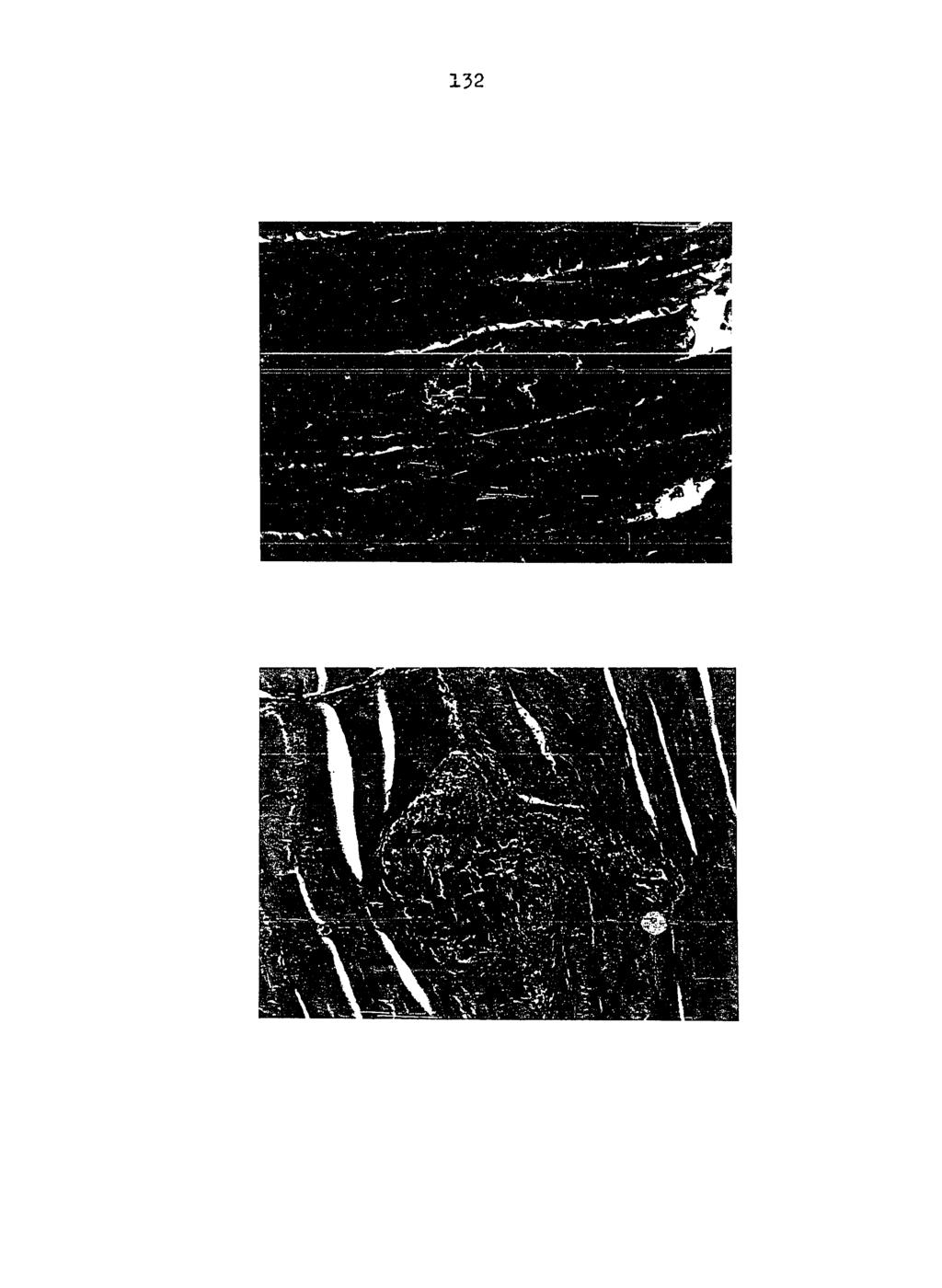
136
137 Plate 9 Sectioned gizzard removed from pigeon infected with Haemoproteus sacharovi 17. Chronic inflammatory condition and pressure atrophy of muscular tissue in gizzard (Mallory's triple stain).

138 134
PLASMODIUM MODULE 39.1 INTRODUCTION OBJECTIVES 39.2 MALARIAL PARASITE. Notes
 Plasmodium MODULE 39 PLASMODIUM 39.1 INTRODUCTION Malaria is characterized by intermittent fever associated with chills and rigors in the patient. There may be enlargement of the liver and spleen in the
Understanding Epidemics Section 3: Malaria & Modelling
 Understanding Epidemics Section 3: Malaria & Modelling PART B: Biology Contents: Vector and parasite Biology of the malaria parasite Biology of the anopheles mosquito life cycle Vector and parasite Malaria
A:Malaria (Plasmodium species) Plasmodium falciparum causes malignant tertian malaria P. malariae: causes Quartan malaria P. vivax: causes benign
 Plasmodium falciparum causes malignant tertian malaria P. malariae: causes Quartan malaria P. vivax: causes benign") A:Malaria (Plasmodium species) Plasmodium falciparum causes malignant tertian malaria P. malariae: causes Quartan malaria P. vivax: causes benign tertian malaria P. ovale: causes benign tertian malaria
A:Malaria (Plasmodium species) Plasmodium falciparum causes malignant tertian malaria P. malariae: causes Quartan malaria P. vivax: causes benign tertian malaria P. ovale: causes benign tertian malaria
BIO Parasitology Spring 2009
 BIO 475 - Parasitology Spring 2009 Stephen M. Shuster Northern Arizona University http://www4.nau.edu/isopod Lecture 10 Malaria-Life Cycle a. Micro and macrogametocytes in mosquito stomach. b. Ookinete
Malaria. This sheet is from both sections recording and includes all slides and diagrams.
 Malaria This sheet is from both sections recording and includes all slides and diagrams. Malaria is caused by protozoa family called plasmodium (Genus) mainly affect blood system specially RBCs and each
Blood protozoan: Plasmodium
 Blood protozoan: Plasmodium Dr. Hala Al Daghistani The causative agent of including Plasmodium vivax P. falciparum P. malariae P. ovale. malaria in humans: four species are associated The Plasmodium spp.
Parasites of Small Mammals in Grand Teton National Park: Babesia and Hepatozoon
 University of Wyoming National Park Service Research Center Annual Report Volume 19 19th Annual Report, 1995 Article 13 1-1-1995 Parasites of Small Mammals in Grand Teton National Park: Babesia and Hepatozoon
Blood protozoan: Plasmodium
 Blood protozoan: Plasmodium The causative agent of including Plasmodium vivax P. falciparum P. malariae P. ovale. malaria in humans:four species are associated The Plasmodium spp. life cycle can be divided
Infecting Anopheles stephensi With Rodent Malaria Parasites Alida Coppi & Photini Sinnis
 Infecting Anopheles stephensi With Rodent Malaria Parasites Alida Coppi & Photini Sinnis A. Reagents: 1. DMEM or RPMI DMEM (4.5g/L glucose) RPMI 1640 Cellgro #MT-10-017-CM Cellgro #MT-10-040-CM 2. Giemsa
A. Effect upon human culture 1. Control of malaria has contributed to world=s population explosion 2. Africans brought to U.S.
 VI. Malaria A. Effect upon human culture 1. Control of malaria has contributed to world=s population explosion 2. Africans brought to U.S. because they were resistant to malaria & other diseases 3. Many
Giardia and Apicomplexa. G. A. Lozano UNBC
 Giardia and Apicomplexa G. A. Lozano UNBC NINE Protozoan diseases/parasites Ciliphora, Ichthyophthirius, Ick Sarcomastigophora, Giardia, giardiasis Apicomplexa: Eimeria, Toxoplasma, Sarcocystis, Cryptosporidium.
Phylum:Apicomplexa Class:Sporozoa
 Phylum:Apicomplexa Class:Sporozoa The most characteristic features of sporozoa are 1-unique appearance of most protozoa makes it possible for knowledge able person to identifiy them to level of genus and
9 Parasitology 9 EXERCISE EQA. Objectives EXERCISE
 0696T_c09_81-90.qxd 07/01/2004 23:19 Page 81 EXERCISE 9 Parasitology Exercise Pre-Test Attempt to answer the following questions before starting this exercise. They will serve as a guide to important concepts.
cyst&' appeared to be of two kinds-one smaller and Smnith "is inclined to regard these epithelial cell parasites as
 COCCIDIA IN SUBEPITHELIAL INFECTIONS OF THE INTESTINES OF BIRDS PHILIP B. HADLEY From the Agricultural Experiment Station of the Rhode Island State College' Received for publication, July 10, 1916 In an
GOJRATI, Hassan Ali Navvab, 1935 EPIZOOTIOLOGICAL SURVEY Of AVIAN I:iALARIA IN THE HAWAIIAN ISLANDS. ' University of Hawaii, Ph.D.
 71.,..12,222 GOJRATI, Hassan Ali Navvab, 1935 EPIZOOTIOLOGICAL SURVEY Of AVIAN I:iALARIA IN THE HAWAIIAN ISLANDS. ' University of Hawaii, Ph.D., 1970 Entomol,ogy University Microfilms, A XEROX Company,
Infection of Haematozoan Parasites Found in Birds of NWFP (Pakistan)
") Pakistan Journal of Biological Sciences 8 (1): 1-5,2005 ISSN 1028-8880 O 2005 Asian Network for Scientific Infomation Infection of Haematozoan Parasites Found in Birds of NWFP (Pakistan) Rukhsana Talat
Pakistan Journal of Biological Sciences 8 (1): 1-5,2005 ISSN 1028-8880 O 2005 Asian Network for Scientific Infomation Infection of Haematozoan Parasites Found in Birds of NWFP (Pakistan) Rukhsana Talat
23 Plasmodium coatneyi Eyles, Fong, Warren, Guinn, Sandosham, and Wharton, 1962
 23 Plasmodium coatneyi Eyles, Fong, Warren, Guinn, Sandosham, and Wharton, 1962 IN the course of studies on simian malaria begun by the late Dr. Don Eyles in Malaya, he and his co-workers isolated a new
Exotic Hematology Lab Leigh-Ann Horne, LVT, CWR Wildlife Center of Virginia
 Exotic Hematology Lab Leigh-Ann Horne, LVT, CWR Wildlife Center of Virginia lhorne@wildlifecenter.org Anne Lynch, LVT Cedarcrest Animal Clinic amllvt9@gmail.com Introduction While the general set-up for
Answer: Europeans risked death by disease when if they left the sea coast and entered the interior of the African continent.
 XXI Malaria [MAL = bad; ARIA = air] 2005 A. Order Haemosporida, Family Plasmodiidae 1. Live in vertebrate tissues and blood 2. SCHIZOGONY (asexual reproduction) in vertebrates 3. SPOROGONY (sexual reproduction)
EFFECTS OF HOST AND SPATIAL FACTORS ON A HAEMOPROTEID COMMUNITY IN MOURNING DOVES FROM WESTERN TEXAS
 Journal of Wildlife Diseases, 26(4), 1990, pp. 435-441 Wildlife Disease Association 1990 EFFECTS OF HOST AND SPATIAL FACTORS ON A HAEMOPROTEID COMMUNITY IN MOURNING DOVES FROM WESTERN TEXAS Ralph 0. Godfrey,
THE ABUNDANCE AND INFECTION STATUS OF ANOPHELES MOSQUITOES IN LOUDOUN COUNTY, VIRGINIA
 THE ABUNDANCE AND INFECTION STATUS OF ANOPHELES MOSQUITOES IN LOUDOUN COUNTY, VIRGINIA Andrew Lima Clarke (Manassas, VA) Priya Krishnan ODU M.S. candidate (Richmond, VA) Objectives To determine: 1) the
XXI. Malaria [MAL = bad; ARIA = air] (Chapter 9) 2008 A. Order Haemosporida, Family Plasmodiidae 1. Live in vertebrate tissues and blood 2.
![XXI. Malaria [MAL = bad; ARIA = air] (Chapter 9) 2008 A. Order Haemosporida, Family Plasmodiidae 1. Live in vertebrate tissues and blood 2.](/thumbs/80/81826026.jpg "XXI. Malaria [MAL = bad; ARIA = air] (Chapter 9) 2008 A. Order Haemosporida, Family Plasmodiidae 1. Live in vertebrate tissues and blood 2.") XXI. Malaria [MAL = bad; ARIA = air] (Chapter 9) 2008 A. Order Haemosporida, Family Plasmodiidae 1. Live in vertebrate tissues and blood 2. SCHIZOGONY (asexual reproduction) in vertebrates 3. SPOROGONY
XXI. Malaria [MAL = bad; ARIA = air] (Chapter 9) 2008 A. Order Haemosporida, Family Plasmodiidae 1. Live in vertebrate tissues and blood 2. SCHIZOGONY (asexual reproduction) in vertebrates 3. SPOROGONY
Malaria parasites of lemurs
 Annales de Parasitologie (Paris), 1975, t. 50, n 4, pp. 409 à 418 Malaria parasites of lemurs by P. C. C. GARNHAM * and G. UILENBERG ** * Imperial College of Science and Technology, Ashurst Lodge, Ascot,
Parasitology Amoebas. Sarcodina. Mastigophora
 Parasitology Amoebas Sarcodina Entamoeba hisolytica (histo = tissue, lytica = lyse or break) (pathogenic form) o Trophozoite is the feeding form o Life Cycle: personfeces cyst with 4 nuclei with thicker
IDENTITY AND PREVALENCE OF BLOOD PARASITES IN WILD-CAUGHT BIRDS FROM MADAGASCAR
 IDENTITY AND PREVALENCE OF BLOOD PARASITES IN WILD-CAUGHT BIRDS FROM MADAGASCAR By AMY FRANCES SAVAGE A THESIS PRESENTED TO THE GRADUATE SCHOOL OF THE UNIVERSITY OF FLORIDA IN PARTIAL FULFILLMENT OF THE
Temperature Gradient in the Egg-Laying Activities of the Queen Bee
 The Ohio State University Knowledge Bank kb.osu.edu Ohio Journal of Science (Ohio Academy of Science) Ohio Journal of Science: Volume 30, Issue 6 (November, 1930) 1930-11 Temperature Gradient in the Egg-Laying
HISTOPATHOLOGY. Introduction:
 Introduction: HISTOPATHOLOGY Goats and sheep are the major domestic animal species in India. Much of the economy of the country has been depend upon the domestication of these animals. Especially economy
Malaria parasites: virulence and transmission as a basis for intervention strategies
 Malaria parasites: virulence and transmission as a basis for intervention strategies Matthias Marti Department of Immunology and Infectious Diseases Harvard School of Public Health The global malaria burden
Arrested oocyst maturation in Plasmodium parasites. lacking type II NADH:ubiquinone dehydrogenase
 Supplemental Information for: Arrested oocyst maturation in Plasmodium parasites lacking type II NADH:ubiquinone dehydrogenase Katja E. Boysen and Kai Matuschewski Contents: - Supplemental Movies 1 and
Keys to the avian malaria parasites
 https://doi.org/10.1186/s12936-018-2359-5 Malaria Journal REVIEW Open Access Keys to the avian malaria parasites Gediminas Valkiūnas * and Tatjana A. Iezhova Abstract Background: Malaria parasites (genus
Sex-specific effects of an avian malaria parasite on an insect vector: support for the resource limitation hypothesis
 Ecology, 93(11), 2012, pp. 2448 2455 Ó 2012 by the Ecological Society of America Sex-specific effects of an avian malaria parasite on an insect vector: support for the resource limitation hypothesis JESSICA
What causes heartworm disease?
 Heartworm Disease: What causes heartworm disease? Heartworm disease (dirofilariasis) is a serious and potentially fatal disease in dogs and cats. It is caused by a blood-borne parasite called Dirofilaria
Heartworm Disease in Dogs
 Kingsbrook Animal Hospital 5322 New Design Road, Frederick, MD, 21703 Phone: (301) 631-6900 Website: KingsbrookVet.com What causes heartworm disease? Heartworm Disease in Dogs Heartworm disease or dirofilariasis
CANINE HEARTWORM DISEASE
 ! CANINE HEARTWORM DISEASE What causes heartworm disease? Heartworm disease (dirofilariasis) is a serious and potentially fatal disease in dogs. It is caused by a blood-borne parasite called Dirofilaria
Malaria in the Mosquito Dr. Peter Billingsley
 Malaria in the Mosquito Senior Director Quality Systems and Entomology Research Sanaria Inc. Rockville MD. 1 Malaria: one of the world s foremost killers Every year 1 million children die of malaria 250
FOOTEDNESS IN DOMESTIC PIGEONS
 FOOTEDNESS IN DOMESTIC PIGEONS I BY HARVEY I. FISHER N studies of the landing forces of Domestic Pigeons (Columba Zivia) it was noted (Fisher, 1956a, 19566) that the birds did not always land si- multaneously
LINKAGE OF ALBINO ALLELOMORPHS IN RATS AND MICE'
 LINKAGE OF ALBINO ALLELOMORPHS IN RATS AND MICE' HORACE W. FELDMAN Bussey Inslitutim, Harvard Univwsity, Forest Hills, Boston, Massachusetts Received June 4, 1924 Present concepts of some phenomena of
EGG production of turkeys is not important
 A Study of Egg Production in Bronze Turkeys S. J. MAESDEN National Agricultural Research Center, Beltsville, Maryland EGG production of turkeys is not important commercially but good egg production during
Some aspects of wildlife and wildlife parasitology in New Zealand
 Some aspects of wildlife and wildlife parasitology in New Zealand Part 3/3 Part three: Kiwis and aspects of their parasitology Kiwis are unique and unusual in many ways. For a comprehensive and detailed
Sporozoae: Plasmodium.
 Sporozoae: Plasmodium. Coccidian. Asexual division in Man (Schizogony), sexual division in the mosquito (sporogony). Plasmodium vivax > P. falciparum > P. malariae > P. ovale as a cause of malaria. P.
HOST INFLUENCES ON SOME HAEMOSPORIDIAN PARASITES "
 HOST INFLUENCES ON SOME HAEMOSPORIDIAN PARASITES " CLAY G. HUFF Naval Medical Research Institute, Bethesdu, Maryla?zd HE effects of microbes on higher animals have certainly and under- T standably received
Malaria parasites of rodents of the Congo (Brazzaville) :
 :") Annales de Parasitologie (Paris), 1976, t. 51, n 6, pp. 637 à 646 Malaria parasites of rodents of the Congo (Brazzaville) : Plasmodium cbabaudi adami subsp. nov. and Plasmodium vinckei lentum Landau, Michel,
Annales de Parasitologie (Paris), 1976, t. 51, n 6, pp. 637 à 646 Malaria parasites of rodents of the Congo (Brazzaville) : Plasmodium cbabaudi adami subsp. nov. and Plasmodium vinckei lentum Landau, Michel,
Burn Infection & Laboratory Diagnosis
 Burn Infection & Laboratory Diagnosis Introduction Burns are one the most common forms of trauma. 2 million fires each years 1.2 million people with burn injuries 100000 hospitalization 5000 patients die
High Mortality of a Population of Cowbirds Wintering at Columbus, Ohio
 The Ohio State University Knowledge Bank kb.osu.edu Ohio Journal of Science (Ohio Academy of Science) Ohio Journal of Science: Volume 67, Issue 1 (January, 1967) 1967-01 High Mortality of a Population
Wheat and Wheat By-Products for Laying Hens
 South Dakota State University Open PRAIRIE: Open Public Research Access Institutional Repository and Information Exchange Bulletins South Dakota State University Agricultural Experiment Station 5-1-1934
Malaria. Malaria is known to kill one child every 30 sec, 3000 children per day under the age of 5 years.
 Malaria Mal-air It is a world wide distribution disease acute or chronic characterized by fever,anemia & spleenomegaly occurs where anopheles mosquito are present & caused by genus plasmodium,which is
Note: The following article is used with permission of Dr. Sonia Altizer.
 PROFESSIONAL BUTTERFLY FARMING PART I - By Nigel Venters (Contributing Author: Dr. Sonia Altizer) Note: The following article is used with permission of Dr. Sonia Altizer. Monarch Health Program, University
Systemic Apicomplexans. Toxoplasma
 Systemic Apicomplexans Toxoplasma Protozoan Groups Historically, protozoa have been grouped by mode of motility. Flagellates Hemoflagellates Trypanosoma cruzi Leishmania infantum Mucoflagellates Tritrichomonas
沖繩産シリケンイモリより発見されたへモグレガリンの 1 新種 Haemogregarina shirikenimori. Citation 熱帯医学 Tropical medicine 19(2). p105-
. p105-") NAOSITE: Nagasaki University's Ac Title Author(s) 沖繩産シリケンイモリより発見されたへモグレガリンの 1 新種 Haemogregarina shirikenimori 宮田, 彬 Citation 熱帯医学 Tropical medicine 19(2). p105- Issue Date 1977-06-30 URL http://hdl.handle.net/10069/4222
NAOSITE: Nagasaki University's Ac Title Author(s) 沖繩産シリケンイモリより発見されたへモグレガリンの 1 新種 Haemogregarina shirikenimori 宮田, 彬 Citation 熱帯医学 Tropical medicine 19(2). p105- Issue Date 1977-06-30 URL http://hdl.handle.net/10069/4222
FOOD HABITS OF NESTING COOPER S HAWKS AND GOSHAWKS IN NEW YORK AND PENNSYLVANIA
 FOOD HABITS OF NESTING COOPER S HAWKS AND GOSHAWKS IN NEW YORK AND PENNSYLVANIA BY HEINZ MENG UCH has been written about the food habits of our birds of prey. M Through crop and stomach content analyses
Laboratory 7 The Effect of Juvenile Hormone on Metamorphosis of the Fruit Fly (Drosophila melanogaster)
") Laboratory 7 The Effect of Juvenile Hormone on Metamorphosis of the Fruit Fly (Drosophila melanogaster) (portions of this manual were borrowed from Prof. Douglas Facey, Department of Biology, Saint Michael's
Laboratory 7 The Effect of Juvenile Hormone on Metamorphosis of the Fruit Fly (Drosophila melanogaster) (portions of this manual were borrowed from Prof. Douglas Facey, Department of Biology, Saint Michael's
Eukaryotic Organisms
 Eukaryotic Organisms A Pictoral Guide of Supportive Illustrations to accompany Select Topics on Eukaryotic Oranisms Bacteria (Not Shown) Agent of Disease Reservoir Vector By Noel Ways Favorable Environmental
$? 479 THE FUNCTION OF M. DEPRESSOR CAUDAE AND M. CAUDOFEMORALIS IN PIGEONS
 Oct.1 $? 479 THE FUNCTION OF M. DEPRESSOR CAUDAE AND M. CAUDOFEMORALIS IN PIGEONS BY HARVEY I. FISHER THE usual method of determining the function of a muscle is by gross dissection and study of attachments.
MclL amqxq Sex Ratio in Wild Birds 85
 Vol. 57] 194o J MclL amqxq Sex in Wild Birds 85 SEX RATIO IN WILD BIRDS BY E. A. MCILHENNY ORNITHOLOGISTS generally concede that there is considerable variation in the sex ratio of some species of wild
4 Life Cycle and the Phenomenon of Relapse
 4 Life Cycle and the Phenomenon of Relapse A. LIFE CYCLE IN the Primate Host. The cycle of malaria in the primate host is initiated by the inoculation of sporozoites by the female mosquito when she punctures
Proteocephalus filicollis (Rud. 1810) in the Netherlands
 in the Netherlands") Proteocephalus filicollis (Rud. 1810) in the Netherlands by J.J. Willemse AND A.L.M. Veltman Zoological Laboratory, University of Amsterdam INTRODUCTION in another glass dish containing about 50 specimens
Proteocephalus filicollis (Rud. 1810) in the Netherlands by J.J. Willemse AND A.L.M. Veltman Zoological Laboratory, University of Amsterdam INTRODUCTION in another glass dish containing about 50 specimens
Ectoparasites Myobia musculi Radfordia affinis Radfordia ensifera
 Ectoparasites Fleas, ticks, and lice are uncommon in modern laboratory facilities, but may be seen on wild or feral rodents. Most ectoparasite infestations seen in rats and mice used for research are various
EUROPEAN STARLING HOUSE FINCH
 EUROPEAN STARLING Scientific Name: Sturnus vulgaris Size: 7.5-8.5 " (19-21 cm) Shape: Short tail; plump body Color: Blackbird with shiny feathers; yellow bill in springtime. Habitat: Cities, parks, farms,
CIWF Response to the Coalition for Sustainable Egg Supply Study April 2015
 CIWF Response to the Coalition for Sustainable Egg Supply Study April 2015 The Coalition for Sustainable Egg Supply study seeks to understand the sustainability impacts of three laying hen housing systems
Apicomplexans Apicomplexa Intro
 Apicomplexans Apicomplexa Intro Cryptosporidium Apicomplexan Select Characteristics Gliding motility Apical Complex organelle for invasion of host cell Life cycle alternates b/w sexual and asexual phases
Pesky Ectoparasites. Insecta fleas, lice and flies. Acari- ticks and mites
 Pesky Ectoparasites Parasite control should be at the forefront of every pet owner s life as all animals have the propensity to contract numerous ones at one stage or another. They are a challenge to the
Comparing Life Cycles
 Image from Wikimedia Commons Pre-Visit Activity Grade Two Comparing Life Cycles Specific Learning Outcomes 2-1-01: Use appropriate vocabulary related to the investigations of growth and changes in animals.
A New Haemoproteus Species (Haemosporida: Haemoproteidae) from the Endemic Galapagos Dove Zenaida galapagoensis
 from the Endemic Galapagos Dove Zenaida galapagoensis") A New Haemoproteus Species (Haemosporida: Haemoproteidae) from the Endemic Galapagos Dove Zenaida galapagoensis, with Remarks on the Parasite Distribution, Vectors, and Molecular Diagnostics Author(s):
A New Haemoproteus Species (Haemosporida: Haemoproteidae) from the Endemic Galapagos Dove Zenaida galapagoensis, with Remarks on the Parasite Distribution, Vectors, and Molecular Diagnostics Author(s):
RABIES CONTROL INTRODUCTION
 RABIES CONTROL INTRODUCTION Throughout human history, few illnesses have provoked as much anxiety as has rabies. Known as a distinct entity since at least 500 B.C., rabies has been the subject of myths
ECHINOCOCCOSIS. By Dr. Ameer kadhim Hussein. M.B.Ch.B. FICMS (Community Medicine).
.") ECHINOCOCCOSIS By Dr. Ameer kadhim Hussein. M.B.Ch.B. FICMS (Community Medicine). INTRODUCTION Species under genus Echinococcus are small tapeworms of carnivores with larval stages known as hydatids proliferating
ECHINOCOCCOSIS By Dr. Ameer kadhim Hussein. M.B.Ch.B. FICMS (Community Medicine). INTRODUCTION Species under genus Echinococcus are small tapeworms of carnivores with larval stages known as hydatids proliferating
DISEASES OF AQUATIC ORGANISMS Dis. aquat. Org.
 Vol. 7: 149-153, 1989 DISEASES OF AQUATIC ORGANISMS Dis. aquat. Org. Published October 26 Developmental cycle of chelonian haemogregarines in leeches with extra-intestinal multiple sporozoite oocysts and
THE MICROSCOPE PATHOGEN IDENTIFICATION
 CONTENTS 5 ABOUT THE AUTHOR 5 ACKNOWLEDGEMENTS 6 OVERVIEW 6 What is the Purpose of this Book? 6 What are the Limitations of Light Microscopy as a Diagnostic Tool? 7 When Should I Contact a Veterinarian?
Life Cycle, Pathogenicity and Prophylaxis of Plasmodium vivax
 Discipline Courses-I Semester-I Paper: Divesity and Evolution of Choradata-I Lesson: Life Cycle, Pathogenicity and Prophylaxis of Plasmodium vivax Lesson Developer: Anubha Das College/Department: Zakir
S7L2_Genetics and S7L5_Theory of Evolution (Thrower)
") Name: Date: 1. Single-celled organisms can reproduce and create cells exactly like themselves without combining genes from two different parent cells. When they do this, they use a type of A. asexual reproduction.
Name: Date: 1. Single-celled organisms can reproduce and create cells exactly like themselves without combining genes from two different parent cells. When they do this, they use a type of A. asexual reproduction.
Diurnal variation in microfilaremia in cats experimentally infected with larvae of
 Hayasaki et al., Page 1 Short Communication Diurnal variation in microfilaremia in cats experimentally infected with larvae of Dirofilaria immitis M. Hayasaki a,*, J. Okajima b, K.H. Song a, K. Shiramizu
Medical and Veterinary Entomology
 Medical and Veterinary Entomology An eastern treehole mosquito, Aedes triseriatus, takes a blood meal. Urbana, Illinois, USA Alexander Wild Photography Problems associated with arthropods 1) Psychological
LABORATORY. The Protozoa. At the Bench
 LABORATORY Laboratory 8, Page 1 8 The Protozoa Introduction: The protozoa are unicellular animals that are classified on the basis of the organelles used for locomotion (flagella, pseudopodia, cilia or
Medical Parasitology (EEB 3895) Lecture Exam #2
 Lecture Exam #2") 1 Name November 2016 Medical Parasitology (EEB 3895) Lecture Exam #2 Read through the exam once before you begin. Read the questions CAREFULLY; be certain to provide all of the information requested. In
1 Name November 2016 Medical Parasitology (EEB 3895) Lecture Exam #2 Read through the exam once before you begin. Read the questions CAREFULLY; be certain to provide all of the information requested. In
Parasitology Departement Medical Faculty of USU
 Malaria Mechanism of infection Parasitology Departement Medical Faculty of USU Introduction Malaria parasites Phylum Order Suborder Family Genus Species : : Apicomplexa : Eucoccidiida : Haemosporida :
Arthropod Parasites of Veterinary importance 2015
 Arthropod Parasites of Veterinary importance 2015 VPM-122 Laboratory 6 Prepared by Spencer J. Greenwood PhD, DVM Dept. of Biomedical Sciences Room 2332N AVC North Annex sgreenwood@upei.ca Office phone:
ON THE HAEMOCYTOZOA OF BIRDS. ]3y EUGENE L. OPIE.
![ON THE HAEMOCYTOZOA OF BIRDS. ]3y EUGENE L. OPIE.](/thumbs/94/121052487.jpg "ON THE HAEMOCYTOZOA OF BIRDS. ]3y EUGENE L. OPIE.") ON THE HAEMOCYTOZOA OF BIRDS. ]3y EUGENE L. OPIE. (From the Clinical Laboratary of the John8 Hopkins Unive~'sity and Hospital.) PLATE IX. Since the discovery of the h~emocytozoa of malaria] fever considerable
ON THE HAEMOCYTOZOA OF BIRDS. ]3y EUGENE L. OPIE. (From the Clinical Laboratary of the John8 Hopkins Unive~'sity and Hospital.) PLATE IX. Since the discovery of the h~emocytozoa of malaria] fever considerable
GROWTH IN THE LARVAE OF TENTHREDINIDAE
 355 GROWTH IN THE LARVAE OF TENTHREDINIDAE BY HERBERT W. MILES, M.Sc, PH.D. (Victoria University of Manchester.) {Received 2 th January, 1931.) FROM a study of the larvae of some twenty-eight species of
New species of haemosporidian parasites (Haemosporida) from African rainforest birds, with remarks on their classification
 from African rainforest birds, with remarks on their classification") Parasitol Res (2008) 103:1213 1228 DOI 10.1007/s00436-008-1118-x ORIGINAL PAPER New species of haemosporidian parasites (Haemosporida) from African rainforest birds, with remarks on their classification
Parasitol Res (2008) 103:1213 1228 DOI 10.1007/s00436-008-1118-x ORIGINAL PAPER New species of haemosporidian parasites (Haemosporida) from African rainforest birds, with remarks on their classification
THE EFFECT OF MUTILATION ON THE TAPEWORM TAENIA TAENIAEFORMIS
 THE EFFECT OF MUTILATION ON THE TAPEWORM TAENIA TAENIAEFORMIS JOE N. MILLER AND WM. P. BUNNER The reader is undoubtedly aware of work which has been done by Child (1910) and others in mutilating certain
ON THE TRANSPLANTABILITY OF THE LARVA OF TEh'IA CRASSICOLLIS AND THE PROBABLE R~LE OF THE LIVER IN CYSTICERCUS DISEASE OF RATS
 ON THE TRANSPLANTABILITY OF THE LARVA OF TEh'IA CRASSICOLLIS AND THE PROBABLE R~LE OF THE LIVER IN CYSTICERCUS DISEASE OF RATS E'. 1). BULLOCI< AND M. 1%. CURTIS (Prom Coltil?lhin Uiiroersity, Iiistitute
BROOD REDUCTION IN THE CURVE-BILLED THRASHER By ROBERTE.RICKLEFS
 Nov., 1965 505 BROOD REDUCTION IN THE CURVE-BILLED THRASHER By ROBERTE.RICKLEFS Lack ( 1954; 40-41) has pointed out that in species of birds which have asynchronous hatching, brood size may be adjusted
A FAMILY OF SPOTTED Q. I. SIMPSON AND W. E. CASTLE1
 A FAMILY OF SPOTTED NEGROES Q. I. SIMPSON AND W. E. CASTLE1 IT is the purpose of this note to put on record an interesting variation in human skin color which made its appearance as a mutation or sport
B SARATCHANDRA Department of Zoology, Andhra University, Waltair , India
 Proc, Indian Acad. Sci. [Anirn. Sci.), Vol. 90, Number 4, July 1981, pp. 365-371. Printed in India. Two new haemogregarines, Haemogregarina waltairensis n, Spa from Caiotes versicolor (Daudin) and H. ganapatii
(Hemorrhagic Septicemia of Fowls) By ROBERT GRAHAM. A Brief Statement of the Cause, Symp" toms, Lesions, and Preventive Measures
 By ROBERT GRAHAM. A Brief Statement of the Cause, Symp toms, Lesions, and Preventive Measures") Fowl Cholera (Hemorrhagic Septicemia of Fowls) By ROBERT GRAHAM A Brief Statement of the Cause, Symp" toms, Lesions, and Preventive Measures Chickens with fowl cholera often sit quietly with necks contracted
Fowl Cholera (Hemorrhagic Septicemia of Fowls) By ROBERT GRAHAM A Brief Statement of the Cause, Symp" toms, Lesions, and Preventive Measures Chickens with fowl cholera often sit quietly with necks contracted
THE POULTRY ENTERPRISE ON KANSAS FARMS
 THE POULTRY ENTERPRISE ON KANSAS FARMS SUMMARY The poultry enterprise in Kansas is taking rank as a major enterprise on an increasingly large number of farms, especially in the eastern two-thirds of the
Avian Reproductive System Female
 extension Avian Reproductive System Female articles.extension.org/pages/65372/avian-reproductive-systemfemale Written by: Dr. Jacquie Jacob, University of Kentucky For anyone interested in raising chickens
The Comparative Study of the Blood Cellular Composition in Muscovy Ducks in Nigeria
 International Journal of Poultry Science 9 (9): 86-841, 2010 ISSN 1682-856 Asian Network for Scientific Information, 2010 The Comparative Study of the Blood Cellular Composition in Muscovy Ducks in Nigeria
INHERITANCE OF BODY WEIGHT IN DOMESTIC FOWL. Single Comb White Leghorn breeds of fowl and in their hybrids.
 440 GENETICS: N. F. WATERS PROC. N. A. S. and genetical behavior of this form is not incompatible with the segmental interchange theory of circle formation in Oenothera. Summary.-It is impossible for the
Selecting Laying Hens
 Selecting Laying Hens Authors Thompson, R. B. Publisher College of Agriculture, University of Arizona (Tucson, AZ) Download date 26/04/2018 15:39:49 Link to Item http://hdl.handle.net/10150/196570 of COLLEGE
Bird-X Goose Chase / Bird Shield Testing Information For Use On: 1. Apples 2. Cherries 3. Grapes 4. Blueberries 5. Corn 6. Sunflowers 7.
 Bird-X Goose Chase / Bird Shield Testing Information For Use On: 1. Apples 2. Cherries 3. Grapes 4. Blueberries 5. Corn 6. Sunflowers 7. Water 8. Structures 9. Rice 10. Turf & Ornamentals 1. Apples Field
PULLORUM DISEASE OF CHICKS
 PULLORUM DISEASE OF CHICKS (Bacillary White Diarrhea) By ROBERT GRAHAM Young chicks fatally affected with pullorum disease UNIVERSITY OF ILLINOIS COLLEGE OF AGRICULTURE : AGRICULTURAL EXPERIMENT STATION
EVALUATION OF A METHOD FOR ESTIMATING THE LAYING RATE OF BROWN-HEADED COWBIRDS
 EVALUATION OF A METHOD FOR ESTIMATING THE LAYING RATE OF BROWN-HEADED COWBIRDS D. M. SCOTT AND C. DAVISON ANKNEY Department of Zoology, University of Western Ontario, London, Ontario, Canada N6A 5B7 AnSTI
Capture and Marking of Birds: Field Methods for European Starlings
 WLF 315 Wildlife Ecology I Lab Fall 2012 Capture and Marking of Birds: Field Methods for European Starlings Objectives: 1. Introduce field methods for capturing and marking birds. 2. Gain experience in
Reedy Fork Dairy Farm Parasitology Report Fall 2016
 Reedy Fork Dairy Farm Parasitology Report Fall 2016 By Catherine Si and Nick De Castro History: Reedy Fork Dairy Farm, certified organic since 2007, has a herd of 200 cattle, which are mostly Jersey/ Holstein
Hepatozoon-Like Parasite (Schizonts) in the Myocardium of the Domestic Cat
 in the Myocardium of the Domestic Cat") Vet. Path. 10: 185-190 (1973) Hepatozoon-Like Parasite (Schizonts) in the Myocardium of the Domestic Cat U. KLOPFER, T.A. NOBEL and F. NEUMANN Department of Pathology, Kimron Veterinary Institute, affiliated
Vet. Path. 10: 185-190 (1973) Hepatozoon-Like Parasite (Schizonts) in the Myocardium of the Domestic Cat U. KLOPFER, T.A. NOBEL and F. NEUMANN Department of Pathology, Kimron Veterinary Institute, affiliated
Chapter 1 COPYRIGHTED MATERIAL. Introduction to Veterinary Pathology. What is pathology? Who does pathology?
 What is pathology? Who does pathology? Chapter 1 Introduction to Veterinary Pathology Anatomic pathology Clinical pathology Microbiology Parasitology Immunology Toxicology Veterinary forensic pathology
UK HOUSE MARTIN SURVEY 2015
 UK HOUSE MARTIN SURVEY 2015 FULL INSTRUCTIONS A one-page summary of these instructions is available from www.bto.org/house-martin-resources SECTION 1: INTRODUCTION & GETTING STARTED The House Martin (Delichon
Mosquito-borne Dog Heartworm Disease 1
 ENY-628 Mosquito-borne Dog Heartworm Disease 1 J. K. Nayar and C. Roxanne Rutledge 2 Each year thousands of dogs become disabled or die from lung, heart or circulatory problems caused by heartworm disease.
Ecology of RMSF on Arizona Tribal Lands
 Ecology of RMSF on Arizona Tribal Lands Tribal Vector Borne Disease Meeting M. L. Levin Ph.D. Medical Entomology Laboratory Centers for Disease Control mlevin@cdc.gov Rocky Mountain Spotted Fever Disease
Biology and Control of Insects and Rodents Workshop Vector Borne Diseases of Public Health Importance
 Vector-Borne Diseases of Public Health Importance Rudy Bueno, Jr., Ph.D. Director Components in the Disease Transmission Cycle Pathogen Agent that is responsible for disease Vector An arthropod that transmits