UC Riverside UC Riverside Electronic Theses and Dissertations
|
|
|
- Felicia Baker
- 5 years ago
- Views:
Transcription
1 UC Riverside UC Riverside Electronic Theses and Dissertations Title Morphology of the Immature Stages of Culicoides sonorensis Wirth and Jones (Diptera: Ceratopogonidae) With Observations on Their Biology Permalink Author Abubekerov, Lucy Anne Publication Date 2014 Peer reviewed Thesis/dissertation escholarship.org Powered by the California Digital Library University of California
2 UNIVERSITY OF CALIFORNIA RIVERSIDE Morphology of the Immature Stages of Culicoides sonorensis Wirth and Jones (Diptera: Ceratopogonidae) With Observations on Their Biology A Thesis submitted in partial satisfaction of the requirements for the degree of Master of Science in Entomology by Lucy Anne Abubekerov December 2014 Thesis Committee: Dr. Bradley A. Mullens, Chairperson Dr. Christiane Weirauch Dr. William Walton
3 The Thesis of Lucy Anne Abubekerov is approved: Committee Chairperson University of California, Riverside
4 Acknowledgements I would first like to thank Dr. Bradley Mullens, who has supported me throughout my thesis with his patience and understanding. He was always there to help and offer advice whenever I was struggling with both research and life. I could not have hoped for a better advisor and mentor and am thankful for the opportunity that he gave me. Thank you also to my committee members, Dr. Christiane Weirauch and Dr. William Walton as well as Dr. Edward Platzer who all spent their time helping me to complete my thesis and providing me with valuable insights. I would also like to thank Ryan Perry and the Hearty Lab for their help with preparing for SEM. Finally, I would like to thank my husband, Mark Abubekerov, for his support and understanding throughout this process and my mother, Mary Gagnon, for her support and for patiently reading through every draft. iii
5 Table of Contents Acknowledgements iii List of Figures....v List of Tables......vii Introduction I. Culicoides-Associated Viruses in North America...1 II. Justification for Studies on Culicoides Immatures...4 III. Historical Progression of Culicoides Immature Studies...5 IV. Historical Work on Subgenus Monoculicoides Immature Morphology...11 V. Immature Biology and Behavior of Culicoides Immatures...15 Materials, Methods, and Results 45 I. Experiments in Behavior of Immature Culicoides sonorensis...45 i. Larval Swimming Speed Analysis..45 ii. Movement of Larvae on a Mud Habitat Slope..47 iii. Lab Pupation Location Experiment..49 iv. Field Pupation Location 52 v. Pupal Behavior...56 vi. Pupal Drowning...58 II. Morphology of Culicoides sonorensis immatures 61 Discussion..76 Conclusions 96 References..99 iv
6 List of Figures 1. Epipharynx morphology of C. sonorensis Culicoides sonorensis larval instar swimming speed comparison (mean + SE) Culicoides sonorensis larval instar speed vs. average body length Culicoides sonorensis pupa in its burrow Locations of field mud samples collected for Culicoides sonorensis field pupation location study The locations of Culicoides sonorensis pupae along the shore of a dairy waste water pond in the field Culicoides sonorensis emergence relative to Chironomid line Male vs. female Culicoides sonorensis emergence based on the distance of the sample from the waterline Experiment cup to test survival of submerged Culicoides sonorensis pupae Survival rate of Culicoides sonorensis pupae submerged underwater Culicoides sonorensis L2 larvae prepared for study of internal structures on SEM pin stub mount Culicoides sonorensis egg Culicoides sonorensis larval instars 1-4 (L1-L4) Culicoides sonorensis L1 larvae (Van Ryn Colony) Culicoides sonorensis L2 larvae (Van Ryn Colony) Culicoides sonorensis L3 larvae (Van Ryn Colony) Culicoides sonorensis L4 larvae (Van Ryn Colony)...73 v
7 18. Size comparison of lab-reared (Van Ryn Colony) and field-collected (S and F Dairy) C. sonorensis fourth instar larvae (L4) Ratio comparison of lab-reared (Van Ryn Colony) and field-collected (S Dairy) C. sonorensis fourth instar larvae (L4) Respiratory horns of Culicoides sonorensis pupae (Van Ryn Colony) Pupal respiratory horn lateral protuberance of C. sonorensis viewed under a light microscope...94 vi
8 List of Tables 1. Historical Studies of Morphology of Culicoides Immature Stages Historic morphological data for the species of the subgenus Monoculicoides larvae Exposure schedule of immature Culicoides sonorensis to KOH to achieve adequate clearing for microscopic examination Comparison of larval feature of last instar C. variipennis s.l. and C. sonorensis..92 vii
9 Introduction Whether they are called no-see-ums, biting midges, gnats, punkies, or one of many other local names, Culicoides Latreille, species often are serious biting pests (Mullen & Durden, 2009). This genus is a part of the family Ceratopogonidae (order Diptera), which has 125 genera containing 6180 extant species (Borkent, 2014). Culicoides is the main one of only four genera which include blood-feeding species; the others are Leptoconops Skuse, Forcipomyia Meigen, and Austroconops Wirth and Lee. I. Culicoides-Associated Viruses in North America Culicoides sonorensis Wirth and Jones is a key North American species and a primary vector of viruses (Tabachnick, 1996). It may be a pest of livestock, but it seldom bites people and is not currently known as a major vector of disease agents which affect humans. Other mammals, particularly ruminants, are not quite so lucky. In the United States alone there are two important arboviruses: Bluetongue Virus (BTV; family Reoviridae, genus Orbivirus) and Epizootic Hemorrhagic Disease Virus (EHDV; family Reoviridae, genus Orbivirus), which plague our domestic and wild ruminant populations. They are common in the western and southern USA wherever C. sonorensis thrives (Tabachnick, 1996; Holbrook et al., 2000). These two viruses (especially BTV) have greatly impacted the agricultural community here in the United States, particularly trade with areas lacking these viruses (historically western Europe) (Tabachnick, 1996; MachLachlan & Osburn, 2006; Carpenter et al., 2008; Carpenter et al., 2009). This has spurred research into various control methods for the main vector, C. sonorensis. 1
10 Although EHDV can have a very high mortality rate in wild ruminants, it has not been as well studied as BTV. This is due to the fact that EDHV primarily affects deer, specifically the white-tailed deer, instead of farmers livestock. Recent emphasis on breeding deer for hunting, however, is changing that (Nol et al., 2008). Once a deer has contracted EHDV, it will usually exhibit a fever, anorexia, respiratory distress, weakness, swelling of the tongue and conjunctivae, and edema of the neck and head. In some cases the individuals will also show signs of extensive internal hemorrhages as well as excessive salivation, nasal discharge, and ulcers on the tongue (Spickler, 2006b). Despite the gruesome nature of EHDV and its ability to wipe out most of an entire population of deer with which C. sonorensis can be closely related (Tabachnick, 1996), Culicoides sonorensis is more famous for transmitting BTV to domestic livestock. The primary reason that BTV is the focus for those interested in C. sonorensis is that BTV affects the agricultural industry. Bluetongue virus is widespread in the various wild and domesticated ruminants which it can infect globally. It mostly can be found between latitude 35 S and 40 N and as far at 50 N in parts of North America (Dulac et al., 1989; Mellor et al., 2000). Recently even Northern Europe has experienced devastating and persistent BTV as far north as southern Sweden (Carpenter et al., 2009; Lewerin et al., 2010). The list of BTV-susceptible ruminants includes everything from sheep, goats, and cattle, to bison, elk, and deer (MacLachlan & Mayo, 2013). BTV was first described as Malarial Catarrhal Fever in 1902 when it was found in sheep in South Africa (Hutcheon, 1902). In the United States it was not found until 1952, first in Texas and then in California at which time it was termed soremuzzle. The name was due to 2
11 the fact that sheep with BTV do appear to have a rather sore snout (Hardy & Price, 1952; McKercher et al., 1953). Since Culicoides sonorensis is known to be the primary vector of BTV in the USA (Tabachnick, 1996), it follows that BTV is primarily found within C. sonorensis range. The BTV serotypes 2, 10, 11, 13, and 17 are historically common throughout the contiguous western and southern United States; it is rare or absent in the northern and northeastern US (Barber, 1979; Tabachnick, 1996). Recently many new BT serotypes have been found in the USA, including Florida, and other vectors are suspect (Johnson, 2011; MacLachlan et al., 2013). When an unfortunate ruminant has been bitten by a BTV-infected female midge, the ruminant has a chance to contract the disease. Once infected, the more susceptible animals, such as certain species of sheep and deer, will begin to show characteristic clinical signs and symptoms. These include fever, depression, excessive salivation, nasal discharge, anemia, facial edema, ulceration of the oral mucosa, coronitis, muscle weakness, secondary pneumonia, and occasionally death (Spickler, 2006a). The mortality rate in sheep typically falls within the range of 0-30% but in highly susceptible breeds of sheep the mortality rate can be as high as 100%. However, less susceptible breeds may not exhibit any symptoms at all when infected: among infected cattle, only about 5% show any signs, while the rest are subclinical and deaths are a rarity (Spickler, 2006a). Cattle can carry BTV for up to about 60 days, and so they are regarded as a reservoir host (Singer et al., 2001). BTV is still very detrimental to cattle producers, since trade restrictions are imposed on any ruminants coming from areas endemic for BTV. 3
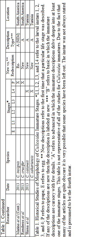
12 II. Justification for Studies on Culicoides Immatures Research into the four larval stages of Culicoides began over 200 years ago. In 1713 Reverend Derham, the Rector of Upminster in Essex, England, published the Physico-Theology: or a demonstration of the being and attributes of God, from His Works of Creation (Murphree & Mullen, 1991). Derham described and illustrated an unknown larval instar of what he called Culex minimus nigricans maculates sanguisuga, a species now known to be a member of the genus Culicoides. Unfortunately, the overall knowledge concerning the larvae of this genus is still sadly incomplete. Murphee and Mullen (1991) noted that larval descriptions exist for 51 of 144 North American species. The lack of detailed data is somewhat surprising due to the relatively large amount of research which has been done on the adults of Culicoides. There are many factors which could cause this disparity, but three likely ones are 1) the small sizes of the immature stages, 2) the fact that they inhabit opaque substrates, and 3) relative ease and glamour in researching blood-feeding adults. There are few associations between the adult and immature stages (Provonsha & McCafferty, 1975; Resh & Unzicker, 1975). It is somewhat surprising that complete descriptions do not exist for Culicoides sonorensis, even though colonies have existed for over 50 years (Jones, 1961). However, these colonies were labeled as C. variipennis until 2000 when Holbrook et al. used both adult morphological and electrophoretic analysis to separate the species C. variipennis into three species including C. occidentalis, C. sonorensis, and C. variipennis. Since 2000 though, none of the three species larval morphologies have been reexamined (Table 1). 4
13 Ignoring the immatures is even more surprising when Culicoides are compared with other insect vectors. For example, when looking to control a mosquito population, researchers will often focus on the larval population for a solution but for Culicoides, managing immatures is not usually considered seriously. This difference is possibly partially due to the simple fact that serious human diseases are not associated with species of Culicoides (Holbrook, 1996). The midges are only considered to be pestiferous instead of dangerous. Despite this, C. sonorensis still affects the agricultural industry. The lack of knowledge of immatures is especially detrimental when one is attempting to understand and control a disease system such as BTV. In 1977 Kettle eloquently explained the importance of studying Culicoides immatures when he noted that Detailed knowledge of the larval biology is essential for a full understanding of bionomics. Without this information it is impossible to construct life tables or to identify key factors limiting pest populations. Such studies are dependent on sound taxonomy, and although the characters separating the immature stages at subfamily level have been known for nearly 50 years, the taxonomy of larvae and pupae, with the exception of Forcipomyinae, is very poorly understood. III. Historical Progression of Culicoides Immature Studies In 1809 the genus Culicoides was erected by Latreille who used Culicoides punctatus (Meigen) as the type species. After Derham s 1713 work (Murphree & Mullen, 1991), it was not until Pratt (1907) that any reference to the larval stages of Culicoides was addressed in scientific articles. The earliest work on Culicoides immatures spanned from approximately 1907 until the late 1940 s. In this period the work was quite sporadic and unorganized. Those 5
14 researchers who did treat the immatures at all did not do so in any great detail and instead focused primarily on the adult stages. There was no standardization of terminology. Their small size, the difficulty in rearing them, as well as the difficulty in finding the immatures probably also contributed to the apparent disinterest. During this time the majority of the descriptions included general morphological features of only the fourth (last) larval instar, occasionally the pupae, and typically included a couple of superficial measurements such as body length or markings. The species, immature stage studied, whether it is a new description or a redescription, whether the description is basic or advanced, the country, and any special techniques used in the study can be seen in Table 1. During this time period only one research group, Carter et al. (1920), showed serious interest in the larvae. Although his observations were not as detailed as many of the more current articles, for the time Carter s work in Africa on five different species of Culicoides was groundbreaking. With no standard terminology or methods for descriptions, pre-1940 researchers decided to measure whatever they thought was relevant. It is also uncertain whether many studies were done using larvae which had been chemically cleared. It was possible to clear insects at this time, so perhaps they were (Carter et al., 1920; Lutz, 1922). Chemically clearing specimen of debris greatly facilitates observation of features such as feeding structures inside of the head capsule. In addition to a lack of detailed methodology, many of the early papers did not mention larval biology or behavior beyond noting the larval habitat. 6
15 By the late 1940 s there was a notable increase in the efforts to better understand both the immature biology and morphology for various Culicoides species. The first of the researchers to try to bring some sort of order to studies of the larval stages was Hill (1947). Although her research into the larval stages was elementary by modern standards, she did attempt to consolidate all of the previously published research into the immature stages and managed to develop a fairly comprehensive index for this. Fortunately, several subsequent researchers began to build on that foundation. Some of the most notable efforts came from Lawson (1951) and Kettle and Lawson (1952). Lawson is probably one of the most influential workers on Culicoides immatures because he did not simply focus his research on the fourth larval instar (L4). Up until his 1951 article, the only stage which had been reported on in much detail usually was the L4. Lawson actually made advanced observations on all four larval stages as well as the pupae. Even to this day very few researchers have focused on anything other than the fourth larval instars, occasionally eggs, or pupae. Lawson s (1951) descriptions of the larval morphology of C. nubeculosus remain some of the most in-depth. Lawson addressed not only the basics of external morphology but also delved into the internal morphology of the head capsule and noted the differences among the four instars. Lawson noted that the pharynges became more complex and sclerotized as the instars progressed from one stage to another. In addition to this, Lawson observed that the first instar of C. nubeculosus had a pro-thoracic pseudopod and that, unlike many other Diptera which have a larval thoracic pseudopod, the pseudopod is lost after the first instar. 7
16 He proposed that this pseudopod was used in order to help with the movement of the first larval instar, particularly when they are attempting to extricate themselves from the egg. In 1952 Kettle and Lawson published data for 28 of the Culicoides species found in Great Britain. By 1979 Kettle and Lawson had augmented this and published descriptions for a total of 46 species of Culicoides in Great Britain which included 88% of all known species found there (Knausenberger 1986, unpub. in Murphree & Mullen, 1991). Across the pond in the United States, Jamnback (1965) published rudimentary descriptions for 23 North American Culicoides species, 17 of which came from New York and of that 17, 15 of the descriptions were entirely new. Jamnback (1965) not only developed a key to species for Culicoides from New York, but also developed keys for the fourth instar larvae as well as the pupae. Jamnback included observations concerning the larval habitats. However, his descriptions of the larvae were quite basic and typically contained only brief descriptions (head capsule shape and color) along with measurements for the frontoclypeus, the total epipharyngeal comb width, and number of teeth per comb. Despite a lack of in-depth exploration into the larval stages, Jamnback s key remains one of the better regional keys to the species level. While Jamnback was working in New York, one of the most influential researchers in Culicoides morphology was working in the Soviet Union. In 1968 Glukhova began to publish the very first comparative morphological studies of Culicoides species. Glukhova began by publishing general morphological descriptions of 29 species of Culicoides found in the Palearctic, and one year later expanded upon the 8
17 descriptions for 8 of these species. By combining both the larval and adult characteristics found in the various Culicoides species, Glukhova (1977a) was able to attempt to redefine the subgenera classifications within Culicoides. Up to this point in time, subgeneric classifications within Culicoides relied solely on adult characteristics (Murphree & Mullen, 1991). By including the larval morphology as well, Glukhova was able to more accurately establish the classifications. Once she had done this, Glukhova (1977b) was then able to also redefine the relationships that Culicoides had with other genera within Ceratopogonidae by not only comparing the adults but also the larvae as well. Not only did Glukhova help to place Culicoides in the appropriate systematic location, she also helped to standardize morphological terminology of Culicoides larvae. Previously, many researchers looking into the larvae of Culicoides simply reported the measurements which they themselves thought were necessary or interesting. Some researchers such as Kettle and Lawson (1952) used some standards such as the head ratio (length:width) but it was not until Glukhova (1968a) that a standardized method of reporting was developed and widely accepted. The measurements chosen by Glukhova are still viewed as the standard today. After Glukhova developed the standards for Culicoides larvae, the research into the genus began to become more sophisticated and uniform. As can be seen in Table 1, from the 1970s until now, the quality of the descriptions began to be more consistently advanced even though there were still some more basic descriptions being produced. In 1978 Kettle and Elson published a larval key to 18 Australian Culicoides species, which was a step up from Jamnback since it included descriptions of both the third and fourth larval instars. One year later Blanton and Wirth 9
18 (1979) developed a key for 12 species using the larvae, but their key was focused on species found in Florida. After Glukhova, the next big event in traditional morphology of Culicoides came from Murphree and Mullen (1991). Before Murphree and Mullen there were some select keys which were used, but the majority of these keys were very restricted to specific areas (e.g. New York state and Florida), did not include a large variety of species in each individual key, or did not have advanced descriptions for the larvae. Murphree and Mullen (1991) developed the first attempted comprehensive key for Culicoides in North America. In their work they described 12 new species, supplemented previously unpublished descriptions for 5 species, and re-described 35 species of Culicoides, totaling 52 species. While still short of the approximately 150 North American Culicoides species, this was a large improvement (Murphree & Mullen, 1991). Not only did they have a large number of species described, Murphree and Mullen also had consistent and detailed descriptions of all of the species. What is probably most notable is the fact that they utilized scanning electron microscopy (SEM) on a few of the larvae in their studies which allowed them to describe various features with more detail. Prior to Murphree and Mullen SEM was not utilized in the study of the internal and external morphology of Culicoides larvae although it was used somewhat on adult insects (Rowley & Cornford, 1972; McKeever et al., 1988). Since Murphree and Mullen began using SEM on the larvae of Culicoides species, the majority of researchers looking into various species began to follow suit. In Argentina, Ronderos et al. (2010) has been working primarily on the fourth larval instar and the pupa of Neotropical species, using 10
19 SEM as an aid. She actually described all four larval instars of C. debilipalpis as well as the pupae, making her only the second researcher to treat all larval instars in any depth. Lawson (1951) described all larval stages of C. obsoletus and C. nubeculosus. Now that researchers have begun using more advanced techniques and are also better trained, the research into the morphology of immature stages of Culicoides can become more detailed. Day et al. (1997) and Breidenbaugh and Mullens (1999a, b) have used SEM to document the structures of the ansulae on eggs. There has been a surge in SEM use on larvae also, in order to better understand and visualize their structures (Murphree & Mullen, 1991; Huerta et al., 2001). Most notable is the recent work on the pupae of various Culicoides species; this has primarily taken place in South America by several different researchers utilizing both SEM and transmission electron microscopy (TEM) to supplement their work (Ronderos, 2010; Marino et al., 2013; Spinelli et al., 2007). All of the new and growing advances in technology have helped in the study of all these various immature stages and hopefully will continue to do so. IV. Historical Work on Subgenus Monoculicoides Immature Morphology Within the genus Culicoides, Culicoides sonorensis joins C. nubeculosus Meigen, C. occidentalis Wirth and Jones, C. parroti Kieffer, C. puncticollis Becker, C. riethi Kieffer, C. stigma Meigen, C. variipennis Coquillett, and the recently added C. grandensis Grogan and Philips to make up the subgenus Monoculicoides Khalaf. Members of subgenus Monoculicoides differ from other members of Culicoides in terms of their size and anatomy, including their internal mouthpart structures of the larvae. As with every other species of Culicoides, the members of Monoculicoides subgenus have 11
20 had their immature stages largely ignored. According to Murphree and Mullen (1991), Monoculicoides subgenus species are known for having relatively very large larvae (among Culicoides) and large mandibles which have 2 subapical teeth. The subgenus Monoculicoides particularly differ in their uniquely massive larval epipharynx with 4 combs. A pictorial representation of the epipharynx of Culicoides sonorensis can be seen in Figure 1. Based on these structures as well as the overall morphology of the head capsule in which the head is both long and narrow with its mouthparts found anteriorly, the Monoculicoides subgenus is considered to be either carnivorous or omnivorous (Thomson, 1937). In addition to this, many researchers also believe that the epipharynges of the various species of the subgenus Monoculicoides are so heavily sclerotized because they have adapted in order to grind and crush their food (Kettle & Lawson, 1952; Jamnback, 1965; Kettle, 1984). Initial research was conducted on the morphology of immature Monoculicoides in 1915, when Malloch studied C. variipennis he collected from the northeastern US. However, his descriptions were not up to modern standards primarily due to a lack of modern technology. In 1965 Jamnback recorded the larval morphology of what he called C. variipennis australis (as per the 5 subspecies described by Wirth and Jones in 1957). The larvae were collected from Missouri and could have been C. variipennis variipennis, or more likely, C. sonorensis. Jamnback did not describe in depth any of the features of the mouthparts of this species and only had two specimens, but he did note the width of the dorsal comb sclerite as well as the number of dorsal (Table 2). A far more complete and detailed description of C. variipennis came much later from Murphree and 12
21 13
22 Mullen (1991). The specimens which were used in this study came from Missouri, California, and Texas and were presumably from C. variipennis, C. sonorensis, as well as C. occidentalis; at that point C. sonorensis and C. occidentalis were still considered to be subspecies of C. variipennis. The large variations in measurements could also indicate that 2 or more species were represented. They also determined that the mandibles and epipharynx were similarly large and that the epipharynx had 4 combs with the dorsal comb (comb 1) having 13 teeth per sclerite, comb 2 and 3 with many rounded tubercles, and a large comb 4 with 6-10 rounded teeth on each side (Table 2). This research into the larvae of C. variipennis is especially important for the research into C. sonorensis due to the fact that the two were only split into separate species very recently (Holbrook et al., 2000) and the data on C. variipennis may give us insights into how C. sonorensis functions as well. Although C. variipennis was described first, the first detailed description of the morphology of the larvae of a species of the subgenus Monoculicoides was for C. nubeculosus. In 1951 Lawson was one of the first researchers to study all four larval instars of a species of Culicoides. His measurements for the L4 can be seen in table 2 and for L1 he found HL = 72 µm and HW = 54 µm, for L2 he found HL = 122 µm and HW = 88 µm, and for L3 he found HL = 200 µm and HW = 122 µm. He also mentioned that the mandibles were heavily sclerotized and that although they could be moved either together or independently, they never met. One particularly interesting point that Lawson made is that C. nubeculosus had very similar mouthparts to species of Tetraphora (= Dasyhelea). When Tetraphora was fed algae or detritus, it made scratching movements with its 14
23 mandibles, detaching pieces and then pushed the pieces into the pre-oral cavity. He postulated that the similarities in their structures suggested that C. nubeculosus larvae might feed in a similar fashion. In 1952 Kettle and Lawson had similar findings and conclusions for C. nubeculosus. Beyond these two important species, other members of the subgenus Monoculicoides have not been studied in depth. Kettle and Lawson (1952) documented the measurements for C. riethi as well as C. stigma, noting that the two species were very similar in size (Table 2). In addition to this, throughout her career, Glukhova supplemented data on some of the species as well as adding the description for C. puncticollis. Unfortunately there have been no studies on the larval stages of C. parroti or C. grandensis. Earlier work on C. stigma may also be suspect; it is very easy to confuse C. stigma with C. parroti adults. Table 2: Historic morphological data for the species of the subgenus Monoculicoides larvae Species Researcher TBL (mm) C. variipennis s.l. Murphree and Mullen 1991 C. nubeculosus C. riethi Kettle and Lawson 1952 C. stigma Kettle and Lawson 1952 HL (µm) 319 ( ) HW (µm) 219 ( ) SGW (µm) 113 (94-156) ML (µm) 64 (45-86) LAW (µm) 129 (98-159) DCW (µm) 30 (25-39) Teeth/ Comb 13 (10-15) Jamnback Lawson V. Immature Biology and Behavior of Culicoides Immatures In addition to a general lack of complete studies of Culicoides immature morphology, there is also an even greater lack of knowledge when it comes to their 15
24 behavior and biology. Some of the first studies into larval biology and behavior were published in the late 1920s and 1930s. Painter (1926) examined the larval habitats of C. furens Poey and C. phlebotomus Williston and considered control methods based on his observations. Myers (1932) studied the ecology of C. furens larvae in the Bahamas in order to help alleviate the economic problems (tourism impacts) caused by this pest. Hull et al. (1934) then delved into the seasonal prevalence and concentrations of C. dovei Hall larvae. Similarly to the studies on the immature morphology of Culicoides, the early studies on behavior and biology have been scattered and lack cohesion. In 1966 Linley first studied the basic biology of larvae of C. furens and gave an account of their feeding habits as well as the activity within a substrate. Of particular interest is Linley and Adam s (1972) work in which he showed that in the field the pupae of C. mellus Coquillett were found to be either at or just below the high tide line and were capable of surviving flooding for 4 days. Linley continued his work with various Culicoides species into the 1980s and 1990s. In 1986 Linley studied how the swimming behavior of C. variipennis larvae varied due to temperature and viscosity and in 1994 he joined Aussel to determine what the larvae of C. furens were eating. Linley examined several species, especially salt marsh biting pest species. Mullens focused his attention primarily on the main BTV vector, C. sonorensis (at the time known as C. variipennis) in southern California. Studies of C. sonorensis immature ecology included the type of dairy wastewater pond the immatures preferred and which factors determined where the highest densities of C. sonorensis were found (Mullens, 1989). Mullens also looked into how the immature stages of C. sonorensis responded to 16
25 experimental variations in the water levels, pond slope, water level in a pond, and manure pollution (Mullens & Rodriguez, 1988; 1989; 1992; Mullens, 1989). These studies showed that fluctuating the water level in a pond should cause the population of C. sonorensis to decrease by stranding larvae above the waterline. Larvae of C. sonorensis also avoid shade (Mullens & Rodriguez, 1985), steep slopes (Mullens, 1989), and are limited by low or high nutrient levels (Mullens & Rodriguez, 1988). Mullens and Lii (1987) monitored immature positions, similar to Linley s work on C. mellus, and determined where along the shoreline the different immature stages of C. sonorensis could be found. They determined that the eggs and the majority of the first larval instars could be found above the waterline, while the second, third, and fourth instars were found below the waterline. His work did not include the location of the pupae, however. Later on he elaborated on this work when he looked into the responses of the third and fourth larval instars of Culicoides sonorensis in drying mud habitats (Mullens & Rodriguez, 1992). Mullens et al. (2008) reviewed all available literature on the relationship between Culicoides species and their main natural enemy group, mermithid nematodes in the genus Heleidomermis Rubtsov. Others have contributed valuable efforts towards understanding the ecology and behaviors of other Culicoides species. While not exhaustively treated here, they include Blackwell and King (1997) who determined the vertical distribution of C. impunctatus Goetghebuer larvae, Uslu and Dik (2006) who looked into the vertical distributions of various species of Culicoides immatures in Turkey, and the work on C. denningi Foote and Pratt by Fredeen (1969) to determine their vertical distribution as well. More recently, 17
26 Swanson (2012) completed his dissertation on the ecology and phylogeny of Culicoides, which includes the first use of molecular approaches to identify immatures in North America. The work on Culicoides immature behavior and ecology is still very incomplete. The ability of Culicoides spp. to transmit devastating disease agents can cause economic problems, particularly in the western part of the United States. This makes it imperative that we learn as much as we can about Culicoides sonorensis ecology. The research which I have done is intended to expand upon the knowledge concerning C. sonorensis. I have specifically filled the gap in terms of the descriptions of larval morphology, especially regarding larval mouthparts of all four larval instars as well as the egg and pupae. In addition to this, I have added to the base of expanding knowledge concerning some aspects of the behavior and the ecology of the immature stages in the laboratory. 18
27 19
28 20
29 21
30 22
31 23
32 24
33 25
34 26
35 27
36 28
37 29
38 30
39 31
40 32
41 33
42 34
43 35
44 36
45 37
46 38
47 39
48 40
49 41
50 42
51 43
52 44
53 Materials, Methods, and Results I. Experiments in Behavior of Immature Culicoides sonorensis A few select behaviors of both the larvae and pupae of C. sonorensis were examined. The swimming speed of the larvae as well as their orientation on a mud slope were studied in the laboratory. The location of the pupae in both the field and laboratory, as well as their responses to fluctuations in water level in the laboratory, were also studied. Understanding the behaviors and biology of the immatures of C. sonorensis will help inform those seeking to utilize various control methods against them. i. Larval Swimming Speed Analysis Purpose: Little is known about the movements of Culicoides sonorensis larvae in the wild. By determining the speed at which the larva is able to move we may be able to discover how well or quickly they respond to environmental changes and predators, as well as whether the various larval instars differ in that regard. Methods: Four 9 cm plastic petri dishes were filled half way (1 cm deep) with 1% water agar. Once the agar had solidified and cooled, 10 ml of deionized (DI) water was added and they were left alone until they reached room temperature. This provided a uniform water layer of about 1 mm depth for larval speed measurements. Concentric circles spaced 5 mm apart were drawn on a black surface under an illuminated magnifier (3x). The black background aided detection of larvae, and the evenly circular fluorescent light avoided directional light cues. The agar dishes were placed on this surface with the center 45
54 of the dish in the center of the circle. Culicoides sonorensis larvae of instars 1-4 collected from the Van Ryn colony were then carefully introduced individually in the center of a dish in about µl of water using a pipette. When the swimming larvae reached the edge of one circle, timing began and the timing (nearest 0.01 sec with a stopwatch) was stopped when the larvae reached the edge of the next designated concentric circle. The times were recorded only if the larva swam steadily in a straight line perpendicular to the two lines, without stopping or slowing during its progress. The younger instars (1, 2) were timed over a smaller distance ( mm) than the older instars (3, 4; mm) in order to obtain more accurate readings. The younger instars were unlikely to travel long distances in a straight line, while the older larvae traveled too quickly for an accurate reading over anything less than a 10 mm distance. The body lengths/second were calculated using the average body length for each larval instar. Results: 14 A 12 Speed (mm/sec) D C B 0 L1 L2 L3 L4 Larval Instar Figure 2. Culicoides sonorensis larval instar swimming speed comparison (mean + SE). The mean speed of each larval instar in mm/sec; n = 10 for L1, 24 for L2, 16 for L3, and 11 for L4. Note how the overall speed increases vs. age. Means with different letters are significantly different (Tukey s HSD, p < 0.05). 46
55 3 Speed (body lengths/sec) Larval Instar Figure 3. Culicoides sonorensis larval instar speed vs. average body length. The mean speed of each larval instar in body lengths/sec. Note how the relative speed decreases vs. age. ii. Movement of Larvae on a Mud Habitat Slope Purpose: Little is known about the movements of Culicoides sonorensis larvae in the wild. Not only is it not known how the various instars orient themselves, it is also unclear as to whether or not the proleg is utilized in order to facilitate locomotion in the first larval instar. This experiment helped to determine how the larvae orient themselves on a slope with water. It also helped to elucidate the use of the proleg. Materials and Methods: Culicoides sonorensis larvae (instars 1-4) were collected from the Van Ryn colony and raised in 1% water agar dishes until they reached the desired instar (Mullens & Velten, 1994). 47
56 Natural C. sonorensis habitat mud was collected from the edge of a waste water pond at a dairy in San Jacinto, CA. It first was frozen (-20ºC for > 1 week) to kill preexisting arthropods. Mud then was placed into four-3.5 cm diameter dishes and arranged into slopes spanning the 3.5 cm, with a maximum height of 1cm. Approximately 2 ml of DI water was then added to each of the dishes, creating a small pool (0.25cm deep) of water at the base of each approximately 15º slope. The dishes were left for 2 hours to allow them to settle and reach room temperature. Live larvae were then individually placed in about µl of water in the middle of each dish (above waterline). They had the choice to move up the slope, down the slope, or laterally. Each larva was observed under a dissecting microscope for 5 minutes or until they buried themselves completely in the mud, whichever came first. Their choice of direction was recorded. This procedure was repeated for five larvae of each instar. In addition to this, the movement of the larvae was observed both on the soil as well as while under a coverslip with a minimal amount of water to determine how or if the L1 proleg was utilized. Results: All five specimens of each earlier instar (L1, L2, and L3) wandered in all directions (apparently randomly) during their time in the dishes. While wandering they appeared to try to enter into any small hole or mud irregularity they came across. Before the 5 minutes were up all of the specimens had buried themselves in the mud. The majority of the larvae managed to bury themselves above the waterline. 48
57 The five L4 specimens tended to go down the slope rather quickly. Only one of the specimens initially went upslope, before turning around and heading down the slope towards the water. Four of the five specimens began to search for a location to bury themselves once they reached the water and had successfully buried themselves below the waterline before the five minutes were up. The fifth specimen continued to travel through the water searching for a suitable site to bury itself but had not succeeded by minute 5. All four instars moved in a sinusoidal manner when there was a sufficient amount of water to move easily. When there was a minimal amount or no free water, the L2-L4 continued to attempt to move in the same sinusoidal manner but they were not very successful. Their movements were very slow and they primarily thrashed around in the one location. The first instar larvae, on the other hand, used a very distinctive movement in which they repeatedly contracted their first few body segments and moved in a straight line on the mud surface. Under the Leitz compound microscope it could be seen that these body movements were causing the proleg to move as well. Although the L1 was not able to move as quickly as it did in free water, its ability to travel was not as impeded by a lack of water as were movements of the L2-L4. iii. Lab Pupation Location Experiment Purpose: Although the field location of C. sonorensis larvae has been well documented by Mullens and Lii (1987), the location of the pupae in a substrate is unknown. By 49
58 determining where the pupae are found, various methods of control can perhaps be altered in order to better manage populations of C. sonorensis. Materials and Methods: Mud was collected from the edges of a waste water pond from a dairy in San Jacinto, CA and frozen at -20C (see above) in order to kill any living arthropods. The mud was then thawed and 150 ml placed into each of twenty plastic containers which were 8.5cm in diameter at the base. The mud was then molded to create a slope of approximately 15 degrees in order to mimic the conditions typically found in an actual waste water pond (McDermott and Mullens 2014). About 15 ml of DI water was then placed in each cup at a level of approximately 1cm from the base (depth) and allowed to settle over a 72-hour period. The water level was checked daily and consistently kept at a 1cm level throughout the experiment. Twenty mature L4 larvae, collected from the Van Ryn colony, were placed in each of 15 containers (300 larvae total). The larvae were then left alone to pupate, and dishes were checked daily using a dissecting microscope. Once a larva had pupated, its position relative to the waterline as well as the position of its respiratory horns (which are found anteriorly on the pupae) in the mud were recorded on a map of the cup. To ensure that pupae were not missed during the inspections, three of the dishes were selected at random after 7 days. Those three dishes were sectioned off into 1 cm sections relative to waterline (e.g. the 1 cm band below, 1 cm band above, etc.). The top layer of soil approximately 3 mm deep for each 1cm section was then carefully removed and placed into a dish filled with DI water. The water and mud were then agitated, 50
59 allowing the buoyant pupae to float to the surface. Pupae were then extracted using a pipette and the number of live pupae per section was recorded. The experiment was then repeated with 10 dishes with 20 larvae in each with a gentler mud slope between 5-10 degrees. In this sandy soil type, it was hypothesized that the gentler slope might create a broader band of penetrable substrate. Results: Steeper (15º) slope: With a 15º slope, 9 out of 13 dishes contained live pupae, and all were found only just below the waterline. Pupae were buried in the soil below water line with either only the top of their anterior end or just the respiratory horns exposed. The distal tip of the respiratory horn was the only structure in contact with the air (Fig. 4). In the sectioned dishes, dish #7 had 3 pupae below the waterline, #11 had 2 pupae below the waterline, and #15 had 6 pupae below the waterline. No pupae were found above the waterline. When disturbed while in their burrows, the pupae would quickly retreat deeper, submerging their respiratory horns. If displaced from their burrows mechanically, the pupae would bury themselves again immediately. Gentler (5-10º) slope: With a 5-10 degree slope, the pupae were all found in the same position as in the 15º slope. In 5 of the 10 dishes, live pupae were found below the waterline and, in 7 of the 10 dishes, the exuvia from the pupae could still be seen still partially in the burrows. 51

60 Two of the dishes were sectioned and in both dishes 2 live pupae were found below the waterline. No pupae were found above the waterline. Figure 4. Culicoides sonorensis pupa in its burrow. Only the anterior end of the pupa can be seen, with the distal tips of the respiratory horns in contact with the air. Purpose: iv. Field Pupation Location Although the locations of the larvae have been well documented by Mullens and Lii (1987), the location of the pupae in a substrate is unknown. By determining where the pupae are found, various methods of control can be altered in order to better manage populations of C. sonorensis. Materials and Methods: Mud was collected from a waste water pond at in San Jacinto, CA (same mud supplying mud for the lab studies). Sections of mud 2 cm wide x 10 cm long x 1 cm deep were collected from the shore of the wastewater pond, which had a slope of approximately 5-6%. These sections were taken parallel to each other and to the waterline. 52
61 Samples were centered at 5 cm and 1 cm below waterline as well as 1 cm, 5 cm, and 9 cm above waterline. This was repeated at six different sites along the shore. As sampling proceeded, added samples centered 13 cm and 17 cm above waterline were also collected from sites 5 and 6 (Fig. 5). This was due to the fact that while collecting pupae the wind ceased and the water line began receding, which could be seen by the high tide line, which was marked by a distinct line of chironomid pupae. Once a sample was collected it was placed into a white Styrofoam cup and clean tap water was added. The sample was allowed to sit for one minute with occasional light stirring. The buoyant pupae which floated to the surface within the minute timeframe were counted and collected. The pupae were placed into 3.5 cm plastic cups with wet cotton in the bottom and labeled with the site number and section. The cups were kept at approximately 22ºC. Every 12 hours the cups were checked for emerged adults. These adults were counted and collected using an aspirator and kept in the freezer (Figs. 5 & 6). The adults were then sexed in order to determine if there was a difference in pupation location between males and females. Results: The pupae were consistently found above the waterline with the majority of the pupae either at or below the chironomid line as can be seen in Figure 6. Adult midges from sections above the chironomid line emerged in greater numbers sooner than those at or below the line (Fig. 7). The number of male vs. female in each section remained constant with more males than females. The only outlier which can be seen in Figure 8 53

62 occurred at 2 0 cm below the waterline where there were 20% males and 80% females with the rest of the sections averaging 65% males and 35% females. Figure 5. Locations of field mud samples collected for Culicoides sonorensis field pupation location study. 54
63 Figure 6. The locations of Culicoides sonorensis pupae along the shore of a dairy waste water pond in the field. The * above a section indicates the location of the chironomid line. Note how the numbers of Culicoides pupae are highest when the location coincided with this line. Percent Emerged Time Elapsed After Collection (hours) 4-8 cm below 0-4 cm below Chironomid Line 2-4 cm Above 6-8 cm Above Figure 7. Adult Culicoides sonorensis emergence relative to Chironomid line. Numbers emerging from all six sites have been combined relative to the chironomid line and the percentage of emergence for each section calculated. Note how the pupae from sections above the chironomid line emerged in greater numbers sooner than those at or below the line. Also note that only one pupa was found 4-8 cm below the line. 55
 may be altered in order to better manage populations of C.")
64 Figure 8. Male vs. female Culicoides sonorensis emergence based on the distance of the sample from the waterline. v. Pupal Behavior Purpose: Little is known about the behaviors of the pupae of C. sonorensis. By determining how the pupae react to fluctuations in water level, various methods of control (i.e. flooding and draining of wastewater ponds) may be altered in order to better manage populations of C. sonorensis. Methods and Materials: Small plastic cups with a base diameter of 2 cm were filled 1cm deep with mud collected from the wastewater pond in San Jacinto, CA. Five pupae from the Van Ryn colony were then placed directly on the dry mud. Their reactions were recorded over a 10 minute period. A thin layer of DI water was then added using a plastic pipette so that the water barely covered the entirety of the pupae. More water was then added so that their 56
65 respiratory horns were submerged completely. More water was then added at so that it reached a depth of 1cm. The water was then partially removed so that there was only a thin layer. Finally all water was removed. After each change in the water, the reactions of the pupae were recorded for 10 minutes. This was repeated three more times (20 pupae total). Step 1: Dry Mud During the entire 10 minutes the pupae attempted to bury themselves unsuccessfully by continually moving their abdomens back and forth while sticking the caudal segment in the mud. They were unable to penetrate the sandy substrate. Step 2: Thin layer of water Once the water was added the pupae began to move much faster and were more active. By moving their abdomens back and forth they were able to quickly bury themselves in the mud with only the tips of their respiratory horns in contact with the surface of the water. All pupae were able to do this within 2 minutes and stayed in their positions for the remainder of the 10 minutes. Step 3: Respiratory horns submerged Within a minute of having their respiratory horns submerged, the pupae began to partially leave their burrows. As soon as the tips of their horns touched the surface of the water they stopped moving and remained in that position, partially in their burrows. They were able to remain connected to their burrows only if the entirety of their caudal segment was still in contact with the mud. When the water was disturbed they retreated back into their burrows underwater, submerging the respiratory horns completely, and 57
66 reemerged to make respiratory contact when the water was still again. They remained there for the rest of the time. Step 4: 1 cm of water Similarly to step 3, the pupae slowly emerged from their burrows in an attempt to make contact with the surface of the water. They were reluctant to leave their burrows entirely. While leaving their burrows, the pupae would swiftly return if the water was disturbed. Within 5 minutes all of the pupae had completely removed themselves from their burrows and floated to the top of the water, again with just their respiratory horns touching the surface. When the water was disturbed the pupae did not go back underwater but instead continued to float. Step 5: Removal of water to a thin layer As soon as the pupae were resting on the surface of the mud, they again quickly buried themselves as they did in step 2. Step 6: Complete water removal When the pupae lacked water they burrowed deeper into the mud and remained in that position for the entire time with their respiratory horns only slightly above the surface of the mud. vi. Pupae Drowning Purpose: Determining how long pupae can survive underwater will help us to understand the behaviors they exhibit. 58

67 Methods: Culicoides sonorensis pupae from the Van Ryn Colony were placed in each of ten small plastic cups with bases 3 cm in width. Eight of these cups were then filled with 15 ml of DI water, while the remaining two cups were used as controls, with a small piece of moist cotton in them on which the pupae rested. Ten identical plastic cups were prepared, with the bases cut off 1/2 cm from the base. Thin, closely knit organdy fabric, cut into 4x4 squares, was placed at on top of all of the cups containing the pupae except for one of the controls. The cut cups were then inserted into the cups containing the pupae, pushing the cloth down so that the pupae were forced underwater (Fig. 9). Any air Figure 9. Experiment cup to test survival of submerged Culicoides sonorensis pupae. Results: remaining under the fabric was removed using a 10 gauge needle attached to a 1 ml syringe. The cut cup was then taped into place using masking tape around the outside of the cups. The pupae were kept at 22ºC. At 2, 4, 6, 8, 10, 12, 14, and 24 hours of pupal submersion, the cut cup and fabric was removed and the water drained from one of the 59
68 cups per time interval. The pupae were then placed onto a wet piece of cotton inside of the cup and left alone to emerge. The number of emerged adults was then recorded. This experiment was done once with 20 pupae per cup (run 3) 10 pupae per cup (run 1) and once with 6 pupae per cup (run 2) due to the limitations of the colony at the time. The survival rate of the Culicoides sonorensis pupae consistently dropped as the number of hours submerged under water increased up to 24 hours. For unknown reasons about 18% of control pupae failed to emerge. Approximately 50% of the pupae failed to emerge when subjected to between 8 10 hours of submersion, a 50% reduction in corrected emergence (versus control pupae) was seen after h of submersion. After the total 24 hours only 13.9% of the pupae survived (Fig. 10), a relative pupal mortality (versus controls) of 83%. Percent Survival Time Submerged (Hours) Figure 10. Survival rate of Culicoides sonorensis pupae submerged underwater. The effect of submersion on mortality was described well (r² = 83.9%) by linear regression (y = ), and the negative slope was highly significant (t=-11.42, 60
69 p<0.001). Using ANOVA and then Tukey s HSD to separate individual time means, submersion of pupae for 10 h or longer was required to result in significant differences from the control. II. Morphology of Culicoides sonorensis immatures Methods: Culicoides sonorensis larvae were collected from multiple sources for these studies in order to determine whether there were differences between populations. Mud containing C. sonorensis larvae was collected from the edges of waste water ponds in summer and fall at 1) the S Dairy (San Jacinto, CA) or 2) F Dairy (Chino, CA), placed into 3.8 l plastic bags, and was transported back to the lab inside of an ice chest. The mud was then stirred and separated (30 ml aliquots) into small plastic cups which were partially filled with water (approximately 50 ml). Saturated MgSO4 was then added via pipette to the cups (approximately 50 ml) in order to aid in larval flotation. The cups were then observed under a dissecting microscope (12-25x), and the L4 larvae which appeared at the top were captured using pipettes and placed in DI water. They were washed three times using DI water and were then separated by larval instar (head width) in order to ensure only L4 larvae were measured. The instars were then separated into two different groups: Group 1 was destined for scanning electron microscopy (SEM) and Group 2 for measurements using a compound microscope (see below). Measurements of L4 structures were compared statistically for Van Ryn Colony vs. Wild, using t-tests and α =
70 Larvae from the Van Ryn Colony were raised from eggs in 1% water agar dishes (Mullens & Velten, 1994). After larvae reached the desired instar they were removed from the dish via pipette, washed three times using DI water, and separated in the same manner as the larvae collected from the field sites. Larvae of instars 1-4 were obtained in this manner. Eggs and pupae were collected from the Van Ryn Colony and washed 3 times using DI water. SEM Specimen Preparation: The specimens which were used for SEM were treated using different concentrations of KOH for different amounts of time, based upon their stage as well as the structure which was to be viewed (Table 3). Those specimens which were to be used to view external structures were typically left in a lower concentration for KOH for a shorter amount of time. This prevented the internal tissue from dissolving so that the specimen would stay largely intact. Specimens which were to be used to view internal structures were left in a higher concentration of KOH for a longer amount of time. This ensured that the soft tissue would dissolve. It allowed the sclerotized internal structures to be easily removed from the head capsule with as little debris remaining as possible, without deforming the structures of interest. After the specimens (all immature stages) were removed from the KOH solutions, they were again washed three times using DI water. They were then dehydrated using a standard ethanol series (15 minutes in each of 30, 50, 70, 80, 85, 90, 95%, and 3 consecutive periods in 100%) to prepare them for hexamethyldisilazane (HMDS) 62
71 Table 3. Exposure schedule of immature Culicoides sonorensis to KOH to achieve adequate clearing for microscopic examination Instar Internal vs. External KOH (%) Time (hours) Structure Examination 1 External External Internal External Internal External Internal Egg External 20 6 Pupa External 20 6 (Drodowitz et al, 1982). The specimens were then placed in HMDS for two 30-minute periods and left to air-dry overnight, with a change of HMDS after each period. Whole specimens were adhered to PELCO Tabs Carbon Conductive Tabs, Double Coated on standard SEM pin stub mounts. To view internal structures, the head capsules were examined under a dissecting microscope and were disrupted using minuten pins. The internal structures thereby were either simply exposed or removed (Fig. 11). If possible, any excess tissue was removed from the internal structures (which would interfere with SEM). For this a small amount of glue obtained from packing tape was adhered to the end of a minuten pin and used to carefully remove the tissue without disturbing the structures of interest. The specimens were then sputter coated with platinum (Pt) for 60 seconds. The SEM images were obtained using an XL30-FEG Scanning Electron Microscope at the University of California, Riverside. The internal structures of first instars were not viewed due to an inability to remove the structures without excessive damage. 63

72 Figure 11. Culicoides sonorensis L2 larvae prepared for study of internal structures on SEM pin stub mount. Note distribution of larval head capsules via rough dissection. Compound Microscope Preparation: Specimens needed for external feature measurements were moved from the DI water to 30% EtOH in order to kill them with minimal structural collapse. After the larvae were dead, they were then placed in that fluid, gently secured on a glass microscope slide under a cover slip and structures were measured using an ocular micrometer in a Leitz Wetzlar compound microscope. Adequate fluid was maintained under the slip to prevent deforming of the specimen. Specimens needed for internal feature measurements were run through the same KOH treatments as those needed for SEM. This dissolved the internal tissues so that the sclerotized structures were more visible. After the KOH treatment, the specimens were washed three times in DI water before being placed in 30% EtOH. They were then 64
73 measured using a Leitz Wetzlar microscope while on a glass slide under a cover slip. Sufficient fluid was maintained on the slide to avoid deforming the specimens via cover slip weight as above. Diagrams of the body structures measured are shown in Figures 11 and 12, using standards terminology from Murphree and Mullen (1991). The total body length (TL), head length (HL), head width (HW) at the widest point, subgenal width (SGW), mandible length (ML), epipharynx lateral arm width (LAW), epipharynx dorsal comb width (DCW), caudal segment length (CSL), and caudal segment width (CW) at the widest point were all measured. The head ratio (HR) was determined by dividing the head length by the head width. The subgenal ratio (SGR), also known as the head-width ratio (Glukhova, 1968a; 1979), was calculated by dividing the head width by the subgenal width. The caudal segment ratio (CSR) was determined by dividing the caudal segment length by the caudal segment width. The height, base width, and the width of the tip of the ansulae found on the eggs of C. sonorensis were measured using the images obtained from SEM. The SEM images were also used in order to determine the arrangement of the ansulae on the surface of the eggs. Four eggs were imaged utilizing SEM and visually segregated into 5 µm² areas. On each egg 10% of the 5 µm² sections were randomly selected using a random number generator without replacement and the number of ansulae within each selected region counted and tested for randomness (variance = mean), versus either clumped (variance: mean >>1) or regular dispersion (variance: mean <<1) patterns ( ntsbarsh/business-stat/otherapplets/randomness.htm). 65

 and lacking secondary features.")
. Ansulae height = 1.29 (1.277-2.142) µm, base width = 0.647 (0.357-0.")

74 For the pupae, the focus of the SEM studies was on the respiratory horns due to their presumed importance for respiration. Results: Egg Cigar-shaped. Average length = ( ) µm, width = 63 µm. Surface covered in ansulae (Fig. 12 a-d) and lacking secondary features. Each individual ansula is narrow along the stalk and widens apically, similar to a tree (Fig. 12a). Ansulae height = 1.29 ( ) µm, base width = ( ) µm, and tip width = ( ) µm. The ansulae were arranged in no discernable pattern and were statistically random with an average of 1.98 (range 0-5) ansulae per 5 µm² of egg surface. A B C D Figure 12. Culicoides sonorensis egg. 66
 µm, HW = 50 (42-60) µm, SGW = 32.67 (28-38) µm; long and narrow, HR = 1.342 (1.333-1.524); triangular, SGR = 1.525 (1.313-1.714). Mandible (Fig.")
75 First larval instar (Van Ryn colony) Total length (Fig. 13) = (range ) µm. Head capsule (Fig. 14A): medium yellowish-brown, small, heavily sclerotized. HL = (64-70) µm, HW = 50 (42-60) µm, SGW = (28-38) µm; long and narrow, HR = ( ); triangular, SGR = ( ). Mandible (Fig. 14C) large, ML = 20 (18-22) µm, curved, pointed apically, mandibular seta near base, 2 subapical teeth, inner tooth greatly reduced. Epipharynx (Fig. 14B): large, wide dorsal comb sclerite, DCW = 8.2 (8-9) µm; number of teeth/sclerite count not be counted accurately; teeth unequal in both width and length, with rounded tips. Other combs were not seen. LAW = 23.6 (22-28) µm. Thoracic segments (Fig. 13): the 3 thoracic and 9 abdominal segments all have a rather uniform, very light, yellowish-brown pigment. A pseudopod (Fig. 14D-E) can be found on the ventral side of the first thoracic segment. It is a complex structure which contains five rows of small setae which vary in their length. Figure 13. Culicoides sonorensis larval instars 1-4 (L1-L4). Note the continuity of body length, but consistency in dimensions of the head capsule within an instar (Dyar s Law). Photo by A. Diniz. 67
76 They are oriented in a triangular fashion. These are accompanied by approximately 8-10 long, strong setae which vary in size but are approximately 18 µm in length and are curved at the tips. This structure is only found on the first instar. Caudal Segment (Fig. 14F): long CSL = (76-140) µm, and narrow CSW = (40-64) µm, CSR = ( ). Four extensible fleshy anal papillae are usually held within the body. They are wide at the base and split into two segments approximately half way along the stalk and the two segments taper to points. Second larval instar (Van Ryn colony) Total length (Fig. 13) = (range ) µm. Head capsule (Fig. 15A): medium yellowish-brown, small, heavily sclerotized. HL = (94-110) µm, HW = (62-82) µm, SGW = (40-54) µm; long and narrow, HR = ( ); triangular, SGR = ( ). Mandible (Fig. 15B) large, ML = 26.4 (24-34) µm, curved, pointed apically, mandibular seta near base, 2 subapical teeth, inner tooth greatly reduced. Epipharynx (Fig. 15A, 15C): large, wide dorsal comb sclerite, DCW = 12.7 (10-16) µm; number of teeth/sclerite could not be accurately counted; teeth unequal in both width and length with rounded tips. Slightly more complex in structure with more defined teeth on the dorsal comb than that seen in the first instar. Other combs were not seen. LAW = 35.7 (32-40) µm. Thoracic segments (Fig. 13): overall body pigments the same as L1. Pseudopod not present. Caudal Segment: long CSL = ( ) µm, and narrow CSW = (60-96) µm, CSR = ( ). Anal papillae are the same as found in L1. 68

. B.")


77 A B C D E F Figure 14. Culicoides sonorensis L1 larvae (Van Ryn Colony). A. Head capsule (dorsal view). B. Epipharynx. C. Overview of anterior head including mandible and antennae. D. Thoracic pseudopod. E. Overview of L1 including pseudopod. F. Caudal segment with anal papillae extended. epy, epipharynx; hyp, hypopharynx; LAW, lateral arm width. 69

 µm, HW = 99.")
. Mandible (Fig. 16B) large, ML = 39.")
: large, wide dorsal comb sclerite, DCW = 24.")
78 A B LAW epy C D Figure 15. Culicoides sonorensis L2 larvae (Van Ryn Colony). A. Epipharynx (dorsal view). B. Hypopharynx C. Epipharynx. D. Overview of external mouthparts including mandible. epy, epipharynx; LAW, lateral arm width. Third larval instar (Van Ryn Colony) - Total length (Fig. 13) = (range 2.2-5) mm. Head capsule: medium yellowish-brown, small, heavily sclerotized. HL = ( ) µm, HW = (92-110) µm, SGW = (58-74) µm; long and narrow, HR = ( ); triangular, SGR = ( ). Mandible (Fig. 16B) large, ML = 39.8 (38-44) µm, curved, pointed apically, mandibular seta near base, 2 subapical teeth, inner tooth greatly reduced. Epipharynx (Fig. 16A, 16C, 16D): large, wide dorsal comb sclerite, DCW = 24.1 (20-28) µm; teeth/sclerite (n=20); teeth unequal in both width and length with rounded tips. Slightly more complex in structure with even better 70

: overall body pigment is the same as L1 and L2. Pseudopod not present.")


79 defined teeth on the dorsal comb sclerite than that seen in the first and second instars. Comb 2 contains only a small number of blunt tubercles. Comb 3 contains many blunted tubercles. A much larger comb 4 is present with a rounded medial process and few blunted tubercles. LAW = 61.8 (56-66) µm. Thoracic segments (Fig 14): overall body pigment is the same as L1 and L2. Pseudopod not present. Caudal Segment: long CSL = ( ) µm, and narrow CSW = (90-171) µm, CSR = (2-3.4). Anal papillae are the same as seen in L1 and L2. LAW A B epy DC HF hyp C D Figure 16. Culicoides sonorensis L3 larvae (Van Ryn Colony). A. Epipharynx (dorsal view). B. Overview of external mouthparts including mandibles and antenna. C. Epipharynx and hypopharynx. D. Dorsal combs. epy, epipharynx; hyp, hypopharynx; LAW, lateral arm width; HF, Hypopharyngeal fringe; DC, dorsal comb. 71
80 Fourth larval instar (Van Ryn Colony) Total length (Fig. 13) = (range 5-6.9) mm. Head capsule (Fig. 17A): medium yellowish-brown, small, heavily sclerotized. HL = ( ) µm, HW = ( ) µm, SGW = ( ) µm; long and narrow, HR = ( ); triangular, SGR = ( Mandible (Fig. 17B) large, ML = 72.4 (68-76) µm, curved, pointed apically, mandibular seta near base, 2 subapical teeth, inner tooth greatly reduced. Epipharynx (Fig. 17C, 17D, 17E): large, wide dorsal comb sclerite, DCW = 48.4 (44-52) µm; teeth/sclerite (n=20); teeth unequal in both width and length with slightly more pointed tips. The structure is more complex with well defined teeth on the dorsal comb sclerite and deeper indentations on the epipharynx than seen in the L1-L3. Comb 2 contains only a few blunt tubercles. Comb 3 contains many blunted tubercles. A much larger comb 4 is present with a rounded medial process and few blunted tubercles. LAW = ( ) µm. Thoracic segments (Fig. 13): overall body pigment is the same as that seen in L1-L3. Pseudopod not present. Caudal Segment (Fig. 17F): long CSL = ( ) µm, and narrow CSW = ( ) µm, CSR = ( ). Anal papillae are the same as those seen in L1-L3. The wild-type L4 were significantly different for TL = 6.4 (6-7) mm, HL = ( ), HW = ( ), SGW = (99-117), SGR = 1.57 ( ), ML = 68.8 (64-74) µm, CSL = ( ), and CSR = 2.37 ( ). They were not significantly different for HR = 1.61 ( ), LAW ( ) µm, DCW 48 (44-52) µm, and CSW = ( ) (Figs. 19 & 20). 72


81 A B DC C LAW D DC E F Figure 17. Culicoides sonorensis L4 larvae (Van Ryn Colony). A. Anterior overview of the head capsule (dorsal view). B. Overview of external mouthparts including antennae and mandibles. C. Epipharynx. D. Epipharynx (Wild). E. Dorsal combs of epipharynx. F. Caudal segment. epy, epipharynx; LAW, lateral arm width; DC, dorsal comb. 73
82 Size (µ) L4 Lab L4 Wild HL HW SGW LAW DCW ML CSL CSW Morphological Feature Figure 18. Size comparison of lab-reared (Van Ryn Colony) and field-collected (S and F Dairy) C. sonorensis fourth instar larvae (L4) Ratio L4 Lab L4 Wild HR (HL/HW) SGR (HW/SGW) CSR (L/W) Body Part Figure 19. Ratio comparison of lab-reared (Van Ryn Colony) and field-collected (S Dairy) C. sonorensis fourth instar larvae (L4). Pupa Respiratory Horns (Fig. 20) Dark brown; lower 1/3 lightly crenulated, middle 1/3 strongly scaled, upper 1/3 smooth; 3 lateral protuberances which lack openings; cross-shaped distal spiracular openings present; color darkens at apex. 74


83 A B C D Figure 20. Respiratory horns of Culicoides sonorensis pupae (Van Ryn Colony). A. Close up of the distal spiracular openings. B. Overview of the respiratory horn including the lateral protuberances. C. Overview of the respiratory horn including the lateral protuberances. D. Overview of the respiratory horn. 75
84 Discussion Egg Ansulae: While the overall shape of the eggs of Culicoides sonorensis is very similar to many other species of Culicoides, the ansulae which are found on the surface of the egg are unique. Various other researchers have utilized SEM in order to help determine not only the structure and placement of the ansulae, but also to guess their purpose (Kwan & Morrison, 1974; Campbell & Kettle, 1975; Nunamaker et al., 1997; Day et al., 1997; Cribb & Chitra 1998; Breidenbaugh & Mullens 1999a; 1999b). In known Californian species, C. freeborni Wirth and Blanton, C. lahontan Wirth and Blanton, C. boydi Wirth and Mullens, C. cacticola Wirth and Hubert, C. vetustus Breidenbaugh and Mullen, C. kettlei Breidenbaugh and Mullen, and C. utahensis Fox, all have either ansulae elongata or ansulae papillae which are arranged in relatively regular formations, typically longitudinal rows, while C. brookmani Wirth appears to completely lack surface structures (Breidenbaugh & Mullens, 1999a; 1999b). Eggs of C. sonorensis, on the other hand, appear to have only ansulae elongata which are not in any discernible formation. While random features typically make it harder to identify a species, the randomness of the ansulae on C. sonorensis compared to the patterns of other species actually helps in identification. The tree-like structure of the ansulae, lack of secondary structures, as well as the size and color of the egg should always also be considered cautiously when identifying species using an egg. This is especially important since the majority of the species within Culicoides have yet to have their eggs described and so what is assumed to be a unique feature may not truly be unique. 76
85 The purpose of the ansulae is not yet known, but two predominant hypotheses have been proposed. The original idea was that the ansulae were used in order to facilitate attachment to a substrate via adhesive secretions (Becker, 1961; Cribb & Chitra, 1998). The second and more recent hypothesis is that the ansulae act as a plastron in order to help in respiration, especially on the concave area of the egg. The idea behind this is that the ansulae will allow for a layer of air to remain on the surface of the egg when it is in a moist environment (Campbell & Kettle, 1975). Throughout the years the plastron hypothesis has gained momentum and in 2013 a textbook, The Encyclopedia of Medical and Veterinary Entomology, ascribed this function to the ansulae (Russell et al, 2013). However, the lack of descriptions for ansulae as well as the environments where the eggs are found makes it impossible to say with any certainty what the function of ansulae is. When looking only at Culicoides sonorensis eggs, it would appear as though the adhesion hypothesis is plausible. The random nature of the ansulae could possibly make the plastron less effective, even in the concave area, due to the increased areas between many of the ansulae. In order to determine the true use of the ansulae, the environment the eggs are found in, along with all spectrums of ansulae, must be analyzed. This should include the random C. sonorensis ansulae, the patterned ansulae of species such as C. freeborni, as well as a lack of ansulae as in C. brookmani. Finally, it is possible that the severe dehydration that is imposed on the eggs for SEM has created an artifact in ours and other researchers images. In some SEM photos (e.g C. utahensis in Breidenbaugh & Mullen, 1999), vestiges of a membrane blanket on the ansulae appear. Perhaps such a membrane is common and involved in plastron 77
86 respiration (e.g. C. molestus Skuse; Cribb and Chitra 1998). Better methods of imaging the eggs are needed in order to better visualize their surface structures. However, this may prove rather difficult due not only to the delicate nature of the eggs, but also to the need to clear the eggs before imaging to eliminate extra debris. Current preparations may not only severely dehydrate the eggs but the clearing of the eggs as is described in Ronderos et al. (2008) using chemicals may also destroy essential structures. Larval Swimming Speed: The swimming speed of the larvae increased with each larval instar, going from mm/sec for the first instar larvae, mm/sec for second instar larvae, mm/sec for third larval instar, and mm/sec for the fourth larval instar. These observations agree with Linley (1986), who determined that within the fourth larval instar of C. variipennis the beat rate declined as the length of the larva decreased while the speed increased as the larvae increased in size. Although there was an increase in swimming speed overall, there was actually a relative decrease in the swimming speed when comparing the number of body lengths traveled per second. The increased beat rate that Linley (1986) noted is probably what contributes to the ability of the earlier, smaller instars to move further relative to their body length while the increased size in itself is what allows the later, larger instars to move at an overall faster rate. It is possible that the earlier instars utilize more of their energy to move due to the fact that they are more at risk than the later instars are. Mullens and Lii (1987), found some eggs of Culicoides sonorensis about 6 cm above the waterline, while the first instar 78
87 is found primarily 2 cm above to 1 cm below the water line, the second instar is found 1 cm below the waterline, the third instar is found 1 to 4 cm below the waterline, and the fourth instar is found 4 cm below the waterline. This means that the first larval instar may need to travel a longer distance in order to reach the area which larvae are most often found after they hatch. A faster swimming speed would help with this. In addition to this, being above and at the waterline and having a thinner cuticle for both the first and second larval instars makes them more exposed to predators as well as the elements (e.g. desiccation). This exposure could also contribute to the behaviors of the earlier instars on a dry slope. This research showed that all instars except for the fourth instar tended to attempt to bury themselves in mud above the waterline as a priority rather than attempting to locate the waterline. It may be more important for the first three instars to hide themselves in the mud than it is for the fourth instar. It could also be that the fourth instar s larger size makes it more difficult to bury themselves in slightly compacted mud and therefore they must seek the looser soil below the waterline before they are capable of burying themselves. Pupation Location: This research has indicated that Culicoides sonorensis prefer to pupate either at or directly below the high tide line both in the lab as well as in the wild. These findings were very similar to Linley and Adam (1972) who found that C. melleus Coquillett also preferred to pupate in this area. 79
88 In the lab, the larvae were allowed to choose a pupation position in conditions very similar to those found in their natural environment except for the lack of water level fluctuation. In these conditions the pupae were always found immediately below the waterline with their bodies completely submerged underwater with only the very tips of their respiratory horns in contact with the surface. This location was ideal for them since they maintained contact with the air, remained in a wet environment, and were almost completely protected in a burrow. By burying their bodies in the mud and only allowing their respiratory horns to show it was extremely difficult to find the pupae even when their location was already known. In the wild at the dairy however, the conditions were not quite as consistent as in the lab and the pupae were forced to adapt to the changes. While the samples in the wild were being taken, the waterline was receding, leaving the pupae stranded above the waterline which made it difficult to quantify where the pupae were located. Because of this, the pupae were most often found between 0 10 cm above the waterline. However, it was fortunate that along the shore there was what we deemed the chironomid line where the exuviae of chironomids formed a distinct line which indicated where the water level had recently been highest. At each of the sites the majority of the C. sonorensis were found at this line. The behavioral aspects of this study also indicate that the pupae prefer to be at or directly below the waterline. It is unlikely that the larvae would chose to pupate far above the waterline for two primary reasons. The first reason is that the fourth instar larvae are very inefficient when attempting to travel across soil which is not at least somewhat 80
89 submerged by water. It would be difficult for the larvae to reach an area above the waterline to pupate and would also risk exposing them to predators. Secondly, the pupae are unable to travel any distance in those same conditions and are also almost incapable of burying themselves without a thin layer of water on the ground. It is also very improbable that they would pupate too far below the waterline primarily due to their tendency to remove themselves from their burrows and float to the surface of the water when they are completely submerged and travel to the waterline. The high tide line is a logical location of the pupae of C. sonorensis to be most common. In a rising water situation, flooded pupae float free and then rebury themselves at the waters edge. They are able to camouflage themselves by being buried and are also able to handle water level fluctuations efficiently from this location, although they apparently don t relocate when water levels drop. This location allows the pupae to remain hidden below the surface of the mud somewhat protected from drying and predators, while still in contact with the air. It also reduces the chance that the pupae will be submerged underwater. Whereas Linley and Adams (1972) found that C. mellus was able to survive up to four days when submerged, the pupae of C. sonorensis do not survive well underwater. Only 50% of pupae are capable of surviving even 8-9 hours underwater. Due to their inability to survive underwater, the pupae extract themselves from their burrows within a minute of submersion and float to the top. This saves them from drowning but also probably exposes them to predators. It usually is far safer for the pupae to remain in a burrow which occasionally dries out rather than risk being submerged and exposed to 81
90 predators as they wriggle free. It is not known how deep the pupae may try to bury themselves as the soil dries, or exactly how well they can tolerate desiccation. Those might be areas of future study. Proleg: One of the most interesting features found on the larvae of Culicoides sonorensis is the pseudopod found on the first larval instar. This feature has not been previously described in depth for many species of Culicoides and its purpose is still debated. The pseudopod, when present, is on the ventral side of the first thoracic segment and is only found on the first larval instar. Very few Culicoides spp. L1 larvae have ever been observed, however. Although Lawson (1951) described the pseudopod of C. obsoletus Meigen as a simple protuberance, he was limited to light microscopy. The pseudopod of C. sonorensis appears to be quite complex. This structure exhibits bilateral symmetry with 4-5 strong setae on each side which are hooked at the ends along with 5 rows of setae which are placed in a triangular fashion. This arrangement is actually somewhat similar to descriptions of C. obsoletus as well as C. nubeculosus and C. scoticus Downes and Kettle. Lawson (1951) described C. nubeculosus as having 4 long setae on each side of the pseudopod. Later in Kettle and Lawson (1952), C. obsoletus is described as having 3 pairs of hooks, 4 median rows of teeth, and 2 pairs of lateral rows of teeth while C. scoticus has 3 pairs of hooks and 2 median rows of fine teeth. It is interesting to note, however, that C. halophilus Kieffer does not have a pseudopod (Kettle & Lawson, 1952). 82
91 These differences in the pseudopod in the various species could help to determine the purpose of this structure. There are currently two ideas on pseudopod function. The first hypothesis is that the larvae use the pseudopod in order to facilitate their escape from the egg (Lawson 1951). This argument is difficult to refute due to the fact that very few researchers have actually witnessed the initial escape of the larvae from the egg. It is true that while escaping from the egg C. sonorensis larvae repeatedly contract their first few body segments in a way that would utilize the hooks on the pseudopod to move forward. However, the lack of a pseudopod in C. halophilus brings into question the necessity of this structure for egg escape. It may be that the true purpose of this structure is to facilitate locomotion. The hypothesis that the pseudopod is utilized in order to facilitate locomotion is reasonable. Looking at the backward-facing arrangement of rows of smaller spinelike structures, they should grip the substrate when the larva uses them to propel itself forward, and in turn should slide readily in the reverse direction. The longer setae also have a hook-like tip that might grip the substrate when moving forward. The research presented here has shown that the first larval instar is more capable than later stage larvae in traveling across dry soil. The L1 utilizes the pseudopod in order to pull itself along incrementally when it is incapable of swimming. In addition to this, as previously stated, the eggs of C. sonorensis are found above the waterline while the second through fourth instars are found below the waterline (Mullens & Lii, 1987). This indicates that the first instar larvae are required to travel over the land in order to reach the water. Since the 83
92 pseudopod is lost in the later instars it follows that the pseudopod may be necessary for the conditions which are only found in the first larval instar. The Ceratopogonidae genera Apelma Kieffer and Austroconops possess a pseudopod in all four larval instars similar to that found in first instar of Culicoides; in both cases the pseudopod is described as being used in locomotion (Saunders, 1925; Borkent & Craig, 2004). It is important to determine if the pseudopod is truly used for this purpose. In order to do this, more research into the egg emergence mechanisms, their movements on various substrates, and the structure of the pseudopod on various species of Culicoides is needed. Of particular interest is C. halophilus, which lacks a pseudopod and is found in shallow, muddy brackish pools as well as salt flats (Kettle & Lawson, 1952). By comparing the structure of the pseudopod with the environment and location of the eggs, first larval instars, and second larval instars it would be possible to hypothesize what factors might influence the structure and presence of the pseudopod over evolutionary time. Unfortunately, there is a lack of data for all of these factors due to the small size of the eggs and the larvae; in many cases even the breeding locations of Culicoides species are unknown. Larval Morphology: The morphology of Culicoides sonorensis is relevant to studies of their behaviors and biology as well as control methods. Unfortunately, the feeding behaviors and the natural diet of Culicoides larvae are not well known. Due to their small size and the opaque environment in which they live, the majority of experimental research into the 84
93 feeding preferences of Culicoides has taken place in artificial environments, which may lead to inaccurate results (Mullen & Hribar, 1988). By knowing the details of their morphology it is possible to pose hypotheses on habitat and food sources. There have been several attempts to place Ceratopogonidae species into categories based on their larval feeding habits. Mayer (1934), separated the Ceratopogonidae into two categories; the coarse detritus feeders, (those which feed on larger particulate matter), and the fine detritus feeders, (those which feed on smaller particulates). He placed Culicoides within the coarse detritus feeders. This was a crude system to separate genera, based primarily on what could be seen in the gut, and did not provide many dependable clues as to larval feeding preferences. Glukhova (1977) suggested placing the larvae into the ecological categories similar to what we use for many other organisms; predators, herbivores, and omnivores. This categorization is probably the easiest to understand but also is not very helpful in terms of the nature of what has been ingested (Mullen & Hribar, 1988). More recently, Teskey (1984) categorized them as scrapers, collector-gatherers, and engulfer-predators with Ceratopogoninae described as primarily engulfers or predators, but this categorization has not been widely used. Mullen and Hribar (1988) felt most ceratopogonids might be opportunistic feeders. There are however a few key structures which have been widely agreed upon as the most important features for determining the feeding preferences of species. These features include the head-ratio, the mandibles (particularly the subapical teeth), the hypopharynx, and most importantly, the epipharynx (Kettle & Lawson, 1952; Neville, 85
94 1969; Glukhova, 1979; 1989; Kettle & Elson, 1976; 1978; Mullen & Hribar, 1988; Murphree & Mullen, 1991; Spinelli et al, 2005; Ronderos et al, 2008b; Ronderos et al, 2010). By comparing these features with other species whose preferences are better known, a researcher can make more accurate assumptions as to what the natural feeding habits may be. Head-Ratio: One of the features which is quite easy to measure, if done correctly, is also one of the most telling clues as to the feeding habits of Culicoides. This is the head-ratio, which is the total length of the head divided by the total width of the head at the widest point. If measurements are done improperly though, this ratio can be deceiving. Many researchers have a tendency to clear their specimen in either phenol or potassium hydroxide prior to slide-mounting the specimen. The problem with this procedure is that clearing may also dissolve some of the internal tissue, distorting the head capsule. That is why in these experiments the larvae were not cleared prior to taking these measurements. This method provided a more accurate measurement without any unnecessary alterations. In addition to this, since the head capsule is quite easy to crush and distort, fresh specimens were used for the measurements. This ensures that preservation methods such as storage in ethanol or slide mounting in Canada Balsam would not have an effect on the dimensions. It is important that all researchers interested in measuring the head capsules dimensions should avoid distortion, so that all comparisons are accurate. 86
95 The head ratio is important because of the correlation of head capsule size with feeding behaviors (Thomsen, 1937). Larvae with shorter heads tend to be more herbivorous while those with longer, narrower heads are more carnivorous. In that categorization, the first, second, third, and fourth larval instars all have head ratios which indicate that the members of Culicoides sonorensis are carnivorous with the head ratios averaging ( ), ( ), ( ), and ( ) respectively. The increasing ratio may also indicate that the third and fourth instars are more likely to be predators than the first and second instars are. Colonies of C. sonorensis feed only on microorganisms, but later stage larvae can readily feed on nematodes (Mullens & Velten, 1994). Mandibles: The mandibles are one of the most commonly described but also one of the most useful in both species identification (as well as feeding preference determination based upon the number of subapical teeth they have). The mandibles are important for identification, since the size of their mandibles, the placement and number of setae on, the number of teeth, and the number and placement of sensory pits differ among Culicoides species (Murphree & Mullen, 1991). In the case of Culicoides sonorensis, this research has shown that the mandible length is fairly consistent. Each larval instar has a mandible which is curved and pointed apically, a single mandibular seta near the base of the mandible, two rounded subapical teeth with the inner tooth greatly reduced in size, and one sensory pit. The only difference between the mandibles of the larval instars is the 87
96 size. These measurements were 20 (18-22) µm, 26.4 (24-34) µm, 39.8 (38-44) µm, and 72.4 (68-76) µm respectively for the four larval instars. These findings place the larvae of Culicoides sonorensis in between Thomsen s (1937) two categories of herbivorous and carnivorous ceratopogonid larvae. According to that, the more herbivorous larvae have mandibles with 3 teeth while the carnivorous larvae have only a single tooth. This system of classification is well known and supported by findings such as those concerning the well known species C. guttipennis Coquillett which was previously shown to feed on small mosquito larvae (Hair & Turner, 1966; Bay, 1974). C. guttipennis has been described as having either two subapical teeth, one of which is reduced (Murphree & Mullen, 1991), or one strong subapical tooth (Ronderos et al, 2010). The Ronderos study was South American, while the Murphree and Mullen study was North American, so there is some possibility they are dealing with two species. Since the second tooth found in Culicoides sonorensis is very reduced and can only truly be seen using SEM, it is likely that they have more carnivorous tendencies similar to C. guttipennis while still feeding on other materials, making them omnivorous. Without question older C. sonorensis larvae can prey on nematodes (Mullens and Velten, 1994). Hypopharnyx: The usefulness of the hypopharynx in identification of Ceratopogonidae larvae has been debated, with some authors choosing to include it in their descriptions while others do not. There are two primary reasons for why this structure is often omitted from descriptions of Culicoides. First this structure is not complex and has very little variation 88
97 between species, making it less useful, presumably, for species identification. It is simply a structure found ventrally, below the epipharynx, and is narrow at its anterior end and widens at the posterior end. Second, in many species this structure is almost completely membranous. As such it is difficult to observe; in order to see the finer details the specimen must be dissected and the epipharynx removed (Murphree & Mullen, 1991). However, it is important to note that this structure can be important in some groups. The members of the subgenus Monoculicoides have a rather unique hypopharynx, which is fairly well-sclerotized, such as was found in this research for Culicoides sonorensis. This feature could perhaps help in determining exactly what C. sonorensis are feeding upon in their natural environment. The heavier sclerotization suggests that C. sonorensis needs either a more resilient or a harder structure in order to be able to process whatever it is they are feeding on. A predator could require this more heavily sclerotized hypopharynx in order to both prevent damage from its prey as well as hold it in place so that that epipharynx could work properly. Epipharynx: The epipharynx has been universally recognized by researchers as the single most important morphological feature in identifying species of Ceratopogonidae currently. This is a complex structure which varies greatly among genera and often between species within a genus. The epipharynx is a heavily sclerotized structure which consists of between 2 and 4 combs with teeth that are directed posteriorly and are held dorsally above the hypopharynx by two lateral arms (Murphree & Mullen, 1991). Fortunately for 89
98 those who research species of Culicoides, the variation in almost all of the features of the epipharynx from the dorsal comb width, the number of teeth on the dorsal comb, the lateral arm width, and the number of combs, all make it far easier to identify a specimen to the species level. This is especially true for members of the subgenus Monoculicoides due to the fact that they have what can only be described as a uniquely massive epipharynx which has been well illustrated by Murphree and Mullen (1991). In the case of Culicoides sonorensis, the epipharynx clearly marks this species as a member of the subgenus Monoculicoides. The structure is well sclerotized with a lateral arm width of 23.6 (22-28) µm, 35.7 (32-40) µm, 61.8 (56-66) µm, and ( ) µm for the first, second, third, and fourth larval instars respectively. There is also a total of four combs with a dorsal comb whose width also increased with each instar with the first instar measuring 8.2 (8-9) µm, the second measuring 12.7 (10-16) µm, the third measuring 24.1 (20-28) µm, and the fourth measuring 48.4 (44-52) µm. It is important to note that the measurements for the dorsal comb followed the method used by Murphree & Mullen (1991) in which both dorsal combs were measured together instead of separately. This increase in size between the instars was expected and follows the pattern of their overall growth. There were, however, differences between the dorsal combs of the instars, primarily in the number and complexity of the teeth. Although for the first and second instar the exact number of teeth was not determined with confidence, it was evident that the numbers of teeth on the first and second instars were fewer than found on the third and fourth instars. In addition to this, with each increase in instar, the complexity of the teeth also increased. One of the reasons that it was difficult to 90
99 determine the exact number of teeth in the youngest instars was that they were not clearly defined. As can be seen in Figures 16, 17, and 18, there are deeper indentations and better defined teeth as the instars progress. This trend was also found by Lawson 1951 for C. nubeculosus. The morphology of this structure is also important in determining both what and how members of Culicoides feed. The massive epipharynx combined with a wellsclerotized hypopharynx is unique to the subgenus Monoculicoides. In the past it has been postulated that these two structures act together as a type of mortar and pestle between which their food is ground up and crushed (Murphree & Mullen, 1991; Kettle & Lawson 1952). This hypothesis stems partially from the movement which can be seen within the head capsule of a live specimen. As a larva feeds on materials, the epipharynx swings from front to back with the dorsal comb swinging across the trough of the hypopharynx. Naturally, any food caught between these two structures could be crushed. However, I would like to present an alternative hypothesis. The larger epipharynx and more heavily sclerotized hypopharynx found in the Monoculicoides subgenus is also found to a lesser extent in other species known to be carnivorous such as C. guttipennis (Ronderos et al, 2010). From my observations, both while the larvae were alive as well as from SEM, it appears as though the two structures do not actually meet and are better suited for another purpose. It may be that instead of acting in a grinding and crushing fashion, the teeth which are found on the dorsal comb may instead be used to perforate the cuticle of their prey items to more easily digest them. This would explain the need for a more massive epipharynx with stronger teeth as well as a more heavily sclerotized 91
100 hypopharynx which would be needed in order to hold the prey item in place as well as protect the larvae. More research needs to be done on the actual mechanism in action in order to discover exactly how these structures are used to process food. Species Comparison: The measurements in this research are a close match to those seen in Murphree and Mullen (1991) for C. variipennis s.l. As previously stated, their research may have included various different species including C. sonorensis. The comparison of features can be seen in Table 4. The only measurement which does not fall within the range found by Murphree and Mullen (1991) is the DCW. This difference could be due to the difference in species or measuring techniques. All other features such as epipharynx and mandible structure are also similar. Table 4. Comparison of larval feature of last instar C. variipennis s.l. and C. sonorensis Species HL HW SGW ML LAW DCW Teeth/Comb ( ) ( ) (94-156) (45-86) (98-159) (25-39) (10-15) C. variipennis (Murphree & Mullen, 1991) C. sonorensis (this study) ( ) ( ) ( ) 72.4 (68-76) ( ) 48.4 (44-52) Pupal Respiratory Horns: One of the most ecologically relevant features of Culicoides pupal morphology is the pair of respiratory horns. In almost every description of the pupae seen in Table 1, the respiratory horns are described. This is because the horns are not only a prominent feature but also because it is recognized that their morphology is some of the most varied as well as the easiest to recognize for the pupa. In addition to this, many keys such as Jamnback (1965) utilize the respiratory horns as one of the primary means to differentiate 92
101 species. The respiratory horns vary in pigmentation, scaling, lateral protrusions, number of spiracular openings, and as can be seen from this study, the shape of the spiracular openings. In the case of Culicoides sonorensis, their respiratory horns are quite unique. The horns in this study were found to be dark brown in color throughout, with the color darkening at the apex, the lower third of the horn lightly crenulated, the middle third strongly scaled, and the upper third smooth. This is all as expected for the genus. However, there were two findings in this study which were quite surprising. First, although it is normal for the horns to have three lateral protuberances as they did, it is surprising that none of the protuberances had spiracular openings. As can be seen in many descriptions of Culicoides, the lateral protuberances are depicted with circular openings (Ronderos et al, 2010; Breidenbaugh & Mullens, 1999a; 1999b; Kettle & Lawson, 1952; Neville et al, 2007). This may however not be true for all of these described species. In the case of C. sonorensis, under a light microscope it appears as though there are internal respiratory horn tracheae with an opening (Fig. 21). However, when viewed using SEM, there are no apparent external openings. It may be that membranes cover the openings, allowing for some exchange of gases while effectively excluding water. The present research discovered that the preferred location for the pupae is at or directly below the waterline with only the tips of the respiratory horns in contact with the air, and it would follow that these structures spend a large amount of time below the surface of the water. In such a situation it would be beneficial not to have any openings on these submerged lateral protuberances. Without SEM it would be very 93
 may have inaccurately reported the presence of openings on these lateral protuberances, as it appears so using a light microscope.")
102 difficult to determine if there were in fact openings on these features. The older research such as Kettle and Lawson (1952) may have inaccurately reported the presence of openings on these lateral protuberances, as it appears so using a light microscope. This illustrates that new research into Culicoides species should include the use of SEM, in order to ensure that small but important differences such as these are properly recorded. In addition to the lateral protuberances lacking actual openings, another surprise in the morphology of the respiratory horns was the presence of cross-shaped distal spiracular openings. Similar to the case of lateral protuberances, previous research on Culicoides has always depicted the horns spiracular openings as circular, which may in fact be the case. However, in the case of C. sonorensis it was immediately apparent when Figure 21. Pupal respiratory horn lateral protuberance of C. sonorensis viewed under a light microscope. utilizing SEM that the distal spiracular openings were cross-shaped. This unique structure could be an adaptation to the environment in which C. sonorensis pupae live. Since, as previously stated, the pupae prefer to be almost completely submerged with only the tips of the respiratory horns in contact with the air, it may follow that a circular opening 94
* * *Determine Culicoides spp. present in the Southeast, including at
 Stacey Vigil, Joseph L. Corn, Mark G. Ruder, and David K. Stallknecht svigil@uga.edu Southeast Cooperative Wildlife Disease Study, College of Veterinary Medicine, University of Georgia United States Animal
Culicoides and the global epidemiology of bluetongue virus infection
 Vet. Ital., 40 (3), 145-150 Epidemiology and vectors Culicoides and the global epidemiology of bluetongue virus infection W.J. Tabachnick Florida Medical Entomology Laboratory, Department of Entomology
SCWDS HD Surveillance 11/8/2016. Update on SCWDS Culicoides Surveys in the Southeast. Common Culicoides species in the Southeast U.S.
 /8/0 Update on SCWDS Culicoides Surveys in the Southeast >00 sites >7,500 trap-nights WMAs, parks, etc July September CDC light traps Stacey Vigil, Mark Ruder, and Joseph L. Corn Southeastern Cooperative
REARING CULICOIDES BAMBUSICOLA (LUTZ, 1913) (DIPTERA, CERATOPOGONIDAE) IN LABORATORY. OBSERVATIONS AND NEW RECORDS
 (DIPTERA, CERATOPOGONIDAE) IN LABORATORY. OBSERVATIONS AND NEW RECORDS") Bol. Mus. Nac. Hist. Nat. Parag. Vol. 14 (1-2), Setiembre 2002, pp. 43-48 REARING CULICOIDES BAMBUSICOLA (LUTZ, 1913) (DIPTERA, CERATOPOGONIDAE) IN LABORATORY. OBSERVATIONS AND NEW RECORDS MARIA M. RONDEROS
Bol. Mus. Nac. Hist. Nat. Parag. Vol. 14 (1-2), Setiembre 2002, pp. 43-48 REARING CULICOIDES BAMBUSICOLA (LUTZ, 1913) (DIPTERA, CERATOPOGONIDAE) IN LABORATORY. OBSERVATIONS AND NEW RECORDS MARIA M. RONDEROS
AUTOGENY AND REARING OF CULlCOlDES FURENS, C. HOLLENSlS AND C. MELLEUS (DIPTERA: CERATOPOGONIDAE) FROM COASTAL NORTH CAROLINAl
 FROM COASTAL NORTH CAROLINAl") 1>1 Reprinted from MOSQUITO NEWS, Vol. 38, No.2, June, 1978 240 MOSQUITO NEWS VOL. 38, No.2 AUTOGENY AND REARING OF CULlCOlDES FURENS, C. HOLLENSlS AND C. MELLEUS (DIPTERA: CERATOPOGONIDAE) FROM COASTAL
1>1 Reprinted from MOSQUITO NEWS, Vol. 38, No.2, June, 1978 240 MOSQUITO NEWS VOL. 38, No.2 AUTOGENY AND REARING OF CULlCOlDES FURENS, C. HOLLENSlS AND C. MELLEUS (DIPTERA: CERATOPOGONIDAE) FROM COASTAL
EBA Series FOOTHILL ABORTION UPDATE: PART I: THE TICK
 EBA Series FOOTHILL ABORTION UPDATE: PART I: THE TICK Foothill abortion in cattle, also known as Epizootic Bovine Abortion (EBA), is a condition well known to beef producers who have experienced losses
Mosquitoes in Your Backyard Diversity, life cycles and management of backyard mosquitoes
 Mosquitoes in Your Backyard Diversity, life cycles and management of backyard mosquitoes Martha B. Reiskind, PhD & Colleen B. Grant, MS North Carolina State University, Department of Applied Ecology, Raleigh,
Comparing Life Cycles
 Image from Wikimedia Commons Pre-Visit Activity Grade Two Comparing Life Cycles Specific Learning Outcomes 2-1-01: Use appropriate vocabulary related to the investigations of growth and changes in animals.
Note: The following article is used with permission of Dr. Sonia Altizer.
 PROFESSIONAL BUTTERFLY FARMING PART I - By Nigel Venters (Contributing Author: Dr. Sonia Altizer) Note: The following article is used with permission of Dr. Sonia Altizer. Monarch Health Program, University
The effects of diet upon pupal development and cocoon formation by the cat flea (Siphonaptera: Pulicidae)
") June, 2002 Journal of Vector Ecology 39 The effects of diet upon pupal development and cocoon formation by the cat flea (Siphonaptera: Pulicidae) W. Lawrence and L. D. Foil Department of Entomology, Louisiana
June, 2002 Journal of Vector Ecology 39 The effects of diet upon pupal development and cocoon formation by the cat flea (Siphonaptera: Pulicidae) W. Lawrence and L. D. Foil Department of Entomology, Louisiana
Feeding Behavior of a Dog, Betta Fish, and Leopard Gecko. Shannon Hutchison
 Feeding Behavior of a Dog, Betta Fish, and Leopard Gecko Shannon Hutchison 05/07/2018 Background The feeding behavior for dogs is greatly influenced by the feeding habits of their wild ancestors. Most
Laboratory 7 The Effect of Juvenile Hormone on Metamorphosis of the Fruit Fly (Drosophila melanogaster)
") Laboratory 7 The Effect of Juvenile Hormone on Metamorphosis of the Fruit Fly (Drosophila melanogaster) (portions of this manual were borrowed from Prof. Douglas Facey, Department of Biology, Saint Michael's
Laboratory 7 The Effect of Juvenile Hormone on Metamorphosis of the Fruit Fly (Drosophila melanogaster) (portions of this manual were borrowed from Prof. Douglas Facey, Department of Biology, Saint Michael's
Characterizing the epidemiology of bluetongue virus serotype one in south Louisiana
 Louisiana State University LSU Digital Commons LSU Master's Theses Graduate School 2008 Characterizing the epidemiology of bluetongue virus serotype one in south Louisiana Michael Edward Becker Louisiana
EGG STAGE. 1. How many eggs does a female Monarch usually lay on one milkweed plant? Given a choice, what age plant, or leaves, does she prefer?
 EGG STAGE 1. How many eggs does a female Monarch usually lay on one milkweed plant? Given a choice, what age plant, or leaves, does she prefer? 2. The egg stage lasts 1-3 days. Look at the egg that you
An Introduction To A Few Of The Most Common Diseases Found In Mammals
 An Introduction To A Few Of The Most Common Diseases Found In Mammals Introduction A disease can be considered something that causes a disturbance to the normal function or structure of an animal. Most
Pesky Ectoparasites. Insecta fleas, lice and flies. Acari- ticks and mites
 Pesky Ectoparasites Parasite control should be at the forefront of every pet owner s life as all animals have the propensity to contract numerous ones at one stage or another. They are a challenge to the
The Armyworm in New Brunswick
 The Armyworm in New Brunswick Mythimna unipuncta (Haworth) Synonym: Pseudaletia unipuncta (Haworth) ISBN 978-1-4605-1679-9 Family: Noctuidae - Owlet moths and underwings Importance The armyworm attacks
Phylum Platyhelminthes Flatworms
 Phylum Platyhelminthes Flatworms The Acoelomates The acoelomates are animals that lack a coelom. Acoelomates lack a body cavity, and instead the space between the body wall and the digestive tract is filled
Emerging Adults BROWARD COUNTY ELEMENTARY SCIENCE BENCHMARK PLAN. SC.F The student describes how organisms change as they grow and mature.
 activity 27 Emerging Adults BROWARD COUNTY ELEMENTARY SCIENCE BENCHMARK PLAN Grade K Quarter 3 Activity 27 SC.F.1.1.3 The student describes how organisms change as they grow and mature. SC.H.1.1.1 The
Engaging Parents in STEAM through the Monarch butterfly. Jacquelyn Ledezma Maricela Martinez El Valor
 Engaging Parents in STEAM through the Monarch butterfly Jacquelyn Ledezma Maricela Martinez El Valor Outcomes Learn about STEAM Learn about the Monarch Butterfly Learn about parental engagement activities
Body Parts and Products (Sessions I and II) BROWARD COUNTY ELEMENTARY SCIENCE BENCHMARK PLAN
 BROWARD COUNTY ELEMENTARY SCIENCE BENCHMARK PLAN") activities 22&23 Body Parts and Products (Sessions I and II) BROWARD COUNTY ELEMENTARY SCIENCE BENCHMARK PLAN Grade K Quarter 3 Activities 22 & 23 SC.F.1.1.1 The student knows the basic needs of all living
activities 22&23 Body Parts and Products (Sessions I and II) BROWARD COUNTY ELEMENTARY SCIENCE BENCHMARK PLAN Grade K Quarter 3 Activities 22 & 23 SC.F.1.1.1 The student knows the basic needs of all living
The Southern Buffalo Gnat (Eusimulium pecuarum) In Mississippi 1937
 In Mississippi 1937") The Southern Buffalo Gnat (Eusimulium pecuarum) In Mississippi 1937 By G. H. Bradley, Associate Entomologist Division of Insects Affecting Man and Animals Bureau of Entomology and Plant Quarantine United
The Southern Buffalo Gnat (Eusimulium pecuarum) In Mississippi 1937 By G. H. Bradley, Associate Entomologist Division of Insects Affecting Man and Animals Bureau of Entomology and Plant Quarantine United
West Nile Virus. Mosquito Control and Personal Protection. West Nile Virus Information - Mosquito Control and Personal Protection
 West Nile Virus Mosquito Control and Personal Protection Objective of the Presentation Description of West Nile Virus Transmission of West Nile Virus Life Cycle of Mosquitoes Controlling Breeding Areas
Guide to the Twelve Most-Important Oklahoma Mosquitoes of Concern for Pest Control Specialists October 2017
 Oklahoma Cooperative Extension Service EPP-7335 Guide to the Twelve Most-Important Oklahoma Mosquitoes of Concern for Pest Control Specialists October 2017 David L. Bradt Entomology and Plant Pathology
WAGENINGEN UNIVERSITY LABORATORY OF ENTOMOLOGY
 WAGENINGEN UNIVERSITY LABORATORY OF ENTOMOLOGY The overwintering behaviour of adult Culicoides species on livestock farms in the Netherlands and the effect of indoor insecticidal treatment on Culicoides
Urbani School Health Kit. A Dengue-Free Me. Urbani School Health Kit TEACHER'S RESOURCE BOOK
 Urbani School Health Kit TEACHER'S RESOURCE BOOK A Dengue-Free Me A Campaign on the Prevention and Control of Dengue for Health Promoting Schools Urbani School Health Kit World Health Organization Western
University of Canberra. This thesis is available in print format from the University of Canberra Library.
 University of Canberra This thesis is available in print format from the University of Canberra Library. If you are the author of this thesis and wish to have the whole thesis loaded here, please contact
Name(s): Period: Date:
: Period: Date:") Evolution in Action: Antibiotic Resistance HASPI Medical Biology Lab 21 Background/Introduction Evolution and Natural Selection Evolution is one of the driving factors in biology. It is simply the concept
Evolution in Action: Antibiotic Resistance HASPI Medical Biology Lab 21 Background/Introduction Evolution and Natural Selection Evolution is one of the driving factors in biology. It is simply the concept
Culicoides DISEASE TRANSMISSION. Arthropod vectors Culicoides
 Culicoides Author: Dr. Gert Venter Licensed under a Creative Commons Attribution license. DISEASE TRANSMISSION In 1943 Du Toit conducted the first successful transmission of BTV from infected Culicoides
DLS Sample Preparation Guide
 DLS Sample Preparation Guide The Leica TCS SP8 DLS is an innovative concept to integrate the Light Sheet Microscopy technology into the confocal microscope. Due to its unique optical architecture samples
LASIUS NIGER (3) COLONY JOURNAL
 COLONY JOURNAL") LASIUS NIGER (3) COLONY JOURNAL 9 September 2007 I brought this colony from Antstore after believing my other Lasius niger colony had died out after I saw what look suspiciously like a segment of Lasius
LASIUS NIGER (3) COLONY JOURNAL 9 September 2007 I brought this colony from Antstore after believing my other Lasius niger colony had died out after I saw what look suspiciously like a segment of Lasius
COOPERATIVE EXTENSION SERVICE
 COOPERATIVE EXTENSION SERVICE Some Common or Important Kentucky Mosquitoes By Lee Townsend, Extension Entomologist Kentucky is home to more than 50 species of mosquitoes. This publication provides information
Mosquito Reference Document
 INTRODUCTION Insects (class Insecta) are highly diverse and one of the most successful groups of animals. They live in almost every region of the world: at high elevation, in freshwater, in oceans, and
X-DISEASE TH. OF CATTLE I AYllG{ - OIS. ~I RCU lar 656 Extension Service in Agriculture and Home Economics INCE
 X-DISEASE TH S INCE OF CATTLE I AYllG{ - OIS By C. C. MORRILL and R. P. LINK UIIJH8SITY OF lluuoij Coll ege of Veterinary Medicine and Agricultural Experiment Station 1941 A DISEASE OF CATTLE which is
Chapter 5: Aquatic Plants, Mosquitoes and Public Health
 Chapter 5: Aquatic Plants, Mosquitoes and Public Health James P. Cuda: University of Florida, Gainesville FL; jcuda@ufl.edu Introduction Approximately 200 species of aquatic plants are classified as weeds
Vol. XIV, No. 1, March, The Larva and Pupa of Brontispa namorikia Maulik (Coleoptera: Chrysomelidae: Hispinae) By S.
 By S.") Vol. XIV, No. 1, March, 1950 167 The Larva and Pupa of Brontispa namorikia Maulik (Coleoptera: Chrysomelidae: Hispinae) By S. MAULIK BRITISH MUSEUM (NATURAL HISTORY) (Presented by Mr. Van Zwaluwenburg
Vol. XIV, No. 1, March, 1950 167 The Larva and Pupa of Brontispa namorikia Maulik (Coleoptera: Chrysomelidae: Hispinae) By S. MAULIK BRITISH MUSEUM (NATURAL HISTORY) (Presented by Mr. Van Zwaluwenburg
Fight The Bite. Mosquito Control on Woodlots. Introduction and Overview. History. Vector. Mosquitoes and Flies
 Fight The Bite Mosquito Control on Woodlots Introduction and Overview Josh Jacobson Assistant Biologist Theresa Micallef Overview District Background/History Mosquito Biology What We Do West Nile Virus
A Survey of Mosquito Populations in the Bryan-College Station Area
 A Survey of Mosquito Populations in the Bryan-College Station Area Sarah Choi, Katie Poulter, Amber Ramirez, and Zeb Thomas Texas A&M University - College Station Edited by Robin Callahan Abstract: Some
Trapped in a Sea Turtle Nest
 Essential Question: Trapped in a Sea Turtle Nest Created by the NC Aquarium at Fort Fisher Education Section What would happen if you were trapped in a sea turtle nest? Lesson Overview: Students will write
Pest Solutions. A Strategy for Flea Control
 Pest Solutions A Strategy for Flea Control A Strategy for Flea Control Fleas are a continuing problem in public health and cases of incomplete control following insecticide treatment are occasionally reported
Science of Life Explorations
 Science of Life Explorations Biological Control and Beneficial Insects Let s Raise Lacewings 1 Beneficial insects are helpful to gardeners and farmers. As you know, insects have three or four stages of
RABIES CONTROL INTRODUCTION
 RABIES CONTROL INTRODUCTION Throughout human history, few illnesses have provoked as much anxiety as has rabies. Known as a distinct entity since at least 500 B.C., rabies has been the subject of myths
Grasshopper Dissection
 Grasshopper Dissection External Observation Locate the head, thorax, and abdomen. Observe the head. Locate the two compound eyes and the three simple eyes. 1. Why do you think grasshoppers have two types
Introduction. Description. Mosquito
 Introduction Mosquito There are about 82 species of mosquitoes in Canada and over 2,500 species throughout the world. The entire cycle from egg to adult of some Canadian species can take less than 10 days,
Chris Kosmos, Division Director, Division of State and Local Readiness, CDC Janet McAlister, Entomologist, CDC
 Discussion of the Interim CDC Recommendations for Zika Vector Control in the Continental United States 03-25-16 Target Audience: Preparedness Directors and National Partners Top 3 Highlights from the Call
Extension Notes. Mosquitoes and the Zika Virus. Beth Wilson Pulaski County Extension Office
 Extension Notes Beth Wilson Pulaski County Extension Office Mosquitoes and the Zika Virus According to the CDC Zika webpage (http://www.cdc.gov/zika/geo/united states.html), 691 travelassociated cases
Selecting Foundation and Replacement Goats
 Selecting Foundation and Replacement Goats G. L. M. Chappell Terry K. Hutchens Department of Animal Sciences College of Agriculture University of Kentucky The selection of goats to begin a flock or add
Mosquitoes and the diseases they spread. An Independent District Protecting Public Health since 1930
 Mosquitoes and the diseases they spread An Independent District Protecting Public Health since 1930 Berkeley City Council Presentation 12/13/2016 What we ll talk about today Overview of ACMAD Mosquito
Description of fourth instar larva and pupa of Atrichopogon delpontei Cavalieri and Chiossone (Diptera: Ceratopogonidae) from Brazilian Amazonia
 from Brazilian Amazonia") Anais da Academia Brasileira de Ciências (2017) 89(3 Suppl.): 2081-2094 (Annals of the Brazilian Academy of Sciences) Printed version ISSN 0001-3765 / Online version ISSN 1678-2690 http://dx.doi.org/10.1590/0001-3765201720150223
Anais da Academia Brasileira de Ciências (2017) 89(3 Suppl.): 2081-2094 (Annals of the Brazilian Academy of Sciences) Printed version ISSN 0001-3765 / Online version ISSN 1678-2690 http://dx.doi.org/10.1590/0001-3765201720150223
What do these strange words mean?
 Bugs What do I need to start? How to draw them Drawing bugs takes practice, so don t expect to draw a perfect picture the first time. Use a notebook and write the date each time you draw to see how your
Urbani School Health Kit. A Malaria-Free Me. Urbani School Health Kit TEACHER'S RESOURCE BOOK
 Urbani School Health Kit TEACHER'S RESOURCE BOOK A Malaria-Free Me A Campaign on the Prevention and Control of Malaria for Health Promoting Schools Urbani School Health Kit World Health Organization Western
DEWORMING PROCESS KRISHIMATE AGRO AND DAIRY PVT LTD NO.1176, 1ST CROSS, 12TH B MAIN, H A L 2ND STAGE, INDIRANAGAR BANGALORE , INDIA
 DEWORMING PROCESS KRISHIMATE AGRO AND DAIRY PVT LTD NO.1176, 1ST CROSS, 12TH B MAIN, H A L 2ND STAGE, INDIRANAGAR BANGALORE-560008, INDIA Email: sales@srisaiagro.com Www.srisaiagro.com INSTRODUCTION According
EC Cattle Grub Control in Nebraska
 University of Nebraska - Lincoln DigitalCommons@University of Nebraska - Lincoln Historical Materials from University of Nebraska- Lincoln Extension Extension 1971 EC71-1528 Cattle Grub Control in Nebraska
LARVAL MOSQUITO SURVEILLANCE. Introduction
 LARVAL MOSQUITO SURVEILLANCE Introduction A mosquito s life cycle includes four stages, three of which often take place in water. 6 Many mosquito species lay their eggs in or near water, where the eggs
Santa Clara County Vector Control District Operations and Surveillance Report October 2018
 Page 1 Santa Clara County Vector Control District Operations and Surveillance Report October 2018 Table of Contents page Manager s Message 1 District Mission To detect and minimize vector-borne diseases,
MALARIA A disease of the developing world
 MALARIA A disease of the developing world Introduction Malaria is an infectious disease and is found mainly in the world s poorest tropical areas, such as Africa, South America and South East Asia. The
Rain and the mosquitoes they bring! Justin Talley, Extension Livestock Entomologist Bruce Noden, Medical/Veterinary Entomologist
 Entomology and Plant Pathology, Oklahoma State University 127 Noble Research Center, Stillwater, OK74078 405.744.5527 Vol. 13, No. 20 http://entoplp.okstate.edu/pddl/ Jun 20, 2014 Rain and the mosquitoes
CANINE HEARTWORM DISEASE
 ! CANINE HEARTWORM DISEASE What causes heartworm disease? Heartworm disease (dirofilariasis) is a serious and potentially fatal disease in dogs. It is caused by a blood-borne parasite called Dirofilaria
ACTIVITY 1 What happened to the holly leaf-miner?
 ACTIVITY 1 Introduction Holly trees (Ilex aquifolium) are common in city squares and urban parks, and several are found in Gordon Square. In this investigation, pupils collect evidence of the food chain
All You Ever Wanted to Know About Hornets and Yellowjackets
 Ages: 8 & up All You Ever Wanted to Know About Hornets and Yellowjackets Contributor: Carolyn Klass, Dept. of Entomology, Cornell University Main idea: The yellowjackets and hornets are social insects
Studying Mechanisms of Inheritance using Drosophila melanogaster
 Revised Fall 2018 Studying Mechanisms of Inheritance using Drosophila melanogaster Learning Objectives: 1. Describe the life cycle of Drospohilia. 2. Explain why the fruit fly is an ideal model organism
TTX - Inject 1: Early warning indicators Part I. Regional Table Top Exercise for Countries of Middle East and North Africa Tunisia; July 2017
 TTX - Inject 1: Early warning indicators Part I Regional Table Top Exercise for Countries of Middle East and North Africa Tunisia; 11 13 July 2017 Background Information The following takes place in YOUR
SOAR Research Proposal Summer How do sand boas capture prey they can t see?
 SOAR Research Proposal Summer 2016 How do sand boas capture prey they can t see? Faculty Mentor: Dr. Frances Irish, Assistant Professor of Biological Sciences Project start date and duration: May 31, 2016
Daylily Leafminer, Ophiomyia kwansonis Sasakawa (Diptera: Agromyzidae), new to North America, including Florida
, new to North America, including Florida") DACS-P-01807 Pest Alert created 22-May-2012 Florida Department of Agriculture and Consumer Services, Division of Plant Industry Adam H. Putnam, Commissioner of Agriculture Daylily Leafminer, Ophiomyia
DACS-P-01807 Pest Alert created 22-May-2012 Florida Department of Agriculture and Consumer Services, Division of Plant Industry Adam H. Putnam, Commissioner of Agriculture Daylily Leafminer, Ophiomyia
Diplurans. Classification Life History & Ecology Distribution. Major Families Fact File Hot Links
 DIPLURA Diplurans The name Diplura, derived from the Greek words "diplo-" meaning two and "ura" meaning tails, refers to the large cerci at the rear of the abdomen. Classification Life History & Ecology
Large Animal Topics in Parasitology for the Veterinary Technician Jason Roberts, DVM This presentation is designed to review the value veterinary
 Large Animal Topics in Parasitology for the Veterinary Technician Jason Roberts, DVM This presentation is designed to review the value veterinary technicians can add to mixed or large animal practices
What causes heartworm disease?
 Heartworm Disease: What causes heartworm disease? Heartworm disease (dirofilariasis) is a serious and potentially fatal disease in dogs and cats. It is caused by a blood-borne parasite called Dirofilaria
Bromeliads, backyards, and mosquitoes
 Bromeliads, backyards, and mosquitoes Roxanne Connelly, PhD Professor University of Florida, IFAS Florida Medical Entomology Laboratory Vero Beach, FL land of swamps, of quagmires, of frogs, and alligators
Abstract Mosquito Control Through Education Directed at Elimination of Container Habitats.
 Abstract Mosquito Control Through Education Directed at Elimination of Container Habitats. David Brackin, Chain of Lakes Middle School, Orlando, Florida 2011 Hypothesis: Mosquitoes are vectors for disease
CSF Position on Blue Tongue and Anaplasmosis Import Regulations with respect to U.S. trade.
 CSF Position on Blue Tongue and Anaplasmosis Import Regulations with respect to U.S. trade. At the Canadian Sheep Federation s 2004 Annual General Meeting the motion was carried to endorse the current
Systematics and taxonomy of the genus Culicoides what is coming next?
 Systematics and taxonomy of the genus Culicoides what is coming next? Claire Garros 1, Bruno Mathieu 2, Thomas Balenghien 1, Jean-Claude Delécolle 2 1 CIRAD, Montpellier, France 2 IPPTS, Strasbourg, France
Alien Population: Game Notes
 The Geneticons: An Alien Population Game Populate Your Alien Planet Breed the Next Generation Migration to New Places Who Will Survive? Dr Gail Davies London IDEAS Education Alien Population: Game Notes
F l e a s. Health Department of We s t e rn Australia. adult flea egg pupa. larva
 P E S T S F l e a s adult flea egg pupa larva Health Department of We s t e rn Australia F l e a s P E S T S Fleas are readily controlled provided you take a few simple steps. The majority of fleas found
Screening Aid. Avocado Seed Moth Stenoma catenifer Walsingham LEPIDOPTERA. Hanna R. Royals 1, Todd M. Gilligan 1 and Steven C.
 Screening Aid Hanna R. Royals 1, Todd M. Gilligan 1 and Steven C. Passoa 2 1) Identification Technology Program (ITP) / Colorado State University, USDA-APHIS-PPQ-Science & Technology (S&T), 2301 Research
Analysis of Sampling Technique Used to Investigate Matching of Dorsal Coloration of Pacific Tree Frogs Hyla regilla with Substrate Color
 Analysis of Sampling Technique Used to Investigate Matching of Dorsal Coloration of Pacific Tree Frogs Hyla regilla with Substrate Color Madeleine van der Heyden, Kimberly Debriansky, and Randall Clarke
Human Impact on Sea Turtle Nesting Patterns
 Alan Morales Sandoval GIS & GPS APPLICATIONS INTRODUCTION Sea turtles have been around for more than 200 million years. They play an important role in marine ecosystems. Unfortunately, today most species
Management of External Parasites on Sheep and Goats 1
 ENY-253 Management of External Parasites on Sheep and 1 P. G. Koehler and J. F. Butler 2 Keys to Pesticide Safety 1. Before using any pesticide, stop and read the precautions. 2. Read the label on each
Dinesh Erram * and Nathan Burkett-Cadena
 Erram and Burkett-Cadena Parasites & Vectors (2018) 11:300 https://doi.org/10.1186/s13071-018-2891-8 RESEARCH Open Access Laboratory studies on the oviposition stimuli of Culicoides stellifer (Diptera:
3. records of distribution for proteins and feeds are being kept to facilitate tracing throughout the animal feed and animal production chain.
 CANADA S FEED BAN The purpose of this paper is to explain the history and operation of Canada s feed ban and to put it into a broader North American context. Canada and the United States share the same
Notes on daytime biting catches of mosquitoes (Diptera: Culicidae) in native forest sites in the Auckland region
 in native forest sites in the Auckland region") 24 The Weta 28: 24-29 (2004) Notes on daytime biting catches of mosquitoes (Diptera: Culicidae) in native forest sites in the Auckland region José G. B. Derraik and Amy E. Snell Ecology and Health Research
24 The Weta 28: 24-29 (2004) Notes on daytime biting catches of mosquitoes (Diptera: Culicidae) in native forest sites in the Auckland region José G. B. Derraik and Amy E. Snell Ecology and Health Research
Epidemiology and vectors Vet. Ital., 40 (3), & R. Meiswinkel
, & R. Meiswinkel") Vet. Ital., 40 (3), 260-265 Entomological surveillance of bluetongue in Italy: methods of capture, catch analysis and identification of Culicoides biting midges M. Goffredo (1) (1, 2) & R. Meiswinkel (1)
Vet. Ital., 40 (3), 260-265 Entomological surveillance of bluetongue in Italy: methods of capture, catch analysis and identification of Culicoides biting midges M. Goffredo (1) (1, 2) & R. Meiswinkel (1)
Animal Care & Selection
 Animal Care & Selection Animal Information & Care This is just a helpful guide and is not required as part of your project. Following these tips does not guarantee that your project will be completed in
FACULTY OF VETERINARY MEDICINE
 FACULTY OF VETERINARY MEDICINE DEPARTMENT OF VETERINARY PARASITOLOGY AND ENTOMOLOGY M.Sc. AND Ph.D. DEGREE PROGRAMMES The postgraduate programmes of the Department of Veterinary Parasitology and Entomology
ODFW LIVESTOCK DEPREDATION INVESTIGATION REPORTS June - August 2018
 ODFW LIVESTOCK DEPREDATION INVESTIGATION REPORTS June - August 2018 This document lists livestock depredation investigations completed by the Oregon Department of Fish and Wildlife since June 1, 2018.
Great Science Adventures
 Great Science Adventures What is complete metamorphosis? Lesson 10 Insect Concepts: Nearly all insects pass through changes in their body form and structure as they grow. The process of developing in stages
Seasonal abundance and detection of West Nile virus in ceratopogonids (Diptera: Ceratopogonidae) in East Baton Rouge Parish, Louisiana
 in East Baton Rouge Parish, Louisiana") Louisiana State University LSU Digital Commons LSU Master's Theses Graduate School 2005 Seasonal abundance and detection of West Nile virus in ceratopogonids (Diptera: Ceratopogonidae) in East Baton Rouge
Louisiana State University LSU Digital Commons LSU Master's Theses Graduate School 2005 Seasonal abundance and detection of West Nile virus in ceratopogonids (Diptera: Ceratopogonidae) in East Baton Rouge
Owl Pellet Dissection A Study of Food Chains & Food Webs
 NAME Owl Pellet Dissection A Study of Food Chains & Food Webs INTRODUCTION: Owl pellets are masses of bone, teeth, hair, feathers and exoskeletons of various animals preyed upon by raptors, or birds of
Medical and Veterinary Entomology
 Medical and Veterinary Entomology An eastern treehole mosquito, Aedes triseriatus, takes a blood meal. Urbana, Illinois, USA Alexander Wild Photography Problems associated with arthropods 1) Psychological
Bluetongue virus serotype 8 in sheep and cattle: a clinical update
 F a r m a n i m a l p r a c t i c e Veterinary surgeons and their farming clients should all be on alert for bluetongue Bluetongue virus serotype 8 in sheep and cattle: a clinical update Daan DERcKSEN
Target Audience. Ed Bynum Extension Entomologist Texas AgriLife Extension Service 6500 Amarillo Blvd. W. Amarillo, TX Page 1
 Evaluation of Miticides and Factors Associated with Chemical Performance When Products are Applied for Control of the Banks Grass Mite and Twospotted Spider Mite 2009-2010 Ed Bynum 1, Pat Porter 1, Monti
Plating the PANAMAs of the Fourth Panama Carmine Narrow-Bar Stamps of the C.Z. Third Series
 Plating the PANAMAs of the Fourth Panama Carmine Narrow-Bar Stamps of the C.Z. Third Series by Geoffrey Brewster The purpose of this work is to facilitate the plating of CZSG Nos. 12.Aa, 12.Ab, 13.A, 14.Aa,
Surveillance of animal brucellosis
 Surveillance of animal brucellosis Assoc.Prof.Dr. Theera Rukkwamsuk Department of large Animal and Wildlife Clinical Science Faculty of Veterinary Medicine Kasetsart University Review of the epidemiology
Rural Training Center Thailand (RTC-TH) REEEPP
 REEEPP") Rural Training Center Thailand (RTC-TH) REEEPP An innovative, non-traditional community-based environmental education program integrating math, science, geography, English language, and technology lessons
Rural Training Center Thailand (RTC-TH) REEEPP An innovative, non-traditional community-based environmental education program integrating math, science, geography, English language, and technology lessons
Mosquito Activity Book
 Mosquito Activity Book Enter Exit A Public Education Program North Shore Mosquito Abatement District The Mosquito Mosquitoes are aquatic insects with a head, thorax, abdomen, two wings and six legs. They
Which came first, The Mosquito. Or the Egg?
 Which came first, The Mosquito Or the Egg? No one really knows for sure. But what we do know is that mosquitoes go through four stages of growth: Eggs hatch into larva, which curl up into pupa, which then
The Ecology of Lyme Disease 1
 The Ecology of Lyme Disease 1 What is Lyme disease? Lyme disease begins when a tick bite injects Lyme disease bacteria into a person's blood. Early symptoms of Lyme disease usually include a bull's-eye
Insect vectors. Dr. Carmen E. Rexach Micro 1 Mt SAC Biology Department Internet version
 Insect vectors Dr. Carmen E. Rexach Micro 1 Mt SAC Biology Department Internet version Biological vs mechanical transmission Mechanical Pathogen is picked up from a source and deposited on another location
Desplaines Valley. Mosquito Abatement District. Prepared by the Desplaines Valley Mosquito Abatement District PROVISO LYONS OAK PARK RIVERSIDE
 Desplaines Valley Mosquito Abatement District PROVISO RIVER FOREST OAK PARK RIVERSIDE LYONS Prepared by the Desplaines Valley Mosquito Abatement District THE DESPLAINES VALLEY Mosquito Abatement District
REPORT TO THE BOARDS OF HEALTH Jennifer Morse, M.D., Medical Director
 Ticks and Tick-borne illness REPORT TO THE BOARDS OF HEALTH Jennifer Morse, M.D., Medical Director District Health Department #10, Friday, May 19, 2017 Mid-Michigan District Health Department, Wednesday,
S7L2_Genetics and S7L5_Theory of Evolution (Thrower)
") Name: Date: 1. Single-celled organisms can reproduce and create cells exactly like themselves without combining genes from two different parent cells. When they do this, they use a type of A. asexual reproduction.
Name: Date: 1. Single-celled organisms can reproduce and create cells exactly like themselves without combining genes from two different parent cells. When they do this, they use a type of A. asexual reproduction.