a&, and inzo SOR IW? SP*"* m -.< ;JP \it*' !«*&***' >*mn. -*&.- t%k K*: -'3TS3 M%f -'if gufvdl '.^»? r '*.:. k» -. -.
|
|
|
- Moses McKinney
- 5 years ago
- Views:
Transcription


1 -. -. New Superfamily and Three New Families of Tetraodontiform Fishes from the Upper Cretaceous: The Earliest and Most Morphologically Primitive Plectognaths T?*'M- $; IW? SP*"*!«*&***' ri m -.< a&, M%f -'if \it*' gufvdl t%k -t--g u >*mn. -*&.- '.^»? r '*.:. k» and inzo SOR ;. ;JP K*: -'3TS3

2 SERIES PUBLICATIONS OF THE SMITHSONIAN INSTITUTION Emphasis upon publication as a means of "diffusing knowledge" was expressed by the first Secretary of the Smithsonian. In his formal plan for the institution, Joseph Henry outlined a program that included the following statement: "It is proposed to publish a series of reports, giving an account of the new discoveries in science, and of the changes made from year to year in all branches of knowledge." This theme of basic research has been adhered to through the years by thousands of titles issued in series publications under the Smithsonian imprint, commencing with Smithsonian Contributions to Knowledge in 1848 and continuing with the following active series: Smithsonian Contributions to Anthropology Smithsonian Contributions to Botany Smithsonian Contributions to the Earth Sciences Smithsonian Contributions to the Marine Sciences Smithsonian Contributions to Paleobiology Smithsonian Contributions to Zoology Smithsonian Folklife Studies Smithsonian Studies in Air and Space Smithsonian Studies in History and Technology In these series, the Institution publishes small papers and full-scale monographs that report the research and collections of its various museums and bureaux or of professional colleagues in the world of science and scholarship. The publications are distributed by mailing lists to libraries, universities, and similar institutions throughout the world. Papers or monographs submitted for series publication are received by the Smithsonian Institution Press, subject to its own review for format and style, only through departments of the various Smithsonian museums or bureaux, where the manuscripts are given substantive review. Press requirements for manuscript and art preparation are outlined on the inside back cover. I. Michael Heyman Secretary Smithsonian Institution


3 SMITHSONIAN CONTRIBUTIONS TO PALEOBIOLOGY NUMBER 82 New Superfamily and Three New Families of Tetraodontiform Fishes from the Upper Cretaceous: The Earliest and Most Morphologically Primitive Plectognaths James C. Tyler and Lorenzo Sorbini SMITHSONIAN INSTITUTION PRESS Washington, D.C. 1996

4 ABSTRACT Tyler, James C, and Lorenzo Sorbini. New Superfamily and Three New Families of Tetraodontiform Fishes from the Upper Cretaceous: The Earliest and Most Morphologically Primitive Plectognaths. Smithsonian Contributions to Paleobiology, number 82, 59 pages, 25 figures, 2 tables, Fishes of the order Tetraodontiformes previously have been known with assurance from as early as the Lower Eocene, about 55 MYA. Two Upper Cretaceous taxa, Protriacanthus d'erasmo (1946) from Comen, Slovenia, about 90 MYA, and Plectocretacicus Sorbini (1979) from Hakel, Lebanon, about 95 MYA, were referred by their authors to the tetraodontiforms, respectively in the vicinity of triacanthoids and ostracioids. Both of these taxa were originally based on single specimens in which insufficient details of critical osteological features were exposed, and their familial and ordinal placement were open to question. Based on additional specimens now available, including acid preparations of several specimens of both species, we believe that they represent two new families (Protriacanthidae and Plectocretacicidae) of tetraodontiforms with numerous features that are more primitive than previously reported for the order. We describe a third taxon, the new genus Cretatriacanthus (and new family Cretatriacanthidae), based on a single specimen from the Upper Cretaceous of Nardd, Italy, about 70 MYA. It shares most of the numerous primitive features of Protriacanthus and Plectocretacicus. Although many of their features are primitive, all three of these Upper Cretaceous taxa possess the most salient derived features of tetraodontiforms (e.g., reduced number of vertebrae, no anal-fin spines, reduced number of pelvic-fin rays, absence of certain skull bones, and, when present, long posterior process of pelvis with its halves in close contact or fused). The three Upper Cretaceous taxa share four derived features (absence of teeth, modified scales around base of pelvic spine, presence of subocular shelf, and diminutive size) that unite them in a clade herein recognized as the superfamily Plectocretacicoidea, whereas all other tetraodontiforms (those from the Lower Eocene to present) are united by seven derived features. Within the Plectocretacicoidea, five derived features support the sister-group relationship of Protriacanthus and Plectocretacicus. Each of the three Upper Cretaceous taxa has a mosaic of primitive and specialized features, with some of the latter being independently derived relative to similar features of triacanthoids, balistoids, and ostracioids. The evidence indicates that the Plectocretacicoidea (presently known from 70 to 95 MYA) are the morphologically primitive sister group of all other tetraodontiforms (presently known from 55 MYA to Recent). OFFICIAL PUBLICATION DATE is handstamped in a limited number of initial copies and is recorded in the Institution's annual report, Smithsonian Year. SERIES COVER DESIGN: The trilobite Phaecops rana Green. Library of Congress Cataloging-in-Publication Data Tyler, James C, New superfamily and three new families of tetraodontiform fishes from the Upper Cretaceous. the earliest and most morphologically primitive plectognaths / James C. Tyler and Lorenzo Sorbini. p. cm. (Smithsonian contributions to paleobiology ; no. 82) Includes bibliographical references. 1. Cretatriacanthus guidotti. 2. Plectocretacicus clarae. 3. Protriacanthus gortanii. 4. Paleontology Cretaceous. 5. Fishes, Fossil. I. Sorbini, Lorenzo. II. Title. III. Series. QE701.S56 no. 82 [QE852.T48] 560 s dc20 [567\5] The paper used in this publication meets the minimum requirements of the American National Standard for Permanence of Paper for Printed Library Materials Z


5 Contents Page Introduction 1 Methods 2 Text and Figure Abbreviations 2 Acknowledgments 3 PLECTOCRETACICOIDEA, new superfamily 3 CRETATRIACANTHIDAE, new family 4 Cretatriacanthus, new genus 4 Cretatriacanthus guidottii, new species 5 PLECTOCRETACICIDAE, new family 16 Plectocretacicus Sorbini, Plectocretacicus clarae Sorbini, PROTRIACANTHIDAE, new family 27 Protriacanthus d'erasmo, Protriacanthus gortanii d'erasmo, Rationale for Outgroup Comparisons 36 Analysis of Characters 40 Synapomorphies of Tetraodontiformes 40 Synapomorphies of Non-plectocretacicoid Tetraodontiformes 44 Synapomorphies of Plectocretacicoidea 46 Synapomorphies of Plectocretacicidae + Protriacanthidae Clade 47 Autapomorphies of Cretatriacanthidae 49 Autapomorphies of Plectocretacicidae 51 Autapomorphies of Protriacanthidae 52 Comments on Other Features 53 Summary of Relationships 55 Conclusion 56 Literature Cited 58 in
6
 fishes was believed to begin in the Eocene.")
 of Monte Bolca, Italy, as do three extinct tetraodontiform families (Spinacanthidae, Eoplectidae, Zignoichthyidae).")
7 New Superfamily and Three New Families of Tetraodontiform Fishes from the Upper Cretaceous: The Earliest and Most Morphologically Primitive Plectognaths James C. Tyler and Lorenzo Sorbini Introduction Until now, the paleontological history of the tetraodontiform (plectognath) fishes was believed to begin in the Eocene. Five (Triacanthidae, Aracanidae, Ostraciidae, Tetraodontidae, Diodontidae) of the 10 extant families of the order have unquestioned representatives in the lower part of the Middle Eocene (about 50 MYA) of Monte Bolca, Italy, as do three extinct tetraodontiform families (Spinacanthidae, Eoplectidae, Zignoichthyidae). Two other families of tetraodontiforms are found in other Eocene localities, the Molidae from the Upper Eocene (about 40 MYA) of the North Caucasus, Russia, and the Triodontidae from the Lower Eocene (about 55 MYA) of the London Clay, southern England, the Lower and Middle Eocene of Belgium and France, and the Middle Eocene of Angola. A primitive balistoid (Eospinus), with features of both balistoids and ostracioids, is known from the Lower Eocene of Turkmenistan (two new balistoid genera, similar to Eospinus, from the Upper Paleocene Moclay of Denmark and the Middle Eocene of Monte Bolca are under study by the authors and N. Bonde, University of Copenhagen). Eocene tetraodontiforms exhibit wide phylogentic and morphological diversity, with all but one of the above taxa distinct from the Recent forms at generic or higher levels. In one case this occurs at the subfamilial level (Protacanthodinae, Triacanthidae), in another, in a genus (Eospinus) that is placed incertae sedis at the family level among balistoids; moreover, there are three extinct families. The single Eocene species of Triodontidae is similar enough to the single Recent species of the family to be accommodated in the same genus, Triodon. Only the Triacanthodidae, Balistidae, and Monacanthidae are not yet known from the Eocene; triacanthodids and balistids are first known from the Oligocene and monacanthids from the Pliocene (Tyler et al., 1993, and contained references; with monacanthids recently found by one of us, L.S., in the Miocene of Mondaino, central Italy). Two Upper Cretaceous fishes that have been described as tetraodontiforms, but have not been widely accepted as such, are Protriacanthus and Plectocretacicus. Protriacanthus gortanii d'erasmo (1946), based on a single specimen (single plate) from the Upper Cenomanian-Lower Turonian of Comen, northwestern Slovenia, near Trieste, Italy (about 90 MYA), was described as related to the Sclerodermi (triacanthoids, balistoids, ostracioids) and especially to triacanthoids. The incompleteness of the holotype lent itself to erroneous descriptions of several critical features (e.g., more vertebrae than in reality). On the basis of the misleading original description, Patterson (1964: ) suggested that Protriacanthus was probably not a tetraodontiform but more likely was related to gasterosteiforms. Patterson's placement of Protriacanthus as incertae sedis among teleosts was followed James C. Tyler, National Museum of Natural History, Smithsonian Institution (MRC-106), Washington, D.C Lorenzo Sorbini, by Tyler (1968, 1980) and by Medizza and Sorbini (1980). Museo Civico di Storia Naturale di Verona, Lungadige Porta Vittoria Sorbini and Guidotti (1984) redescribed Protriacanthus on the 9, Verona, Italia. basis of the holotype and seven additional specimens. They 1
.")
, based on a single specimen (in counterpart) from the Lower Cenomanian of Hakel, Lebanon (about 95 MYA), was described as the earliest known tetraodontiform,")
8 SMITHSONIAN CONTRIBUTIONS TO PALEOBIOLOGY added much to our knowledge of its anatomical features but nevertheless were unable to resolve its relationships, leaving it incertae sedis among teleosts. We further redescribe Protriacanthus herein on the basis of these specimens (two of which we acid prepared). We recognize it as the new family Protriacanthidae and show that it is most closely related to Plectocretacicus, below. Plectocretacicus clarae Sorbini (1979), based on a single specimen (in counterpart) from the Lower Cenomanian of Hakel, Lebanon (about 95 MYA), was described as the earliest known tetraodontiform, perhaps representing a new family of ostracioids. Several critical areas of the skeleton, however, were poorly preserved, and, on the basis of its few described features, its placement among the tetraodontiforms has been uncertain. Four additional specimens (one in counterpart and three acid prepared plates) from Hakel reveal much new information on the osteology of Plectocretacicus. We redescribe Plectocretacicus on the basis of the five specimens now available, recognize it as the new family Plectocretacicidae, and propose that it is the sister group of the Protriacanthidae on the basis of five derived features. We also describe a new genus and species of Upper Cretaceous tetraodontiform, Cretatriacanthus guidottii, based on a single specimen from the Upper Campanian-Lower Maastrichtian (about 70 MYA) of Nard6, southeastern Italy, which we place in the new family Cretatriacanthidae. The latter has many plesiomorphic features typical of triacanthoids but is shown to be the sister group of the clade composed of the Plectocretacicidae and Protriacanthidae. In the "Summary of Relationships," we postulate that the three new families of Upper Cretaceous tetraodontiforms are a monophyletic clade (the new superfamily Plectocretacicoidea) that is the morphologically primitive sister group of all other tetraodontiforms (the triacanthoid, balistoid + ostracioid, and tetraodontoid clades), which are known only as early as the Lower Eocene (Upper Paleocene for one of the new Eospinuslike taxa of primitive balistoids under study). METHODS. All measurements are in mm and all proportions are in percent of standard length (SL; not to be confused with some MCSNV specimen catalog numbers that incorporate the letters S.L.); TL is total length. Data on osteological conditions in the various families of tetraodontiforms with which the new taxa are compared are from Tyler (1980), supplemented by that from Tyler and Bannikov (1992a) on fossil and Recent balistoids, from Tyler et al. (1993) on fossil and Recent triacanthoids, and from Matsuura (1979) for Recent balistoids. Statements about the presence or absence of features in outgroups are based on both fossil and extant species. We agree with Patterson (1994:653) that the report by Gayet et al. (1984) of ostracioid-like scale plates from the Lower Paleocene and Upper Cretaceous requires confirmation; we do not consider these further, although it is possible that they could be scale plates from plectocretacicoids. Likewise, we agree with Patterson (1994:653) that the isolated teeth from the Upper Cretaceous to Middle Eocene referred by many authors to the Eotrigonodontidae are neither tetraodontiform nor teleost but probably pycnodonts. We use the term gasterosteiform in the inclusive sense of Johnson and Patterson (1993), i.e., including the gasterosteoids and syngnathoids of Pietsch (1978) as well as Indostomus. Statements about anatomical conditions in gasterosteiforms are based on Johnson and Patterson (1993), which includes not only their original data but summaries of that of other authors, especially Pietsch (1978) and Jungersen (1908, 1910). Two morphological features (complete branchiostegal series and presumed vomerine teeth) can be seen in one of the three taxa of plectocretacicoids, but they are unknown in the other two because of either lack of preservation or exposure. These two features tentatively are considered to be present in all three taxa and are putative primitive features common for the superfamily. We use the term "primitive sister group" simply to indicate which of two sister groups has a striking predominance of morphologically primitive characters relative to the other sister group, even though each group also has a suite of derived features. Characters said to be of equivocal polarity are those for which there are two equally parsimonious interpretations of ancestrally primitive versus derived. Equivocal synapomorphies are those that are derived under one of two equally parsimonious scenarios. In those few cases in which there are two equally parsimonious scenarios for the distribution of a specialized feature and we propose one of them as an equivocal synapomorphy, our preferred hypothesis is based on what we consider the evolutionary reasonableness of the ancestor having the primitive condition of the outgroups. Abbreviations of museum collections are as follows: AMS, Australian Museum, Sydney; IGPUB, Istituto di Geologia e Paleontologia della Universita di Bologna; MCSNV, Museo Civico di Storia Naturale di Verona; MNHN, Mus6um National d'histoire Naturelle, Paris; USNM, former collections of the United States National Museum now deposited in the National Museum of Natural History, Smithsonian Institution, Washington, D.C. Osteological terminology is that of Tyler (1980) except that we follow Johnson and Patterson (1993) and Patterson and Johnson (1995) in using the term epineurals for what previously have been called epipleural intermuscular bones. TEXT AND FIGURE ABBREVIATIONS. Osteological abbreviations used in the text and illustrations are as follows: Aa Act AECar ASP Bo BP BR Bra Ch CI anguloarticular actinosts anterior edge of carapace accessory scale plate basioccipital basal pterygiophore branchiostegal ray(s) branched caudal-fin rays ceratohyal cleithrum
")
 pelvic-fin ray(s) pelvic girdle (pelvis) parhypural premaxilla")
9 NUMBER 82 Co De DH DP DS E En Eo Ep Etp Ex F Fr FR GS H Hp HS I Im Io L LE Lt M MeFl NS 0 P Pal Pel PECar PECo-Cl PecR PF PG Ph Pm Po PPPG Pr PR ProR PS Pt Ptt PU Q Ra RP Rt s Sc Sci So Sp SP SS u U, and U 2 Un Unb Ver VH X-Sec coracoid dentary dorsal hypohyal distal pterygiophore dorsal-fin spine ethmoid epineural epiotic epural ectopterygoid exoccipital frontal fracture in matrix Fin ray(s) guard scale hyomandibular hypural haemal spine infraorbital impression of missing bony part interopercle lachrymal lateral ethmoid left maxilla medial flange of basal pterygiophore neural spine opercle parasphenoid palatine postcleithrum posterior edge of carapace posterior edge of coracoid-cleithrum pectoral-fin ray(s) pelvic-fin ray(s) pelvic girdle (pelvis) parhypural premaxilla preopercle posterior process of pelvic girdle prootic principal caudal-fin ray(s) procurrent caudal-fin ray(s) pelvic-fin spine pterotic posttemporal preural centrum quadrate retroarticular rostral process of ethmoid right supraoccipital scapula supracleithrum subopercle sphenotic scale plate subocular shelf urohyal ural centra one and two uroneural unbranched caudal-fin ray(s) vertebra(e) ventral hypohyal cross section at two magnifications (x2) ACKNOWLEDGMENTS. We thank Guido Guidotti, Collaboratore del Museo Civico di Storia Naturale, Verona, for his discovery and gift of the specimen of Cretatriacanthus; Flavio Bacchia, of Trieste, for his discovery and gift of specimens of Protriacanthus; Jack W. Pierce, former Chairman of the Department of Paleobiology, National Museum of Natural History, Smithsonian Institution, for a contribution from the Walcott Fund to purchase specimens of Plectocretacicus obtained by professional collectors in Lebanon; Gaetano Bonato, Museo Civico di Storia Naturale, Verona, for acid preparations and photography of Plectocretacicus and Protriacanthus; Mireille Gayet, Universite de Lyon, for loaning us a specimen of Plectocretacicus that she obtained from Lebanon and had acid prepared; Ortwin Schultz, Naturhistorisches Museum, Wien, for arrangements to examine a specimen of Plectocretacicus in the private collection of Kurt Weiss; Diane M. Tyler, Smithsonian Institution Press, for editorial suggestions and preparation of the tables; George C. Steyskal, Gainesville, Florida, for advice on the terminations of the new family names; Mary Parrish, Department of Paleobiology, National Museum of Natural History, for advice on and help with our illustrations; Alexandre F. Bannikov, Paleontological Institute, Moscow, for suggestions that improved our descriptions; G. David Johnson and Carole C. Baldwin, Department of Vertebrate Zoology, National Museum of Natural History, for discussions of zeiform anatomy and phylogeny; James W. Orr and Theodore W Pietsch, College of Fisheries, University of Washington, for discussions of gasterosteiform anatomy and phylogeny; Steven J. Zehren, University of Alabama, for discussions and data on zeiform and caproid anatomy; and Colin Patterson, The Natural History Museum, London, for discussions of the anatomy and interpretation of tlie caudal skeleton and infraorbitals and for good advice on our descriptions and interpretations. We are especially grateful to Richard P. Vari, National Museum of Natural History, for the many times he interrupted his own research over a period of several years to answer a flow of questions from us on phylogenetic interpretations of the unusual mosaic of primitive and advanced features of the new taxa described herein, and for thoroughly reviewing an early draft of the manuscript. The manuscript was further improved by the many helpful suggestions received during the preacceptance peer review process from Colin Patterson, The Natural History Museum, London, and Richard P. Vari. We appreciate the careful copy editing and composition of the final paper by Cheryl S. Roesel, Smithsonian Institution Press. PLECTOCRETACICOIDEA, new superfamily DIAGNOSIS. Tetraodontiform fishes with a subocular shelf on second and third infraorbitals (sometimes onto fourth infraorbital as well); no teeth in jaws; modified carapace scales forming a guard around base of pelvic-fin spine; diminutive adult size.


10 SMITHSONIAN CONTRIBUTIONS TO PALEOBIOLOGY These characters, except adult size, are shared derived features not found in other tetraodontiforms (first outgroup) or in the zeiform extraordinal outgroup. Diminutive adult size is shared by all three taxa of plectocretacicoids, whereas no zeiform taxa and only two of three species of a single genus of specialized monacanthid tetraodontiform have such small adult size. DISCUSSION. As detailed in the "Analysis of Characters" below, the most trenchant ordinal-level derived features of plectocretacicoids that show them to be tetraodontiforms are as follows. The number of vertebrae is reduced to about There are no anal-fin spines. The pelvic fin is reduced to a spine of long to short length and no more than two rudimentary rays. The two halves of the posterior process of the pelvis are medially consolidated or fused. Parietals, nasals, extrascapulars, and pleural ribs are absent. There is no beryciform foramen. In two of three taxa the principal caudal-fin rays are reduced to 12 (14 rays in the other taxon). Plectocretacicoids otherwise have many primitive features typical of zeiforms, beryciforms, and lower percomorphs that are not found in other tetraodontiforms, or they have features hypothesized as primitive for the ancestry of tetraodontiforms. These features, along with their four shared derived features, serve to easily distinguish plectocretacicoids from all other tetraodontiforms; the latter are characterized by seven derived states of the primitive conditions in plectocretacicoids (see "Analysis of Characters"). The most important features of the new superfamily are as follows. The spiny dorsal fin is either positioned anteriorly and well developed (about six spines, first spine rudimentary but second long), positioned posteriorly with a reduced number of spines (large first spine and short second spine), or absent entirely. The ventral shaft of the first basal pterygiophore of the spiny dorsal fin (if present) is positioned either toward the rear of the skull between the neural spines of the first and second vertebrae or far posteriorly between the neural spines of the last two abdominal vertebrae. The pelvic fin is positioned thoracically in about the middle of the length of the pelvis (which has a long posterior process) or abdominally at the posterior end of the pelvis (which therefore has no posterior process). The caudal fin has one to several procurrent rays. The mouth is large. The jaws are without teeth. The dentary has a prominent groove for the sensory canal system, either open or with bridged areas between openings. Scales are mostly absent except for a few slightly enlarged ones as hexagonal plates on the top of the rear of the head; when present, scales are moderately to greatly enlarged hexagonal plates sutured into a carapace over the body from just behind the head to the origins of the soft dorsal and anal fins. Posterior to the dorsal and anal fins there may be less-consolidated scale plates between these fins and extending onto the caudal peduncle. The carapace plates around the base of the pelvic fin are modified into a crest of guard scales. Infraorbitals are present, with a prominent subocular shelf formed by the second, third, and sometimes fourth infraorbitals. There are seven branchiostegal rays. The caudal skeleton is partially consolidated, with two hypural plates representing fusions of the first to fourth hypurals, and a free fifth hypural is either present or absent. There are one to three free epurals. The posterodorsal urostylar process (Uj) is united with the last vertebral centrum (PUj) and is truncate distally in two of the three taxa and perhaps so in the other. The posttemporal is long and relatively free from the skull, being attached only at its dorsal end to the epiotic-pterotic region of the cranium. The ascending premaxillary process is either long or essentially absent. The premaxilla and maxilla are unfused and apparently movably articulated. Epineural intermuscular bones are present, but pleural ribs are absent. In addition to their synapomorphies, the most notable characteristic of the Plectocretacicoidea is the large number of primitive features either not otherwise found in tetraodontiforms (e.g., presence of infraorbitals, vomerine teeth, groove on dentary for sensory canal, seven branchiostegals) or not found in the basal clades of the order (large mouth) but present in zeiforms (see "Analysis of Characters"). Two other primitive features of the Plectocretacicoidea, the truncate posterior end of the posterodorsal urostylar process of the complex terminal centrum (PU t + U t ) of at least two of the taxa and the long posttemporal relatively free from the epiotic-pterotic region of the cranium, are even more primitive than in zeiforms. These two features may be derived reversals in plectocretacicoids, or the derived conditions found in zeiforms and nonplectocretacicoid tetraodontiforms are independently acquired. The contrasting derived conditions of most of these features (posttemporal short and firmly attached over most of its inner surface to skull, PUj + Uj tapered or hypurally fused, absence of infraorbitals, absence of vomerine teeth, six branchiostegal rays, absence of groove on dentary for sensory canal, and small mouth) characterize at least the more morphologically primitive non-plectocretacicoid tetraodontiforms (the successive triacanthoid and balistoid -I- ostracioid clades) and comprise the defining synapomorphies of that group. This establishes the sister-group relationship of plectocretacicoids with the clade composed of all other tetraodontiforms (with mouth size reversal to large in tetraodontoids). CRETATRIACANTHIDAE, new family DIAGNOSIS. See that of its only representative, Cretatriacanthus. Cretatriacanthus, new genus TYPE SPECIES. Cretatriacanthus guidottii, new species, by monotypy. DIAGNOSIS (see diagnoses of Plectocretacicus and Protriacanthus for contrasting conditions). Plectocretacicoids with a prominent, anteriorly positioned spiny dorsal fin of about six spines interpreted as a rudimentary first spine, a second spine of great length (-50% SL), and other spines decreasing in length


11 NUMBER 82 FIGURE 1. Photograph of the holotype of Cretatriacanthus guidottii, new genus and species from the Upper Cretaceous, MCSNV 1377, 24.5 mm SL; Upper Campanian-Lower Maastrichtian, Nardd, Italy. A 1.0 mm wide fracture separates most of the vertebral column from the rest of the body and head; see Figure 2 for a reconstruction. to probably a very short last element; about 10 dorsal-fin rays (anal fin unknown); pelvic fin with a stout spine of great length (47% SL) and two rudimentary rays; pelvic fin positioned thoracically below pectoral-fin base alongside middle region of pelvis; pelvic-fin spine without a basal-flange locking mechanism; posterior process of pelvis long and shaft-like, not much expanded anteriorly between pelvic-fin spines; 12 principal caudal-fin rays; probably only one epural; probably 21 vertebrae; neural and haemal spines of posterior caudal vertebrae relatively well developed; premaxilla with a long ascending process, without serrations along outer edge at angle of ascending and alveolar processes; maxilla long, with a prominent posterodorsally oriented process articulating with ascending process of premaxilla; subocular shelf formed by second through fourth infraorbitals, with most of shelf formed from second infraorbital; postcleithrum enormously expanded; body scaleless except for a few slightly enlarged hexagonal scale plates on top of rear of head; head bones without tubercles. ETYMOLOGY. The generic epithet is from creta (for the Cretaceous age) and triacanthus (for the superfamily Triacanthoidea, with which the new taxon shares many similarities that are ancestral for tetraodontiforms, such as the 1,2 pelvic fin, shaft-like posterior process of the pelvis, and six dorsal-fin spines); the gender is masculine. Cretatriacanthus guidottii, new species FIGURES 1-8 HOLOTYPE. A relatively complete skeleton (Figure 1), single plate, head to right, MCSNV 1377, 24.5 mm SL. NONTYPE SPECIMENS. None; known only from the holotype. TYPE LOCALITY AND HORIZON. Canale, Nardo, Lecce, Region Apulia, southeastern Italy; Upper Cretaceous (Upper Campanian-Lower Maastrichtian), about 70 MYA. DIAGNOSIS. As for the monotypic genus. DESCRIPTION. A tetraodontiform fish of more-or-less tria-

.")
12 SMITHSONIAN CONTRIBUTIONS TO PALEOBIOLOGY 4 mm FIGURE 2. Reconstruction of the skeleton of the holotype of Cretatriacanthus guidottii, MCSNV 1377, 24.5 mm SL (adjusted to eliminate the fractured zone through the upper body; two smaller fractures in the upper region of the postcleithral plate and a grooved impression, in its lower region, of the displaced pelvic-fin spine are not shown; the lateral ethmoids and infraorbitals from the right side are shown in their proper place rather than displaced; the guard scales around the pelvis are reconstructed in part from the fractured parts above the base of the intact left pelvic-fin spine). The bases of the presumed fourth to sixth spines are shown in dashed but unstippled outline as hypothesized on the basis of the size and shape of the three basal pterygiophores posterior to the two pterygiophores bearing the rudimentary first spine and the long second and third spines. canthoid-like appearance except for the much larger edentulous mouth, the presence of infraorbitals, a shorter snout, a rudimentary first dorsal-fin spine, relatively long second and third dorsal-fin spines, an enormously expanded postcleithrum, and a few slightly enlarged hexagonal scale plates on the top of the rear of the head. The skeleton in the single plate is moderately well preserved, except for the region of the anal fin and the dorsal part of the caudal fin and its supports.

 and is discounted in the measurement of the depth of the body; two smaller fractures in the postcleithral plate also are")

13 NUMBER 82 A fracture runs along the length of the body from the top of the head just below the bases of the anterior dorsal-fin spines to the region just below the last few caudal vertebrae. Those bones that have been broken along the fracture line have not lost much of their substance, and the parts are in good alignment to either side of the 1.0 mm gap between the two parts of the plate, which have been glued together. This 1.0 mm gap is eliminated in the reconstruction of the entire skeleton (Figure 2) and is discounted in the measurement of the depth of the body; two smaller fractures in the postcleithral plate also are eliminated in the reconstructions. Median Fins: There are probably six dorsal-fin spines, but only two are relatively fully preserved as bone (Figures 3, 4). What we interpret as the rudimentary first spine is a short, poorly preserved bony element just above the anterodorsal edge of the first basal pterygiophore and just in front of the medial flange on that pterygiophore, around which the base of the long spine just behind it articulates. The base of the rudimentary spine is clearer than its short and fractured distal end, and, although we cannot be certain of it, we believe this element can reasonably be interpreted only as a small spine. The second spine is exceptionally long and well preserved, except that its extreme distal tip is missing; its base is on the vertical through what we estimate to be the rear of the gill opening. Judging from its broad overlapping of the median flange of the basal pterygiophore, the base of the second spine is deeply concave. The third spine is somewhat shorter but only slightly less robust than the second, and it is completely preserved. No spines are preserved or represented by impressions posterior to the long third spine, but at least two and probably three additional spines must have been present based on the size and position of the basal pterygiophores. The two pterygiophores (sequentially third and fourth) behind that bearing the long third spine are of only slightly decreasing size posteriorly, and they are typical of pterygiophores that bear spines in other tetraodontiforms with well-developed spiny dorsal fins (tria- FIGURE 3. Photograph of the spiny dorsal-fin base of the holotype of Cretatriacanthus guidottii, MCSNV 1377, 24.5 mm SL; see Figure 4 for a reconstruction.

.")
14 SMITHSONIAN CONTRIBUTIONS TO PALEOBIOLOGY DS3 DS2 :>- Ver5-6 FIGURE 4. Reconstruction of the basal region of the spiny dorsal fin and basal pterygiophores above the fracture in the holotype of Cretatriacanthus guidottii, MCSNV 1377, 24.5 mm SL (the rudimentary first spine is poorly preserved). From the size and shape of the last three basal pterygiophores shown here, we presume that they supported the missing fourth to sixth dorsal-fin spines. canthoids). The next more posterior basal pterygiophore (sequentially fifth) is substantially smaller but is apparently in continuous series with the larger, more anterior pterygiophores, and it is slightly removed from the first pterygiophore that seems to be part of the soft dorsal fin; a pterygiophore like this in triacanthoids bears a small spine, and we presume that this one in Cretatriacanthus did likewise. Therefore, it seems obvious that the third and fourth pterygiophores, and probably the fifth pterygiophore as well, bore spines that are missing in this single plate. The first two spines (short first spine and long second spine) are borne in supernumerary association on the first basal pterygiophore; the third and, if our interpretation is correct, the successive spines are each borne on their own basal pterygiophore. The five basal pterygiophores of the spiny dorsal fin are positioned between the neural spines of the first to eighth abdominal vertebrae. The ventral shaft of the first basal pterygiophore is vertically oriented and close to the rear edge of the neural spine of the first vertebra. There is no evidence of the ventral end of the shaft of the first pterygiophore being slanted forward between the two halves of the neural arch and spine of the first vertebra to make close contact with the rear of the skull, as is the case in triacanthoids. This region in Cretatriacanthus, however, is poorly preserved, and the forward slant typical of primitive tetraodontiforms could be present but undetected. The soft dorsal fin has 10 basal pterygiophores, which are positioned between the neural spines of the eighth abdominal to the fifth caudal vertebrae. The bases of only a few of the dorsal-fin rays are preserved, with those preserved each borne on its own pterygiophore. Based on the number of pterygiophores, there were probably about 10 dorsal-fin rays present (possibly 11 if the last pterygiophore bore two separate rays, rather than a single ray split to the base). The anal fin is unknown, this region of the body not being well preserved. The caudal fin is incomplete dorsally, but it clearly is divided into upper and lower lobes (Figure 5). The ventral lobe has at least the basal regions of six principal rays borne on what we interpret as the parhypural and the lower hypural plate, and there are about two procurrent rays at the region between the

, there would be a total of 12 principal caudal-fin rays plus one")
15 NUMBER 82 distal ends of the parhypural and the haemal spine of the penultimate vertebra (PU 2 ). It is possible that the procurrent elements are the two halves of a single procurrent ray that are separated and slightly displaced. The dorsal lobe of the caudal fin has only the lower two rays preserved, borne on the upper hypural plate. Presuming that the dorsal lobe of the caudal fin is basically the same as the ventral lobe (as is true of all other tetraodontiforms), there would be a total of 12 principal caudal-fin rays plus one or two procurrent rays above and below. Pelvic Fin and Pelvis: It is clear that the pelvic fin has a large spine and two rudimentary rays (Figures 6, 7) and that it is thoracic in position, directly below the pectoral-fin base. The enlarged guard scales and the region just above the single complete pelvic-fin spine are much fractured and displaced, and the following is an interpretation that we hope can be improved by the next specimen that becomes available for study. The long intact pelvic spine seems to be that of the left side seen in medial view. The pelvis just above this pelvic spine is somewhat fractured, and we believe it to be shifted almost at a right angle so that it is seen in ventral view (perhaps slightly obliquely). This is often the case with fossil triacanthodids, in which the pelvic bones are turned and exposed in dorsoventral view (Tyler et al., 1993). The left pelvic spine is articulated against the left edge of the pelvis, seen, as exposed, as the lower edge of the pelvis, with the medial part of the deeply indented base (as seen in the base of the fragmented right pelvic spine displaced above it) of the spine clasping the edge of the pelvis. The first pelvic-fin ray is a short but well-preserved rod of bone without visible segmentation that lies along the lower posteroventral edge of the left pelvic spine, the ray being erected along with the spine (in triacanthoids with short, rudimentary pelvic-fin rays, these also are unsegmented rods). What we interpret as the smaller second ray is a similar rod of bone recumbent against the edge of the posterior process of the pelvis (the second rudimentary ray in triacanthoids also is often recumbent against the posterior process). Although the pelvic fins, as preserved, have the spine and two rays of what we interpret to be the left fin intact and in normal position, the right fin and the associated portion of the pelvis are much fractured and are displaced slightly dorsal to the base of the left spine. Hp3-4l+U 2 ) PRlBra PU 1 PU 2 PU 3 l+u-,] FIGURE 5. Reconstruction of the last few vertebrae and caudal-fin supporting structures of the holotype of Cretatriacanthus guidottii, MCSNV 1377, 24.5 mm SL (most of the bony substance of the hypural region is absent and the dashed lines are based only on impressions).


16 10 SMITHSONIAN CONTRIBUTIONS TO PALEOBIOLOGY FIGURE 6. Photograph of the pelvic region of the holotype of Cretatriacanthus guidottii, MCSNV 1377, 24.5 mm SL; see Figure 7 for an interpretation of the parts of the fractured guard scales and right pelvic-fin spine. The right pelvic spine seems to be represented by two fragments of its base and a long impression of its missing parts on the postcleithral plate and beyond in the matrix. The combined length of the basal fragments and impression are essentially the same as the length of the intact left pelvic spine. The more basal of the two pieces of the base of the right spine (i.e., the more anterior of the two fragments labeled RtPS in Figure 7) has a deep median concavity that presumably clasped the edge of the right side of the pelvis, which as preserved is turned in position above the left side. The extreme base of this basal fragment, however, is so poorly preserved that we cannot determine whether its bifurcate lobes had inner-directed processes enclosing a foramen for a ring-link articulation with the pelvis. Whereas both of the rudimentary fin rays are preserved in the left pelvic fin, neither is clearly preserved or exposed in the displaced right pelvic fin. We believe that most of the length of the missing distal part of the right pelvic spine is represented by a posteriorly and slightly dorsally directed groove of appropriate width that begins at the distal end of the fragmented base of the spine and courses along the length of the postcleithral plate and beyond it into the matrix (this groove can be seen in the photographs but is not shown in the reconstructions). A median ridge along the length of the impression represents a groove on the posterior surface of the spine. The base of the pelvic fin is surrounded by enlarged scales forming a guard or shield, the exposed surface of which is tuberculate. This tuberculate surface is similar to that of the enlarged scales on the top of the head. The guard scales (GS in Figure 7) are somewhat fractured as preserved, and only those from the front of both fins and from the region lateral to the right fin are exposed.

 obliquely")
17 NUMBER Ver4 Ver1 LtPS FIGURE 7. Interpretation of the pectoral-fin and pelvic-fin girdles, and interpretation of the fractured guard scales and upwardly displaced parts and impressions of the right pelvic-fin spine in the holotype of Cretatriacanthus guidottii, MCSNV 1377, 24.5 mm SL. Most of the length of the right pelvic-fin spine is indicated only by an impression on, and posterior to, the postcleithral plate, from the fractured base of the spine (labeled RtPS) obliquely posterodorsally (impression with deep median groove on postcleithral plate labeled ImRtPS), with both this and the part of the impression posterior to the plate being apparent in Figures 1,2,6. Not indicated are two posterodorsally oriented fractures in the postcleithral plate that can be seen in Figure 1.

 pelvic spine displaced above the intact one, we assume that the long rod-like structure behind the")
18 12 SMITHSONIAN CONTRIBUTIONS TO PALEOBIOLOGY The pelvis is slightly expanded between the pelvic spines, but this wider region could be some combination of the pelvis and the inner surface of the guard scales. There is nothing about the base of either the intact pelvic spine or thefragmentedone to suggest an expansion into a locking flange like that found in triacanthoids. Extending posteriorly from the basal region of the intact long (left) pelvic spine, and similar to it in length, is a sturdy, rod-like structure that might be thought to be the other pelvic spine. Because we believe that we have correctly identified fragments and impressions of the other (right) pelvic spine displaced above the intact one, we assume that the long rod-like structure behind the base of the intact spine is the posterior process of the pelvis. This interpretation is supported by two other factors. First, the rod-like structure is slightly but broadly expanded in the region of the base of the intact pelvic spine, and such an expansion (slight to great) is typical of the pelvis and posterior process of many fishes, including triacanthoids, but it is not typical of pelvic spines. Second, and even more persuasive, the cross section of the process at its place of fracture is symmetrical and has either a longitudinal cavity or a weakly ossified central region, and the top one-third of the shaft is narrower than the ventral region (the cross section of the shaft at the place of fracture is shown in Figure 7 at x 2). This kind of shape, symmetry, and cavity is not typical of a pelvic spine in tetraodontiforms but is typical of the fusion product of the two halves of a posterior process of a tetraodontiform pelvis (see Tyler, 1968, for cross sections of the pelvis in numerous triacanthoids), with the cavity representing an area of incomplete fusion. Because we have only the single plate of this species and not its counterpart, we note that we could be wrong in our interpretation of the various parts of the pelvic complex. The main alternative is that what we interpret as the fragmented base of the right pelvic spine could be some other fin spine, such as one displaced from the dorsal fin where spines are missing, and what we interpret as the posterior process of the pelvis could be the left pelvic spine, with the intact pelvic spine being from the right side. The posterior process of the pelvis in this scenario would be represented by the groove extending posterodorsally along the postcleithral plate and beyond. We have rejected this possible interpretation not only because of the structure observed at the cross section and anterior end of what we identify as the posterior process of the pelvis but also because of the length of the fractured base and impression of what we identify as the right pelvic spine. The length of our putative right pelvic spine is the same as that of the intact pelvic spine. The intact pelvic spine (47% SL) is about the same length as the second dorsal spine, estimated to be 50% SL when its missing extreme distal tip is included, but is distinctly longer than the third dorsal spine (39% SL). It is highly unlikely that the fragments and impression of a spine above the intact pelvic spine represent one of the missing dorsal spines because even the longest of these can be expected to be at least slightly shorter than the third spine; such series of spines typically decrease in length posteriorly and that is surely the case in Cretatriacanthus, as evidenced by the slightly decreasing size of its third and fourth basal pterygiophores. Additionally, it seems unlikely that a single displaced dorsal spine would become positioned so low on the body, both being among the fractured pelvic guard scales and having its base close to, and in good alignment with, that of the intact pelvic spine. Regardless of the interpretative details of the pelvic region, it is still apparent that Cretatriacanthus has a long pelvic-fin spine, two rudimentary pelvic-fin rays, and a long, rod-like posterior process of the pelvis, and these are the only features of the pelvic complex of phyletic importance as used in the "Analysis of Characters." A region along the ventral edge of the postcleithral plate that superficially has a tapering spine appearance (Figure 6) is described below. Pectoral Fin and Girdle: The pectoral fin has weak indications of only the lower approximately eight rays, and the total number of rays is unknown. Poorly preserved remains of several actinosts and the scapula are present. The pectoral girdle (Figure 7) is incomplete dorsally and ventrally. The dorsal end of the supracleithrum is relatively far removed from the pterotic-epiotic region of the cranium to which the girdle typically articulates, and the girdle does not seem to be displaced. Therefore, we presume mat a posttemporal, similar in length to the supracleithrum, must have been present but was not preserved in the single plate, and that it linked the supracleithrum to the pterotic-epiotic region and did not contact the cranium except at its anterodorsal end. The postcleithrum is enormously expanded into a plate that covers most of the anterior half of the abdominal region, reaching posteriorly to the level of the first caudal vertebra. As preserved, there are several fractures in limited regions of the postcleithral plate (the two largest fractures being directed posterodorsally) and the gaps are slightly separated by matrix, but this plate is a single, continuous piece of bone; there is no indication that the edges of the bone along the gaps were sutures. Along its lower region, the postcleithral plate has a posterodorsally directed groove that we interpret as the impression of most of the length of the displaced right pelvic-fin spine. The ventral edge of the postcleithral plate has a thickening that tapers to a point posteriorly. This thickening seems to be of the bone of the postcleithral plate itself, and we interpret this as a strengthening ridge rather than some impression of displaced pelvic-fin parts. Our interpretation of the expanded plate covering most of the lateral surface of the abdominal region being the postcleithrum is based on it being a single bone attached to the rear edge of the pectoral arch just above the region of the pectoral fin, which is the position of attachment of the single postcleithrum in tetraodontiforms, including those in which the postcleithrum is greatly expanded (aracanids). The coracoid and cleithrum are poorly preserved, but we have no reason to believe that they form any portion of the expanded plate. We considered but rejected the possibility that the plate is part of a scale-derived carapace because it


19 NUMBER FIGURE 8. Reconstruction of the anterior part of the head of the holotype of Cretatriacanthus guidottii, MCSNV 1377, 24.5 mm SL, with the lateral ethmoids and infraorbitals of the right side shown in proper position rather than displaced. seems to be a single piece of bone with a smooth surface and has neither a hexagonal pattern of plates nor any evidence of tuberculations, features found in the carapace of all other tetraodontiforms, and because it seems to have a postcleithrumlike attachment to the pectoral girdle. Head: The upper region of the head is relatively well preserved, but the infraorbitals and lateral ethmoids from both sides are mixed together and slightly displaced; these displaced elements are shown in the reconstruction in their proper alignment for the right side only (Figures 2, 8). The frontal and the lateral ethmoid are broad and sturdy. The ethmoid apparently is of moderate length, originating posteriorly between the estimated anterior end of the frontals and the medial edges of the lateral ethmoids and extending anteriorly to the posterior end of the ascending process of the premaxilla. None of the head bones has tuberculate ornamentation. The premaxilla has a prominent ascending process abutting the apparently obliquely vertical anterior end of the ethmoid, but much of the ventral arm (alveolar process) of the premaxilla is obscured by the overlying maxilla and its length is indetermi-


20 14 SMITHSONIAN CONTRIBUTIONS TO PALEOBIOLOGY nate. The maxilla has a strong, rounded dorsal head articulating with a concavity on the palatine, whereas the ventral process of the maxilla overlies most of the length of the upper lateral surface of the dentary. Teeth are absent on both the premaxilla and the dentary. The palatine is mostly obscured by the overlying lachrymal, but its exposed anterodorsal end is concave to articulate with the dorsal head of the maxilla. The dentary is large and bears about four large, bridged openings along the length of its groove for the sensory canal. The separation between the anguloarticular and retroarticular is unclear, but together they form about half the length of the lower edge of the lower jaw. The anguloarticular articulates in about the middle of its upper surface with the head of the quadrate. The retroarticular is connected posteriorly with the anterior end of the interopercle. The parasphenoid is not preserved. The vomerine region is obscured by the pterygoid bones and infraorbitals, especially by the subocular shelf, and it is impossible to determine if vomerine teeth were present. The quadrate has its main strengthening lateral ridge oriented vertically, as is its edge of articulation with the ectopterygoid; the latter is oriented distinctly anterodorsally. The anterior end of the preopercle articulates between the ventral edge of the quadrate and the dorsal edge of the interopercle. The branchiostegal rays and hyoid arch are not preserved. Only the dorsal head of the hyomandibular is well preserved, articulating with what we interpret as the sphenotic anterodorsally and, with less assurance, the pterotic posterodorsally, of which only the anterior region is preserved. The other bones on the top of the skull are obscured by the slightly enlarged scale plates, some of which are hexagonal whereas others are irregularly polygonal. The edges of at least some of the hexagonal plates are in close contact and are probably sutured to one another, forming a relatively solid covering over the rear of the top of the head. No other scales are evident on the body except those of the shield around the bases of the pelvic-fin spines. The infraorbitals are well developed, with a relatively large and deep lachrymal and an only slightly smaller and less deep second infraorbital. The third and fourth infraorbitals are less distinct but are apparently tube-like, whereas the most posterior element that can be distinguished in the series is far longer and may be a combination of the fifth and sixth infraorbitals, either fused or with indistinct limits. The lateral surfaces of the lachrymal and second infraorbital are sculptured. A large subocular shelf is present under the middle of the orbit; it is composed of medial extensions of the second and what we interpret as the third and fourth infraorbitals, with the second infraorbital forming most of the shelf (Figure 24A). Vertebral Column: The vertebrae are probably 9+12 = 21 (Figure 2). The first vertebra is partially indistinct but apparently closely attached along its anterior end to the rear of the skull. The region of the basioccipital and first centrum is not well preserved, and there could be an additional centrum between that described here as the first centrum and the basioccipital. If that were the case, there would bel0+12 = 22 vertebrae and the shaft of the first basal pterygiophore would be positioned between the neural spines of the second and third vertebrae. We doubt that this is the case, not only because of our interpretation of the structure in the specimen, but also because all other tetraodontiforms with a well-developed spiny dorsal fin (triacanthoids, balistoids, eoplectids) have the shaft of the first basal pterygiophore directed toward the skull and either placed medially between the right and left halves of the bifid first neural spine or placed even more anteriorly, high on the rear of the skull. What we interpret as the first and second vertebrae apparently have short neural spines, between which lies the ventral end of the first basal pterygiophore of the spiny dorsal fin. The neural spines of the third and subsequent vertebrae are longer and more deeply interspaced with the lower regions of the basal pterygiophores of the spiny and soft dorsal fins. The last three of the nine abdominal vertebrae have progressively larger haemal processes and evidence of attached epineurals. There are no pleural ribs. The first caudal vertebra is considered to be that with the first especially long haemal spine as judged from the large size of its basal region, which is preserved even though its distal end is missing. The second to fourth caudal vertebrae have most of the haemal spines missing, but the haemal spine of the fifth caudal vertebra is relatively complete and short. The haemal spines of the subsequent caudal vertebrae are progressively somewhat longer. Therefore, we presume that the haemal spines of the first to fifth caudal vertebrae were of decreasing length and supported the basal pterygiophores of the missing anal fin, just as the neural spines of these vertebrae support most of the soft dorsal-fin basal pterygiophores (as is the case in other tetraodontiforms with opposed soft dorsal and anal fins). The penultimate (11th caudal or PU 2 ) vertebra has long neural and haemal spines, with the distal end of the haemal spine helping to support the procurrent caudal-fin rays. The terminal (12th caudal) vertebra is represented by the ural centrum complex (PUj fused with Uj), which has an hour-glass shape, with a distinctly truncate or squared-off posterodorsal termination (Figure 5). Along the dorsal surface of PUj + \J l there seems to be a small, apparently separate ossification that we interpret as an uroneural, and above this is the basal region of a long bone that we interpret as an epural. Most of the bony substance of the hypural region is missing, but impressions of various quality lead us to the following interpretations. Behind and below PUj + Uj are four relatively separate caudal-fin supporting elements. We believe these represent, from below to above, a parhypural that is perhaps partially consolidated with the lower of the two large plates above it; the lower hypural plate composed of fused hypurals one and two; the upper hypural plate composed of fused hypurals three and four and, presumably, the second ural centrum; and, most dorsally, a more elongate rod-like fifth hypural. In addition, at the middle of the caudal-fin base is a relatively distinct and horizontally oriented separation between the comparably sized lower

; horizontal diameter of the orbit 2.6 mm (10.6%); length of the ventral process of the maxilla from the external angle with its posterodorsal head 2.5 mm (10.")
21 NUMBER hypural plate (fused hypurals one and two) and the upper hypural plate (fused hypurals three and four). Measurements: Measurements are followed by percentage of standard length, in parentheses. They are as follows: standard length 24.5 mm; greatest depth of the body, between the spiny dorsal-fin base and pelvic-fin origin, 10.8 mm (44.1%); length of the head 8.8 mm (35.9%); length of the snout 1.5 mm (6.1%); horizontal diameter of the orbit 2.6 mm (10.6%); length of the ventral process of the maxilla from the external angle with its posterodorsal head 2.5 mm (10.2%), this also being for practical purposes the length of the upper jaw, because the ventral process of the premaxilla begins at the same level as the external angle of the maxilla; length of the lower jaw along the lower edge between the most distant ends of the dentary and retroarticular 3.9 mm (15.9%); length of the lower jaw from the articular head of the quadrate to the anterior end of the dentary 3.1 mm (12.7%); greatest dimension of the enlarged scale plates on the top of the rear of the head 1.4 mm (5.7%); greatest length of the expanded postcleithral plate, from the anterior edge of the middle region of its attachment at the pectoral arch to its most posteroventral extent just above the posterior process of the pelvis, 9.5 mm (38.8%); snout to the origin of the spiny dorsal fin 7.6 mm (31.0%); length of the vertebral column 17.0 mm (69.4%); spiny dorsal-fin base 8.6 mm (35.1%); soft dorsal-fin base 4.3 mm (17.6%); length of the rudimentary first dorsal spine 0.7 mm (2.9%); length of the preserved portion of the second dorsal spine, excluding the missing distal end for which there is no impression, 11.6 mm (47.3%); length of the complete third dorsal spine 9.6 mm (39.2%); length of the sixth dorsal spine 0.3 mm (1.2%); length of the pelvic spine, including its knob-like base, 11.6 mm (47.3%); length of the first pelvic ray 1.7 mm (6.9%); length of the second pelvic ray 1.3 mm (5.3%); length of the preserved posterior process of the pelvis, behind the base of the pelvic spine, 6.1 mm, length of the impression indicating its missing distal end 4.0 mm, the total length of the posterior process therefore 10.1 mm (41.2%). SIMILARITIES AND DIFFERENCES TO OTHER TETRAODONTI FORMS. As represented by Cretatriacanthus guidottii, the Cretatriacanthidae have many morphological similarities to the Triacanthoidea, especially to the Triacanthodidae, among other tetraodontiforms outside of the Plectocretacicoidea. The most important of these are the thoracic pelvic fin with a large spine and two rudimentary rays and the long posterior process of the pelvis. These are specializations relative to the condition in zeiforms (which have five or more pelvic rays and a spine, when present, of moderate length, and, at least primitively, a shorter, flatter posterior process of the pelvis). Cretatriacanthid pelvic features, however, are plesiomorphic relative to the condition in other tetraodontiforms, which have a similar length of posterior process (when present) of the pelvis and further reduction in the pelvic fin in all groups except eoplectids (see "Analysis of Characters," characters 3, 4). In addition to its pelvic fin and the length of the posterior process of the pelvis, the Cretatriacanthidae are similar to one or both of the two families of the Triacanthoidea in the following features. The six dorsal-fin spines are positioned just behind the rear of the skull, far behind the vertical through the rear of the orbit, as in both families of triacanthoids (in the balistoid Spinacanthidae, the five or six dorsal-fin spines are positioned further forward on the head over the middle of the orbit). The first four basal pterygiophores of the spiny dorsal fin are large and sturdy, as in all but the more derived genera of triacanthodids (only the first basal pterygiophore is large and sturdy in triacanthids). The ventral shaft of the first basal pterygiophore of the spiny dorsal fin is positioned between the neural spines of the first two abdominal vertebrae (as in zeiforms, triacanthoids, and, with modification, in balistoids). The spiny dorsal-fin base is much longer than the soft dorsal-fin base, as in triacanthodids (the soft dorsal-fin base is longer than the spiny dorsal-fin base in triacanthids). The long posterior process of the pelvis is shaft-like, as in hollardiin triacanthodids and triacanthids (the posterior process is basin-like in triacanthodin triacanthodids). There are 12 principal caudal-fin rays, as in both families of triacanthoids (and in balistids, monacanthids, and primitive Eocene eoplectid and zignoichthyid tetraodontoids, all other tetraodontoids having fewer than 12 principal caudal-fin rays, and only one of the three taxa of plectocretacicids having more than 12). The hypurals and parhypural are relatively consolidated, as in triacanthodids (the hypurals and parhypural are fused together in triacanthids). These similarities of Cretatriacanthus with triacanthoids are, however, synplesiomorphies. The Cretatriacanthidae share four synapomorphies with the other two Upper Cretaceous taxa of tetraodontiforms, and the clade formed by these three taxa lacks the numerous synapomorphies that define the Eocene to Recent tetraodontiforms, including triacanthoids. Moreover, the differences between the Cretatriacanthidae and Triacanthoidea are numerous, with cretatriacanthids differing most notably from both families of triacanthoids as follows. The postcleithrum is greatly expanded (versus not expanded in triacanthoids). The main structural ridge of the quadrate as well as the border of the quadrate and ectopterygoid is oriented vertically (versus the ridge orientation being horizontal and the border orientation being posterodorsal). The first dorsal-fin spine is rudimentary (versus well developed). There are probably = 21 vertebrae (versus = 20). There is no prominent posteromedially directed flange at the base of the pelvic spine to act as a locking mechanism against the side of the pelvis (versus the presence of such a flange and a locking mechanism). PALEOECOLOGY AND ICHTHYOFAUNAL ASSOCIATIONS. The holotype and only known specimen is from Canale, near Cascina Donnadonata, Nardb, in southeastern Italy, where one of us (L.S.) carried out excavations in 1977 and The holotype was found with many other fossils in the walls of a


22 16 SMITHSONIAN CONTRIBUTIONS TO PALEOBIOLOGY construction site for a drainage canal; hence the name, Canale, for the locality. The outcropping series is about 9 m thick and consists of the following, from bottom to top: stratified and locally banded micritic limestone about 4.5 m thick, with only rare, poorly preserved fishes; parallel-banded, soft, whitish, micritic limestone about 1.5 m thick, with numerous complete fishes in good states of preservation, and the level at which the excavations were concentrated and the holotype obtained; coarse, yellowish calcarenites about 3 m thick, with many Middle and Upper Pleistocene pelecypods and gastropods. The numerous nannofossils in the fish-bearing layers establish an age between the Upper Campanian and Lower Maastrichtian (Sorbini, 1981), with the fish beds having an age of about 70 MYA. The beds were deposited on an interior marine platform separated from the open sea by an organogenic reef (primarily bivalve), as inferred from the strata with rudist fragments that alternate with the fish beds. There is no sign of bioturbation in the fish beds, and the indigenous benthic fauna is almost totally absent in these anoxic beds; the extraordinary abundance of fossils, including benthic elements, in this deposit is probably due to transportation following death in cyclical phenomena of mass mortality. Hundreds of specimens of fishes have been found at Canale and the neighboring sites of Porto Selvaggio and Cava (both of which also date from the Upper Campanian-Lower Maastrichtian), as well as algae, medusae, cephalopods, crustaceans, and marine reptiles. The fossil fishes from Canale, Cava, and Porto Selvaggio include representatives of sharks, rays, Pycnodontiformes, Aspidorhynchiformes, Elopiformes, Clupeiformes, Salmoniformes, Myctophiformes, Lampridiformes, Beryciformes, Gasterosteiformes, Perciformes, and the single tetraodontiform reported here; some of these are as yet undescribed but are under study by one of us (L.S.). Given that the fishes were obtained as part of a systematic excavation in which all specimens were retained, we note the frequency of various groups: the majority of specimens belong to species of morphologically primitive orders, such as Elopiformes, Clupeiformes, and Salmoniformes, whereas the more advanced orders, such as Beryciformes, Gasterosteiformes, Perciformes, and Tetraodontiformes, are represented by very few specimens. ETYMOLOGY. The specific epithet guidottii is in honor of Dr. Guido Guidotti, the paleoichthyologist who collected the holotype, recognized its importance, and donated it to the Museo Civico di Storia Naturale di Verona, and in recognition of his contributions to our knowledge of Protriacanthus, one of the other Upper Cretaceous tetraodontiforms. PLECTOCRETACICIDAE, new family DIAGNOSIS. See that of its only representative, Plectocretacicus. Plectocretacicus Sorbini, 1979 Plectocretacicus Sorbini, 1979:1 [new genus]. Patterson, 1993:47 [discussed as example of Upper Cretaceous putative tetraodontiform]. Tyler et al., 1993:18 [mentioned as Upper Cretaceous tetraodontiform]. Patterson, 1994:652 [listed among tetraodontiforms incertae sedis as the earliest member of the order]. TYPE SPECIES. Plectocretacicus clarae Sorbini, 1979, by monotypy. DIAGNOSIS (see diagnoses of Cretatriacanthus and Protriacanthus for contrasting conditions). Plectocretacicoids without dorsal-fin spines; having about six dorsal- and anal-fin rays; pelvic fin with a robust short spine (14%-18% SL) and no fin rays; pelvic fin positioned abdominally at posterior end of pelvis just in front of anal fin; pelvic-fin spine without a basal-flange locking mechanism; no posterior process of pelvis; 14 principal caudal-fin rays; three epurals; probably 20 vertebrae; neural and haemal spines of posterior caudal vertebrae poorly developed, short; premaxilla with a long ascending process and with thom-like serrations present along outer edge at angle of ascending and alveolar processes; maxilla short and relatively straight, its dorsal end articulating along alveolar arm of premaxilla well below ascending process; subocular shelf formed by second, third, and fourth infraorbitals, with most of shelf from fourth infraorbital; postcleithrum not expanded; body from just behind head to origins of dorsal and anal fins covered by greatly enlarged hexagonal scale plates sutured into a carapace; head bones with tubercles. Plectocretacicus clarae Sorbini, 1979 FIGURES 9-17 Plectocretacicus clarae Sorbini, 1979:2, figs. 2, 3 [original description]. HOLOTYPE. A relatively complete skeleton (Figure 9), part and counterpart, MCSNV S.L.I and 2 (head to left in S.L.2), 23.6 mm SL. NONTYPE SPECIMENS. MCSNV 1374 and MCSNV 1376, part and counterpart, MCSNV 1374 (head to left), acid prepared, 16.2 mm SL; USNM , single plate, acid prepared, 18.9 mm SL; MCSNV 1375, single plate, 16.5 mm SL; MNHN HAK509, single plate, acid prepared, 17.2 mm SL. Through the cooperation of Ortwin Schultz, Naturhistorisches Museum Wien, we have seen, and briefly examined in order to verify its identification, one other specimen of P. claraefromhakel, 20.5 mm SL, in the private collection of Mr. Kurt Weiss of Vienna. We have seen, but not examined, three specimens of this species from Hakel in a private collection in Trieste, Italy, and during the peer review process of this paper we were informed by Colin Patterson, Natural History Museum, London, that he had seen six specimens of it from Hakel in a private collection in Lebanon; all of these specimens are of similar small size as those listed above. TYPE LOCALITY AND HORIZON. All of the above specimens are from Hakel, Lebanon; Upper Cretaceous (Lower Cenomanian), about 95 MYA.


23 NUMBER FIGURE 9. Photographs of the part and counterpart of the holotype of Plectocretacicus clarae Sorbini from the Upper Cretaceous, MCSNV S.L.I (head to right) and S.L.2, 23.6 mm SL; Lower Cenomanian, Hakel, Lebanon. DIAGNOSIS. As for the monotypic genus. DESCRIPTION. A tetraodontiform fish of more-or-less ostracioid, especially aracanid, appearance except for the far larger edentulous mouth, the tuberculate head bones, the presence of infraorbital bones and a prominent, short pelvic-fin spine, the absence of the carapace on the head, a greater number of caudal-fin rays and branchiostegal rays, and numerous other internal osteological differences. Because the carapace does not cover the head and rear of the body, many osteological features can be observed in these regions, especially in the acidprepared materials. Median Fins: There are no dorsal-fin spines. The soft dorsal and anal fins have only two or three partially preserved rays intact. About five soft dorsal-fin basal pterygiophores are preserved, as interpreted from all specimens, positioned between the neural spines of what are herein considered (see below under "Vertebral Column") the 9th to 14th vertebrae (Figure 11). The dorsal fin was very short-based and had a low number of rays, which is apparent both from the number and placement of preserved soft dorsal-fin basal pterygiophores and from the space available for a dorsal fin behind the carapace and in front of the caudal peduncle vertebrae with


24 18 SMITHSONIAN CONTRIBUTIONS TO PALEOBIOLOGY more posterodorsally oriented neural spines. It seems likely that the dorsal rays were only one more than the number of preserved basal pterygiophores; therefore, about six rays were probably present. There are about six anal-fin basal pterygiophores preserved, as interpreted from all specimens, positioned between the haemal spines of the 11th to 15th vertebrae. If the first two of these basal pterygiophores are associated with the haemal spine directly anterodorsal to their proximal ends, then the sequentially 11th vertebrae is the first of the caudal series. The anal fin is apparently short-based and, like the dorsal fin, has few rays. There is no evidence of anal-fin spines. The caudal fin has what we interpret as 14 principal rays and two procurrent rays above and below, based on all specimens but best shown in USNM (Figure 12, which is also the caudal region shown in Figure 10). The bases of the rays in USNM are apparently somewhat displaced in the middle of the fin, so that only six principal rays are supported along the upper hypural plate (fused hypurals 3-4+ U 2 ), whereas eight principal rays are supported along the lower hypural plate (fused hypurals 1-2) and the distal ends of the parhypural and haemal spine of the penultimate vertebra (PU 2 ). Based on conditions in other tetraodontiforms, it seems likely that the principal rays were supported symmetrically before displacement, with seven above and seven below the center line of articulation between the two hypural plates. The upper two procurrent rays are supported by the three epurals and the lower two procurrent rays by the distal end of the haemal spine of the penultimate vertebra. Pelvic Fin and Pelvis: The pelvic fin has a prominent spine of moderate length on each side of the body (Figures 10, 11). The fin is abdominal in position, much closer to the caudal-fin base than to the pectoral-fin base. The base of the spine is surrounded on the sides and front by a curved (horseshoeshaped) crest of the modified carapace scale plates, which form a guard. The pelvic fins in all specimens are preserved with the left and right spines pressed together in a single plane along with fragments of the guard scales, making their interpretation difficult without the benefit of acid preparations. In the original description of the species, based on a single unprepared specimen, it was thought that these structures might represent rudimentary anal-fin spines, but it is now clear that these are pelvic spines and that no anal spines were present. There is no evidence of pelvic rays; however, rays, if small, could be present but not readily observable medial to the base of the spine. The base of the pelvic spine is concave for rotation around the presumed pelvis, but we cannot determine whether the base of the spine has a foramen and a ring-link articulation with the pelvis. The lateral surface of the spine external to the guard scales has shallow longitudinal grooves and low tubercles. The pelvis is obscured by scale plates, but a ridge along the lower region of the side of the carapace from below the pectoral-fin base to the base of the pelvic spine indicates its probable course. The indistinct bony material at the base of the concave articular facet of the pelvic spine presumably is part of the pelvis. Pectoral Fin and Girdle: The pectoral fin is not completely preserved in any specimen, but we estimate that there are 11 or 12 rays based on a composite of all specimens, especially MCSNV 1376 and MCSNV The uppermost ray is much shorter than the second ray. The pectoral girdle has a long arch of bone connecting the cranium with the cleithrum (Figures 10, 13). Although it is not preserved well enough in any specimen for us to be certain of it, we interpret the upper part of the bone as the posttemporal and the lower part as the supracleithrum, but with only weak evidence of an articulation between the two. If this is correct, then most of the length of the posttemporal is out of contact with the cranium. The postcleithrum has an expanded anterior head articulating with the posterior edge of the upper region of the cleithrum. The posterior extent of the postcleithrum behind the level of the pectoral-fin base is unclear, perhaps because it is closely held as a thin sheet of bone to the inner surface of the thick carapace in this region. The cleithrum is broad and sturdy anteroventrally and has an anteromedial flange in the region below the lower arm of the opercle. The scapula apparently has a complete foramen. The lower region of the coracoid is indistinct in most specimens, but MCSNV 1374 has indications of a posteriorly directed process below the fin rays and internal to the lateral carapace crest (not shown in Figure 10). There are four actinosts, the upper three being relatively distinct in some specimens. Head: The head is well preserved (Figures 10, 13-15). The supraoccipital is broad posteriorly and has a welldeveloped, high median crest along most of its length, the crest beginning at the region of the middle of the orbit. Thefrontalis broad and sturdy posteriorly and moderately tapered anteriorly where it overlies the lateral ethmoid. The broad posterior end of the frontal contacts the sphenotic and, above that, what we interpret as a large epiotic. There is no evidence of a parietal. The lateral ethmoid is relatively large and has a posterior process. The ethmoid is sturdy and obliquely oriented; its posterior portion is positioned between the anterior ends of the frontals above the lateral ethmoids, and its anterior portion protrudes forward below the ascending process of the premaxilla. The premaxilla has a long ascending process at about 90 to the alveolar process. There are five or six strong thorn-like serrations present along the external rounded surface of the angle of the premaxilla in all specimens with this region of the bone well preserved; these deep serrations in the premaxilla are especially clear in MCSNV 1374 and USNM , which are both acid prepared. We considered the possibility that these serrations might be teeth, but we are confident that they are part of the bony substance of the premaxilla. The ventral arm of the premaxilla is about as long as the ascending process, with the maxilla seemingly articulated along the lower half of its posterolateral surface well below the ascending process; this is


25 NUMBER os P i. f - - "S.5 J2 t^lf 1 I u w a T5«BS u & ass a


26 20 SMITHSONIAN CONTRIBUTIONS TO PALEOBIOLOGY PECar PECar FIGURE 11. Reconstruction of the posterior region of the body of Plectocretacicus clarae, USNM ,18.9 mm SL; same age and locality as the holotype. Only two of the accessory scale plates at the posterior edge of the carapace are indicated.
 there is an indication that the maxilla is broad ventrally and tapered dorsally to a narrower")

27 NUMBER a unique arrangement between these two bones if our interpretation is correct. In the specimen in which the articulation of the maxilla is best preserved (USNM , acid prepared) there is an indication that the maxilla is broad ventrally and tapered dorsally to a narrower head with a concavity for articulation with the premaxilla (Figure 14), but this is somewhat speculative. Teeth are absent on both the premaxilla and dentary. The dentary is large. There are a few large, bridged openings evident in the groove on the dentary for the sensory canal in some of the specimens, but the surface of the bone is relatively poorly preserved in other specimens. The anguloarticular is large and bears prominent bridged openings to the sensory canal groove. The retroarticular is not evident. Minute teeth (-0.1 mm in MCSNV 1374) are present in the vomerine region of the oral cavity anterior to the lateral ethmoid and posterior to the dorsal head of the premaxilla, and we interpret these as being from the vomer. Somewhat larger (-0.4 mm in MCSNV 1374) and more elongate teeth are present in the region above the ceratohyal, these probably being from either the last ceratobranchials (lower pharyngeals) or pharyngobranchials (upper pharyngeals). The prootic and parasphenoid are clearly exposed within the lower part of the orbital cavity. There is no indication of a ventral flange on the parasphenoid. The vomer is only weakly indicated at the anterior end of the parasphenoid where minute teeth are present. The epiotic is indicated at the rear of the skull. Below the epiotic some specimens have indications of a sphenotic anteriorly and a pterotic posteriorly, both articulating with the dorsal margin of the hyomandibular (Figure 15), which is a stout curved shaft supporting the relatively well-preserved opercle and preopercle. The subopercle is positioned under the ventral end of the opercle. The interopercle is indistinct below the lower portion of the preopercle. The symplectic, quadrate, and some of the pterygoid bones are variously preserved (Figure 14). The frontal and the bones of the occipital and opercular series are extensively ornamented with tubercles similar to those of the carapace. The hypohyals, urohyal, ceratohyal, and epihyal are variously preserved (Figure 13). PRiUnbl Hp3-4 PRlBra] PR lunb) ProR FIGURE 12. Reconstruction of the caudal-fin supporting structures of Plectocretacicus clarae, USNM , 18.9 mm SL; same age and locality as the holotype.


28 22 SMITHSONIAN CONTRIBUTIONS TO PALEOBIOLOGY AECar Pm BR1-3 AECar FIGURE 13. Reconstruction of the head of Plectocretacicus clarae, MCSNV 1374,16.2 mm SL; as in Figure 10, except the tubercles on the surface of the bones are not shown. The ceratohyal apparently lacks a beryciform foramen. Three branchiostegal rays are evident in an anterior group (somewhat displaced forward alongside the hypohyals) and four are in a posterior group below the ceratohyal. The last branchiostegal ray seems to be especially elongate. Even though the branchiostegals from both sides are pressed together in a single plane and are not complete in all specimens, we are confident that the branchiostegal count is = 7. The infraorbitals are well developed. The lachrymal is poorly preserved anteriorly but is followed by what appears to be a small second infraorbital, a slightly larger third infraorbital, and a much larger fourth infraorbital. The last infraorbital element is long

; the vertebrae posterior to the carapace can be seen in their entirety, whereas those within the carapace have only their neural")
29 NUMBER and perhaps composed of the fifth and sixth elements, either fused or with indistinct limits. Some sculpturing is evident on the lateral surfaces of the second to fourth infraorbitals. A large subocular shelf is present under the middle of the orbit; it is composed of medial extensions of the second through fourth infraorbitals, with the fourth infraorbital forming most of the shelf (Figures 13,24B). Vertebral Column: The vertebrae are probably = 20 (Figure 10); the vertebrae posterior to the carapace can be seen in their entirety, whereas those within the carapace have only their neural spines clearly visible. In some specimens there are clearly nine neural spines within the carapace and nine other vertebrae seen more completely posterior to the carapace, and therefore a total of no less than 18 vertebrae. The first neural spine evident within the carapace, however, is positioned relatively far behind what we believe to be the rear of the FIGURE 14. Reconstruction of the head and anterior part of the carapace of Plectocretacicus clarae, USNM , 18.9 mm SL; same age and locality as the holotype. The tubercles on the surface of the bones are not shown.

 and 10 caudal vertebrae (the last vertebra within the carapace, the haemal spine of which is")
30 24 SMITHSONIAN CONTRIBUTIONS TO PALEOBIOLOGY cranium, and there is space for an estimated two additional centra (based on a size equal to that estimated for those in the anterior abdominal series) between the rear of the cranium and the centrum with the first visible neural spine. Therefore, we estimate that there are a total of 11 vertebrae within the carapace. If the first vertebra of the caudal series is that with which the first two evident basal pterygiophores of the anal fin are associated, then there are 10 abdominal vertebrae (the estimated first two, without evident neural spines, and the eight with evident neural spines within the carapace) and 10 caudal vertebrae (the last vertebra within the carapace, the haemal spine of which is associated with the anal-fin basal pterygiophores, and the nine more fully seen vertebrae behind the carapace). The last few abdominal and the first caudal vertebrae have indications of progressively longer haemal spines, several of which have epineurals attached. There is no evidence of pleural ribs. The caudal vertebrae behind the carapace have progressively shorter neural and haemal spines. The last few centra in the caudal peduncle are progressively shorter anteroposteriorly, but none of them is abruptly or greatly shorter than the preceding centra. The terminal (10th caudal) vertebra is represented by the hour-glass-shaped ural centrum complex (PU, fused with U t ), with a distinctly truncate or squared-off posterodorsal termination (Figure 12). There is a large uroneural over the top of the terminal centrum Eo+Ex FIGURE 15. Reconstruction of the orbital region of Plectocretacicus clarae, MCSNV 1375, 16.5 mm SL; same age and locality as the holotype. The tubercles on the surface of the bones are not shown. (PTJj + U,), posterodorsal to which are three long, rod-like epurals and, along the posterior edge of the uroneural, an equally rod-like uppermost fifth hypural. The truncate posterior end of the terminal centrum articulates with the anterior end of the upper hypural plate, which is composed of fused hypurals three and four and, presumably, the second ural centrum. Along its ventral edge the terminal centrum articulates with the anterodorsal end of the lower hypural plate, which is composed of fused hypurals one and two. The parhypural is positioned between the lower hypural plate and the haemal spine of the penultimate vertebra. The haemal spine of the penultimate vertebra is either autogenous to the arch and centrum or fracturedfromit (condition unclear even in the specimen with this region best preserved). Carapace: The carapace is composed of greatly enlarged (up to 36% SL) and mostly hexagonal scale plates sutured together with delicate interdigitations. The external surface of most of the plates has a large central tubercle from which smaller tubercles radiate out to the periphery of the plate. One of the largest plates, that behind the pectoral fin, has a large posteriorly directed spiny process, whereas another scale plate below and behind it is modified into a crest or guard ridge around the front and sides of the pelvic-fin spine. The fully consolidated carapace is present dorsally from the anterior curve of the pectoral arch to just behind the head, from which it is separated by a distinct scaleless gap, and posteriorly to just in front of the origins of the dorsal and anal fins, at the level of the centra of the first and second caudal vertebrae. There is no carapace on the head. Posterior to the fully consolidated carapace, the rest of the body between the dorsal and anal fins and onto the caudal peduncle is extensively but incompletely covered by less-enlarged scales having rounded posterior outlines. These subsidiary scales seem to be only in close contact rather than sutured, presumably being somewhat flexible at their articulations. Three of these smaller and nonhexagonal scales are present along the posterior edge of the main carapace. These are followed by two others and then by one or two more on the caudal peduncle (Figures 16, 17). Measurements: Measurements for the holotype and the additional four specimens are given in Table 1, with the following definitions: greatest body depth, at about the middle of the carapace; head length, from the tip of the snout to the estimated line of contact between the opercle and cleithrum; orbit diameter, the horizontal distance between the vertical shaft of the lateral ethmoid and the posteroventral end of the frontal; upper-jaw length, from the top of the serrated dorsal knob of the premaxilla to the ventral end of the maxilla; lower-jaw length, from the anterior end of the dentary to the posterior end of the combined anguloarticular-retroarticular; pelvic-spine length, from the rounded base inside the guard scales to the distal tip; caudal-fin length, from the base of the rays at the edge of the hypural plate to the distal tips of the middle rays, which is usually at least vaguely indicated even if
, 16.2 mm SL; same age and locality as the holotype.")
; scale-plate length, the greatest dimension of the largest scale plate, which is variously just")
31 NUMBER FIGURE 16. Reconstruction of the carapace and external appearance of the holotype of Plectocretacicus clarae, MCSNV S.L.I and 2, 23.6 mm SL (counterparts). FIGURE 17. Reconstruction of the carapace and external appearance of Plectocretacicus clarae, based on MCSNV 1374 and MCSNV 1376 (counterparts), 16.2 mm SL; same age and locality as the holotype. many individual rays are not distinct distally; carapace length, from the front of the cleithrum to the nearest point on the concave curve of the posterior edge of the main carapace, usually over the centra of the 11th or 12th vertebrae (1st or 2nd caudal vertebrae); scale-plate length, the greatest dimension of the largest scale plate, which is variously just above the

 (37.7) (11.0) (18.6) (16.1) (15.")
32 26 SMITHSONIAN CONTRIBUTIONS TO PALEOBIOLOGY TABLE 1. Measurements of holotype and four additional specimens of Plectocretacicus clarae. Measurements are in mm followed in parentheses by values in percent of standard length. Characters Holotype MCSNV S.L.I & 2 mm (% SL) USNM mm (% SL) Additional specimens MNHN HAK509 MCSNV 1375 mm (% SL) mm (% SL) MCSNV 1374 & 1376 mm (%SL) Average value %SL Standard length Body depth Head length Orbit diameter Upper-jaw length Lower-jaw length Pelvic-spine length Caudal-fin length Carapace length Scale-plate length (74.6) (37.7) (11.0) (18.6) (16.1) (15.7) (19.1) (37.3) (31.4) (68.3) (39.7) (12.2) (20.1) (15.3) (14.3) (18.0) (42.9) (36.5) (71.5) (41.9) (12.8) - - (14.5) (19.2) (40.1) (29.7) (72.7) (38.2) (12.7) - (16.4) (15.8) (20.6) (37.0) (33.9) (59.3) (38.9) (11.7) (17.9) (15.4) (17.9) (18.5) (42.6) (36.4) pectoral fin, just behind the pectoral-fin base (for the plate that bears the posteriorly directed carapace spine), or in the middle of the ventral edge of the carapace. SIMILARITIES AND DIFFERENCES TO OTHER TETRAODONTI FORMS. As represented by Plectocretacicus clarae, the Plectocretacicidae have a few morphological similarities to several groups within the balistoid + ostracioid clade of tetraodontiforms outside of the Plectocretacicoidea. The spiny dorsal fin is absent in plectocretacicids and ostracioids alone among the non-tetraodontoid tetraodontiforms. A welldeveloped carapace with large, regularly hexagonal, thick, sutured scale plates is present in plectocretacicids and all ostracioids, but, as detailed in the "Analysis of Characters" (character 28), fully to partially developed carapaces are independently acquired in single species within groups of balistoids, tetraodontoids, and molids that otherwise have no carapace. The soft dorsal and anal fins are short-based and positioned relatively far posteriorly in plectocretacicids and ostracioids, whereas the moderate size and abdominal position of the pelvic-fin spine at the end of the pelvis, as found only in plectocretacicids among tetraodontiforms, can be considered the hypothetical primitive condition for the Balistoidea (at least for balistoids and ostracioids; pelvic-fin condition unknown in spinacanthids). As with the other similarities between plectocretacicids and ostracioids, however, these are shown in the "Analysis of Characters" (see characters 25, 33, 38) to be homoplasies. The differences between the Plectocretacicidae and the Aracanidae + Ostraciidae are numerous, with plectocretacicids differing most notably from ostracioids as follows. The scale plates are huge, up to about 37% SL (versus 12% maximum in both families of ostracioids, except up to 25% in the Eocene aracanid Proaracana). The anterodorsal knob of the premaxilla has a well-developed spiny ornamentation of deep serrations (versus smooth surface). The carapace is present only on the body, not on the head (versus present continuously on the head and body). The left and right pelvic fins are separate, each with a robust spine of short length (versus the pelvic fins absent in aracanids and ostraciids, althoughrepresentedby a single fused median rudimentary spine in balistids and monacanthids). There are about 20 vertebrae (versus 18, or secondarily 19 in one species). The parhypural, hypurals, epurals, and uroneural are relatively separate (versus all elements fully fused into a large plate). None of the vertebral centra of the caudal peduncle are especially compressed anteroposteriorly (versus one to four compressed). The neural and haemal spines of the more anterior caudal vertebrae are long and slender (versus shorter and broader). There are 14 principal caudal-fin rays plus procurrent rays (versus 10 or 11 principal and no procurrent rays). Epineurals are present (versus absent). The ventral region of the parasphenoid in front of the orbit is not thickened and expanded ventrolaterally (versus thickened and expanded, forming a partial to complete hard palate over the roof of the mouth). The prootic does not have a large anterior process sutured alongside the parasphenoid under the orbit (versus such a process present). The pterosphenoid and parasphenoid do not have prominent median flanges uniting in the interorbital septum (versus such flanges present). The supraoccipital has a high vertical crest over the rear half of the cranium, from the level of the middle of the orbit (versus crest essentially absent in ostraciids and present only as a low flange behind the cranium in aracanids). The premaxilla has a long ascending process (versus the process short or essentially absent). The premaxilla and maxilla are movably articulated (versus immovably sutured). PALEOECOLOGY AND ICHTHYOFAUNAL ASSOCIATIONS. The fish beds at Hakel, Lebanon, were thought to be Upper Cenomanian by d'erasmo (1946) but now have been well demonstrated to be Lower Cenomanian (Huckel, 1970), which in terms of radiometric age corresponds to about 95 MYA.
 interbedded with coarse breccias.")

33 NUMBER According to Hemleben and Swinburne (1991), these beds are portions of small basins that were each a few hundred meters in width but now are filled to a depth of about 270 m with plattenkalk (micritic limestone with parallel, thin bedding) interbedded with coarse breccias. The basins were located on the outer part of the Lebanese carbonate platform on which there were rudist oyster mounds and patch reefs. The plattenkalk beds were deposited under the quiet, stratified, stagnant water conditions that prevailed in these basins, with the anoxic bottom waters leading to the excellent preservation of the fishes that lived wholly or in part in these basins. The fishes died in association with periodic planktonic blooms (annual red tides) induced by local upwellings in short periods of time. The Hakel ichthyofauna is very diverse (Hay, 1903), with representatives of sharks, skates, rays, Pycnodontiformes, Elopiformes, Ctenothrissiformes, Clupeiformes, Myctophiformes, Gonorhynchiformes, Anguilliformes, and Beryciformes. PROTRIACANTHIDAE, new family DIAGNOSIS. See that of its only representative, Protriacanthus. Protriacanthus d'erasmo, 1946 Protriacanthus d'erasmo, 1946:116 [new genus]. Patterson, 1993:48 [briefly discussed as a putative gasterosteiform relative of such uncertain affinity that it remains incertae ordinis]. TYPE SPECIES. Protriacanthus Gortanii d'erasmo, 1946, by monotypy. DIAGNOSIS (see diagnoses of Cretatriacanthus and Plectocretacicus for contrasting conditions). Plectocretacicoids with a posteriorly positioned prominent spiny dorsal fin of only two spines, first spine long and second spine short; about five to seven dorsal- and anal-fin rays; pelvic fin with a stout spine of great length (39%-48% SL) and no fin rays; pelvic fin positioned thoracically somewhat behind pectoral-fin base alongside middle region of pelvis; pelvic-fin spine probably with some sort of basal-flange locking mechanism; posterior process of pelvis long and flattened, wider anteriorly between pelvic spines than posteriorly; 12 principal caudal-fin rays; probably one epural; probably 18 vertebrae; neural and haemal spines of posterior caudal vertebrae moderately developed; maxilla curved anteriorly, its dorsal head and that of premaxilla articulating together, with no posterodorsal process from either (i.e., no ascending premaxillary process) and no serrations along outer edge of upper end of premaxilla; subocular shelf formed by second and third infraorbitals, with most of shelf from the second infraorbital; postcleithrum not expanded; body from just behind head to origins of soft dorsal and anal fins covered by moderately enlarged hexagonal scale plates sutured into a carapace; head bones with tubercles. Protriacanthus gortanii d'erasmo, 1946 FIGURES Protriacanthus gortanii d'erasmo, 1946:116, 117, fig. 33 [original description]. Patterson, 1964: , fig. 94 [analysis of its tetraodontiform and gasterosteiform characteristics, with incertae sedis status among teleosts]. Tyler, 1968:15 [follows Patterson (1964) in excluding it from tetraodontiforms]; 1980:98 [follows Patterson (1964) in excluding it from tetraodontiforms]. McAllister, 1968:149 [putative number of branchiostegals suggests gasterosteiform relationship]. Medizza and Sorbini, 1980:115 [discussion of gasterosteiform characteristics and retention of incertae sedis status]. Sorbini and Guidotti, 1984: , figs. 1-4, pis. 1-3 [redescription and illustration on basis of holotype and seven additional specimens, with analysis of gasterosteiform and tetraodontiform characteristics but retention of incertae sedis status]. Patterson, 1993:48 [incertae ordinis status among teleosts]. HOLOTYPE. A relatively complete skeleton (Figure 18), except missing some parts of the snout and jaws, single plate, IGPUB 1FDC29, 20.5 mm SL. NONTYPE SPECIMENS. All are single plates from MCSNV: T913, 14.5 mm SL; T914, -13 mm SL; T915, acid prepared, -14 mm SL; T916, acid prepared, -16 mm SL; T917, -10 mm SL; T918, -12 mm SL; T919, -18 mm SL. TYPE LOCALITY AND HORIZON. All of the above specimens are from Comen, Slovenia, near Trieste, Italy; Upper Cretaceous (Upper Cenomanian-Lower Turanian), about 90 MYA. DIAGNOSIS. As for the monotypic genus. DESCRIPTION. A tetraodontiform fish with an ostracioidlike carapace on the anterior half of the body behind the head and with a rriacanthoid-like pelvic fin and pelvis. It differs in appearance from both groups by the far larger edentulous mouth, the tuberculate head bones, the presence of infraorbital bones, the absence of the carapace from the head, and the posterior position of the spiny dorsal fin. The carapace and the heavy tubercles of the upper bones of the skull obscure many of the osteological features in these regions, but the bones of the lower part of the head (including the mouth) and the rear of the body (last eight vertebrae) are relatively well exposed and preserved in most specimens. This species was first described by d'erasmo (1946) on the basis of a single specimen with what we now know to be an incomplete mouth. The original description contains several misinterpretations (especially the overestimation of the number of vertebrae exposed posterior to the carapace) that led to questions concerning the placement of the species within the tetraodontiforms. These misinterpretations were compounded by some misleading errors (especially the supposed presence of large teeth). The species was redescribed in far more detail and accurately illustrated (with the large toothless mouth intact and the proper number of vertebrae posteriorly) by Sorbini and Guidotti (1984) on the basis of the holotype and seven additional specimens (all single plates). We have made acid preparations of two of these additional specimens, and we provide additional details and illustrations of its anatomy.
 from the Upper Cretaceous, IGPUB 1FDC29, 20.")


34 28 SMITHSONIAN CONTRIBUTIONS TO PALEOBIOLOGY 1 : FIGURE 18. Photograph of the holotype of Protriacanthus gortanii d'erasmo (1946) from the Upper Cretaceous, IGPUB 1FDC29, 20.5 mm SL; Upper Cenomanian-Lower Turanian, Comen, Slovenia The rostral process and most of the jaws are absent. The carapace scale plates in front of and below the pelvic spines are displaced and seen in dorsoventral view, with both the left and right pelvic spines exposed, one above the other. \ * u M i L * FIGURE 19. Photograph of a relatively well-preserved and intact specimen of Protriacanthus gortanii, MCSNV T913, 14.5 mm SL: same age and locality as the holotype. The carapace scale plates in front of the dorsal spines and in front of and below the pelvic spines are somewhat displaced and are seen in dorsoventral view, with both the left and right pelvic spines exposed, one above the other, and the spinous carapace scale plate is seen just above the distal third of the pelvic spines. The jaws and rostral process are intact.
.")

35 NUMBER Because we are only certain of vertebral conditions posterior to the carapace, we describe these in preural terms. Median Fins: There are two dorsal-fin spines (Figure 20). The first spine is well developed and placed far posteriorly, back more than two-thirds along the length of the head and body, along the vertical through PU 9 (ninth from last vertebral centrum). The second spine is much shorter and placed very close to the base of the first spine. The second spine is thick basally where it is close to the posterior edge of the first spine. These two spines may have some sort of a locking mechanism; however, there is no evidence of a complex balistoid-like mechanism in which a rounded anterior bulge on the second spine fits into a concavity on the rear of the first spine. The base of the first spine, as seen in several specimens (especially the holotype) in which it is exposed in anteroposterior view (i.e., with the spine rotated 90 from its normal position), has a foramen above a central longitudinal concavity. The surface of the concavity bears ridges that apparently articulate against a medial flange on the basal pterygiophore. We presume that the apposition of the ridged basal surface of the first spine against the flanged distal end of the basal pterygiophore provides a frictional locking function, similar to that of triacanthoids and many other acanthomorphs. Because of the foramen in the base of the first spine, we presume that the articulation of the first spine with the median flange on the pterygiophore was of the ring-link type. Although the carapace somewhat obscures the area below the dorsal spines, it is clear from several specimens (especially MCSNV T913 and T915, the latter acid prepared) that the two spines were supported in supernumerary association on a long sturdy basal pterygiophore oriented obliquely downward toward what we estimate to be PU 1(M,. The anterior and lateral surfaces of the first dorsal spine bear tubercles like those of the scale plates; the second dorsal spine is without tubercles. The soft dorsal fin has five rays, with the last ray split to the base. The rays are borne on five basal pterygiophores positioned between the neural spines of PU 5 _ 9. The anal fin is positioned opposite the soft dorsal fin, and its more anterior rays are obscured by the posteriorly directed spinous process of the preanal carapace scale plate. The last three anal-fin rays are closely grouped together and are supported by a complex series of pterygial elements (Figure 21; Sorbini and Guidotti, 1984, fig. 4). There are five basal pterygiophores; the fifth probably is a fusion product with a sixth element. The proximal end of the first pterygiophore is indistinct but is directed toward what we presume is the haemal spine of PU 9 just in front of the posterior edge of the carapace. The proximal end of the second pterygiophore is directed toward the haemal spine of PU 8, whereas the other pterygiophores articulate with the haemal spines of PU 5 _ 7. There is a short, relatively horizontal rod of bone at the distal end of the third basal pterygiophore that we interpret as the distal element of the series. This distal element is just below the distal end of the fourth basal pterygiophore and contacts the distal end of the fifth basal pterygiophore. The fifth basal pterygiophore has a long shaft, the proximal end of which contacts the sixth from last vertebral centrum. Along the lower half of its posterior edge, the fifth basal pterygiophore is only marginally distinct from a shorter but broader bone (articulated proximally with the haemal spine of the fifth from last vertebra) with a thickened posterior edge that could represent a sixth basal pterygiophore partially fused to the fifth basal pterygiophore. Between the posterodistal end of the fifth basal pterygiophore and the closely grouped last three anal-fin rays are what seem to be two short, separate ossifications. We interpret these as distal pterygiophores, one probably associated with the fifth basal pterygiophore and the other probably originally associated with either the fourth or putative sixth basal pterygiophore, with these distal elements now positioned in sequence at the base of the last three rays along the long axis of the latter. We presume that there were fin rays borne on each of the first to fourth pterygiophores; therefore, there were a total of about seven rays. These putative anterior rays, however, are neither well preserved nor exposed (this region is mostly obscured by the preanal spinous scale plate), with the exception of what we think are the bases of about three rays exposed along the upper edge of the preanal spinous plate. There is no evidence of anal-fin spines. The caudal fin has 12 principal rays, six above and six below the juncture between the upper and lower hypural plates (Figure 22). There are faint indications of one or two procurrent rays in one specimen (lower edge of fin in MCSNV T913). The posterior edge of the caudal fin is apparently gently rounded. Pelvic Fin and Pelvis: The pelvic fin has a large spine; no rays are evident. The fin is thoracic in position, somewhat behind the pectoral-fin base. On one side of one specimen (MCSNV T918), the pelvic-fin spine is detached from the side of the pelvis and rotated so that what we interpret as the posteromedial region of its base is exposed. This region is of increased width and may represent an extension or flange that formed a locking mechanism against the side of the pelvis. This basal expansion does not seem to be as distinctly set off from the base of the spine as is the well-delimited locking flange found in triacanthoids. The pelvic spine has tuberculate surfaces like those of the first dorsal spine. The base of the pelvic spine is surrounded by a thickened crest of modified carapace scales forming a guard around it. The medial surface of the base of the spine is not exposed in any specimen, and the details of its articulation with the pelvis are unknown, including whether it had a foramen and a ring-link articulation with the pelvis. The pelvis has a long flat posterior process, somewhat wider anteriorly and tapering posteriorly. The two halves of the pelvis are in close contact along the midline and are probably fused, with no evidence of a medial suture. As exposed in dorsoventral view in some specimens, the pelvis has the shape of a long, flat, narrow, and gently tapered triangle, and even

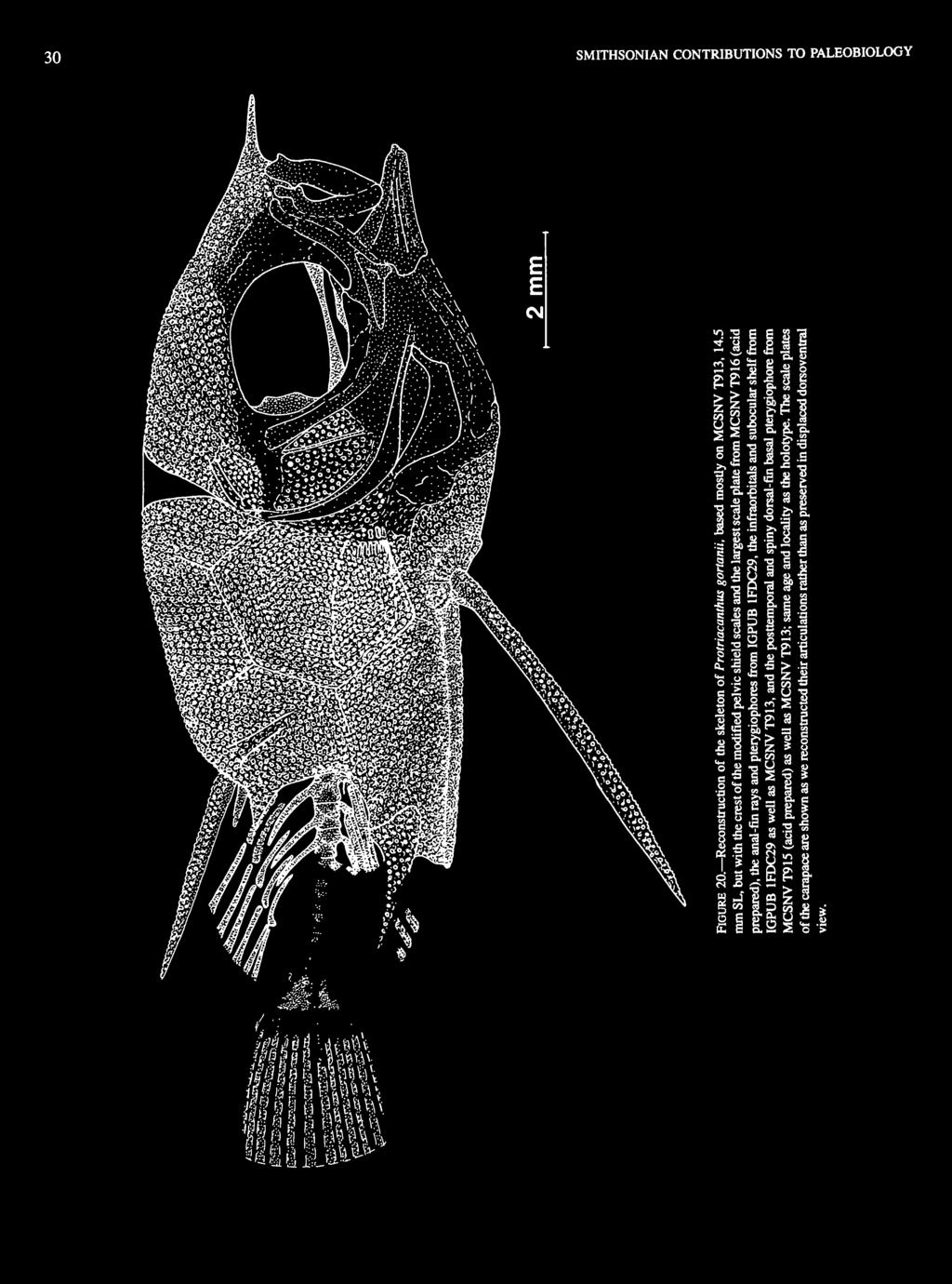
36 30 SMITHSONIAN CONTRIBUTIONS TO PALEOBIOLOGY llllli^


37 NUMBER though it is not exposed in cross-section in any specimen, it is clear that the posterior process is a relatively flattened structure rather than a shaft-like one. Pectoral Fin and Girdle: The pectoral fin is not completely preserved in any specimen. The pectoral girdle is relatively elongate anteroventrally. The distinction between the cleithrum and supracleithrum is not apparent, but the upper region of these combined bones is oriented relatively vertically and reaches dorsally to the level of the dorsal ends of the opercle and hyomandibular. At this dorsal end, the element articulates with a much more horizontally oriented element positioned between it and the skull, which we interpret as the posttemporal. The anterior attachment of the posttemporal to the skull is obscured by the tubercles on the bones, but the posttemporal was obviously free from the skull for most of its length. The coracoid is incompletely preserved below the lower part of the cleithrum. A large bone with a curved lower edge lies below the anterior region of the cleithrum and coracoid and behind the dentary; this could be some fragment of the lower pectoral girdle. The postcleithrum is not apparent. The lower actinosts and pectoral-fin rays are preserved in a few specimens, but none has the fin and its supports complete. The actinosts are hour-glass shaped. Head: The surface of the bones of the occipital, frontal, ethmoid, and opercular regions have strong tubercles like those of the carapace, with the tubercles obscuring much of the peripheral limits of the bones. The posterodorsal region of the head is separated from the carapace by an unomamented region with no scales or scutes evident. The bones of the mouth and the region behind and below the orbit, including the anterior infraorbitals, are relatively well preserved in several specimens and are without surface ornamentation. The frontal is apparently broad posteriorly above the rear of the orbit and tapers anteriorly to the deep ethmoid region, with the latter continued anteriorly above and beyond the mouth as a pointed rostrum. The basal region of the rostrum has a surface ornamentation that is continuous with the tubercles on the ethmoid and frontal. A lateral ethmoid is present but its details are indistinct; however, it does not appear to be ornamented. The premaxilla has no ascending process, and its posterodorsal region is apparently simply indented for articulation with the maxilla. The maxilla and premaxilla are clearly separated from one PU 5 FIGURE 21. Reconstruction of the anal-fin pterygiophores and last three anal-fin rays of the holotype of Protriacanthus gortanii, IGPUB 1FDC29, 20.5 mm SL.


38 32 SMITHSONIAN CONTRIBUTIONS TO PALEOBIOLOGY another along most of their lengths and presumably are movably articulated with one another (Figure 23). Teeth are absent on both the premaxilla and the dentary. The palatine is evident in one specimen (MCSNV T919) as a curved bone, the anterior end of which abuts the posterodorsal region of the maxilla behind its articulation with the premaxilla, and with the posterior end meeting the upper end of the ectopterygoid. The dentary is large, deep posteriorly, and with a crest along its length, below which is an open sensory canal groove without apparent foramina. The posterior end of the dentary is concave to accommodate the anguloarticular, which has a posterodorsal indentation for articulation with the head of the quadrate. The retroarticular is separated from the dentary along the ventral edge of the lower jaw but is only indistinctly separatedfromthe anguloarticular and dentary more dorsally. The quadrate has its anterior edge oriented relatively vertically or somewhat anterodorsally for articulation with the ectopterygoid. The articular head of the quadrate is oriented obliquely anteroventrally. The parasphenoid is clearly preserved as a strong shaft without a ventral flange in the region of the orbit. The parasphenoid curves upward posteriorly to join the prootic in the rear of the orbit. The vomer is not visible in any specimen, and it is impossible to determine if vomerine teeth were present. The ectopterygoid is elongate, joining the anterodorsal region of the quadrate and the posterior region of the palatine. The opercle is broad dorsally, with its posterior edge evenly curved to its tapered ventral end. The preopercle is poorly preserved ventrally but is tapered dorsally in the region between the opercle and hyomandibular. The articular head of the hyomandibular is positioned behind the middle of the orbit, and its ventral shaft is angled obliquely forward. The branchiostegal rays and hyoid arch are not preserved. The infraorbital series is incomplete or indistinct posteriorly, but the first four elements are relatively well preserved in a few specimens (especially MCSNV T913). The lachrymal is oblong, although its anterior end is indistinct. The second infraorbital has a relatively short lateral surface, positioned mostly above the posterodorsal edge of the lachrymal and, to a lesser extent, the anterodorsal edge of the third infraorbital. The third infraorbital is about as long as the lachrymal but is slightly deeper, with a tapered anterior end contacting the posterior margin of the second infraorbital just above the posterior end of the lachrymal. The posterior end of the third infraorbital has a poorly demarked junction with the anterior end of the fourth infraorbital. The posterior region of the fourth infraorbital is too indistinct at the lower rear of the orbit to determine how many other infraorbitals might be present more dorsally. No surface ornamentation is apparent on the infraorbitals. A subocular shelf is apparent in two specimens (IGPUB 1FDC29 (holotype) and MCSNV T913) under the middle of the orbit. The shelf is H3-5 l+u 2 ) PR (Unb) FIGURE 22. Reconstruction of the last three vertebrae and caudal-fin supporting structures of the holotype of Protriacanthus gortanii, IGPUB 1FDC29, 20.5 mm SL.
, but with the upper jaw bones in what we believe is a morerealisticposition, as found in MCSNVT913,14.")

39 NUMBER FIGURE 23. Reconstruction of the mouth region in Protriacanthus gortanii, based mostly on MCSNV T919, -18 mm SL (same specimen illustrated in Sorbini and Guidotti, 1984, fig. 3, in which the upper jaw bones are probably somewhat unnaturally rotated outward), but with the upper jaw bones in what we believe is a morerealisticposition, as found in MCSNVT913,14.5 mm SL; same age and locality as the holotype. basal pterygiophores. The haemal spine of PU 4 is directed toward the basal region of the group of last three anal-fin rays but probably did not support them. There is no evidence of pleural ribs, but epineurals are present on at least PU 7 _g (just behind the carapace; Figure 21; see also Sorbini and Guidotti, 1984, fig. 4), with others presumably present more anteriorly in the same series on the vertebrae obscured by the carapace. The relative length of the centra of the caudal vertebrae decreases only slightly posteriorly, the centrum of PU 2 not being markedly shorter or smaller than those immediately preceding it. The neural spine of PU 2 is expanded posterodorsally to broadly contact the neural arch of the terminal centrum (PU, + U t ) and the element above the latter. This element is probably the anterior end of the fragmented epural (Figure 22; Sorbini and Guidotti, 1984, fig. 5), but this also could be interpreted as an uroneural. The haemal spine of PU 2 is a posteriorly directed shaft in broad contact with the expanded anterior end of the parhypural, which becomes much narrower posteriorly below the hypurals. The hypurals are consolidated into an upper and lower plate with an uniquely oblique orientation, the division between the two plates coursing distinctly anteroventrally from the indentation at their posterior margin of contact to the ventral region of the centrum of of small to moderate size (its full extent may not be exposed) and is formed by the second and third infraorbitals, with the greater portion from the second infraorbital (Figure 24c). Vertebral Column: The vertebrae are probably 18; in one of the acid-prepared specimens (MCSNV T915), eight vertebrae are fully exposed behind the posterior edge of the carapace, and 10 others are indicated under the carapace. Our count of the vertebrae under the carapace, however, may not be exact, especially anteriorly near the base of the skull, and the total number of vertebrae could be 19 or, at a maximum, 20. The proximal end of the first basal pterygiophore of the anal fin apparently articulates with the haemal spine of PU 9 (the centrum is just anterior to the posterior edge of the carapace). If there are, as seems most likely, a total of 18 vertebrae, then there are nine abdominal and nine caudal vertebrae. Only the centra of the vertebrae under the carapace are evident (and only in one acid-prepared specimen, MCSNV T915), but the vertebrae posterior to the carapace are relatively well preserved in several specimens (especially in the holotype). The neural spines of PU 2 _ 8 decrease in length posteriorly, and the haemal spines are relatively shorter. The neural and haemal spines of PU 5 _ 9 support, respectively, the soft dorsal-fin and anal-fin B FIGURE 24. Reconstruction of the infraorbitals and subocular shelf of A, Cretatriacanthus guidottii, B, Plectocretacicus clarae, and C, Protriacanthus gortanii (B and C are composites based on all material).


40 34 SMITHSONIAN CONTRIBUTIONS TO PALEOBIOLOGY PUj + Uj. The lower hypural plate is presumably composed of fused hypurals one and two. The upper hypural plate is composed of fused U 2 and hypurals three and four and perhaps hypural five, the latter because a separate fifth element is found in Cretatriacanthus and Plectocretacicus and in all other morphologically primitive tetraodontiforms. There are six caudal-fin rays articulated along the posterior edge of each of the two hypural plates. The upper hypural plate is apparently fused to the centrum of PUj + U p but the type of articulation of the lower hypural plate to that centrum is unclear. The exact configuration of the region of the terminal centrum and urostylar region is unclear, but there is a short oblique mark posteriorly in the urostylar region of the holotype that is suggestive of the truncate termination of PUj + Uj found in the other two Upper Cretaceous genera of tetraodontiforms. Carapace: The carapace is composed of moderately enlarged (up 25% SL) and mostly hexagonal scale plates sutured together with delicate interdigitations. The outlines of individual scale plates are not clear in most specimens because of fracturing, and the hexagonal shape is only apparent in its entirety in one of the acid-prepared specimens, MCSNV T916. The external surfaces of most plates have tubercles of similar size, which are variously either evenly distributed or arranged more-or-less in rows parallel to the peripheral edges of the plates. The linear arrangement is most apparent on the guard scales around the pelvic-fin spine. The fully consolidated carapace is present from the curve of the pectoral arch to about the origins of the soft dorsal and anal fins, with the posterolateral edge of the carapace slightly curved forward to the level of the posterior end of PU 9. The anterodorsal edge of the carapace is distinctly separated from the head by a scaleless area. Lateral to the anterior region of the anal fin, the carapace has one of the scale plates modified into a posteriorly directed process (preanal carapace spine). A crest of modified scales forms an arch around the base of the pelvic-fin spine, with the arch being between 2.5 and 3.5 mm long in the three specimens in which it is best exposed (MCSNV T913, MCSNV T916, and IGPUB 1FDC29 (holotype), which are mm SL). There is no carapace on the head, and there are no isolated scale plates along the region of the caudal peduncle. Measurements: Measurements for the holotype and six additional specimens (meaningful measurements are not obtainable from MCSNV T917, -10 mm SL) are given in Table 2; they were made in the same manner as given for the measurements of Cretatriacanthus guidottii and Plectocretacicus clarae, with the following additions: width of the pelvis is the distance between the estimated positions of the bases of the pelvic spines, because in both specimens in which this region is exposed and undistorted these spines are displaced from the side of pelvis and the width of the pelvis is clearly shown; length of the rostral process from the level of the upper posterior end of the premaxilla, excluding most of the basal plate, to the distal tip; length of the snout excludes the rostral process, being from the front of the upper end of the premaxilla to the nearest point on the orbit; length of the preanal scale plate from the anterior end of its flat basal surface to the posterior end of the spiny process. SIMILARITIES AND DIFFERENCES TO OTHER TETRAODONTI FORMS. As represented by Protriacanthus gortanii, the Protriacanthidae have several morphological specializations similar to those of various other families of tetraodontiforms outside of the Plectocretacicoidea. In protriacanthids the thoracic pelvic fin has a large spine with what appears to be a basal flange that could lock the spine erect against the side of the pelvis, much as in triacanthoids. Nevertheless, this putative basal flange can be seen on only one side of one specimen, and it is possible that it is more of an enlargement of the base of the spine than it is the well-defined locking flange found in triacanthoids. None of the features that distinguish the locking mechanisms of triacanthodids (basal flange and its surface of articulation on the pelvis smooth or with numerous grooves and a single or multiple positions of spine erection) and triacanthids (an oblique ridge on the side of the pelvis and two positions of erection) can be detected in the single specimen of Protriacanthus that seems to have a basal expansion of the spine; therefore, we are unsure of its similarity to the locking mechanism of triacanthoids. In any case, it is most parsimonious to propose that the possible locking mechanism of protriacanthids was acquired independently of that in triacanthoids (see "Comments on Other Features"). In both Protriacanthus and triacanthoids, the base of the first dorsal spine has a foramen for ring-link articulation with a flange on the first basal pterygiophore, but this derived condition (relative to zeiforms) is interpreted as primitive for tetraodontiforms. The well-developed carapace and the posterior placement of the soft dorsal and anal fins of protriacanthids (as well as of plectocretacicids) are similar overall to those conditions in ostracioids (except for the continuity of the carapace onto the head in the latter), but these are proposed as independent acquisitions (see "Analysis of Characters," characters 25, 28). The posterior position of the reduced spiny dorsal fin in protriacanthids is similar to that condition in triodontids among tetraodontoids, but this condition is most parsimoniously interpreted as an independent specialization in each of these two taxa (see "Analysis of Characters," character 41). The rostral process in protriacanthids, which we believe to be formed by the bones of the snout, is superficially similar to the more dorsally positioned snout spine of Eospinus (an incertae sedis balistoid) and larval molids, but the spiny processes in the latter two groups clearly are modified scale plates and are not anatomically homologous to the rostral process of protriacanthids. Because protriacanthids are most similar to triacanthoids, we cite the numerous differences between the two groups. In addition to their defining synapomorphies and large array of especially primitive features, protriacanthids differ most notably from triacanthoids by the following: the presence of a carapace (versus no carapace); probably 18 vertebrae (versus


41 s m n O -1 o «* 1-1 in 01 oo in N q m od o >d ci r^ «' " n i o i d o o H i n ' d C<-1 ^ ** ~ ri. «t en r- r^ ts 35 i r~ in r» o r* in cs o <t o\ d I I \ j * q - ^f» Tt ^ vi ri - - «p t ~~. r ": '"i! i r^ n r^ i oi ci «c> "t * ' I I CM I 2 2 <n ri ii.9 g r-; * p Tf tn o d vo in -^ it d»$ r^ h - -; «O f, 5 in «i pi H ri "' I I I I r-i I ' d rn e 4) "O 1)»fr--v ~-p-'tcsov - N * f n ' d c i ^ i i ( 5 *-* tn -' <N d I ro y s g. f ^ fc> o "3 -.s Us.a «s e 111 -II g 3 ill K -o -o 3 u u S I *? <2 ^ c4 5.» &8 inoh^gigjoooio; ' t ' d i d r i - r i ^ d n lo VO 7 OO \Q 7 r- O ^o o o m vo m c r-; <s en in - - in d * > in oo O -t t t -* en c-> vo O vo m r- *tf <N CT> r- 00 m i i r^ in d oo in oo al oo \ in \q in r~; d r^ r^ ri "1. S CM T}- -^ OO r; \0 rn rn ft Sff a -e JJ =3 o u t nxo;
; the short-based soft dorsal and anal fins with few rays (five to seven) and basal pterygiophores (versus moderate- to moderately long-based fins with 11-26 rays and pterygiophores);")
; a rostral process present (versus no rostral process); many head bones with tuberculate ornamentation (versus no")
42 36 SMITHSONIAN CONTRIBUTIONS TO PALEOBIOLOGY 20); the spiny dorsal fin positioned posteriorly and reduced to two spines (versus positioned just behind head and usually with six, rarely only four or five, spines); the short-based soft dorsal and anal fins with few rays (five to seven) and basal pterygiophores (versus moderate- to moderately long-based fins with rays and pterygiophores); the last three anal-fin rays closely grouped together and specially articulated on a complex series of pterygial elements (versus last few rays not so grouped and articulated); essentially no ascending premaxillary process (versus ascending process well developed); a rostral process present (versus no rostral process); many head bones with tuberculate ornamentation (versus no ornamentation); the fifth hypural absent or fused with the upper hypural plate (versus fifth hypural free); no uroneurals (versus one or two uroneurals); the articulation between the upper and lower hypural plates oblique (versus articulation or area of fusion horizontal). PALEOECOLOGY AND ICHTHYOFAUNAL ASSOCIATIONS. The Comen, Slovenia, site (located between Gorizia and Trieste, Italy) has been known for its Upper Cretaceous fossil fishes since the description and illustration of a variety of species by Heckel (1850, 1856). The most extensive work on these fishes is that of d'erasmo (1946), who reported on the numerous specimens from Comen now in the Istituto di Geologia e Paleontologia della Universita di Bologna. Other fossils from Comen are housed in the Museo Civico di Storia Naturale in Trieste; one of us (L.S.) has identified a specimen of Tselfatia formosa Arambourg among these materials in Trieste, a species not previously reported from the Comen site. The fossil fishes of Comen are found in the Monte Coste Member of the dark gray to blackish, compact, and sometimes bituminous limestones, in which there is some evidence of centrimetric-polydecimetric bedding (Cucchi, Pirini Radrizzani, and Pugliese, 1987). On the basis of both geological data and his detailed faunal analyses of a wide variety of Cretaceous deposits around the Mediterranean, d'erasmo (1946) dated the Comen ichthyofauna as Upper Cenomanian. Radovcic (1973) thought that the Comen strata were deposited between the Cenomanian and Senonian, whereas Hemleben and Swinburne (1991) date them between the Upper Cenomanian and Lower Turanian. The presence of Tselfatia formosa among the Comen fishes and the fact that this species also has been found at the Cinto Euganeo fossil site (Sorbini, 1976), which has been dated on the basis of planktonic foraminifera as Upper Cenomanian-Lower Turanian, leads us to consider the latter date (about 90 MYA) as the more probable one. Regional geological data indicate that the Comen limestones had a sedimentation environment in an inner carbonate platform that was sometimes rather restricted. Among the Comen fishes are reef-associated pycnodontids and numerous reef-associated or shallow benthic mollusks and echinoderms, although there are many groups of fishes (e.g., clupeiforms, beryciforms) with species having wide-ranging habitats. Rationale for Outgroup Comparisons The overall phylogeny of the Recent Tetraodontiformes followed here is that established in Winterbottom's (1974) myologically based cladistic analysis, amended with the phylogeny of the balistoid + ostracioid clade by Winterbottom and Tyler (1983) and the phylogenies of a variety of fossil taxa by Tyler and Gregorova (1991, for ostracioids), Tyler and Bannikov (1992a, for the balistoid + ostracioid clade; 1992b, for molids), and Tyler et al. (1993, for triacanthoids). Based on those analyses and the new data presented herein, the higher classification (using the family levels of Tyler, 1980) of the order adopted here is as follows (see Figure 25 for a cladogram of the order): Order TETRAODONTIFORMES tsuperfamily PLECTOCRETACICOIDEA (plectocretacicoids) tfamily CRETATRIACANTHIDAE (Upper Cretaceous) tfamily PLECTOCRETACICIDAE (Upper Cretaceous) tfamily PROTRIACANTHIDAE (Upper Cretaceous) Superfamily TRIACANTHOIDEA (triacanthoids) Family TRIACANTHODIDAE (Oligocene to Recent) Subfamily TRIACANTHODINAE (Oligocene to Recent) Subfamily HOLLARDHNAE (Oligocene to Recent) Family TRIACANTHIDAE (Eocene to Recent) tsubfamily PROTACANTHODINAE (Eocene) Subfamily TRIACANTHINAE (Oligocene to Recent) fcryptobalistes incertae sedis at family level within either triacanthoids or balistoids (Oligocene) Superfamily BALISTOIDEA (balistoids and ostracioids) balistoids ^Eospinus incertae sedis at family level within Balistoidea, probably with balistoids (Eocene) tfamily SPINACANTHIDAE (Eocene) Family BALISTIDAE (Oligocene to Recent) Family MONACANTHIDAE (Miocene to Recent) ostracioids Family ARACANIDAE (Eocene to Recent) Family OSTRACIIDAE (Eocene to Recent) Superfamily TETRAODONTOIDEA (tetraodontoids) tfamily EOPLECTIDAE (Eocene) Family TRIODONTIDAE (Eocene to Recent) tfamily ZlGNOlCHTHYlDAE (Eocene) Family TETRAODONTIDAE (Eocene to Recent) Family DlODONTIDAE (Eocene to Recent) Family MOLIDAE (Eocene to Recent) We do not believe that anything is to be gained at present in ranking the four monophyletic superfamilial clades (plectocretacicoids, triacanthoids, balistoids + ostracioids, tetraodontoids) as suborders, which would be informationally empty categories redundant to the superfamilies. This is especially the case now, when the order Tetraodontiformes is being expanded both by the addition of new groups, such as plectocretacicoids, and by the likely superfamilial revisions that will result from studies underway, such as those being conducted on fossil balistoids by N. Bonde and the authors. For comparisons with the Plectocretacicoidea, we use the clade composed of all other tetraodontiforms as the first outgroup and use zeiforms as the second or extraordinal
 in accepting zeiforms as the primitive sister group of tetraodontiforms on the basis of 11 derived features shared by zeiforms and tetraodontiforms, seven of which are")
 related to acanthuroids (Bannikov, 1991) that have independently acquired the seven derived features shared by caproids,")
43 NUMBER outgroup. The latter follows Rosen (1984) in accepting zeiforms as the primitive sister group of tetraodontiforms on the basis of 11 derived features shared by zeiforms and tetraodontiforms, seven of which are also shared by caproids. Although caproids are probably percomorphs (Johnson and Patterson, 1993) related to acanthuroids (Bannikov, 1991) that have independently acquired the seven derived features shared by caproids, zeiforms, and tetraodontiforms, that does not invalidate the 11 derived features proposed by Rosen as applying to zeiforms and tetraodontiforms. We have no reason to believe that the sister group of zeiforms + tetraodontiforms is one of the taxa among the higher percomorphs, and, given the nature of the truncate posterodorsal end of the PU t + U, terminal centrum in plectocretacicoids, we suspect that the sister group of zeiforms + tetraodontiforms will prove to be a taxon among either beryciform-like fishes or lower percomorphs. The preliminary results of a study of the phylogeny of zeiform families based on osteology and myology (R. Winterbottom et al., unpublished) are in agreement with the conclusion of Johnson (1993) and Johnson and Patterson (1993) that zeiforms (exclusive of caproids) are not percomorphs, but we are not yet prepared to propose a sister group for the putative zeiform + tetraodontiform clade. Because the phylogenetic relationships of thefivefamilies of zeiforms are still undeciphered, we use representatives of all five families in our comparisons with tetraodontiforms. Among zeiforms, however, we focus on what have been suggested by Johnson and Patterson (1993) to be the two most primitive families in the order, the Parazenidae (single species, Parazen pacificus Kamohara) and Macrurocyttidae (or Zeniontidae, comprising the several species of Zenion, of which we consider Cyttula to be a synonym, and the problematical Macrurocyttus acanthopodus Fowler), and on the morphologically primitive members of the speciose family Zeidae, unless we know of contrary evidence in the more specialized families (Grammicolepididae and Oreosomatidae) (see Tyler et al., 1993, for a list of the zeiform genera, representing all five families, for which we have examined skeletons and also for literaturereferencesto zeiform osteology). Data on the osteology of beryciforms is from Zehren (1979). One of the taxa (Protriacanthus) that we include in the plectocretacicoids has previously been tentatively referred to the gasterosteiforms, but this was at least in part on the basis of the somewhat misleading original description. Patterson (1964) doubted that Protriacanthus was a tetraodontiform, partially because of the erroneously high number of posterior vertebrae illustrated by d'erasmo (1946); Patterson suggested that it was perhaps a gasterosteiform relative and placed it incertae sedis among teleosts. Tyler (1968, 1980) agreed with Patterson's analysis of the original description of Protriacanthus, dismissing it as a possible tetraodontiform, and Medizza and Sorbini (1980) likewise followed Patterson in listing it incertae sedis among the fishes of Comen. Even with its accurate redescription on the basis of additional specimens, the combination of gasterosteiform- and tetraodontiform-like features in Protriacanthus led Sorbini and Guidotti (1984) to retain its incertae sedis status. Based on the characters in our redescription of Plectocretacicus (especially as seen in the acid-prepared specimens), along with those of the new taxon Cretatriacanthus, the numerous specialized features that Protriacanthus shares in combination with those taxa, and the synapomorphies common to Protriacanthus and Plectocretacicus, we are convinced that Protriacanthus is a tetraodontiform. However, we outline below our rationale for believing that Protriacanthus and the other plectocretacicoids are not gasterosteiforms (characteristics of the latter from Johnson and Patterson, 1993). Most of the similarities between Protriacanthus and gasterosteiforms are reductive, such as reduced numbers of both vertebrae and fin spines, including the lack of anal-fin spines, and we interpret these reductions as independently acquired (like similar reductions in such major groups as lophiiforms). Derived features of all or nearly all gasterosteiforms that are not shared by Protriacanthus and the other plectocretacicoids include the following. Gasterosteiforms have a single supernumerary dorsal-fin spine, whereas the two genera of plectocretacicoids with spiny dorsal fins (Protriacanthus and Cretatriacanthus) have the primitive acanthomorph condition of two supernumerary spines, as do the primitive clades of Eocene to Recent tetraodontiforms. Gasterosteiforms have five or fewer branchiostegal rays (except six in Indostomus, of uncertain position among syngnathoid gasterosteiforms), whereas the only plectocretacicoid in which the branchiostegals are known, Plectocretacicus, has seven and the Eocene to Recent tetraodontiforms have, primitively, six. Gasterosteiforms have one to three infraorbitals, whereas all three taxa of plectocretacicoids have at least four infraorbitals from the lachrymal back to about the middle of the orbit; posterior to this there are probably one or two others less distinctly preserved in plectocretacicoids. With the exception of two genera (at least one of which is a specialized form), all gasterosteiforms have some form of complex suturing between the ceratohyal and epihyal, whereas the only plectocretacicoid in which this articulation can be seen (Plectocretacicus) lacks such suturing, as do at least the more primitive clades of Eocene to Recent tetraodontiforms. Moreover, when plectocretacicoids are compared with the more morphologically primitive groups of gasterosteiforms (aulorhynchids, Hypoptychus, and gasterosteids; pers. comm., G.D. Johnson, August 1994: see three columns on left in table 1 of Johnson and Patterson, 1993:576) the following differences are evident. Primitive gasterosteiforms have the derived condition of lacking postcleithra, whereas plectocretacicoids have a single postcleithrum (unknown for Protriacanthus), as do Eocene to Recent tetraodontiforms. Primitive gasterosteiforms have the following primitive conditions: parietals present, pleural ribs present in most species, extrascapulars present in aulorhynchids and Hypoptychus, relatively large
 cr CD c _) a. u h 0 n rtl c > > m m 0 0 > -i CT LU CD > CD DC CO LU cr 0.2 i to! o I.")
lubobu10jd E Q o <9 co js 4.")
44 38 SMITHSONIAN CONTRIBUTIONS TO PALEOBIOLOGY 9BP! 0 /\ CO ^ Q) co E Q co c c t o O- 3 CO 2 eepiiuopoja aep!}uopoej;ei 4. eep!ai }ip!0ii6!z eepiiuopoui I 8Bpip9 d03 E CO CD Q-.9> F 0 Q. CO c_ C/J >> to 0 0 > -) cr CD c _) a. u h 0 n rtl c > > m m 0 0 > -i CT LU CD > CD DC CO LU cr 0.2 i to! o I. 9Bpipej}so eepiubobjv LUt- < DC H LU E to t CD Q. D CO CO g o I < CO _Q aepmiubobuoiaj aep!}si Bg 4. SBpiqiuBOBUjds CO.-. > CD «= "O 5 E o o co.c 1 9BU!L ;UB0BU1 1 1 ebpiljjuboeijl < 1 I abujpoliluboblojcl 9BUMpjB 0H abpipolilubobul < 9BU!p0L }UB0BUl!?! 4- ebpil)lubobu10jd E Q o <9 co js 4. abpppbiajoopaici CO «o CO V. 4. BBpjiuueoeiJiBieJO LU N
.")
-ir)vor- a -a i i... SI.")
45 NUMBER S SL -.5 <N -a C 3 fc C «? OJ.2 > 1 I O 4).Sbg w _ c io,5 n J5 a 8231 fc 2 fi "3 4> o O E O PQ Z -H (sm, *>o\or~-ooctno^csfot)-ir)vor- a -a i i... SI. a o I 111 gife S "> c u 3 s as ~ S * -C?: sa Sis a s a O VS 3 00 Q co


46 40 SMITHSONIAN CONTRIBUTIONS TO PALEOBIOLOGY numbers of vertebrae (28-56, from Pietsch, 1978), one anal spine in aulorhynchids and gasterosteids (absent in Hypoptychus), and no ring-link articulation of the dorsal spines with their pterygiophores. By contrast, plectocretacicoids and the Eocene to Recent tetraodontiforms lack parietals, extrascapulars, and anal spines, have the vertebrae reduced to 21 or less, and have pleural ribs absent in the two genera of plectocretacicoids in which they might be observed. Pleural ribs also are absent in all Eocene to Recent tetraodontiforms except as rare reversals in three species, although there is a ring-link dorsal spine articulation in Protriacanthus, the only plectocretacicoid in which the spine is appropriately turned 90 in exposure to reveal a foramen through its base. We note that gasterosteiforms lack what we consider to be two of the more innovative derived features of tetraodontiforms. In tetraodontiforms (and zeiforms) the bifid neural spine of the first vertebra is closely applied to the rear of the skull, and, at least in primitive groups with a well-developed spiny dorsal fin, the ventral shaft of the first basal pterygiophore slants forward to contact the skull between the first neural spine (or, in the case of Cretatriacanthus, to at least be in the first interneural space; Protriacanthus has a specialized posterior position of the spiny dorsal fin and its pterygiophore). In tetraodontiforms with a pelvis there is a long posterior extension beyond the pelvic fins, and this is a single consolidated piece, closely sutured or fused in the midline. Many gasterosteiforms have toothless mouths, but because the gasterosteiform small mouth is nearly always at the end of a tubular snout, we consider the absence of teeth in these gasterosteiforms to be a specialization very different from the absence of teeth in the large mouths of plectocretacicoids. The posterior placement and enlargement of one of the dorsal-fin spines in macrorhamphosids and the Upper Cretaceous Gasterorhamphosus (which Sorbini, 1981, considered a macrorhamphosid sensu lato and Patterson, 1993, considered a stem-group gasterosteiform) is superficially similar to the condition in Protriacanthus; however, the second spine is elongate in macrorhamphosids rather than, as in Protriacanthus, the first spine. Moreover, there are additional moreposterior spines fused to the basal pterygiophores in macrorhamphosids that seem to us to be a different specialization from that in protriacanthids. Macrorhamphosids have enlarged scale plates forming a partial carapace-like structure around parts of the head and body, but these plates never have what we consider the more specialized condition of being regularly hexagonal and delicately interdigitated as in plectocretacicoids. Macrorhamphosids have the first five vertebrae elongate and inflexibly sutured, features absent in plectocretacicoids (also absent in Gasterorhamphosus). Moreover, macrorhamphosids have several other derived features (e.g., specialized series of median-scale scutes at the isthmus and complex semicircular ridges basally on the large dorsal spine and its basal pterygiophore; J.W. Orr, pers. comm. and in litt, May 1993) that one could expect to be preserved and exposed in fossil materials like those of Protriacanthus but which are absent in the latter. Therefore, given the numerous synapomorphies that unite plectocretacicoids with other tetraodontiforms and unite protriacanthids with plectocretacicids among plectocretacicoids, and given the several derived features of all gasterosteiforms that are absent in plectocretacicoids, as well as the derived features of plectocretacicoids and other tetraodontiforms that are absent in gasterosteiforms, the few specialized similarities between protriacanthids and some families of gasterosteiforms (especially macrorhamphosids) are considered to be the result of independent acquisitions. Analysis of Characters The diagnostic and distinguishing features of the order Tetraodontiformes as a whole are followed by those of the superfamily Plectocretacicoidea and its three contained families, with the numbers corresponding to those in the cladogram (Figure 25). The cladogram includes the distribution of the defining characteristics of tetraodontiforms as a whole and those of plectocretacicoids and its contained families, but it does not include the numerous other features that distinguish the other three superfamilies of tetraodontiforms (for which see Winterbottom, 1974, and Tyler, 1980). SYNAPOMORPHIES OF TETRAODONTIFORMES Many of the following 12 specializations in the bony structures and external features have been recognized to one extent or another and have been variously used by most authors dealing with the classification of the plectognath or tetraodontiform fishes since the establishment of the order by Cuvier (1817), with more recent utilizations by Tyler (1980) and Rosen (1984). Nevertheless, proper documentation for the diversity of these conditions in zeiforms and tetraodontiforms has not been provided previously, and therefore we do so below. Winterbottom (1974) documented the myological specializations of the families of tetraodontiforms, but these are not discussed here because they are not available in fossils of the order. Rosen (1984) has proposed that the sister-group relationship of zeiforms and tetraodontiforms is supported by 11 derived features, seven of which also are found in caproids, with six other derived features shared by tetraodontiforms but not zeiforms (characters 2, 4-6, 8, 14, herein). 1. Number of Vertebrae. Plectocretacicoids have about 18 to 21 vertebrae, as follows: probably 21 in Cretatriacanthus, 20 in Plectocretacicus, and 18 in Protriacanthus. With a few exceptions, all other tetraodontiforms (first outgroup), including all of the more basal groups, have 20 or fewer vertebrae, as follows: triacanthoids 20; spinacanthids probably 20; balistids 18; monacanthids mostly 19 or 20, with a few specialized species, such as those with elongate bodies,


47 NUMBER having up to 30; aracanids 18; ostraciids 18, except one species with 19; eoplectids 20; triodontids 20; tetraodontids mostly 17-20, with a few species with up to 23 and one specialized species, with many rays in the long-based dorsal and anal fins, with up to 30; diodontids 18-23; molids 16-18; number unknown in the tetraodontoid zignoichthyids. Zeiforms (second outgroup) have far higher vertebral numbers, 25-46, as do most beryciforms and lower acanthomorphs. Therefore, low vertebral numbers of 21 or less, and usually 20 or less, are considered a synapomorphy of tetraodontiforms. Within tetraodontiforms, the occasional increases to a larger number of vertebrae in a few species that are well nested within several families are most parsimoniously considered independent secondary specializations. 2. Anal-Fin Spines. Plectocretacicoids and all other tetraodontiforms lack anal-fin spines. Anal spines are present in all five families of zeiforms (short and inconspicuous in Zeniontidae and perhaps absent in a few of its species; anal spines often are stated to be absent in Parazen, the only representative of its family, but we find a buried rudimentary anal spine in several cleared and stained specimens of species of this genus), and anal spines are a common feature of most other acanthomorphs. The complete absence of anal spines in a few species of one family of zeiforms is most parsimoniously interpreted as independent of the absence in all tetraodontiforms. Therefore, the absence of anal spines is considered a synapomorphy of tetraodontiforms. 3. Pelvic-Fin Elements. Plectocretacicoids have a pelvic-fin spine and either two rudimentary rays or no rays. Triacanthoids have a pelvic spine and one or two rudimentary rays. In balistoids, the pelvic rays are lost and the spines from each side are reduced to a single, small, median rudiment usually surrounded by specialized encasing scales, with the rudimentary spine and encasing scales lost altogether in some monacanthids. All ostracioids have lost the entire pelvic fin. Among tetraodontoids, only eoplectids have a pelvic fin; it is composed of a slender spine of modest length and four well-developed branched rays, the rays being far more numerous and far longer than the unbranched rays in plectocretacicoids and triacanthoids. In zeiforms the pelvic fin either has a spine and five to seven well-developed branched rays (1,5-7) or the spine is absent and there are nine to 10 rays. If a pelvic-fin reduction by one ray from the zeiform condition of 1,5-7 (rays well developed) to an eoplectidlike 1,4 (rays well developed) is primitive for tetraodontiforms, then four additional steps are required in an ordered transformation series to account for the following: independent reduction to 1,2 (rays rudimentary) as primitive for the ancestor of the plectocretacicoid clade (as found in cretatriacanthids, and then further reduction of the rays in plectocretacicids and protriacanthids); independent reduction to 1,2 (rays rudimentary) as primitive for the ancestor of the triacanthoid clade (as found in triacanthodids, and then further reduction of the second ray in most triacanthids); independent reduction to a rudimentary spine in the ancestor of the balistoidostracioid clade (as in balistids, with further reduction in monacanthids and loss in ostracioids); loss of the fin in all tetraodontoids except eoplectids. The most-parsimonious hypothesis is that a pelvic-fin reduction to a plectocretacicoid- and triacanthoid-like 1,2 (rays rudimentary and further reduced within some taxa of each group) from the zeiform condition is primitive for tetraodontiforms because 1,2 (rays rudimentary) can be considered primitive for the ancestors of the plectocretacicoid, triacanthoid, and balistoid + ostracioid clades, with only two additional steps required to account for the reversal in eoplectids to a 1,4 (rays well developed) fin and the loss of the fin in all other tetraodontoids. Therefore, the reduction of the pelvic fin to the condition of 1,2 (rays rudimentary) or less is considered a synapomorphy for tetraodontiforms (reversed in eoplectids). 4. Posterior Process of Pelvis. Both Cretatriacanthus and Protriacanthus have long posterior pelvic processes either fused or otherwise indistinguishably articulated medially throughout their length. In Plectocretacicus the pelvis is obscured by the carapace, but in any case the specialized posterior positioning of the pelvic fin is concomitant with the specialized loss of the posterior process, essentially all of the length of the pelvis being anterior to the pelvic fin. In triacanthoids the two halves of the posterior process are sutured or partially fused together medially throughout their length. In balistoids the two halves of the posterior process are fused medially throughout their length, whereas in ostracioids the pelvis is absent. The condition of the pelvis is unknown in the most morphologically primitive tetraodontoid, the Eocene Eoplectus, but in Triodon, the sister group of all other extant tetraodontoids and the only Recent tetraodontoid to possess a pelvis, the two halves of the pelvis are sutured together medially throughout their length (there is no posterior process as such because there is no pelvic fin) and may be fused posteriorly. Thus, all tetraodontiforms with a pelvis and posterior process have the two halves sutured or fused medially at least throughout the portion posterior to the level of the pelvic fins (or the posterior half of the length of the pelvis when the pelvic fin is absent, as in Triodon). In zeiforms the halves of the pelvis are relatively well separated throughout most of their length, although sometimes broadly and flexibly overlapped in the middle region, but the posterior processes are never sutured or fused along the midline. This unconsolidated condition


48 42 SMITHSONIAN CONTRIBUTIONS TO PALEOBIOLOGY also is the case in most other acanthomorphs. Therefore, the medial suturing or fusion of the posterior processes is considered a synapomorphy of tetraodontiforms. 5. Parietal. All Recent tetraodontiforms lack a parietal. This region of the skull is only rarely well preserved or exposed in fossil tetraodontiforms, usually making it impossible to determine whether the parietal is present. Although this is the case with all of the numerous Eocene taxa, the two Oligocene taxa of triacanthodids both clearly lack the parietal (Tyler et al., 1993), and in one of the three Upper Cretaceous taxa (Plectocretacicus) the upper rear part of the skull is sufficiently well preserved to indicate that the parietal is probably absent. All zeiforms have a parietal, as do most other acanthomorphs, and the loss of the parietal is considered a synapomorphy of tetraodontiforms. 6. Gill Opening. In all Recent tetraodontiforms, the gill opening is greatly reduced in length (to between 5%-15% SL), being a short slit in front of the pectoral-fin base (only rarely extending even slightly below that base), and the opercular apparatus cannot be laterally flared. The degree of gill opening restriction has not been well documented in most fossil tetraodontiforms, and the precise dimension of this soft tissue character is likely to remain poorly known in fossil materials. We have, however, reexamined some of the tetraodontiforms from the Eocene of Monte Bolca, the triacanthid Protacanthodes ombonii (Zigno) (small scales with upright spinules), and the ostracioids Proaracana dubia (de Blainville) and Eolactoria sorbinii Tyler (enlarged hexagonal carapace plates), and it is clear from the relatively uninterrupted and complete covering of scales in the region anteroventral to the pectoral-fin base in such species that the gill opening was restricted and did not extend much, if at all, below the level of the pectoral-fin base. The gill openings in the especially well-preserved species of Pliocene monacanthids reported by Sorbini (1988) are as restricted as in extant species of that family. Although the gill opening itself is not apparent in the three Upper Cretaceous taxa of tetraodontiforms, the opercle and subopercle are of about the same relatively small size (Cretatriacanthus) or are only very slightly larger (Plectocretacicus and Protriacanthus) than in triacanthoids. Therefore, we think it safe to assume that the gill opening was restricted in these taxa. In zeiforms and most other acanthomorphs, the gill opening extends down to the isthmus, well below the pectoral-fin base, and the opercular apparatus can be laterally flared. Therefore, the restricted gill opening is considered a synapomorphy of tetraodontiforms. 7. Pleural Ribs. Pleural ribs are absent in plectocretacicoids and triacanthoids, the two basal clades of tetraodontiforms, and also are absent in all members of the balistoid + ostracioid clade except one highly specialized species deeply nested among monacanthids, Pseudalutarius nasicornis (Schlegel) (with dorsal spine far forward on head, loss of pelvic fin rudiment and encasing scales, and increased number of vertebrae). Among tetraodontoids, pleural ribs are present in only two species, Triodon macropterus Lesson, the only Recent triodontid (condition of ribs unknown in the single fossil species of triodontid) and Eotetraodon pygmaeus (Zigno), an Eocene species of tetraodontid. Most zeiforms, like most other acanthomorphs, have ossified pleural ribs (although often of reduced number and sometime only present on the last abdominal vertebra), and even when pleural ribs are not ossified in zeiforms there are usually ligaments representing unossified pleural elements. The pleural rib series of zeiforms is specialized by being present no more anteriorly than on the fifth abdominal vertebra (with the exception of Grammicolepis, with ribs from the third vertebra posteriorly), but pleural ribs are primitively present in acanthomorphs in one form or the other (Johnson and Patterson, 1993; Patterson and Johnson, 1995). Therefore, the absence of pleural ribs in all but single species of three different families of tetraodontiforms is considered a synapomorphy of the order. Because the diodontid sister group of tetraodontids lacks pleural ribs and the molid sister group of tetraodontids + diodontids also lacks pleural ribs, it is most parsimonious to hypothesize that the presence of pleural ribs in Eotetraodon and in Triodon represents independent reversal in each to the ancestral zeiform-like condition, as is also the case in Pseudalutarius. 8. Number of Caudal-Fin Rays. Plectocretacicids have 14 principal caudal-fin rays (branched rays plus two), whereas its protriacanthid sister group has 12 principal rays, as does the cretatriacanthid sister group of protriacanthids + plectocretacicids. All other tetraodontiforms have 12 or fewer principal caudal rays (12 in triacanthoids, balistoids, eoplectids, triodontids; 10 or 11 in ostracioids; 11 or fewer in tetraodontids, diodontids, molids). Zeiforms usually have 13 or 15 principal caudal rays, with the exception of 11 in Parazen, a genus with relatively poorly developed fins (dorsal and anal fins with weak spines and low number of rays), and we thus hypothesize that 13 to 15 rays is primitive for zeiforms, especially because lower percomorphs and beryciforms tend to have higher caudal-fin numbers than zeiforms. It is most parsimonious to consider the reduced number of 12 principal rays as ancestral for tetraodontiforms, with reversal to the higher number of 14 in Plectocretacicus (two steps including the initial reduction to 12), rather than 14 being ancestral, which would require the independent reduction to 12 in Protriacanthus, in Cretatriacanthus, and in all non-plectocretacicoid tetraodontiforms (a minimum of three steps even


49 NUMBER if no change to 14 were required from the ancestral zeiform condition). Therefore, the reduction to 12 principal caudal-fin rays is considered a synapomorphy of tetraodontiforms. 9. Beryciform Foramen. The condition of the ceratohyal is unknown in Cretatriacanthus and Protriacanthus, but in Plectocretacicus it is preserved and there is no evidence of either a beryciform foramen or a long deep groove along its upper lateral surface. The beryciform foramen also is absent in all other tetraodontiforms, and there is never a deep groove along the surface of the ceratohyal. In zeiforms the ceratohyal usually has a beryciform foramen, although this is sometimes lost with increasing specimen size, or it has a long groove along the upper lateral surface (including the long groove in large specimens after the loss of the foramen). The foramen may be absent in some species of zeiforms, but this seems to be a secondary loss from the presence of the foramen or deep groove as the primitive condition. The foramen is also the primitive condition for beryciforms and other lower acanthomorphs. The absence of the foramen or longitudinal groove, therefore, is considered a synapomorphy of tetraodontiforms. 10. Nasals. Nasals are absent in plectocretacicoids and all other tetraodontiforms, but they are present in all zeiforms and most other lower acanthomorphs, including most beryciforms. The absence of nasals, therefore, is considered a synapomorphy of tetraodontiforms. 11. Scale Bones. Supratemporal scale bones (extrascapulars or tabulars) are not evident in plectocretacicoids and are absent in all other tetraodontiforms, but one or two are present in all zeiforms and are commonly found in other lower acanthomorphs, including beryciforms. The absence of scale bones, therefore, is considered a synapomorphy of tetraodontiforms. 12. Ring-link Fin-Spine Articulation. The first long dorsal-fin spine is in place in Cretatriacanthus and is therefore only seen in lateral view, but its base deeply embraces a high medium crest on the basal pterygiophore, and we presume that the base of the spine is deeply concave. In Protriacanthus the first dorsal spine is disarticulated and is seen in anteroposterior view in one specimen. The basal region of this spine has a foramen, and the basal pterygiophore has a medium flange; we presume that these interlocked in a ring-link articulation. The spiny dorsal fin is absent in Plectocretacicus. Both families of triacanthoids, the most primitive extant clade of tetraodontiforms, also have a foramen in the base of the first dorsal spine, and the spine rotates around a medium flange or prong on the pterygiophore (Tyler, 1968, fig. 7; Tyler, 1980, figs. 13, 38) in a ring-link articulation. In other extant tetraodontiforms with a well-developed spiny dorsal fin (balistoids), the ring-link arrangement is lost in concert with the development of the complex locking mechanism between the first and second spines. The ring-link condition of the articulation of the supernumerary first dorsal spine is absent in zeiforms and most other acanthomorphs. Zeiforms have a primitive type of articulation between a relatively unmodified base of the first dorsal spine (rounded with a slight median indentation) and the concave surface of the proximal-middle radial and small distal radial (see Johnson and Patterson, 1993, fig. 23d). The ring-link articulation of the first dorsal spine in tetraodontiforms is a different specialization from the chain-link articulation typical of beryciforms and percomorphs and is not present in zeiforms. Therefore, we consider the ring-link first dorsal spine articulation a synapomorphy of tetraodontiforms relative to zeiforms as well as to beryciforms and perciforms. The details of the articulation of the pelvic-fin spine against the pelvis cannot be determined in the present plectocretacicoid materials. In extant tetraodontiforms with well-developed pelvic spines, a foramen is present through the base of the spine, and the spine rotates over a flange on the side of the pelvis in a ring-link arrangement (i.e., triacanthoids; Tyler, 1968, 1980). Abundant evidence has been presented by Mok and Shen (1983), Mok and Chang (1986), and Johnson and Patterson (1993) that at least the more complex form of ring-link pelvic spine arrangement is derived relative to the morphologically simpler articulation of a bifurcate pelvic-spine base fitting against the side of the pelvis as found in many other acanthomorphs, including beryciforms and zeiforms. (Stiassny and Moore, 1992, question the phylogenetic utility of this character at certain levels, but we do not think that this applies to the tetraodontiform condition relative to zeiforms.) Therefore, the ring-link arrangement of the pelvic-spine articulation is considered a synapomorphy of at least extant tetraodontiforms. Because we do not know, however, whether it occurs in plectocretacicoids, and therefore at what level among all tetraodontiforms it is synapomorphic, we do not list it separately from the dorsal-spine ring-link synapomorphy. We have a similar uncertainty about the interpretation of the occurrence of the basisphenoid. A basisphenoid is present in all zeiforms except the species of Zeus and Zenopsis, a specialized clade of zeids. A basisphenoid is consistently absent in all families of tetraodontiforms except for triodontids and molids among the tetraodontoids; however, a basisphenoid is at least rarely present in one species of triacanthodid (Tyler, 1980:42). The posterior region of the orbit is not well-enough exposed or preserved in plectocretacicoids and in the most basal clade of tetraodontoids (eoplectids) for it to be known whether a basisphenoid was present in these groups or not. Therefore, we are uncertain whether the basisphe-
, balistoids + ostracioids, and some tetraodontoids.")
 13. Sensory Canal in Dentary.")
50 44 SMITHSONIAN CONTRIBUTIONS TO PALEOBIOLOGY noid was lost by the ancestor of all tetraodontiforms and was regained independently by a few clades of tetraodontoids or if it was present in the tetraodontiform ancestor and independently lost by triacanthoids (with rare reversal), balistoids + ostracioids, and some tetraodontoids. We speculate that the basisphenoid will prove to be primitive for the ancestors of both zeiforms and tetraodontiforms. SYNAPOMORPHIES OF NON-PLECTOCRETACICOID TETRAODONTIFORM ES (triacanthoids, balistoids + ostracioids, tetraodontoids) 13. Sensory Canal in Dentary. A prominent groove for the sensory canal system is present in the dentary of plectocretacicoids. A sensory canal groove is absent from the dentary of all other tetraodontiforms but is present in zeiforms and most beryciforms and lower percomorphs. Therefore, the absence of a prominent sensory canal groove in the dentary is considered a synapomorphy of all non-plectocretacicoid tetraodontiforms. The groove is either open or has bridged pores in plectocretacicoids (see character 46). 14. Infraorbitals. Infraorbitals are present in plectocretacicoids. Infraorbitals are absent in all other tetraodontiforms but are present in all zeiforms and are a common feature of beryciforms and lower percomorphs. Therefore, the absence of infraorbitals is considered a synapomorphy of all non-plectocretacicoid tetraodontiforms. 15. Mouth Size. Plectocretacicoids have a large mouth and gape, with the length of the upper jaw being 10% SL in Cretatriacanthus, 11%-13% SL in Protriacanthus, and 18%-20% SL in Plectocretacicus, whereas the length of the lower jaw is between 15% and 20% SL in all three taxa. The mouth and gape are much smaller in triacanthoids and balistoids + ostracioids, i.e., tetraodontiforms with relatively normal jaw structure. Measurements of mouth size in these tetraodontiforms are not available in the literature, but the relative length of the jaws can be approximated from the illustrations in Tyler (1980). For example, in triacanthoids, which are the most morphologically primitive superfamily among the nonplectocretacicoid tetraodontiforms, the upper-jaw length is 4%-5% SL and the lower-jaw length is 4%-9% SL (exclusive of the even smaller jaws in the two specialized long-snouted genera of triacanthodids), these sizes being substantially smaller than in plectocretacicoids. The mouth is just as small in balistoids and ostracioids as it is in triacanthoids. The specialized jaws of tetraodontoids, however, are large and massive, with the jaws and incorporated teeth forming a parrot-like beak. In zeiforms except grammicolepidids, the jaws are large, of a similar size to those of plectocretacicoids. We have reason to believe that grammicolepidids are not a basal group among zeiforms, and we assume that the small grammicolepidid mouth is a derived feature within zeiforms independent of the small mouth in triacanthoids and balistoids + ostracioids. Beryciforms and most lower acanthomorphs have jaws of moderate to large size. Therefore, the large mouth of plectocretacicoids, as found in the morphologically primitive families of zeiforms, is considered to be primitive, and the small mouth of triacanthoids and balistoids + ostracioids is considered to be a derived feature that is reversed in tetraodontoids. 16. Number of Branchiostegal Rays. The total number of branchiostegal rays among plectocretacicoids is only known for Plectocretacicus, but, for purposes of this analysis, we presume that seven branchiostegal rays are general for plectocretacicoids. There are six or fewer branchiostegal rays in all other tetraodontiforms. There are seven branchiostegal rays in zeiforms (Johnson and Patterson, 1993:597). Beryciforms have seven to nine branchiostegals and lower percomorphs often have seven (McAllister, 1968). Therefore, seven branchiostegal rays, as found in plectocretacicoids, is considered primitive for tetraodontiforms, and the reduced number of six or fewer branchiostegals is considered a synapomorphy of all non-plectocretacicoid tetraodontiforms. 17. Vomerine Teeth. It is our interpretation that there are vomerine teeth in Plectocretacicus based on the presence of isolated teeth in the appropriate region of the oral cavity. Although this region is obscured in the other two genera of plectocretacicoids, we tentatively presume that they had vomerine teeth. Vomerine teeth are absent in all other tetraodontiforms but are present in nearly all zeiforms and are a common feature among beryciforms and lower percomorphs. Therefore, the absence of vomerine teeth is considered a synapomorphy of all non-plectocretacicoid tetraodontiforms. 18. Posttemporal. We interpret the posttemporal of plectocretacicoids as probably being a relatively elongate bone that articulates with the otic region of the cranium only anterodorsally, with most of the bone not in contact with the cranium. In triacanthoids and balistoids + ostracioids among the first outgroup, the posttemporal is closely applied to the cranium by sutures along most of its internal surface, being structurally integrated into the otic region of the cranium. The posttemporal is lost in all tetraodontoids except triodontids, in which it is fully sutured to the otic region along all of its length at smaller sizes (-100 mm SL; Tyler and Patterson, 1992) and is essentially fused to the cranium at larger sizes. In zeiforms the posttemporal is sutured to the otic region along all of its internal surface and is structurally integrated with the cranium in somewhat the same


51 NUMBER manner as non-plectocretacicoid tetraodontiforms. In most beryciforms and lower percomorphs, the attachment of the pectoral girdle to the cranium is flexible and is mediated through a posttemporal bone that is largely free from the cranium and articulated to it by connective tissue at its often forked anterodorsal region. Thus, if our interpretation of the plectocretacicoid condition of the posttemporal as being attached to the skull only anterodorsally is correct, it is similar to that of most other acanthomorphs and is considered plesiomorphic, whereas the structural integration of the posttemporal with the cranium in zeiforms and nonplectocretacicoid tetraodontiforms is considered derived. It is, however, equally parsimonious for either structural integration to be the ancestral condition for zeiforms + tetraodontiforms, with a reversal to the more primitive acanthomorph condition of a free posttemporal in plectocretacicoids (two steps), or for the free posttemporal to be ancestral for zeiforms + tetraodontiforms, with structural integration gained independently by zeiforms and all non-plectocretacicoid tetraodontiforms (also two steps). Because an unconsolidated posttemporal is such a common primitive feature of so many acanthomorph lineages, our preferred hypothesis is that the unconsolidated posttemporal of plectocretacicoids is likewise the primitive condition for tetraodontiforms, one of the many retained by plectocretacicoids from its ancestry prior to the level of the zeiform sister group. Therefore, based on what we believe to be evolutionary reasonableness, we consider the unconsolidated posttemporal of plectocretacicoids primitive for tetraodontiforms and consider the structurally integrated posttemporal an equivocal synapomorphy of all other tetraodontiforms, independently acquired in zeiforms. 19. Terminal Centrum (PU, + U,). In Cretatriacanthus and Plectocretacicus, the terminal or urostylar centrum is apparently composed of the combined PU, and Up the latter continuous with the centrum as a posterodorsal extension that has a truncate or squared-off posterior end facing the anterior region of the upper hypural plate (fused hypurals 3-4). We presume that the upper hypural plate incorporates U 2, with this region in Plectocretacicus being as truncate as the U, region. In Protriacanthus the caudal skeleton is far more specialized than it is in the other two taxa of plectocretacicoids, and, although there is no clearly defined truncate posterior termination to PUj + Uj, there is a linear mark in the appropriate place that is suggestive of a comparable condition in Protriacanthus. In all other tetraodontiforms and in zeiforms, the terminal centrum is either prolonged posterodorsally as a tapering process with a pointed posterior end, which presumably incorporates U 2, or the centrum is fused with the hypurals into a composite plate. In neither case is there a posterior process with a truncate posterior end. In lower (nonpercomorph) acanthomorphs the terminal centrum (PUj + Uj) has a truncate posterior end that articulates with either a separate U 2 (e.g., Polymixia, Monocentris, Anomalops) or with a plate composed of U 2 fused with one or more of the upper hypurals (e.g., Percopsis, Anoplogaster, Velifer) (for this primitive condition of PU, + U, being truncate posteriorly see the following: Schultze and Arratia, 1989, for fossil and Recent lower teleosts; Patterson, 1968, for Mesozoic fishes, including many beryciforms and some lampridiforms and salmoniforms; Fujita, 1990, for Recent fishes, including many elopiforms and beryciforms and some osteoglossiforms and lampridiforms; Oelschlager, 1983, for lampridiforms). We presume that the truncate termination of PU, + U, is primitive for acanthomorphs and that the tapered condition (whether separate from or fused with hypurals) found in zeiforms, non-plectocretacicoid tetraodontiforms, and percomorphs is derived. This presumption is supported by the report in Johnson and Patterson (1993, fig. 21) that a second ural centrum appears to develop separately in the larval stages of some zeiforms (Zeus and Zenion), even though it is fused to the upper hypural plate in adults, and that the juncture between the fused PU, + Uj and U 2 is marked by a bony truncate partition. Just as with the uniquely primitive posttemporal in plectocretacicoids, it is equally parsimonious either for the tapered or hypurally fused PU, + Uj to be the ancestral condition for zeiforms + tetraodontiforms, with reversal to the more primitive lower acanthomorph condition of truncate termination in plectocretacicoids (two steps), or for the truncate termination to be ancestral for zeiforms + tetraodontiforms, with tapering or fusion gained independently by zeiforms and all nonplectocretacicoids (also two steps). Under the same presumption of evolutionary reasonableness used in relation to the posttemporal, our preferred hypothesis is that the truncate termination of PUj + Uj in plectocretacicoids is the primitive condition for tetraodontiforms, and that the tapered or hypurally fused last centrum is an equivocal synapomorphy of all other tetraodontiforms, independently acquired from the latter condition in zeiforms. This seems especially likely because a reversal from a consolidated PUj + U, + U 2 to a free U 2 is unknown among teleosts. The above preferred hypothesis presumes that the ancestor of zeiforms + tetraodontiforms is a lower acanthomorph with a truncate PU, + Uj process. If, however, the ancestor is a percomorph with a tapering process, then the truncate condition would become a synapomorphy for plectocretacicoids.
 to moderate size in triacanthoids, moderate size in ostracioids, and large in balistoids.")
52 46 SMITHSONIAN CONTRIBUTIONS TO PALEOBIOLOGY SYNAPOMORPHIES OF PLECTOCRETACICOIDEA 20. Teeth in Jaws. The jaws of plectocretacicoids lack discernable teeth. Among other tetraodontiforms, the teeth are small (but readily visible without magnification) to moderate size in triacanthoids, moderate size in ostracioids, and large in balistoids. All tetraodontoids have the teeth highly modified and incorporated into the jaw bones to form a parrot-like beak, but the teeth, either as small rounded units or as long rods that can be seen in the outer surface of the jaws (or larger trituration teeth on the inner surface of the jaws), retain their individual identity, except in molids, in which discrete teeth cannot be distinguished in the outer jaws (but with trituration teeth present in the inner jaws). The reduction in discrete outer jaw teeth in molids (and their presumed incorporation into the massive bone during developmental stages) is not comparable to the loss of teeth in the relatively normal jaw bones of plectocretacicoids, and in any case molids always have large trituration teeth. Zeiforms have small teeth in bands, and most beryciforms and lower percomorphs also have discrete teeth. The loss of teeth in plectocretacicoids, therefore, is considered a synapomorphy. 21. Pelvic-Fin Shield. In plectocretacicoids with carapaces, Plectocretacicus and Protriacanthus, the scale plates around the base of the pelvic-fin spine are modified into a thickened crest described herein as guard scales. In Cretatriacanthus, which does not have a carapace, the base of the pelvic fin is surrounded by an isolated crest of enlarged scales with tuberculate surfaces, similar to the guard scales in the two carapaced taxa, and we interpret these crests of guard scales around the pelvic fin as being homologous within plectocretacicoids. No such enlarged scales are present around the pelvic-fin base in triacanthoids and eoplectids, the only other groups of tetraodontiforms with a well-developed pelvic fin, and none are present in zeiforms. In balistoids, in which the pelvic fin is rudimentary, there are small, modified scales that encase the rudiment in a complexly articulated and flexible sheath (primitively) at the end of the pelvis. We do not consider these to be homologous with the enlarged scales around the base of the far better developed pelvic fin that is along the middle of the pelvis in plectocretacicoids. In Eospinus, an incertae sedis but morphologically primitive Eocene balistoid, a carapace and a relatively long rudimentary pelvic-fin are present at the end of the pelvis, but there are neither carapace guard scales nor encasing scales. Guard scales around the base of the well-developed pelvic spine are unique to plectocretacicoids among tetraodontiforms; therefore, we consider the crested shield a synapomorphy. 22. Body Size. All three taxa of plectocretacicoids are relatively very small or diminutive; the seven examined specimens of Plectocretacicus are mm SL (nine others in unexamined private collections are of similar size); the eight specimens of Protriacanthus are about mm SL, and the single specimen of Cretatriacanthus is 24.5 mm SL. All three of the fossil localities at which these taxa have been collected have yielded many species of other orders of fishes of far larger size (for example, at Comen, several trachinoids of up to 80 mm SL, several beryciforms of up to 70 mm SL, and one species of beryciform of 125 mm SL) and there is nothing to indicate that these localities have a significant array of larval or juvenile specimens of fishes. Because there are reasonably good series of two of the three Upper Cretaceous tetraodontiform taxa, we consider their very small size significant and tentatively conclude that they do not obtain much larger size. Even if subsequent specimens are obtained that are mm larger than those presently known for these three taxa, they still can be considered to be relatively small species. Nearly all other tetraodontiforms obtain at least 70 mm SL, and most species are over 100 mm SL. Only two families of tetraodontiforms contain many species that do not reach at least 70 mm SL. A few genera of tetraodontids, like Canthigaster, have a few species as small as 40 mm SL, a few others are mm SL, but most species are 80 mm SL and larger, to over 200 mm SL (Allen and Randall, 1977). A few genera of fresh-water tetraodontids also have species as small as some of those of Canthigaster, but a far more typical example of size in tetraodontids is Sphoeroides, whose many Atlantic species, for example, range from mm SL maximum size (Shipp, 1974). Although most monacanthids obtain over 100 mm SL, there are many genera with adult sizes of mm SL, and one genus, Rudarius, with one species of about 60 mm SL (R. ercodes Jordan and Fowler (Matsuura, 1984)). Two other species of Rudarius, however, are the only diminutive extant species of tetraodontiforms, with both R. minutus Tyler and R. excelsus Hutchins having maximum known sizes of 24 mm SL and reaching sexual maturity at about mm SL (Tyler, 1970; Hutchins, 1977; Matsuura, 1989). Nearly all species of zeiforms reach sizes well over 150 mm SL (up to about 710 mm SL or 900 mm TL for one species of Zeus, Z. capensis Valenciennes (Heemstra, 1980:10)), although a few species have adult sizes smaller than about 150 mm SL. Among the Macrurocyttidae, for example, Zenion has several species as small as 100 mm SL (Heemstra, 1986; Paxton et al., 1989) but has other species of 150 mm SL and larger. The largest of the few specimens of the only species of the other genus of this family, Macrurocyttus acanthopodus Fowler (which could be the sole representative of a distinct family; C. Karrer, in litt., June 1994),
 mention that fragments of ovarian tissue with large eggs are loose in the container with the type specimens (all")
, which perhaps obtains only a little more than 80 mm SL (Machida, 1984,")
53 NUMBER is only 43 mm TL (= 39 mm SL based on the illustration). Tighe and Keene (1984) thought that M. acanthopodus was probably a juvenile or presettlement form, but Karrer and John (1994) mention that fragments of ovarian tissue with large eggs are loose in the container with the type specimens (all but one of which is disintegrated); thus, this species may have a small adult size, but we do not believe that it is a basal zeiform. Although most species of Zeidae are over 200 mm SL, there are a few smaller species, including Capromimus stelgis (Gilbert), which perhaps obtains only a little more than 80 mm SL (Machida, 1984, gives 70 mm SL as the largest size, but we have examined a 81 mm SL specimen, AMS ); this species and Macrurocyttus acanthopodus may have the smallest adult size among zeiforms. No zeiforms are diminutive, and such extreme reduction in size is rare among beryciforms and lower percomorphs. Therefore, the diminutive size of no more than about 25 mm SL presently known for plectocretacicoids is considered a synapomorphy. 23. Subocular Shelf. The infraorbitals of plectocretacicoids bear a prominent subocular shelf. In all three taxa the second and third infraorbitals contribute to the shelf, as does the fourth infraorbital in two (Cretatriacanthus and Plectocretacicus) of the taxa (Figure 24A-C). Although the relative contribution of the various infraorbitals to the shelf differs between the taxa (mostly from the second in Cretatriacanthus and Protriacanthus and mostly from the fourth in Plectocretacicus), a contribution from the second and third infraorbitals is common to all three taxa, and we consider this portion of the shelf to be homologous. Infraorbitals are absent in all other tetraodontiforms but are present in zeiforms, none of which, however, has a subocular shelf. The subocular shelf of plectocretacicoids, therefore, is considered a synapomorphy. SYNAPOMORPHIES OF PLECTOCRETACICIDAE + PROTRIACANTHIDAE CLADE 24. Head Bone Ornamentation. The frontal, occipital, and opercular bones of Plectocretacicus and Protriacanthus bear prominent tubercles, whereas these bones in Cretatriacanthus lack tubercles. No other tetraodontiforms have tubercles on any of the head bones, and surface ornamentation of any kind is minor except for grooves and ridges. Zeiforms also lack tubercles on any of the head bones, although surface ornamentation by pits, honeycombing, spiny processes, and deep grooves and ridges are common. The highly tuberculate head bones of the two taxa of plectocretacicoids is considered a synapomorphy of plectocretacicids + protriacanthids. 25. Number of Dorsal- and Anal-Fin Rays. In Plectocretacicus and Protriacanthus there are no more than five to seven rays in the very short-based dorsal and anal fins. In Cretatriacanthus there are 10 (perhaps 11) rays in the moderate-based dorsal fin. Although the anal-fin rays are not preserved, it is reasonable to presume on the basis of the space available for an anal fin that the presumably opposed dorsal and anal fins had a similar number of rays, as is the case with other tetraodontiforms of similar configuration. Nearly all other tetraodontiforms, and all of the more basal clades and morphologically primitive groups, have at least 10 dorsal- and anal-fin rays in moderately short- to moderately long-based fins, as follows. In triacanthodids, the most morphologically primitive non-plectocretacicoid tetraodontiforms, the fins are moderate-based and have a moderate number of rays (usually dorsal and 12 or 13 anal). In triacanthids there are more rays (usually dorsal and anal) in moderately long-based fins that extend further forward on the body. Among balistoids, there are many rays in balistids and monacanthids (usually more than 20) in moderately long-based or long-based fins that extend far forward on the body (with the spiny dorsal-fin base being restricted), whereas spinacanthids have short-based soft dorsal and anal fins with nine or 10 rays. Ostracioids usually have 9-13 rays in the short-based soft dorsal and anal fins. Among tetraodontoids, eoplectids, the morphologically primitive sister group of all other tetraodontoid families, have the soft dorsal and anal fins of both moderate length and number of rays (about 16 or 17). Most of the other tetraodontoid families have fins with shorter bases and fewer rays (usually 10-12; but with two closely related specialized genera, Chonerhinos and Xenopterus, having secondarily elongated fins with numerous rays, more than 30 in the dorsal fin of one of these). In zeiforms the moderately long-based soft dorsal and anal fins have many rays (usually more than 20). If moderately long-based fins with 20 or more rays, as in zeiforms, were primitive for tetraodontiforms, shortening of the base and reduction in the number of rays would be required in plectocretacicoids, triacanthodids among triacanthoids, in spinacanthids among balistoids, in ostracioids (either independently or with short-based being primitive for balistoids + ostracioids and reversal in balistids + monacanthids), and in tetraodontoids, with reversal in the two genera with long-based fins (six steps). If, however, moderately short-based fins were primitive for tetraodontiforms, it would require a change from the moderately long-based condition of zeiforms and independent extreme shortening of the bases in plectocretacicoids, elongation of the bases in triacanthids among triacanthoids and in balistids + monacanthids among balistoids, and in the two genera of tetraodontoids mentioned above (five steps). Thus, we consider moderate- to moderately short-based fins with about 10-15


54 48 SMITHSONIAN CONTRIBUTIONS TO PALEOBIOLOGY rays to be primitive for tetraodontiforms, with slight reductions for spinacanthids and some ostracioids and a slight increase for eoplectids. Therefore, among plectocretacicoids, 10 dorsal-fin rays in Cretatriacanthus is plesiomorphic, and the great reduction in number of rays and extreme shortening of the bases of the fin is considered a synapomorphy of plectocretacicids + protriacanthids. 26. Number of Pelvic-Fin Rays. The rationale for considering a pelvic-fin of one spine and two rays as primitive for tetraodontiforms is given under character 3, with a reversal to a larger number of rays in only one taxon (eoplectids) and the loss of one or both of the rays as specializations of triacanthids, balistoids + ostracioids, and of tetraodontoids other than eoplectids. Because one of the three taxa of plectocretacicoids, Cretatriacanthus, has the primitive 1,2 number of pelvic-fin elements also found in triacanthodids, and given the numerous derived features uniting the plectocretacicoids on the one hand and all other tetraodontiforms on the other, the absence of pelvic rays in Protriacanthus and Plectocretacicus is most parsimoniously interpreted as being independent of that in balistoids + ostracioids and in most tetraodontoids, and is therefore a synapomorphy of plectocretacicids + protriacanthids. 27. Number of Dorsal-Fin Spines. There are about six dorsal-fin spines in cretatriacanthids (presuming our interpretation of a rudimentary first element and of the missing last three elements is correct), two in protriacanthids, and the spiny dorsal fin is absent in plectocretacicids. There are six dorsal spines in triacanthoids. Among balistoids there are five or six spines in spinacanthids, three in balistids, and two (rarely one) in monacanthids, whereas in ostracioids the spiny dorsal fin is absent. Among tetraodontoids, the morphologically primitive eoplectids have six dorsal spines and triodontids have two or three rudimentary dorsal spines (or none in some populations), but in all other tetraodontoids the dorsal spines are absent. Zeiforms have five to 10 dorsal spines (with only one spine on the first basal pterygiophore, versus two in all tetraodontiforms, the former condition probably being derived; Patterson, 1992). Therefore, the primitive number of spines for tetraodontiforms is considered to be six. The specialized reduction in number of spines to two or three in balistoids and the loss of spines in ostracioids has been hypothesized to be independent of that in tetraodontoids (i.e., in all tetraodontoids except eoplectids and triodontids; documentation in Tyler and Bannikov, 1992a). Because one of the three taxa of plectocretacicoids, Cretatriacanthus, has the primitive number of six dorsal spines also found in triacanthoids, and given the numerous derived features uniting the three families of plectocretacicoids on the one hand and all other tetraodontiforms on the other, the specialized reduction to two dorsal spines in Protriacanthus and the loss of the spiny dorsal fin in Plectocretacicus is most parsimoniously interpreted as being independent of that in balistoids + ostracioids and in most tetraodontoids, and it is therefore a synapomorphy of protriacanthids + plectocretacicids. 28. Carapace. In plectocretacicids and protriacanthids there is a well-developed and fully consolidated carapace composed of large, hexagonal, sutured scale plates of 25% to 36% SL maximum dimension covering the body from just behind the head and pectoral girdle to the level of the origins of the dorsal and anal fins. A comparable well-developed, fully consolidated carapace of enlarged, thickened, regularly hexagonal, and sutured scale plates otherwise occurs among tetraodontiforms only in ostracioids, and no zeiforms have a carapace. Thus, a well-developed carapace is most parsimoniously hypothesized to be the derived condition of two of the three families of plectocretacicoids and of all ostracioids. The ostracioid carapace differs, however, from that of plectocretacicids and protriacanthids in being continuous over all of the head as well as the body and in having distinctly less enlarged hexagonal plates, which are of about 12% SL maximum dimension in all species except the Eocene Proaracana, in which they are of up to 25% SL. In aracanid ostracioids the carapace extends posteriorly to the same level as in plectocretacicids and protriacanthids. In both aracanids and plectocretacicids, but not protriacanthids, there are numerous ancillary scales extending onto the caudal peduncle (in aracanids this is hypothesized as the primitive condition, with secondary reduction in number of peduncular scales in some genera; Winterbottom and Tyler, 1983). In ostraciids the carapace extends further back behind the origins of the dorsal and anal fins, usually to behind the bases of the fins. One other tetraodontiform, the molid Ranzania, has a relatively complete carapace over nearly all of the head and body, but the carapace in Ranzania differs from that of plectocretacicids, protriacanthids, and ostracioids in being composed of relatively far smaller and thinner scales that are less regularly hexagonal. Given the distant relationship of molids to these other carapace-bearing groups (Winterbottom, 1974; Tyler, 1980), and no other specialized features of similarity, we will not belabor the point that the carapace of Ranzania is an independent development from that in plectocretacicoids. A few other tetraodontiforms in diverse families (i.e., the spinacanthid Protobalistum, the tetraodontid Ephippion, and Eospinus (incertae sedis among balistoids)) have developed enlarged scale plates that suture together, at least in large adults, and some of them may even be hexagonal (Protobalistum). As detailed in Tyler and Bannikov (1992a), all of these partial carapaces are most parsimoniously considered independent acquisitions.

 and ostracioids.")
55 NUMBER The development of a fully consolidated carapace in plectocretacicids and protriacanthids also must be considered a synapomorphy within plectocretacicoids and an independent development from that in ostracioids for two reasons. First, only two characters (loss of dorsal spines; posterior position of pelvic-fin spine as ancestral for balistoids + ostracioids) besides the questionably homologous similarity of the carapaces are potential shared derived features that might link plectocretacicids (but not protriacanthids) and ostracioids. This evidence, however, is outweighed by the numerous homoplasies that would be required for plectocretacicids to be the sister group of ostracioids rather than of protriacanthids among plectocretacicoids, as detailed in the section "Summary of Relationships," as well as by the numerous synapomorphies that link balistoids and ostracioids (Winterbottom and Tyler, 1983). Second, the carapace of plectocretacicids and protriacanthids, with large scale plates and less extensive coverage of the body (absent on the head), is rather different and is perhaps nonhomologous to the more extensive one in ostracioids (carapace always present on head and scale plates smaller except in Proaracana). AUTAPOMORPHIES OF CRETATRIACANTHIDAE 29. Length of Posterior Process of Pelvis. The posterior process of the pelvis is extremely long in Cretatriacanthus, 41% SL, relative to Protriacanthus, in which it averages 31% SL. In Plectocretacicus there is essentially no posterior process because the pelvic fin is positioned at the end of the pelvis. Among triacanthoids, the posterior process averages 18%-34% SL in triacanthodids and 19%-22% SL in triacanthids, the only other tetraodontiforms with a posterior process of the pelvis (exclusive of Eoplectus, the primitive tetraodontoid that has a pelvic fin but an unknown condition of pelvis). Zeiforms lack a tetraodontiform-like posterior process of the pelvis because the process in zeiforms is composed of paired halves (either flattened plates or rods) that are not sutured or fused together medially. These paired posterior processes of zeiforms are never more than 22% SL (usually much less) and are not considered homologous to those of tetraodontiforms, as discussed by Tyler et al. (1993). Therefore, based on the condition in the triacanthoid basal clade of the sister group of plectocretacicoids, the 31% SL length of the posterior process in protriacanthids is plesiomorphic and the increased length of 41% SL is an autapomorphy of cretatriacanthids. 30. Postcleithrum. An enormously expanded postcleithrum is found among tetraodontiforms in cretatriacanthids and in aracanids, but not ostraciids, the sister group of the latter. The expanded postcleithrum is a single piece in cretatriacanthids but is composed of separate dorsal and ventral postcleithra in aracanids; the bones in the two groups otherwise are similar. Our reservations about the homology of the postcleithral structures between aracanids and cretatriacanthids are discussed below, but the major consideration is that the expanded postcleithrum is the only derived feature of similarity that would be a potential synapomorphy linking cretatriacanthids and aracanids. This evidence, however, is outweighed by the four synapomorphies linking cretatriacanthids with plectocretacicids + protriacanthids and by the numerous homoplasies that would be required if cretatriacanthids were the sister group of aracanids rather than of plectocretacicids + protriacanthids (as detailed in the section "Summary of Relationships"), as well as by the numerous synapomorphies that link balistoids and ostracioids (Winterbottom and Tyler, 1983). It is obvious that the expanded postcleithra in cretatriacanthids and aracanids must be considered independent acquisitions. In any case, we doubt that the postcleithrum expansion in aracanids and cretatriacanthids is homologous. In aracanids the carapace is attached firmly to the surface of the skull and to the pectoral girdle but its posterior edge, at the level of the origin of the dorsal and anal fins, is not directly supported by the axial skeleton, and there are no processes of the neural and haemal arches extending out to contact the inner surface of the carapace. In ostraciids the carapace extends relatively far posteriorly, to behind the dorsal- and anal-fin bases, and the posterior region of the carapace is directly supported by processes from the neural and haemal arches and spines. Thus, the carapace in ostraciids is attached both anteriorly to the rear of the cranium and the pectoral arch and posteriorly to the caudal vertebrae and surrounding dense muscle mass, and the postcleithrum is only slightly expanded into a relatively narrow area of attachment on the inner surface of the mid-lateral region of the carapace. Because the carapace in aracanids is not attached posteriorly to the caudal vertebrae, we propose that the greatly expanded postcleithrum of aracanids, which is closely held by fibrous tissue to the inner surface of the side of the carapace, forms a broad attachment by which the pectoral arch and the anterior part of the skeleton help support the carapace. In cretatriacanthids there is no carapace, and the expanded postcleithrum must have some function other than that hypothesized for it in aracanids. Perhaps the left and right postcleithra in cretatriacanthids help protect the two sides of the abdominal cavity, which is seemingly vulnerable because of the lack of scales on the body other than on the top of the rear of the head. We have considered but rejected the idea that cretatriacanthids may have had a carapace similar to that

. This is the case even in cleared and stained specimens where the connective tissue is weakened.")
56 50 SMITHSONIAN CONTRIBUTIONS TO PALEOBIOLOGY of plectocretacicids and protriacanthids or of aracanids, and that it fell away from the body during preservation. Our rejection is based on the observation that the postcleithrum in all extant ostracioids is so firmly bound by fibrous connective tissue to the inner surface of the carapace that it is almost impossible to separate the two in attempts to dissect the carapace away from the body (the illustrations of intact ostracioid postcleithra in Tyler, 1980, are reconstructions with all the missing regions filled in for the parts of the bone that remained attached to the carapace). This is the case even in cleared and stained specimens where the connective tissue is weakened. Therefore, we are confident that if cretatriacanthids had a carapace it would have remained in place and been preserved. We speculate that the ancestor of the plectocretacicoid clade had a partial covering of slightly to moderately enlarged hexagonal scale plates, and that this covering became more extensive in the plectocretacicid + protriacanthid clade and became somewhat reduced in cretatriacanthids. 31. Length of First Dorsal-Fin Spine. In cretatriacanthids the first spine is rudimentary and the second spine is the longest of the probably six dorsal-fin spines, whereas in protriacanthids there are only two spines and the first spine is much longer than the second; in plectocretacicids dorsal spines are absent. In all other tetraodontiforms with a spiny dorsal fin, the first spine is well developed and at least as long as, and usually longer than, the second. One exception to this generalization occurs in triodontids, where the entire spiny dorsal fin is rudimentary, and, although the first spine is usually longer than the second, in some specimens of the single extant species the first spine may be a buried nubbin. The single supernumerary dorsal spine of zeiforms is not comparable to the condition of two supernumerary spines in tetraodontiforms; in any case, the first spine in many families of zeiforms is usually robust but of such variable length (very short to elongate but never as rudimentary as in Cretatriacanthus) and relationship to the second spine (shorter than or of equal length) that it would not aid in establishing the polarity of first spine length in tetraodontiforms. The data do, however, indicate that a rudimentary first dorsal spine is not a primitive feature of zeiforms. Because the first spine is long in protriacanthids, triacanthoids, balistoids, and eoplectids, the rudimentary first dorsal spine of cretatriacanthids is considered an autapomorphy. 32. Length of Second Dorsal-Fin Spine. The second spine in the dorsal fin of cretatriacanthids is relatively long, about 50% SL when the missing distal tip is considered. Only some fossil balistoids among all other tetraodontiforms have the first few dorsal spines similarly elongate: 54%-92% SL in spinacanthids (first two spines about equal in length); 73% SL for the first spine and 63% SL for the second spine in Eospinus, incertae sedis. Tyler and Bannikov (1992a) have documented that moderate dorsal-spine length of about 17%-43% SL can be considered primitive for tetraodontiforms, and that the great length of the spines is a synapomorphy of spinacanthids, along with the origin of the spiny dorsal fin far forward on the head over the small and dorsally positioned orbit (as first suggested by Winterbottom, 1974). Cretatriacanthids do not share any of these other synapomorphies (forward position of spiny dorsal; small and dorsally positioned orbit) of spinacanthids, nor are spinacanthids known to have any of the specializations of cretatriacanthids, with the single exception of the relatively long second dorsal spine. It is more parsimonious to presume that the increased dorsal-spine length in cretatriacanthids is independently acquired and not a single common feature indicating relationship with balistoids, because this single feature is outweighed by the four synapomorphies uniting cretatriacanthids with plectocretacicids + protriacanthids, by the numerous homoplasies that would be required under the latter scenario, as detailed in the section "Summary of Relationships," and by the numerous synapomorphies that link balistoids with ostracioids (Winterbottom and Tyler, 1983). 33. Length of Pelvic-Fin Spine. Although the pelvic-fin spine is short in plectocretacicids (16% SL average), in the other two families of plectocretacicoids it is relatively long; 47% SL in Cretatriacanthus and 42% SL average in Protriacanthus. Other tetraodontiforms with a relatively well-developed pelvic fin have the pelvic spine shorter than in Cretatriacanthus and Protriacanthus; pelvic spine length (average for all sizes) ranges from 23%-36% SL in triacanthodids and from 21%-27% SL in triacanthids. In the most basal clade of tetraodontoids, the Eocene Eoplectus, the pelvic spine is incomplete distally, but, based on its slenderness, it is reasonable to presume that it was somewhat shorter than in triacanthoids (about 17% SL if the missing distal portion is about 25% of the total length of the spine). In zeiforms the pelvic spine when well developed is about 20%-30% SL and is never as prominent and robust as in triacanthoids and in plectocretacicoids with long pelvic spines. Therefore, the moderate pelvic spine of about 20%-35% SL found in most triacanthoids and in some zeiforms is considered primitive for tetraodontiforms, with the increased length in Cretatriacanthus and Protriacanthus derived. Nevertheless, because four synapomorphies link Protriacanthus with Plectocretacicus, it is equally parsimonious to hypothesize that the ancestral condition for plectocretacicoids was a moderate pelvic spine (like that of triacanthoids), with the increased length in cretatriacanthids and protriacanthids independ-


57 NUMBER ently derived, or to hypothesize that increased length occurred in the plectocretacicoid ancestor, with length of the spine secondarily decreased in plectocretacicids. We think it more likely that the primitive plectocretacicoid condition is one of moderate spine length as found in its sister groups, with the increases in length being equivocal autapomorphies of cretatriacanthids and protriacanthids. Under either of the two equally parsimonious hypotheses for increased length, the reduction in length is autapomorphic for plectocretacicids (see character 39). 34. Vertical Orientation of Quadrate. In cretatriacanthids and protriacanthids, but not in plectocretacicids, the anterior edge of the quadrate is relatively vertically oriented or inclined anterodorsally and bears the main strengthening ridge just behind its articulation with the ectopterygoid. In plectocretacicids and all other tetraodontiforms, the main supporting ridge on the quadrate is oriented more horizontally, more-or-less along its ventral edge, whereas the anterior edge of the quadrate that articulates with the ectopterygoid is obliquely oriented posterodorsally. In zeiforms the main supporting ridge varies from horizontal to oblique. It is clear that the vertical orientation of the quadrate supporting ridge and its articulation with the ectopterygoid, as well as the anterodorsal orientation of the latter, must be considered derived in Cretatriacanthus and Protriacanthus. Nevertheless, because four synapomorphies link Protriacanthus with Plectocretacicus and there is only one other derived feature shared by Cretatriacanthus and Protriacanthus (long pelvic spine, see character 33), it is equally parsimonious to propose that the ancestral condition for plectocretacicoids was a horizontal orientation like that of triacanthoids, with the vertical orientation in cretatriacanthids and protriacanthids independently derived, or to hypothesize that the vertical orientation occurred in the plectocretacicoid ancestor, with a secondary reversal to the horizontal orientation in plectocretacicids. We think it more likely that the primitive plectocretacicoid condition is one of horizontal orientation as found in its sister groups, with the vertical orientations being equivocal autapomorphies of cretatriacanthids and protriacanthids. AUTAPOMORPHIES OF PLECTOCRETACICIDAE 35. Reversal of Number of Caudal-Fin Rays. As documented under character 8, the reduction in the number of principal caudal-fin rays to 12 is primitive for tetraodontiforms, and the reversal in Plectocretacicus to the higher number of 14 as found in some zeiforms is autapomorphic. 36. Premaxillary Serrations. The external surface of the angle of the premaxilla bears prominent deep serrations or spiny processes in plectocretacicids but not in cretatriacanthids, protriacanthids, other tetraodontiforms, or zeiforms. Therefore, these serrations are considered to be an autapomorphy of plectocretacicids. The only other occurrence of such premaxillary serrations known among acanthomorphs is along a ridge on the anterior surface of the ascending process in larval acanthuroids (in all families except siganids), both extant (Johnson and Washington, 1987) and fossil (Blot and Tyler, 1991). Because the specimens of Plectocretacicus are all relatively small (16-24 mm SL), one could propose that the premaxillary serrations are a larval or early juvenile feature that might be reduced or lost in larger specimens. We have no reason, however, to believe that the specimens of Plectocretacicus are larval stages because they have fully formed thick carapaces and all of the bones seem fully ossified, giving the impression that they are at least young adults if not fully adult specimens of a diminutive species. 37. Maxillary Articulation Dorsally. In plectocretacicids the dorsal end of the maxilla seems to articulate along the rear edge of the ventral arm of the premaxilla and does not extend dorsally to lie alongside the inner edge of the anterodorsal end of the premaxilla at the origin of its ascending process. If our interpretation of these bones is correct, this is a highly unusual and perhaps unique arrangement among teleosts. In tetraodontiforms with movably articulated upper jaw bones (cretatriacanthids and triacanthoids) and in zeiforms, the dorsal end of the maxilla reaches the level of the inner edge of the anterodorsal end of the premaxilla and is expanded into a rounded facet (often oriented posterodorsally). The medial surface of the facet articulates in this region with the ascending process of the premaxilla, whereas its lateral surface articulates with the anterior end of the palatine. Protriacanthids are exceptional because, although the dorsal end of the maxilla has an expanded head for articulation with the upper end of the premaxilla, the latter has no ascending process. Therefore, the more ventral placement of the dorsal end of the maxilla and the absence of a rounded dorsal head for articulation with the upper end and/or the ascending process of the premaxilla and the palatine is considered an autapomorphy of plectocretacicids; however, this putatively unique arrangement needs to be verified in specimens better preserved in this region Pelvic -Fin Position. The pelv ic fin in plectocretacicids is abdominal in position, whereas it is thoracic in cretatriacanthids, protriacanthids, and all other tetraodontiforms with a well-developed pelvic fin (triacanthoids and the primitive eoplectids among tetraodontoids). Zeiforms have thoracic pelvic fins (origin varying from well in front of the level of the pectoral-fin base to slightly behind it, although about halfway between the pectoral-fin base and the anus in Parazen). Therefore, thoracic pelvic position is considered primitive for

, and")
58 52 SMITHSONIAN CONTRIBUTIONS TO PALEOBIOLOGY tetraodontiforms. The only other tetraodontiforms with abdominal pelvic fins are balistoids, in which the highly modified and rudimentary single median spine is positioned at the end of the pelvis and is encased (at least primitively) by complexly articulated segments of small specialized scales, a very different condition than the far better developed pelvic spine in plectocretacicids. Given the large number of synapomorphies respectively uniting balistoids with spinacanthids and ostracioids on the one hand (Winterbottom and Tyler, 1983; Tyler and Bannikov, 1992a), and plectocretacicids with plectocretacicoids on the other, the derived abdominal position in plectocretacicids is considered autapomorphic. The alternative hypothesis of abdominal position being ancestral for tetraodontiforms would require an initial change to abdominal from the essentially thoracic position found in all zeiforms and related groups and would require the independent acquisition of thoracic position in cretatriacanthids, protriacanthids, triacanthoids, and eoplectids. As an aside, we note that a pelvic-fin condition of an abdominal spine of only moderate length (and either no rays or one or two rudimentary rays) at the posterior end of the pelvis, similar to that in plectocretacicids, is what could reasonably be expected to be the primitive condition of the ancestor of the balistoid + ostracioid clade. This hypothetical ancestral balistoid + ostracioid pelvic fin would then have the spines from both sides further reduced and combined into a single median rudiment that is surrounded by encasing scales in balistoids, lost entirely by ostracioids, and perhaps independently lost by spinacanthids (in which the pelvic spine, if present, must have been rudimentary or lost in this poorly preserved region of the single specimen of each of the included two taxa). 39. Reduced Pelvic-Spine Length. As documented under character 33, a moderate pelvic-spine length of about 20%-35% SL is considered primitive for tetraodontiforms, and irrespective of whether the derived increased length of the pelvic spine in cretatriacanthids and protriacanthids is the ancestral plectocretacicoid condition or is independently acquired, the reduction in size of the spine in plectocretacicids is considered autapomorphic. 40. Absence of Dorsal-Fin Spines. As documented under character 27, six dorsal-fin spines is considered primitive for tetraodontiforms, and the reduction to two or fewer spines is considered a synapomorphy of the plectocretacicid + protriacanthid clade among plectocretacicoids. Within that clade, the two dorsal-fin spines of protriacanthids is considered plesiomorphic, and the complete loss of the spines is considered an autapomorphy of plectocretacicids. AUTAPOMORPHIES OF PROTRIACANTHIDAE In addition to the two derived features (increased length of pelvic-fin spine and vertical orientation of anterior edge of quadrate, see characters 33, 34) of equivocal independent acquisition in protriacanthids and cretatriacanthids, the following are unequivocal autapomorphies of protriacanthids. 41. Position of Spiny Dorsal Fin. The origin of the well-developed spiny dorsal fin of cretatriacanthids is just behind the head, whereas the well-developed first dorsal spine in protriacanthids is positioned far behind the head, about two-thirds back along the length of the body; the spiny dorsal fin is absent in plectocretacicids but presumably was lost after posterior migration, as was the case in triodontids. In all other tetraodontiforms with a well-developed spiny dorsal fin, the fin origin is just behind the head or over the rear of the head (and sometimes further forward on the head). In the only other tetraodontiforms besides protriacanthids with a posteriorly positioned spiny dorsal fin, the tetraodontoid triodontids, the spines are rudimentary. Furthermore, so many derived features unite triodontids with other tetraodontoids, and protriacanthids with other plectocretacicoids, that the posterior position of the dorsal-fin spines in protriacanthids is considered autapomorphic and independent of the condition in triodontids. 42. Articulation of Last Few Anal-Fin Rays. In protriacanthids the bases of the last three anal-fin rays are closely grouped together and articulated to a complex series of distal pterygiophores between them and the last basal pterygiophore(s). The anal fin is poorly preserved in plectocretacicids, but there is no evidence of close grouping and complex articulation of the last few rays, whereas the anal fin is unknown in cretatriacanthids. In all other tetraodontiforms and in zeiforms, the last few anal-fin rays are spaced in the same sequence and articulated in the same fashion as those more anteriorly (except that the last ray is often split to the base). Therefore, the unique grouping and articulation of the last few rays in protriacanthids is considered autapomorphic. 43. Rostral Process. In protriacanthids the bones of the snout are prolonged as a rostral process, which is probably composed mostly of the ethmoid. No other tetraodontiform or zeiform has such a bony snout process, there being only a superficial similarity between the rostral process formed from bone in protriacanthids and the rostral spine borne on an enlarged scale plate on the snouts of several other tetraodontiforms (the incertae sedis balistoid Eospinus and larval molids). Therefore, the rostral process of plectocretacicoids is considered autapomorphic. 44. Absence of Ascending Process of Premaxilla. In protriacanthids there is no ascending premaxillary proc-


59 NUMBER ess and, even though the premaxilla and maxilla are separate from one another and apparently rotatable, it would seem that the jaw is not protrusile. In cretatriacanthids and plectocretacicids there is a long ascending premaxillary process, the premaxilla and maxilla are separate from one another, and the upper jaw apparently is protrusile. In triacanthoids the premaxilla also has a long ascending process and is movably articulated to the maxilla in a slightly protrusile upper jaw. In all other tetraodontiforms, the premaxilla lacks a prominent ascending process and is sutured immovably to the maxilla in a nonprotrusile jaw. Zeiforms have a long ascending premaxillary process, and the premaxilla is movably articulated to the maxilla in a protrusile upper jaw. It is most parsimonious to hypothesize that a long ascending process on a premaxilla that is separate from the maxilla in a protrusile jaw, as in zeiforms, is primitive for tetraodontiforms and that loss of both the ascending process and the ability to protrude the upper jaw has occurred independently in protriacanthids and in the ancestor of the balistoid + ostracioid + tetraodontoid clade (two steps). The alternative hypothesis, the loss of the ascending process and the consolidation of the premaxilla and maxilla into a nonprotrusile jaw being primitive for tetraodontiforms, requires three independent reversals to the primitive condition in cretatriacanthids, plectocretacicids, and triacanthoids. Therefore, the loss of the ascending premaxillary process in protriacanthids is considered autapomorphic. 45. Flat Posterior Process of Pelvis. As documented under character 4, all tetraodontiforms with a pelvis share the specialization of medial suturing or fusion of the two halves of the long portion of the pelvis posterior to the pelvic fin. This posterior process is relatively flat in protriacanthids and in the subfamily Triacanthodinae among triacanthodids, but it is shaft-like in all other tetraodontiform taxa with a posterior process, these being cretatriacanthids, the subfamily Hollardiinae among triacanthodids, all three families of balistoids, and triodontids among tetraodontoids (condition of pelvis unknown in eoplectid tetraodontoids and doubtful in Cryptobalistes incertae sedis among balistoids or triacanthoids, but probably shaft-like based on recent reexamination of the holotype, Tyler and Winterbottom, unpublished). Some zeids among zeiforms have a pair of rod-like posterior processes to either side of the midline of the abdomen, but most have the two halves of the pelvis as shorter, flattened plates. Tyler et al. (1993) reasoned that the paired posterior processes in some zeids are homoplastic to the thicker, consolidated, shaft-like structures in tetraodontiforms, given the large number of derived features uniting tetraodontiforms on the one hand and zeiforms on the other. It has previously been hypothesized that the basin-like posterior process of the pelvis in triacanthodins is derived (Winterbottom, 1974; Tyler et al., 1993). Among plectocretacicoids, the posterior process is shaft-like in cretatriacanthids, basin-like in protriacanthids, and absent in plectocretacicids. It is most parsimonious to propose that the shaft-like posterior process is primitive for tetraodontiforms and that the basin-like process was acquired independently by protriacanthids among plectocretacicoids and by triacanthodins among triacanthodids, rather than a basin-like process being primitive for tetraodontiforms and the shaft-like process acquired independently by cretatriacanthids among protriacanthids, hollardiins among triacanthodids, balistoids, and triodontids among tetraodontoids. Therefore, the basinlike process of protriacanthids is considered autapomorphic. 46. Absence of Pores in Dentary Sensory Canal Groove. In cretatriacanthids and plectocretacicids, the sensory canal system on the dentary is borne in a groove with bridged pores, but in protriacanthids the groove is open and without bridged pores. No other tetraodontiforms have a groove in the dentary for the sensory canal system. Bridged pores are present in the dentary sensory canal groove in zeiforms and in most beryciforms and lower percomorphs. Therefore, the absence of bridged pores in the dentary groove of protriacanthids is considered autapomorphic. COMMENTS ON OTHER FEATURES SUPRAOCCIPITAL SHAPE. In plectocretacicids the supraoccipital is broad and flat and bears a moderate median crest along most of its length (condition of supraoccipital unknown in cretatriacanthids and protriacanthids). In most other tetraodontiforms and in zeiforms, the supraoccipital also is basically flat, although variously modified and with or without a crest. A dome-like supraoccipital is found among the more morphologically primitive tetraodontiforms only in hollardiin triacanthodids and in triacanthids, although it is also dome-like in molids, the most derived family among tetraodontoids. Tyler et al. (1993) reasoned that the flattened supraoccipital with a low crest is primitive for tetraodontiforms but that it is as parsimonious for a flat condition to be ancestral in the triacanthoid clade (with independent acquisition of the domelike condition in hollardiins and triacanthids) as it is for the dome-like condition to be ancestral (acquired by ancestor and lost by triacanthodins). The flat supraoccipital of plectocretacicids is congruent with this flattening being the hypothesized primitive condition for tetraodontiforms. SPINY DORSAL-FIN BASAL PTERYGIOPHORES. In cretatriacanthids the basal pterygiophores of the spiny dorsal fin decrease only moderately in relative size (both in horizontal


60 54 SMITHSONIAN CONTRIBUTIONS TO PALEOBIOLOGY and vertical length) posteriorly. In the more morphologically primitive triacanthodids (i.e., exclusive of the long-snouted genera) and in primitive eoplectid tetraodontoids, the first basal pterygiophore is by far the largest and the others decrease gradually in relative size posteriorly. In both cretatriacanthids and triacanthodids, the first vacant interneural space is between the neural spines of the second and third abdominal vertebrae. In triacanthids, in which the second and more posterior dorsal spines are shorter and far less robust than the first, the second and more posterior basal pterygiophores are greatly reduced in size relative to the first, whereas in triodontids all of the basal pterygiophores supporting the rudimentary fin are reduced in size. The basal pterygiophores in balistids + monacanthids form a highly specialized carina that attaches directly to the rear and top of the skull and cannot meaningfully be compared to the conditions under discussion here. In the other two families of tetraodontiforms with a spiny dorsal fin, the posteriorly positioned first basal pterygiophore of protriacanthids is a specialization much different from that of any of the other families, whereas the arrangement of the basal pterygiophores in eoplectids is another type of specialization described by Tyler (1973) that does not need to be detailed here. Basal pterygiophore size is obviously correlated with the size of the spines supported, and the primitive condition of a well-developed spiny dorsal fin in cretatriacanthids and triacanthodids, as in zeiforms, accounts for the plesiomorphic similarity in the size of the well-developed basal pterygiophores in these two most morphologically primitive groups of tetraodontiforms. The vacant interneural space between the second and third neural spines also can be taken as primitive for tetraodontiforms. Comparisons to zeiforms are not helpful because the phylogeny of the families in that order is not yet established, and the placement of the vacant interneural space is highly diverse, including occasionally, but not usually, between the second and third neural spines. CAUDAL-FIN SUPPORTING SKELETON. In plectocretacicids and cretatriacanthids there are one to three epurals, an uroneural, a free parhypural (perhaps partially consolidated with the adjacent hypural in cretatriacanthids), and three free hypural elements. Of the three hypural elements, the lower large plate represents fused hypurals 1-2, the upper large plate represents fused hypurals 3-4, and the upper rod-like element corresponds to hypural 5. The caudal skeleton of protriacanthids is more specialized by consolidation of parts and by the oblique articulation between the two hypural plates. In triacanthodids, the most morphologically primitive nonplectocretacicoid tetraodontiforms, the caudal skeleton is more generalized than in plectocretacicoids in the number of separate elements behind the terminal centrum, with five free hypurals, a free parhypural, one epural, and an uroneural (but at least two of the three taxa of plectocretacicoids have a more primitive posterior truncation to the terminal centrum). In triacanthids the lower hypurals and the parhypural are fused to the centrum and only the upper rod-like element is free, along with an epural and uroneural. In balistids + monacanthids, the caudal skeleton is as consolidated as in triacanthids except that the parhypural is free and the uroneural sometimes is lost. In aracanids + ostraciids, the hypurals, parhypural, and epural are all fused to the centrum and there is no uroneural. Among tetraodontoids, the caudal skeleton is relatively unconsolidated in eoplectids, which have five separate hypurals and a separate parhypural and epural (uroneural unknown), and in triodontids, which have four separate hypurals, a separate parhypural and epural, and two uroneurals. In all other tetraodontoids the caudal skeleton is more consolidated. For example, in tetraodontids the lower hypurals are fused to the centrum and the upper hypurals are fused together into a plate that is articulated with but not fused to the centrum, whereas there is a free parhypural and epural but no uroneural. In diodontids all of the hypurals are fused to the centrum, the epural and parhypural are fused to either the centrum or to the preceding neural and haemal spines, and the uroneural is absent. In molids all of these elements are absent from the truncate rear end of the body. Zeiforms have one or two separate epurals, a separate parhypural, no free uroneural, and hypurals that are always at least partially consolidated, with no more than three separate elements: a lower hypural plate from fused hypurals 1-2, an upper plate from fused hypurals 3-4, and a rod-like uppermost hypural 5. In many zeiforms, the two hypural plates fuse to the centrum (Fujita, 1990). The generalized zeiform condition of three separate hypural elements, a separate parhypural, and two epurals is remarkably similar to that of plectocretacicids, except that the latter have three epurals. In spite of the fact that the relatively unconsolidated caudal skeleton in triacanthodids, eoplectids, and triodontids is more like that of primitive percomorphs, it is, as explained below, more parsimonious to hypothesize that a partially consolidated caudal skeleton of no more than three separate hypural elements is primitive for zeiforms and tetraodontiforms. If one assumes that the condition of three separate hypural elements is primitive for tetraodontiforms, only three steps are required to account for the reversal to four or five separate hypurals in triacanthodids among triacanthoids and in the common ancestor of eoplectids and triodontids, followed by the consolidation of hypurals in all other tetraodontoids. If the ancestor of non-plectocretacicoid tetraodontiforms had four or five separate hypurals, a change from the primitive partially consolidated zeiform condition (three or fewer separate hypural elements) is required, followed by the independent consolidation of hypurals in triacanthids among triacanthoids, in the balistoid + ostracioid clade, and in all tetraodontoids except eoplectids and triodontids (four steps). Therefore, we propose that the caudal skeleton as found in cretatriacanthids and plectocretacicids (but not in plectocretacicoids) is primitive for tetraodontiforms.


61 NUMBER EPINEURALS. Plectocretacicoids have epineural intermuscular bones (the epipleurals of Tyler, 1980). Among other tetraodontiforms, epineurals are present in triacanthoids, in balistoids but not ostracioids, and in triodontids alone among tetraodontoids. Most zeiforms have epineurals. Therefore, epineurals are considered primitive for tetraodontiforms and their absence is considered an independent specialization in ostracioids and non-triodontid tetraodontoids. SIZE OF POSTERIOR VERTEBRAE. In cretatriacanthids the posterior vertebrae in the caudal peduncle are about the same size (including both centrum and neural and haemal spines) as the more anterior vertebrae. By contrast, in plectocretacicids and protriacanthids these posterior vertebrae have gradually reduced centra and neural and haemal spines (e.g., neural and haemal spines are well developed in cretatriacanthids, moderately developed in protriacanthids, and poorly developed in plectocretacicids). In all other groups of tetraodontiforms except ostraciids, these posterior vertebrae are about the same size or only slightly decreased in size relative to the more anterior caudal series vertebrae, whereas in ostraciids one to four of the caudal peduncular vertebrae have the centra much compressed anteroposteriorly and have shortened neural and/or haemal spines. This decrease in size of the posterior vertebrae is relatively abrupt in ostraciids but is gradual in plectocretacicids and protriacanthids. In addition, the vertebrae in plectocretacicids and protriacanthids do not have the neural and haemal spines as low and as broadly attached to the centra as in ostraciids. Although the posterior caudal vertebrae are reduced in size in both plectocretacicids + protriacanthids and ostraciids, the several differences between the two conditions leads us to consider them as different synapomorphies. These distinctions, however, are so subjective that we do not use this feature as a synapomorphy of plectocretacicids + protriacanthids. DORSAL- AND PELVIC-SPINE LOCKING MECHANISMS. In protriacanthids there is an enlargement at the base of the pelvic spine that may be associated with some sort of locking mechanism. The base of first dorsal spine and the upper surface of its basal pterygiophore are grooved and flanged in such a manner that it is likely that this also is a locking mechanism in protriacanthids. There is no pelvic-spine locking mechanism in cretatriacanthids and no evidence of such in the dorsal spines, although none of the dorsal spines are displaced in such a manner that a locking mechanism, if present between the base of the spine and its pterygiophore, would be readily detected. The base of the pelvic spine in plectocretacicids is deeply concave and rotates over a flange from the pelvis, and although a locking mechanism cannot be observed, it is entirely possible that one is present when the surfaces of the pelvis and the base of the pelvic spine are in close contact. None of these diverse mechanisms of plectocretacicoids seems closely similar to those of the dorsal and pelvic spines in either triacanthoids or balistoids, other than the presumably primitive frictional contact between spine and pterygiophore surfaces found in many acanthomorphs. Given the synapomorphies that unite the three families of plectocretacicoids and those that unite all non-plectocretacicoid tetraodontiforms, it is most parsimonious to propose that any similarities between either the putative or the poorly known locking mechanisms of various plectocretacicoids and those of various triacanthoids and balistoids result from independent specializations. SNOUT LENGTH. In cretatriacanthids the length of the snout is especially short, about 6% SL. The snout length is about 9% SL in plectocretacicids and is about 11% SL in protriacanthids. In triacanthodids, the most morphologically primitive non-plectocretacicoid family of tetraodontiforms, the snout length averages 11%-16% SL (except longer in the two specialized long-snouted genera), whereas in triacanthids the snout averages 19%-24% SL. In balistoids the snout is about 17%-26% SL; in ostracioids 20%-30% SL; and in tetraodontoids usually 12%-25% SL, although it may be as short as about 10% SL in diodontids. Snout lengths in zeiforms are usually greater than 15% SL, except that the snout is short in grammicolepidids, about 9%-10% SL, which can be considered one of the several specializations of this family that we do not believe is basal among zeiforms. Therefore, moderate snout lengths of at least 10%-15% SL are hypothesized to be primitive for tetraodontiforms, with the uniquely short snout of cretatriacanthids considered derived; however, this character is not strongly enough demarked for us to list it as an autapomorphy. Because the snout of plectocretacicids and protriacanthids is somewhat longer than in cretatriacanthids and is only slightly shorter or no shorter than in some triacanthodids, we do not consider this a significant specialization. Summary of Relationships The superfamily Plectocretacicoidea, a clade of three Upper Cretaceous families described herein, possesses all 11 of the hard-tissue synapomorphies given here to define the order Tetraodontiformes (21 or fewer vertebrae; no anal spines; no more than two pelvic-fin rays; posterior process of pelvis consolidated along midline; parietal absent; pleural ribs absent; 12 caudal-fin rays, primitively; beryciform foramen absent; nasal absent; extrascapular scale bones absent; ring-link fin-spine articulation present) and probably also possesses the only external soft tissue synapomorphy (restricted gill opening). The members of the plectocretacicoid clade share four derived features (presence of subocular shelf; absence of teeth in the jaws; crest of modified shield scales around base of pelvic-fin spine; diminutive size), whereas the other three major clades of tetraodontiforms (triacanthoids, balistoids + ostracioids, and tetraodontoids), known from the Eocene to present, are linked by five unequivocal synapomorphies (absence of grooves in dentary for sensory canal; absence of infraorbitals; small mouth, at least primitively; six or fewer branchiostegal rays; absence of vomerine teeth) and two others


62 56 SMITHSONIAN CONTRIBUTIONS TO PALEOBIOLOGY that are equivocal (posttemporal consolidated with skull; terminal centrum with tapered urostylar process, or process fused with hypurals). Among the three Upper Cretaceous families, the Plectocretacicidae and Protriacanthidae share five derived features (tuberculate head bones; 5-7 dorsal- and anal-fin rays; absence of pelvic-fin rays; two or fewer dorsal-fin spines; presence of carapace) that indicate that they are more closely related to one another than to the other family, the Cretatriacanthidae. The Plectocretacicoidea possess the plesiomorphic states for the seven synapomorphies that define the clade composed of all other tetraodontiforms, and, thereby, they are the most morphologically primitive group within the order. Cretatriacanthids, plectocretacicids, and protriacanthids each have one or two derived features that are similar to those of various triacanthoids, balistoids, ostracioids, and tetraodontoids. In each case, however, evidence is presented showing that these similar specializations are homoplasies because a greater number of synapomorphies unite the clade of the three families of plectocretacicoids (four) and the clade of the other three major lineages of non-plectocretacicoid tetraodontiforms (seven). In cretatriacanthids the enormous expansion of the postcleithrum is similar to that of aracanids, and its great dorsal-spine length is similar to that of the balistoid spinacanthids and Eospinus incertae sedis. In plectocretacicids and protriacanthids, the carapace is similar to that in ostracioids, with plectocretacicids also being similar to ostracioids in the loss of the spiny dorsal fin. Protriacanthids are perhaps similar to triacanthodids in some aspects of spine-locking mechanisms and to triacanthodins in the flat posterior process of the pelvis. Nevertheless, if any one of these Upper Cretaceous plectocretacicoid families were to be proposed as the sister group of any of the Eocene to Recent families of tetraodontiforms on the basis of the one or two features similar to one or the other of these, it would require the independent acquisition of the four derived features that unite plectocretacicoids and the reversal to primitive conditions of the seven features that unite all non-plectocretacicoid families. Thus, it is more parsimonious to hypothesize that the three Upper Cretaceous families of tetraodontiforms form a clade, each family of which has a few independently acquired derived features similar to those of one or more of the Eocene to Recent families of the order. Conclusion All of the major lineages of extant tetraodontiforms (triacanthoids, balistoids + ostracioids, tetraodontoids) are first known in the Eocene. Some of these Eocene representatives are judged to be morphologically distinct enough from the extant species to warrant recognition at the familial level (Eoplectidae and, from what little is known of them, Spinacanthidae and Zignoichthyidae). Other Eocene species of tetraodontiforms are similar enough to extant species to be accommodated in the same genus (Triodon, for the single species of both fossil and extant Triodontidae) or family (Triacanthidae, Aracanidae, Ostraciidae, Tetraodontidae, Diodontidae, Molidae; from what little is known of the internal features of the fossils of most of these), although the Eocene triacanthids are distinct at the subfamilial level (Protacanthodinae). Some of the Eocene forms are relatively generalized morphologically (e.g., triacanthids and tetraodontoid eoplectids, with well-developed spiny dorsal fins and pelvic fins), and others are morphologically highly specialized (ostracioids and most tetraodontoids, with independent loss of fin spines in both, development of a carapace in the former, and beak-like consolidated jaws in the latter). With such wide phylogenetic and morphological diversity among the Eocene tetraodontiforms, it is reasonable to expect that the order originated substantially prior to that period (Tyler, 1968,1980; Winterbottom, 1974). The new superfamily described herein confirms that speculation. The Upper Cretaceous taxa not only extend the frontiers of the age of origin and initial radiation of the order Tetraodontiformes to no less than about 95 MYA, they also shed light on the phylogeny and morphological diversity of the order. Although the three families of Upper Cretaceous Plectocretacicoidea have many specializations, some of which are unique to tetraodontiforms, this clade has a preponderance of features that are more primitive than previously reported for the order. We consider the Plectocretacicoidea to be the morphologically primitive sister group of the rest of the order, as well as an intriguing example of a major clade of tetraodontiforms with a mosaic of as many derived as primitive features. Plectocretacicoids, especially Plectocretacicus and Protriacanthus, have short-based soft dorsal and anal fins with low to extremely low numbers of rays. Based on the relatively small size of these fins and the modest size of the caudal peduncle and caudal fin in all three taxa, it seems reasonable to assume that plectocretacicoids were relatively weak swimmers, with their propulsion mainly from some combination of the rear of the body and the pectoral fins. Given the edentulous mouth and few soft-fin rays, we envision plectocretacicoids swimming by slow sculling fin-movements along ocean coasts and bays to feed on soft-bodied invertebrates like coelenterates and worms, as well as larval stages of many kinds of animals. These factors, along with the long and heavy spines in the dorsal and pelvic fins in Cretatriacanthus and Protriacanthus and the heavy carapace in Plectocretacicus and Protriacanthus, indicate that the earliest known tetraodontiforms already were diversifying along lines that emphasized defensive mechanisms and protection of the body in slow-swimming inshore species. This Cretaceous theme of decreased swimming speed combined with the acquisition of protective armaments continued to be followed through the Eocene to the present by forms with a wide variety of carapaces and fixed or erectile spiny scales and fin spines. We emphasize that all three of the Upper Cretaceous taxa of tetraodontiforms are well ossified but are much smaller than nearly all other Eocene to Recent
.")

63 NUMBER members of the order, and the earliest evolutionary processes in the order may have had a substantial paedomorphic component. The hallmark in the evolution of both the earliest known Upper Cretaceous tetraodontiforms and those that followed in the Eocene to present is reduction (Tyler, 1962, 1980). The fins in most cases have decreased in size and spininess (balistoids, with long-based soft dorsal and anal fins, being the main exception). There also have been pervasive simplifications of the internal osteological features involving reduction and loss of elements. These reductive tendencies are evident in the early evolutionary origins of the order in the Upper Cretaceous, with most of the 12 synapomorphies that distinguish the tetraodontiforms from zeiforms and other acanthomorphs being complete losses of elements or decreases in numbers of serial elements. Likewise, most of the synapomorphies that distinguish the Upper Cretaceous plectocretacicoids from the Eocene to Recent tetraodontiforms, and within the latter the successive triacanthoid, balistoid + ostracioid, and tetraodontoid clades, are reductive. The single greatest step in the loss of elements was sometime between the Upper Cretaceous and the Eocene with the elimination of many of the primitive features found in plectocretacicoids (e.g., groove in dentary for sensory canal, infraorbitals, vomerine teeth, truncate termination of urostylar centrum). Even though there are many innovative specializations among and between all of these clades, especially in fin-spine locking mechanisms, squamation, dentition and jaw supports, gut inflation, pelvic bone mobility and dewlap flaring, reductive processes have predominated in the order since its earliest known origins in the Upper Cretaceous.
:47-58.")
![[Russian original in Voprosy Ikhtiologii, 31(2): 179-188.] Blot, J., and J.C. Tyler 1991.](/docs-images/81/83351557/images/64-2.jpg "New Genera and Species of Fossil Surgeon Fishes and Their Relatives (Acanthuroidei, Teleostei) from the Eocene of Monte Bolca, Italy, with Application of the Blot Formula to Both Fossil and Recent")
64 Literature Cited Allen, G.R., and J.E. Randall Review of the Sharpnose Pufferfishes (Subfamily Canthigasterinae) of the Indo-Pacific. Records of the Australian Museum, 30(17): Bannikov, A.F On the Systematic Position of the Family Caproidae with Reference to the Eocene Genus Acanthonemus. Journal of Ichthyology, 31(5): [Russian original in Voprosy Ikhtiologii, 31(2): ] Blot, J., and J.C. Tyler New Genera and Species of Fossil Surgeon Fishes and Their Relatives (Acanthuroidei, Teleostei) from the Eocene of Monte Bolca, Italy, with Application of the Blot Formula to Both Fossil and Recent Forms. Museo Civico di Storia Naturale di Verona, Studi e Ricerche sui Giacimenti Terziari di Bolca, 6: Cucchi, F., C. Pirini Radrizzani, and N. Pugliese The Carbonate Stratigraphy Sequence of the Karst of Trieste (Italy). Memorie Societa Geologica Italiana, 40: Cuvier, G Le Rigne Animal. Volume 2, 532 pages. Paris: Deterville. d'erasmo, G L'ittiofauna Cretacea dei dintorni di Comeno nel Carso Triestino. Atti delta Reale Accademia delta Scienze Fisiche e Matematiche (Societd Reale di Napoli), series 3, 2(8): Fujita, K The Caudal Skeleton ofteleostean Fishes. 897 pages. Tokyo: Tokai University Press. Gayet, M., J.C. Rage, and R.S. Rana Nouvelles ichthyofaune et herp&ofaune de Gitti Khadan, le plus ancien gisement connu du Deccan (CreaceTPateocene) a Microvert6br6s; Implications paldogeographiques. Mimoires Sociiti Geologique de France, new series, 147: Hay, O.P On a Collection of Upper Cretaceous Fishes from Mount Lebanon, Syria, with Descriptions of Four New Genera and Nineteen New Species. Bulletin of the American Museum of Natural History, 19(10): Heckel, J.J Beirtrage zur Kenntniss der fossilen Fische Osterreichs; Abhandlung I. Denkschriften der Kaiserlichen Akademie der Wissenschaften (Mathematisch-Naturwissenschafiliche Classe), 1: Beirtrage zur Kenntniss der fossilen Fische Osterreichs; Abhandlung II. Denkschriften der Kaiserlichen Akademie der Wissenschaften (Mathematisch-Naturwissenschaftliche Classe), 11: Heemstra, P.C A Revision of the Zeid Fishes (Zeiformes: Zeidae) of South Africa. Ichthyological Bulletin, J.L.B. Smith Institute of Ichthyology, Rhodes University (Grahamstown), 41: Zeniontidae. In M.M. Smith and P.C. Heemstra, editors, Smiths' Sea Fishes, page 441. Johannesburg: Macmillan South Africa. Hemleben, C, and N.H.M. Swinburne Cyclical Deposition of the Plattenkalk Facies. In G. Einsele, W. Ricken, and A. Seilacher, editors, Cycles and Events in Stratigraphy, pages Berlin and Heidelberg: Springer Verlag. HUckel, U Die Fischschiefer von Haqel und Hjoula in der Oberkreide des Libanon. Neues Jahrbuch fuer Geologie und Palaeontologie, Abhandlungen, 135(2): Hutchins, J.B Descriptions of Three New Genera and Eight New Species of Monacanthid Fishes from Australia. Records of the Western Australian Museum, 5(l):3-58. Johnson, G.D Percomorph Phylogeny: Progress and Problems. Bulletin of Marine Science, 52(l):3-28. Johnson, G.D., and C. Patterson Percomorph Phylogeny: A Survey of Acanthomorphs and a New Proposal. Bulletin of Marine Science, 52(l): Johnson, G.D., and B.B. Washington Larvae of the Moorish Idol, Zanclus cornutus. Including a Comparison with Other Larval Acanthuroids. Bulletin of Marine Science, 40(3): Jungersen, H.F.E Ichthyotomical Contributions, I: The Structure of the Genera Amphisile and Centriscus. Mimoires de I'Acadimie Royale des Sciences et des Lettres de Danemark, 7 sirie. Section des Sciences, 2: Ichthyotomical Contributions, II: The Structure of the Aulostomidae, Syngnathidae and Solenostomidae. Mimoires de I'Acadimie Royale des Sciences et des Lettres de Danemark, 7 sirie. Section des Sciences, 8: Karrer, C, and H.-C. John Dories and Their Allies. In J.R. Paxton and W.N. Eschmeyer, editors, Encyclopedia of Fishes, pages Sydney: University of New South Wales Press. Machida, Y Zeiformes. In H. Masuda, K. Amaoka, C. Araga, T. Uyeno, and T. Yoshino, editors, The Fishes of the Japanese Archipelago, pages Tokyo: Tokai University Press. Matsuura, K Phylogeny of the Balistoidea (Pisces: Tetraodontiformes). Memoirs of the Faculty of Fisheries, Hokkaido University, 26(l/2): Tetraodontiformes. In H. Masuda, K. Amaoka, C. Araga, T. Uyeno, and T. Yoshino, editors, The Fishes of the Japanese Archipelago, pages Tokyo: Tokai University Press First Record of an Extremely Small Filefish Rudarius excelsus from the North Pacific. Japanese Journal of Ichthyology, 35(4): McAllister, D.E The Evolution of Branchiostegals and Associated Opercular, Gular, and Hyoid Bones and the Classification of Teleostome Fishes, Both Living and Fossil. Bulletin of the National Museum of Canada, 221 (Biological Series 77): Medizza, F., and L. Sorbini II giacimento di Comeno (Carso). In / vertebrati fossili Italiani, pages Verona: Catalogo della Mostra. Mok, H., and H. Chang Articulation of the Pelvic Spine in Acanthopterygian Fishes, with Notes on Its Phylogenetic Implications. Japanese Journal of Ichthyology, 33(2): Mok, H., and S. Shen Osteology and Phylogeny of Squamipinnes. Special Publications of the Taiwan Museum, Zoology Series, 1: 82 pages. OelschlSger, H.A Vergleichende und Funktionelle Anatomie der Allotriognathi (= Lampridiformes), ein Beitrag zur Evolutionsmorphologie der Knochenfische. Abhandlungen der Senckenbergischen Naturforschenden Gesellschaft, 541:

:29-59. 1994. Osteichthyes: Teleostei. In M.J. Benton, editor, Fossil Record 2, pages 621-656.")
65 NUMBER Patterson, C A Review of Mesozoic Acanthopterygian Fishes, with Special Reference to Those of the English Chalk. Philosophical Transactions of the Royal Society of London, series B, Biological Science, 247: The Caudal Skeleton in Mesozoic Acanthopterygian Fishes. Bulletin of the British Museum (Natural History). Geology, 17(2): Supernumerary Median Fin-Rays in Teleostean Fishes. Zoological Journal of the Linnean Society, 106: An Overview of the Early Fossil Record of Acanthomorphs. Bulletin of Marine Science, 52(l): Osteichthyes: Teleostei. In M.J. Benton, editor, Fossil Record 2, pages London: Chapman and Hall. Patterson, C, and G.D. Johnson The Intermuscular Bones and Ligaments of Teleostean Fishes. Smithsonian Contributions to Zoology, 559: 85 pages. Paxton, J.R., D.F. Hoese, G.R. Allen, and J.E. Hanley Zoological Catalogue of Australia; Volume 7: Pisces. 665 pages. Canberra: Australian Biological Resources Study, Australian Government Publishing Service. Pietsch, T.W Evolutionary Relationships of the Sea Moths (Teleostei: Pegasidae) with a Classification of Gasterosteiform Families. Copeia, 1978: Radovcic, J About the Age and the Localities of the Cretaceous Fishes in the Adriatic Zone. Geoloski Vjesnik (Zagreb). 1973: Rosen, D.E Zeiforms as Primitive Plectognalh Fishes. American Museum Novitates, 2782:1-45. Schultze, H-R, and G. Arratia The Composition of the Caudal Skeleton of Teleosts (Actinopterygii: Osteichthyes). Zoological Journal of the Linnean Society, 97: Shipp, R.L The Pufferfishes (Tetraodontidae) of the Atlantic Ocean. Publications of the Gulf Coast Research Laboratory (Ocean Springs, Mississippi), 4: Sorbini, L LTttiofauna Cretaceo di Cinto Euganeo (Padova, Nord Italia). Bollettino del Museo Civico di Storia Naturale di Verona, 3: Segnalazione di un plettognato Cretacico Plectocretacicus nov. gen. Bollettino del Museo Civico di Storia Naturale di Verona, 6: The Cretaceous Fishes of Nardd; 1 Order Gasterosteiformes (Pisces). Bollettino del Museo Civico di Storia Naturale di Verona, 8: Biogeography and Climatology of Pliocene and Messinian Fossil Fish of Eastern-Central Italy. Bollettino del Museo Civico di Storia Naturale di Verona, 14:1-85. Sorbini, L., and G. Guidotti Nuovi dati sull'osteologia di Protriacanthus gortanii d'erasmo (Pisces). Bollettino del Museo Civico di Storia Naturale di Verona, U-.2A Stiassny, M.L.J., and J.A. Moore A Review of the Pelvic Girdle of Acanthomorph Fishes, with Comments on Hypotheses of Acanthomorph Intrarelationships. Zoological Journal of the Linnean Society, 104: Tighe, K.A., and M.J. Keene Zeiformes: Development and Relationships. In H.G. Moser, W.J. Richards, D.M. Cohen, M.P. Fahay, A.W. Kendall. Jr., and S.L. Richardson, editors, Ontogeny and Systematics of Fishes. American Society of Ichthyologists and Herpetologists, Special Publication, 1: Tyler. J.C The Pelvis and Pelvic Fin of Plectognath Fishes; a Study in Reduction. Proceedings of the Academy of Natural Sciences of Philadelphia, 114: A Monograph on Plectognath Fishes of the Superfamily Triacanthoidea. Academy of Natural Sciences of Philadelphia, Monograph, 16: An Especially Small, Sexually Dimorphic New Species of Filefish (Monacanthidae) from Australian Reefs. Proceedings of the Academy of Natural Sciences of Philadelphia, 122(5): A New Species of Triacanthodid Fish (Plectognathi) from the Eocene of Monte Bolca, Italy, Representing a New Subfamily Ancestral to the Triodontidae and the Other Gymnodonts. Museo Civico di Storia Naturale di Verona, Studi e Ricerche sui Giacimenti Terziari di Bolca, 2: Osteology, Phylogeny, and Higher Classification of the Fishes of the Order Plectognathi (Tetraodontiformes). National Oceanic and Atmospheric Administration, Technical Report, National Marine Fisheries Service, Circular, 434: Tyler, J.C, and A.F. Bannikov 1992a. A Remarkable New Genus of Tetraodontiform Fish with Features of Both Balistids and Ostraciids from the Eocene of Turkmenistan. Smithsonian Contributions to Paleobiology, 72: 14 pages. 1992b. New Genus of Primitive Ocean Sunfish with Separate Premaxillae from the Eocene of S.W. Russia (Molidae, Tetraodontiformes). Copeia, 1992(4): Tyler, J.C, and R. Gregorova A New Genus and Species of Boxfish (Tetraodontiformes: Ostraciidae) from the Oligocene of Moravia, the Second Fossil Representative of the Family. Smithsonian Contributions to Paleobiology, 71: 20 pages. Tyler, J.C, A. Jerzmanska, A.F. Bannikov, and J. Swidnicki Two New Genera and Species of Oligocene Spikefishes (Tetraodontiformes: Triacanthodidae), the First Fossils of the Hollardiinae and Triacanthodinae. Smithsonian Contributions to Paleobiology, 75:27 pages. Tyler, J.C, and C. Patterson The Skull of the Eocene Triodon antiquus (Triodontidae; Tetraodontiformes): Similar to tiiat of the Recent Threetooth Pufferfish, T. macropterus. Proceedings of the Biological Society of Washington, 104(4): Winterbottom, R The Familial Phylogeny of the Tetraodontiformes (Acanthopterygii: Pisces) as Evidenced by Their Comparative Myology. Smithsonian Contributions to Zoology, 155: 201 pages. Winterbottom, R., and J.C. Tyler Phylogenetic Relationships of Aracanin Genera of Boxfishes (Ostraciidae: Tetraodontiformes). Copeia, 1983(4): Zehren, S J The Comparative Osteology and Phylogeny of the Beryciformes. Evolutionary Monographs, 1: to U.S. GOVERNMENT PRINTING OFFICE: /20010
66


67 REQUIREMENTS FOR SMITHSONIAN SERIES PUBLICATION Manuscripts intended for series publication receive substantive review (conducted by their originating Smithsonian museums or offices) and are submitted to the Smithsonian Institution Press with Form SI-36, which must show the approval of the appropriate authority designated by the sponsoring organizational unit. Requests for special treatment use of color, foldouts, case-bound covers, etc. require, on the same form, the added approval of the sponsoring authority. Review of manuscripts and art by the Press for requirements of series format and style, completeness and clarity of copy, and arrangement of all material, as outlined below, will govern, within the judgment of the Press, acceptance or rejection of manuscripts and art. Copy must be prepared on typewriter or word processor, double-spaced, on one side of standard white bond paper (not erasable), with 1 1 /4 W margins, submitted as ribbon copy (not carbon or xerox), in loose sheets (not stapled or bound), and accompanied by original art. Minimum acceptable length is 30 pages. Front matter (preceding the text) should include: title page with only title and author and no other information; abstract page with author, title, series, etc., following the established format; table of contents with indents reflecting the hierarchy of heads in the paper; also, foreword and/or preface, if appropriate. First page of text should carry the title and author at the top of the page; second page should have only the author's name and professional mailing address, to be used as an unnumbered footnote on the first page of printed text. Center heads of whatever level should be typed with initial caps of major words, with extra space above and below the head, but no other preparation (such as all caps or underline, except for the underline necessary for generic and specific epithets). Run-in paragraph heads should use period/dashes or colons as necessary. Tabulations within text (lists of data, often in parallel columns) can be typed on the text page where they occur, but they should not contain rules or numbered table captions. Formal tables (numbered, with captions, boxheads, stubs, rules) should be submitted as carefully typed, double-spaced copy separate from the text; they will be typeset unless otherwise requested. If camera-copy use is anticipated, do not draw rules on manuscript copy. Taxonomic keys in natural history papers should use the aligned-couplet form for zoology and may use the multi-level indent form for botany. If cross referencing is required between key and text, do not include page references within the key, but number the keyed-out taxa, using the same numbers with their corresponding heads in the text. Synonymy in zoology must use the short form (taxon, author, year:page), with full reference at the end of the paper under "Literature Cited." For botany, the long form (taxon, author, abbreviated journal or book title, volume, page, year, with no reference in "Literature Cited") is optional. Text-reference system (author, yeartpage used within the text, with full citation in "Literature Cited" at the end of the text) must be used in place of bibliographic footnotes in all Contributions Series and is strongly recommended in the Studies Series: "(Jones, 1910:122)" or "...Jones (1910:122)." If bibliographic footnotes are required, use the short form (author, brief title, page) with the full citation in the bibliography. Footnotes, when few in number, whether annotative or bibliographic, should be typed on separate sheets and inserted immediately after the text pages on which the references occur. Extensive notes must be gathered together and placed at the end of the text in a notes section. Bibliography, depending upon use, is termed "Literature Cited," "References," or "Bibliography." Spell out titles of books, articles, journals, and monographic series. For book and article titles use sentence-style capitalization according to the rules of the language employed (exception: capitalize all major words in English). For journal and series titles, capitalize the initial word and all subsequent words except articles, conjunctions, and prepositions. Transliterate languages that use a non-roman alphabet according to the Library of Congress system. Underline (for italics) titles of journals and series and titles of books that are not part of a series. Use the parentheses/colon system for volume (number):pagination: "10(2):5-9." For alignment and arrangement of elements, follow the format of recent publications in the series for which the manuscript is intended. Guidelines for preparing bibliography may be secured from Series Section, SI Press. Legends for illustrations must be submitted at the end of the manuscript, with as many legends typed, double-spaced, to a page as convenient. Illustrations must be submitted as original art (not copies) accompanying, but separate from, the manuscript. Guidelines for preparing art may be secured from the Series Section, SI Press. All types of illustrations (photographs, line drawings, maps, etc.) may be intermixed throughout the printed text. They should be termed Figures and should be numbered consecutively as they will appear in the monograph. If several illustrations are treated as components of a single composite figure, they should be designated by lowercase italic letters on the illustration; also, in the legend and in text references the italic letters (underlined in copy) should be used: "Figure 9b." Illustrations that are intended to follow the printed text may be termed Plates, and any components should be similarly lettered and referenced: "Plate 9b." Keys to any symbols within an illustation should appear on the art rather than in the legend. Some points of style: Do not use periods after such abbreviations as "mm, ft, USNM, NNE." Spell out numbers "one" through "nine" in expository text, but use digits in all other cases if possible. Use of the metric system of measurement is preferable; where use of the English system is unavoidable, supply metric equivalents in parentheses. Use the decimal system for precise measurements and relationships, common fractions for approximations. Use day/month/ year sequence for dates: "9 April 1976." For months in tabular listings or data sections, use three-letter abbreviations with no periods: "Jan, Mar, Jun," etc. Omit space between initials of a personal name: "J.B. Jones." Arrange and paginate sequentially every sheet of manuscript in the following order: (1) title page, (2) abstract, (3) contents, (4) foreword and/or preface, (5) text, (6) appendices, (7) notes section, (8) glossary, (9) bibliography, (10) legends, (11) tables. Index copy may be submitted at page proof stage, but plans for an index should be indicated when the manuscript is submitted.


68 ', «*2ji C* V JP. / ' '... ', : J. 1, I "--'"^w^wftjl^^ ^ \V 1 mill s : im ** ' ' K-JjTi y'-'y- JaRy'v* fvjsl w i, ;:aggg JMKjCfl "*%lll II *" > ^ j ^*****w=;v, r ^ ' ^ 3H ^^>r., JN^^jf *TT 1 : /*'$. ;. m > * * '
Phylogenetic Revision of the Fish Families Luvaridae and fkushlukiidae j&^j $it (Acanthuroidei), with a New Genus and rf^;'
, with a New Genus and rf^;'") *J J& ^m^ "*" Phylogenetic Revision of the Fish Families Luvaridae and fkushlukiidae j&^j $it (Acanthuroidei), with a New Genus and rf^;',j Two New Species of Eocene Luvarids PMl 4B3lRli» - I ^ V' SERIES
*J J& ^m^ "*" Phylogenetic Revision of the Fish Families Luvaridae and fkushlukiidae j&^j $it (Acanthuroidei), with a New Genus and rf^;',j Two New Species of Eocene Luvarids PMl 4B3lRli» - I ^ V' SERIES
Exceptional fossil preservation demonstrates a new mode of axial skeleton elongation in early ray-finned fishes
 Supplementary Information Exceptional fossil preservation demonstrates a new mode of axial skeleton elongation in early ray-finned fishes Erin E. Maxwell, Heinz Furrer, Marcelo R. Sánchez-Villagra Supplementary
Osteology of the Clupeiform fish, genus Hyperlophus (II)
") Bull. Kitakyushu Mas. Nat. Hist., 4: 77-102. December 31, 1982 Osteology of the Clupeiform fish, genus Hyperlophus (II) Yoshitaka Yabumoto Kitakyushu Museum of Natural History, Nishihonmachi, Yahatahigashiku,
Bull. Kitakyushu Mas. Nat. Hist., 4: 77-102. December 31, 1982 Osteology of the Clupeiform fish, genus Hyperlophus (II) Yoshitaka Yabumoto Kitakyushu Museum of Natural History, Nishihonmachi, Yahatahigashiku,
Anatomy. Name Section. The Vertebrate Skeleton
 Name Section Anatomy The Vertebrate Skeleton Vertebrate paleontologists get most of their knowledge about past organisms from skeletal remains. Skeletons are useful for gleaning information about an organism
VERTEBRATA PALASIATICA
 VERTEBRATA PALASIATICA ONLINE SUPPLEMENTARY MATERIAL Panxianichthys imparilis gen. et sp. nov., a new ionoscopiform (Halecomorphi) from the Middle Triassic of Guizhou Province, China XU Guang-Hui 1,2 SHEN
Two New Genera and Species of Oligocene Spikefishes (Tetraodontiformes: Triacanthodidae), the First Fossils of the Hollardiinae and Triacanthodinae
, the First Fossils of the Hollardiinae and Triacanthodinae") JrfVQQp^^ Two New Genera and Species of Oligocene Spikefishes (Tetraodontiformes: Triacanthodidae), the First Fossils of the Hollardiinae and Triacanthodinae HraBu JaB^^Oud P.: ~f B^BPftt5wPaKcyir^3y^y"^MMIBBBBBLa
JrfVQQp^^ Two New Genera and Species of Oligocene Spikefishes (Tetraodontiformes: Triacanthodidae), the First Fossils of the Hollardiinae and Triacanthodinae HraBu JaB^^Oud P.: ~f B^BPftt5wPaKcyir^3y^y"^MMIBBBBBLa
AMERICAN MUSEUM NOVITATES Published by
 AMERICAN MUSEUM NOVITATES Published by Number 782 THE AmzRICAN MUSEUM OF NATURAL HISTORY Feb. 20, 1935 New York City 56.81, 7 G (68) A NOTE ON THE CYNODONT, GLOCHINODONTOIDES GRACILIS HAUGHTON BY LIEUWE
.56 m. (22 in.). COMPSOGNATHOID DINOSAUR FROM THE. Medicine Bow, Wyoming, by the American Museum Expedition
. COMPSOGNATHOID DINOSAUR FROM THE. Medicine Bow, Wyoming, by the American Museum Expedition") Article XII.-ORNITHOLESTES HERMANNI, A NEW COMPSOGNATHOID DINOSAUR FROM THE UPPER JURASSIC. By HENRY FAIRFIELD OSBORN. The type skeleton (Amer. Mus. Coll. No. 6I9) of this remarkable animal was discovered
Article XII.-ORNITHOLESTES HERMANNI, A NEW COMPSOGNATHOID DINOSAUR FROM THE UPPER JURASSIC. By HENRY FAIRFIELD OSBORN. The type skeleton (Amer. Mus. Coll. No. 6I9) of this remarkable animal was discovered
Relationships of the Fossil and Recent Genera of Rabbitfishes (Acanthuroidei: Siganidae)
") ».«L"WHB' vn«74mv /ir, ^/j" -w irjur- Relationships of the Fossil and Recent Genera of Rabbitfishes (Acanthuroidei: Siganidae) R - 5Vf^> ES C. TYLt and fdref.ban ->: m ^ 1 "-. *6$B O PALEO * i SERIES PUBLICATIONS
».«L"WHB' vn«74mv /ir, ^/j" -w irjur- Relationships of the Fossil and Recent Genera of Rabbitfishes (Acanthuroidei: Siganidae) R - 5Vf^> ES C. TYLt and fdref.ban ->: m ^ 1 "-. *6$B O PALEO * i SERIES PUBLICATIONS
SOME LITTLE-KNOWN FOSSIL LIZARDS FROM THE
 PROCEEDINGS OF THE UNITED STATES NATIONAL MUSEUM issued SWsK \ {^^m ^V ^^ SMITHSONIAN INSTITUTION U. S. NATIONAL MUSEUM Vol. 91 Washington : 1941 No. 3124 SOME LITTLE-KNOWN FOSSIL LIZARDS FROM THE OLIGOCENE
ONLINE APPENDIX 1. Morphological phylogenetic characters scored in this paper. See Poe (2004) for
 for") ONLINE APPENDIX Morphological phylogenetic characters scored in this paper. See Poe () for detailed character descriptions, citations, and justifications for states. Note that codes are changed from a
ONLINE APPENDIX Morphological phylogenetic characters scored in this paper. See Poe () for detailed character descriptions, citations, and justifications for states. Note that codes are changed from a
New Carnivorous Dinosaurs from the Upper Cretaceous of Mongolia
 1955 Doklady, Academy of Sciences USSR 104 (5):779-783 New Carnivorous Dinosaurs from the Upper Cretaceous of Mongolia E. A. Maleev (translated by F. J. Alcock) The present article is a summary containing
Osteology and Relationships of the Eel Diastobranchus capensis (Pisces, Synaphobranchidae) I
 I") Pacific Science (1975), Vol. 29, No.2, p. 159-163 Printed in Great Britain Osteology and Relationships of the Eel Diastobranchus capensis (Pisces, Synaphobranchidae) I P. H. J. CASTLE2 ABSTRACT: An osteological
Pacific Science (1975), Vol. 29, No.2, p. 159-163 Printed in Great Britain Osteology and Relationships of the Eel Diastobranchus capensis (Pisces, Synaphobranchidae) I P. H. J. CASTLE2 ABSTRACT: An osteological
A NEW AUSTROSQUILLA (STOMATOPODA) FROM THE
 FROM THE") A NEW AUSTROSQUILLA (STOMATOPODA) FROM THE MARQUESAS ISLANDS BY ALAIN MICHEL Centre O.R.S.T.O.M., Noumea, New Caledonia and RAYMOND B. MANNING Smithsonian Institution, Washington, U.S.A. The At s,tstrosqzlilla
A NEW AUSTROSQUILLA (STOMATOPODA) FROM THE MARQUESAS ISLANDS BY ALAIN MICHEL Centre O.R.S.T.O.M., Noumea, New Caledonia and RAYMOND B. MANNING Smithsonian Institution, Washington, U.S.A. The At s,tstrosqzlilla
A Fossil Snake (Elaphe vulpina) From A Pliocene Ash Bed In Nebraska
 From A Pliocene Ash Bed In Nebraska") University of Nebraska - Lincoln DigitalCommons@University of Nebraska - Lincoln Transactions of the Nebraska Academy of Sciences and Affiliated Societies Nebraska Academy of Sciences 198 A Fossil Snake
University of Nebraska - Lincoln DigitalCommons@University of Nebraska - Lincoln Transactions of the Nebraska Academy of Sciences and Affiliated Societies Nebraska Academy of Sciences 198 A Fossil Snake
THE family Carangidae (jacks, trevallies, pompanos,
 Copeia 2010, No. 2, 312 333 Osteology and Systematics of Parastromateus niger (Perciformes: Carangidae), with Comments on the Carangid Dorsal Gill-Arch Skeleton Eric J. Hilton 1, G. David Johnson 2, and
HONR219D Due 3/29/16 Homework VI
 Part 1: Yet More Vertebrate Anatomy!!! HONR219D Due 3/29/16 Homework VI Part 1 builds on homework V by examining the skull in even greater detail. We start with the some of the important bones (thankfully
TRACHEMYS SCULPTA. A nearly complete articulated carapace and plastron of an Emjdd A NEAKLY COMPLETE SHELL OF THE EXTINCT TURTLE,
 A NEAKLY COMPLETE SHELL OF THE EXTINCT TURTLE, TRACHEMYS SCULPTA By Charles W. Gilmore Curator of Vertebrate Paleontology, United States National Museum INTRODUCTION A nearly complete articulated carapace
SUPPLEMENTARY INFORMATION
 Character 155, interdental ridges. Absence of interdental ridge (0) shown in Parasaniwa wyomingensis (Platynota). Interdental ridges (1) shown in Coniophis precedens. WWW.NATURE.COM/NATURE 1 Character
Title: Phylogenetic Methods and Vertebrate Phylogeny
 Title: Phylogenetic Methods and Vertebrate Phylogeny Central Question: How can evolutionary relationships be determined objectively? Sub-questions: 1. What affect does the selection of the outgroup have
FIELDIANA GEOLOGY NEW SALAMANDERS OF THE FAMILY SIRENIDAE FROM THE CRETACEOUS OF NORTH AMERICA
 FIELDIANA GEOLOGY Published by CHICAGO NATURAL HISTORY MUSEUM Volume 10 Sbftember 22, 1968 No. 88 NEW SALAMANDERS OF THE FAMILY SIRENIDAE FROM THE CRETACEOUS OF NORTH AMERICA Coleman J. Coin AND Walter
AUSTRALIAN MUSEUM SCIENTIFIC PUBLICATIONS
 AUSTRALIAN MUSEUM SCIENTIFIC PUBLICATIONS Riek, E. F., 1964. Merostomoidea (Arthropoda, Trilobitomorpha) from the Australian Middle Triassic. Records of the Australian Museum 26(13): 327 332, plate 35.
The comparative osteology of the trunk skeletons of three species of Paralichthys, family Bothidae, from North Carolina
 University of Richmond UR Scholarship Repository Master's Theses Student Research Summer 1967 The comparative osteology of the trunk skeletons of three species of Paralichthys, family Bothidae, from North
A NEW SPECIES OF A USTROLIBINIA FROM THE SOUTH CHINA SEA AND INDONESIA (CRUSTACEA: BRACHYURA: MAJIDAE)
") 69 C O a g r ^ j^a RAFFLES BULLETIN OF ZOOLOGY 1992 40(1): 69-73 A NEW SPECIES OF A USTROLIBINIA FROM THE SOUTH CHINA SEA AND INDONESIA (CRUSTACEA: BRACHYURA: MAJIDAE) H P Waener SMITHSONIAN INSTITUTE
69 C O a g r ^ j^a RAFFLES BULLETIN OF ZOOLOGY 1992 40(1): 69-73 A NEW SPECIES OF A USTROLIBINIA FROM THE SOUTH CHINA SEA AND INDONESIA (CRUSTACEA: BRACHYURA: MAJIDAE) H P Waener SMITHSONIAN INSTITUTE
A NEW GENUS AND SPECIES OF AMERICAN THEROMORPHA
 A NEW GENUS AND SPECIES OF AMERICAN THEROMORPHA MYCTEROSAURUS LONGICEPS S. W. WILLISTON University of Chicago The past summer, Mr. Herman Douthitt, of the University of Chicago paleontological expedition,
2. Skull, total length versus length of the presacral vertebral column: (0); extremely elongated neck (e.g. Tanystropheus longobardicus).
; extremely elongated neck (e.g. Tanystropheus longobardicus).") Character list of the taxon-character data set 1. Skull and lower jaws, interdental plates: absent (0); present, but restricted to the anterior end of the dentary (1); present along the entire alveolar
Character list of the taxon-character data set 1. Skull and lower jaws, interdental plates: absent (0); present, but restricted to the anterior end of the dentary (1); present along the entire alveolar
A new species of sauropod, Mamenchisaurus anyuensis sp. nov.
 A new species of sauropod, Mamenchisaurus anyuensis sp. nov. by Xinlu He, Suihua Yang, Kaiji Cai, Kui Li, and Zongwen Liu Chengdu University of Technology Papers on Geosciences Contributed to the 30th
REVISION OF THE GENUS MARTINICHTHYS, MARINE FISH (TELESOSTEI, TSELFATIIFORMES) FROM THE LATE CRETACEOUS OF KANSAS (UNITED STATES)
 FROM THE LATE CRETACEOUS OF KANSAS (UNITED STATES)") 1 REVISION OF THE GENUS MARTINICHTHYS, MARINE FISH (TELESOSTEI, TSELFATIIFORMES) FROM THE LATE CRETACEOUS OF KANSAS (UNITED STATES) TAVERNE L., 2000. Revision of the genus Martinichthys, marine fish (Teleostei,
1 REVISION OF THE GENUS MARTINICHTHYS, MARINE FISH (TELESOSTEI, TSELFATIIFORMES) FROM THE LATE CRETACEOUS OF KANSAS (UNITED STATES) TAVERNE L., 2000. Revision of the genus Martinichthys, marine fish (Teleostei,
CRANIAL OSTEOLOGY OF SCHIZOTHORAICHTHYS NIGER (MECKEL) MISRA (CYPRINIDAE: SCHIZOTHORACINAE). L NEUROCRANIUM
 MISRA (CYPRINIDAE: SCHIZOTHORACINAE). L NEUROCRANIUM") CRANIAL OSTEOLOGY OF SCHIZOTHORAICHTHYS NIGER (MECKEL) MISRA (CYPRINIDAE: SCHIZOTHORACINAE). L NEUROCRANIUM A. R. YousuF, A. K. PANDIT AND A. R. KHAN Postgraduate Department of Zoology, University of Kashmir,
CRANIAL OSTEOLOGY OF SCHIZOTHORAICHTHYS NIGER (MECKEL) MISRA (CYPRINIDAE: SCHIZOTHORACINAE). L NEUROCRANIUM A. R. YousuF, A. K. PANDIT AND A. R. KHAN Postgraduate Department of Zoology, University of Kashmir,
SUPPLEMENTARY ONLINE MATERIAL FOR. Nirina O. Ratsimbaholison, Ryan N. Felice, and Patrick M. O connor
 http://app.pan.pl/som/app61-ratsimbaholison_etal_som.pdf SUPPLEMENTARY ONLINE MATERIAL FOR Nirina O. Ratsimbaholison, Ryan N. Felice, and Patrick M. O connor Ontogenetic changes in the craniomandibular
Mammalogy Laboratory 1 - Mammalian Anatomy
 Mammalogy Laboratory 1 - Mammalian Anatomy I. The Goal. The goal of the lab is to teach you skeletal anatomy of mammals. We will emphasize the skull because many of the taxonomically important characters
LABORATORY EXERCISE 6: CLADISTICS I
 Biology 4415/5415 Evolution LABORATORY EXERCISE 6: CLADISTICS I Take a group of organisms. Let s use five: a lungfish, a frog, a crocodile, a flamingo, and a human. How to reconstruct their relationships?
YANGCHUANOSAURUS HEPINGENSIS - A NEW SPECIES OF CARNOSAUR FROM ZIGONG, SICHUAN
 Vol. 30, No. 4 VERTEBRATA PALASIATICA pp. 313-324 October 1992 [SICHUAN ZIGONG ROUSHILONG YI XIN ZHONG] figs. 1-5, pl. I-III YANGCHUANOSAURUS HEPINGENSIS - A NEW SPECIES OF CARNOSAUR FROM ZIGONG, SICHUAN
290 SHUFELDT, Remains of Hesperornis.
 290 SHUFELDT, Remains of Hesperornis. [ Auk [July THE FOSSIL REMAINS OF A SPECIES OF HESPERORNIS FOUND IN MONTANA. BY R. W. SHUFELD% M.D. Plate XI7III. ExR,¾ in November, 1914, Mr. Charles W. Gihnore,
List of characters used in the phylogenetic analysis. Capital letters T, R, and L, refer to
 1 Supplementary data CHARACTER LIST List of characters used in the phylogenetic analysis. Capital letters T, R, and L, refer to characters used by Tchernov et al. (2000), Rieppel, et al. (2002), and Lee
complex in cusp pattern. (3) The bones of the coyote skull are thinner, crests sharper and the
 The bones of the coyote skull are thinner, crests sharper and the") DISTINCTIONS BETWEEN THE SKULLS OF S AND DOGS Grover S. Krantz Archaeological sites in the United States frequently yield the bones of coyotes and domestic dogs. These two canines are very similar both
DISTINCTIONS BETWEEN THE SKULLS OF S AND DOGS Grover S. Krantz Archaeological sites in the United States frequently yield the bones of coyotes and domestic dogs. These two canines are very similar both
Gladiopycnodontidae, a new family of pycnodontiform fishes from the Late Cretaceous of Lebanon, with the description of three genera
 European Journal of Taxonomy 57: 1-30 ISSN 2118-9773 http://dx.doi.org/10.5852/ejt.2013.57 www.europeanjournaloftaxonomy.eu 2013 Taverne L. & Capasso L. This work is licensed under a Creative Commons Attribution
A NEW SPECIES OF EXTINCT TURTLE FROM THE UPPER PLIOCENE OF IDAHO
 A NEW SPECIES OF EXTINCT TURTLE FROM THE UPPER PLIOCENE OF IDAHO By Charles W. Gilmore Curator, Division of Vertebrate Paleontology United States National Museum Among the fossils obtained bj^ the Smithsonian
BREVIORA LEUCOLEPIDOPA SUNDA GEN. NOV., SP. NOV. (DECAPODA: ALBUNEIDAE), A NEW INDO-PACIFIC SAND CRAB. Ian E. Efford 1
, A NEW INDO-PACIFIC SAND CRAB. Ian E. Efford 1") ac lc BREVIORA CAMBRIDGE, MASS. 30 APRIL, 1969 NUMBER 318 LEUCOLEPIDOPA SUNDA GEN. NOV., SP. NOV. (DECAPODA: ALBUNEIDAE), A NEW INDO-PACIFIC SAND CRAB Ian E. Efford 1 ABSTRACT. Leucolepidopa gen. nov.
ac lc BREVIORA CAMBRIDGE, MASS. 30 APRIL, 1969 NUMBER 318 LEUCOLEPIDOPA SUNDA GEN. NOV., SP. NOV. (DECAPODA: ALBUNEIDAE), A NEW INDO-PACIFIC SAND CRAB Ian E. Efford 1 ABSTRACT. Leucolepidopa gen. nov.
Osteological description of Barbus lacerta Heckel, 1843 (Cyprinidae) from Tigris basin of Iran
 from Tigris basin of Iran") 2016; 4(4): 473-477 E-ISSN: 2320-7078 P-ISSN: 2349-6800 JEZS 2016; 4(4): 473-477 2016 JEZS Received: 18-05-2016 Accepted: 19-05-2016 Nasrin Nikmehr Soheil Eagderi Pariya Jalili Osteological description
2016; 4(4): 473-477 E-ISSN: 2320-7078 P-ISSN: 2349-6800 JEZS 2016; 4(4): 473-477 2016 JEZS Received: 18-05-2016 Accepted: 19-05-2016 Nasrin Nikmehr Soheil Eagderi Pariya Jalili Osteological description
FURTHER STUDIES ON TWO SKELETONS OF THE BLACK RIGHT WHALE IN THE NORTH PACIFIC
 FURTHER STUDIES ON TWO SKELETONS OF THE BLACK RIGHT WHALE IN THE NORTH PACIFIC HIDEO OMURA, MASAHARU NISHIWAKI* AND TOSHIO KASUYA* ABSTRACT Two skeletons of the black right whale were studied, supplementing
Introduction to Cladistic Analysis
 3.0 Copyright 2008 by Department of Integrative Biology, University of California-Berkeley Introduction to Cladistic Analysis tunicate lamprey Cladoselache trout lungfish frog four jaws swimbladder or
A Short Report on the Occurrence of Dilophosaurus from Jinning County, Yunnan Province
 A Short Report on the Occurrence of Dilophosaurus from Jinning County, Yunnan Province by Hu Shaojin (Kunming Cultural Administrative Committee, Yunnan Province) Vertebrata PalAsiatica Vol. XXXI, No. 1
Vol. XIV, No. 1, March, The Larva and Pupa of Brontispa namorikia Maulik (Coleoptera: Chrysomelidae: Hispinae) By S.
 By S.") Vol. XIV, No. 1, March, 1950 167 The Larva and Pupa of Brontispa namorikia Maulik (Coleoptera: Chrysomelidae: Hispinae) By S. MAULIK BRITISH MUSEUM (NATURAL HISTORY) (Presented by Mr. Van Zwaluwenburg
Vol. XIV, No. 1, March, 1950 167 The Larva and Pupa of Brontispa namorikia Maulik (Coleoptera: Chrysomelidae: Hispinae) By S. MAULIK BRITISH MUSEUM (NATURAL HISTORY) (Presented by Mr. Van Zwaluwenburg
Fig. 5. (A) Scaling of brain vault size (width measured at the level of anterior squamosal/parietal suture) relative to skull size (measured at the
 Scaling of brain vault size (width measured at the level of anterior squamosal/parietal suture) relative to skull size (measured at the") Fig. 5. (A) Scaling of brain vault size (width measured at the level of anterior squamosal/parietal suture) relative to skull size (measured at the distance between the left versus right temporomandibular
Fig. 5. (A) Scaling of brain vault size (width measured at the level of anterior squamosal/parietal suture) relative to skull size (measured at the distance between the left versus right temporomandibular
by Louis TAVERNE Abstract Résumé Introduction
 BULLETIN DE L INSTITUT ROYAL DES SCIENCES NATURELLES DE BELGIQUE BULLETIN VAN HET KONINKLIJK BELGISCH INSTITUUT VOOR NATUURWETENSCHAPPEN SCIENCES DE LA TERRE, 78: 209-228, 2008 AARDWETENSCHAPPEN, 78: 209-228,
Williston, and as there are many fairly good specimens in the American
 56.81.7D :14.71.5 Article VII.- SOME POINTS IN THE STRUCTURE OF THE DIADECTID SKULL. BY R. BROOM. The skull of Diadectes has been described by Cope, Case, v. Huene, and Williston, and as there are many
PARMOPS CORUSCANS, A NEW GENUS AND SPECIES OF FLASHLIGHT FISH (BERYCIFORMES: ANOMALOPIDAE) FROM THE SOUTH PACIFIC
 FROM THE SOUTH PACIFIC") 18 June 1991 PROC. BIOL. SOC. WASH. 104(2), 1991, pp. 328-334 PARMOPS CORUSCANS, A NEW GENUS AND SPECIES OF FLASHLIGHT FISH (BERYCIFORMES: ANOMALOPIDAE) FROM THE SOUTH PACIFIC Richard H. Rosenblatt and
18 June 1991 PROC. BIOL. SOC. WASH. 104(2), 1991, pp. 328-334 PARMOPS CORUSCANS, A NEW GENUS AND SPECIES OF FLASHLIGHT FISH (BERYCIFORMES: ANOMALOPIDAE) FROM THE SOUTH PACIFIC Richard H. Rosenblatt and
THE SKULLS OF ARAEOSCELIS AND CASEA, PERMIAN REPTILES
 THE SKULLS OF REOSCELIS ND CSE, PERMIN REPTILES University of Chicago There are few Permian reptiles of greater interest at the present time than the peculiar one I briefly described in this journal' three
8/19/2013. Topic 4: The Origin of Tetrapods. Topic 4: The Origin of Tetrapods. The geological time scale. The geological time scale.
 Topic 4: The Origin of Tetrapods Next two lectures will deal with: Origin of Tetrapods, transition from water to land. Origin of Amniotes, transition to dry habitats. Topic 4: The Origin of Tetrapods What
A new species of Hsisosuchus (Mesoeucrocodylia) from Dashanpu, Zigong Municipality, Sichuan Province
 from Dashanpu, Zigong Municipality, Sichuan Province") A new species of Hsisosuchus (Mesoeucrocodylia) from Dashanpu, Zigong Municipality, Sichuan Province Yuhui Gao (Zigong Dinosaur Museum) Vertebrata PalAsiatica Volume 39, No. 3 July, 2001 pp. 177-184 Translated
A new species of Hsisosuchus (Mesoeucrocodylia) from Dashanpu, Zigong Municipality, Sichuan Province Yuhui Gao (Zigong Dinosaur Museum) Vertebrata PalAsiatica Volume 39, No. 3 July, 2001 pp. 177-184 Translated
Postilla PEABODY MUSEUM OF NATURAL HISTORY YALE UNIVERSITY NEW HAVEN, CONNECTICUT, U.S.A.
 Postilla PEABODY MUSEUM OF NATURAL HISTORY YALE UNIVERSITY NEW HAVEN, CONNECTICUT, U.S.A. Number 117 18 March 1968 A 7DIAPSID (REPTILIA) PARIETAL FROM THE LOWER PERMIAN OF OKLAHOMA ROBERT L. CARROLL REDPATH
Phylogeny Reconstruction
 Phylogeny Reconstruction Trees, Methods and Characters Reading: Gregory, 2008. Understanding Evolutionary Trees (Polly, 2006) Lab tomorrow Meet in Geology GY522 Bring computers if you have them (they will
Geo 302D: Age of Dinosaurs LAB 4: Systematics Part 1
 Geo 302D: Age of Dinosaurs LAB 4: Systematics Part 1 Systematics is the comparative study of biological diversity with the intent of determining the relationships between organisms. Humankind has always
MUNIDOPSIS ALBATROSSAB, A NEW SPECIES OF DEEP-SEA GALATHEIDAE (DECAPODA, ANOMURA) FROM THE EASTERN PACIFIC OCEAN
 FROM THE EASTERN PACIFIC OCEAN") MUNIDOPSIS ALBATROSSAB, A NEW SPECIES OF DEEP-SEA GALATHEIDAE (DECAPODA, ANOMURA) FROM THE EASTERN PACIFIC OCEAN BY WILLIS E. PEQUEGNAT and LINDA H. PEQUEGNAT Department of Oceanography, Texas A & M University,
MUNIDOPSIS ALBATROSSAB, A NEW SPECIES OF DEEP-SEA GALATHEIDAE (DECAPODA, ANOMURA) FROM THE EASTERN PACIFIC OCEAN BY WILLIS E. PEQUEGNAT and LINDA H. PEQUEGNAT Department of Oceanography, Texas A & M University,
THE GORGONOPSIAN GENUS, HIPPOSAURUS, AND THE FAMILY ICTIDORHINIDAE * Dr. L.D. Boonstra. Paleontologist, South African Museum, Cape Town
 THE GORGONOPSIAN GENUS, HIPPOSAURUS, AND THE FAMILY ICTIDORHINIDAE * by Dr. L.D. Boonstra Paleontologist, South African Museum, Cape Town In 1928 I dug up the complete skeleton of a smallish gorgonopsian
First Ornithomimid (Theropoda, Ornithomimosauria) from the Upper Cretaceous Djadokhta Formation of Tögrögiin Shiree, Mongolia
 from the Upper Cretaceous Djadokhta Formation of Tögrögiin Shiree, Mongolia") First Ornithomimid (Theropoda, Ornithomimosauria) from the Upper Cretaceous Djadokhta Formation of Tögrögiin Shiree, Mongolia Tsogtbaatar Chinzorig¹, ³ *, Yoshitsugu Kobayashi², Khishigjav Tsogtbaatar³,
First Ornithomimid (Theropoda, Ornithomimosauria) from the Upper Cretaceous Djadokhta Formation of Tögrögiin Shiree, Mongolia Tsogtbaatar Chinzorig¹, ³ *, Yoshitsugu Kobayashi², Khishigjav Tsogtbaatar³,
muscles (enhancing biting strength). Possible states: none, one, or two.
. Possible states: none, one, or two.") Reconstructing Evolutionary Relationships S-1 Practice Exercise: Phylogeny of Terrestrial Vertebrates In this example we will construct a phylogenetic hypothesis of the relationships between seven taxa
Reconstructing Evolutionary Relationships S-1 Practice Exercise: Phylogeny of Terrestrial Vertebrates In this example we will construct a phylogenetic hypothesis of the relationships between seven taxa
UPOGEBIA LINCOLNI SP. NOV. (DECAPODA, THALASSINIDEA, UPOGEBIIDAE) FROM JAVA, INDONESIA
 FROM JAVA, INDONESIA") NOTES AND NEWS UPOGEBIA LINCOLNI SP. NOV. (DECAPODA, THALASSINIDEA, UPOGEBIIDAE) FROM JAVA, INDONESIA BY NGUYEN NGOC-HO i) Faculty of Science, University of Saigon, Vietnam Among material recently collected
NOTES AND NEWS UPOGEBIA LINCOLNI SP. NOV. (DECAPODA, THALASSINIDEA, UPOGEBIIDAE) FROM JAVA, INDONESIA BY NGUYEN NGOC-HO i) Faculty of Science, University of Saigon, Vietnam Among material recently collected
PUBLISHED BY THE AMERICAN MUSEUM OF NATURAL HISTORY CENTRAL PARK WEST AT 79TH STREET, NEW YORK, NY 10024
 PUBLISHED BY THE AMERICAN MUSEUM OF NATURAL HISTORY CENTRAL PARK WEST AT 79TH STREET, NEW YORK, NY 10024 Number 3455, 21 pp., 10 figures, 1 table October 28, 2004 Redescription of Santanichthys diasii
SUBFAMILY THYMOPINAE Holthuis, 1974
 click for previous page 29 Remarks : The taxonomy of the species is not clear. It is possible that 2 forms may have to be distinguished: A. sublevis Wood-Mason, 1891 (with a synonym A. opipara Burukovsky
( M amenchisaurus youngi Pi, Ouyang et Ye, 1996)
") 39 4 2001 10 V ERTEBRATA PALASIATICA pp. 266 271 fig. 1,pl. I ( 643013), ( M amenchisaurus hochuanensis),,, Q915. 864 1995 12 31 (ZDM0126) ( M amenchisau rus hochuanensis Young et Chao, 1972),,, ZDM0126
39 4 2001 10 V ERTEBRATA PALASIATICA pp. 266 271 fig. 1,pl. I ( 643013), ( M amenchisaurus hochuanensis),,, Q915. 864 1995 12 31 (ZDM0126) ( M amenchisau rus hochuanensis Young et Chao, 1972),,, ZDM0126
A REDESCRIPTION OF THE HOLOTYPE OF CALLIANASSA MUCRONATA STRAHL, 1861 (DECAPODA, THALASSINIDEA)
") Crustaceana 52 (1) 1977, E. J. Brill, Leiden A REDESCRIPTION OF THE HOLOTYPE OF CALLIANASSA MUCRONATA STRAHL, 1861 (DECAPODA, THALASSINIDEA) BY NASIMA M. TIRMIZI Department of Zoology, University of Karachi,
Crustaceana 52 (1) 1977, E. J. Brill, Leiden A REDESCRIPTION OF THE HOLOTYPE OF CALLIANASSA MUCRONATA STRAHL, 1861 (DECAPODA, THALASSINIDEA) BY NASIMA M. TIRMIZI Department of Zoology, University of Karachi,
v:ii-ixi, 'i':;iisimvi'\>!i-:: "^ A%'''''-'^-''S.''v.--..V^'E^'-'-^"-t''gi L I E) R.ARY OF THE VERSITY U N I or ILLINOIS REMO
 R.ARY OF THE VERSITY U N I or ILLINOIS REMO") "^ A%'''''-'^-''S.''v.--..V^'E^'-'-^"-t''gi v:ii-ixi, 'i':;iisimvi'\>!i-:: L I E) R.ARY OF THE U N I VERSITY or ILLINOIS REMO Natural History Survey Librarv GEOLOGICAL SERIES OF FIELD MUSEUM OF NATURAL
"^ A%'''''-'^-''S.''v.--..V^'E^'-'-^"-t''gi v:ii-ixi, 'i':;iisimvi'\>!i-:: L I E) R.ARY OF THE U N I VERSITY or ILLINOIS REMO Natural History Survey Librarv GEOLOGICAL SERIES OF FIELD MUSEUM OF NATURAL
Origin and Evolution of Birds. Read: Chapters 1-3 in Gill but limited review of systematics
 Origin and Evolution of Birds Read: Chapters 1-3 in Gill but limited review of systematics Review of Taxonomy Kingdom: Animalia Phylum: Chordata Subphylum: Vertebrata Class: Aves Characteristics: wings,
NAUSHONIA PAN AMEN SIS, NEW SPECIES (DECAPODA: THALASSINIDEA: LAOMEDIIDAE) FROM THE PACIFIC COAST OF PANAMA, WITH NOTES ON THE GENUS
 FROM THE PACIFIC COAST OF PANAMA, WITH NOTES ON THE GENUS") 5 October 1982 PROC. BIOL. SOC. WASH. 95(3), 1982, pp. 478-483 NAUSHONIA PAN AMEN SIS, NEW SPECIES (DECAPODA: THALASSINIDEA: LAOMEDIIDAE) FROM THE PACIFIC COAST OF PANAMA, WITH NOTES ON THE GENUS Joel
5 October 1982 PROC. BIOL. SOC. WASH. 95(3), 1982, pp. 478-483 NAUSHONIA PAN AMEN SIS, NEW SPECIES (DECAPODA: THALASSINIDEA: LAOMEDIIDAE) FROM THE PACIFIC COAST OF PANAMA, WITH NOTES ON THE GENUS Joel
UNIVtKSlT v C p. ILLINOIS srary AT URBANA-CHAMPAIGN L ZOLOGY
 UNIVtKSlT v C p ILLINOIS srary I AT URBANA-CHAMPAIGN L ZOLOGY CO CO /kjjuh^^i IUHMT FIELDIANA: GEOLOGY A Continuation of the GEOLOGICAL SERIES of FIELD MUSEUM OF NATURAL HISTORY VOLUME 41 FIELD MUSEUM
SUPPLEMENTARY INFORMATION
 In comparison to Proganochelys (Gaffney, 1990), Odontochelys semitestacea is a small turtle. The adult status of the specimen is documented not only by the generally well-ossified appendicular skeleton
LABORATORY EXERCISE 7: CLADISTICS I
 Biology 4415/5415 Evolution LABORATORY EXERCISE 7: CLADISTICS I Take a group of organisms. Let s use five: a lungfish, a frog, a crocodile, a flamingo, and a human. How to reconstruct their relationships?
OSTEOLOGICAL NOTE OF AN ANTARCTIC SEI WHALE
 OSTEOLOGICAL NOTE OF AN ANTARCTIC SEI WHALE MASAHARU NISHIWAKI* AND TOSHIO KASUYA* ABSTRACT This is a report of measurements on the skeleton of a male se1 whale caught in the Antarctic. The skeleton of
Descriptive anatomy of Iso rhothophilus (Ogilby), with a phylogenetic analysis of Iso and a redefinition of Isonidae (Atheriniformes)
, with a phylogenetic analysis of Iso and a redefinition of Isonidae (Atheriniformes)") aqua, Journal of Ichthyology and quatic Biology Descriptive anatomy of Iso rhothophilus (Ogilby), with a phylogenetic analysis of Iso and a redefinition of Isonidae (theriniformes) Basim Saeed, Walter
aqua, Journal of Ichthyology and quatic Biology Descriptive anatomy of Iso rhothophilus (Ogilby), with a phylogenetic analysis of Iso and a redefinition of Isonidae (theriniformes) Basim Saeed, Walter
PUBLISHED BY THE AMERICAN MUSEUM OF NATURAL HISTORY CENTRAL PARK WEST AT 79TH STREET, NEW YORK, NY 10024
 PUBLISHED BY THE AMERICAN MUSEUM OF NATURAL HISTORY CENTRAL PARK WEST AT 79TH STREET, NEW YORK, NY 10024 Number 3383, 43 pp., 21 figures, 3 color plates December 27, 2002 Paradox Lost : Skeletal Ontogeny
Reprinted from: CRUSTACEANA, Vol. 32, Part 2, 1977 LEIDEN E. J. BRILL
 Reprinted from: CRUSTACEANA, Vol. 32, Part 2, 1977 LEIDEN E. J. BRILL NOTES AND NEWS 207 ALPHE0PS1S SHEARMII (ALCOCK & ANDERSON): A NEW COMBINATION WITH A REDESCRIPTION OF THE HOLOTYPE (DECAPODA, ALPHEIDAE)
SOME NEW AMERICAN PYCNODONT FISHES.
 SOME NEW AMERICAN PYCNODONT FISHES. By James Williams Gidley, Assistant Curator of Fossil Mammals, United States National Museum. In the United States National Museum are several specimens representing
Origin and Evolution of Birds. Read: Chapters 1-3 in Gill but limited review of systematics
 Origin and Evolution of Birds Read: Chapters 1-3 in Gill but limited review of systematics Review of Taxonomy Kingdom: Animalia Phylum: Chordata Subphylum: Vertebrata Class: Aves Characteristics: wings,
Jesús Alvarado-Ortega 1,* and María del Pilar Melgarejo-Damián 2. REVISTA MEXICANA DE CIENCIAS GEOLÓGICAS v. 34, núm. 3, 2017, p.
 Alvarado-Ortega and Melgarejo-Damián REVISTA MEXICANA DE CIENCIAS GEOLÓGICAS v. 34, núm. 3, 2017, p. 234-249 Paraclupea seilacheri sp. nov., a double armored herring (Clupeomorpha, Ellimmichthyiformes)
Recently Mr. Lawrence M. Lambe has described and figured in the
 56.81,9C(117:71.2) Article XXXV.-CORYTHOSAURUS CASUARIUS, A NEW CRESTED DINOSAUR FROM THE BELLY RIVER CRETA- CEOUS, WITH PROVISIONAL CLASSIFICATION OF THE FAMILY TRACHODONTIDA1X BY BARNUM BROWN. PLATE
Erycine Boids from the Early Oligocene of the South Dakota Badlands
 Georgia Journal of Science Volume 67 No. 2 Scholarly Contributions from the Membership and Others Article 6 2009 Erycine Boids from the Early Oligocene of the South Dakota Badlands Dennis Parmley J. Alan
A DESCRIPTION OF CALLIANASSA MARTENSI MIERS, 1884 (DECAPODA, THALASSINIDEA) AND ITS OCCURRENCE IN THE NORTHERN ARABIAN SEA
 AND ITS OCCURRENCE IN THE NORTHERN ARABIAN SEA") Crustaceana 26 (3), 1974- E. J. BiiU, Leide A DESCRIPTION OF CALLIANASSA MARTENSI MIERS, 1884 (DECAPODA, THALASSINIDEA) AND ITS OCCURRENCE IN THE NORTHERN ARABIAN SEA BY NASIMA M. TIRMIZI Invertebrate
Crustaceana 26 (3), 1974- E. J. BiiU, Leide A DESCRIPTION OF CALLIANASSA MARTENSI MIERS, 1884 (DECAPODA, THALASSINIDEA) AND ITS OCCURRENCE IN THE NORTHERN ARABIAN SEA BY NASIMA M. TIRMIZI Invertebrate
1 EEB 2245/2245W Spring 2014: exercises working with phylogenetic trees and characters
 1 EEB 2245/2245W Spring 2014: exercises working with phylogenetic trees and characters 1. Answer questions a through i below using the tree provided below. a. The sister group of J. K b. The sister group
A NEW PLIOCENE FOSSIL CRAB OF THE GENUS (Trichopeltarion) FROM NEW ZEALAND
 FROM NEW ZEALAND") De/i & I f f n 8 t 0 * of Orustac^ A NEW PLIOCENE FOSSIL CRAB OF THE GENUS (Trichopeltarion) FROM NEW ZEALAND by R. K. DELL Dominion Museum, Wellington, New Zealand ABSTRACT A new Pliocene species of Trichopeltarion
De/i & I f f n 8 t 0 * of Orustac^ A NEW PLIOCENE FOSSIL CRAB OF THE GENUS (Trichopeltarion) FROM NEW ZEALAND by R. K. DELL Dominion Museum, Wellington, New Zealand ABSTRACT A new Pliocene species of Trichopeltarion
Lab 2 Skeletons and Locomotion
 Lab 2 Skeletons and Locomotion Objectives The objectives of this and next week's labs are to introduce you to the comparative skeletal anatomy of vertebrates. As you examine the skeleton of each lineage,
TWO NEW SPECIES OF WATER MITES FROM OHIO 1-2
 TWO NEW SPECIES OF WATER MITES FROM OHIO 1-2 DAVID R. COOK Wayne State University, Detroit, Michigan ABSTRACT Two new species of Hydracarina, Tiphys weaveri (Acarina: Pionidae) and Axonopsis ohioensis
Museum. National. Proceedings. the United States. A New Genus and isew Species SMITHSONIAN INSTITLTION WASHINGTON, D.C.
 Proceedings of the United States National Museum SMITHSONIAN INSTITLTION WASHINGTON, D.C. Volume 122 1967.Numbf^r 3398 A New Genus and isew Species Of Zoarcid Fish From the North Pacific Ocean By Leonard
Jesús Alvarado-Ortega 1,* and Bruno Andrés Than-Marchese 2
 Revista Mexicana de Ciencias Geológicas, v. 29, Zoqueichthys núm. 3, 2012, carolinae p. 735-748 gen. and sp. nov. 735 A Cenomanian aipichthyoid fish (Teleostei, Acanthomorpha) from America, Zoqueichthys
Test one stats. Mean Max 101
 Test one stats Mean 71.5 Median 72 Max 101 Min 38 30 40 50 60 70 80 90 100 1 4 13 23 23 19 9 1 Sarcopterygii Step Out Text, Ch. 6 pp. 119-125; Text Ch. 9; pp. 196-210 Tetrapod Evolution The tetrapods arose
Biology 340 Comparative Embryology Lecture 12 Dr. Stuart Sumida. Evo-Devo Revisited. Development of the Tetrapod Limb
 Biology 340 Comparative Embryology Lecture 12 Dr. Stuart Sumida Evo-Devo Revisited Development of the Tetrapod Limb Limbs whether fins or arms/legs for only in particular regions or LIMB FIELDS. Primitively
Giant croc with T. rex teeth roamed Madagascar
 Giant croc with T. rex teeth roamed Madagascar www.scimex.org/newsfeed/giant-croc-with-t.-rex-teeth-used-to-roam-in-madagascar Embargoed until: Publicly released: PeerJ A fossil of the largest and oldest
CRANIAL ANATOMY OF ENNATOSAURUS TECTON (SYNAPSIDA: CASEIDAE) FROM THE MIDDLE PERMIAN OF RUSSIA AND THE EVOLUTIONARY RELATIONSHIPS OF CASEIDAE
 FROM THE MIDDLE PERMIAN OF RUSSIA AND THE EVOLUTIONARY RELATIONSHIPS OF CASEIDAE") Journal of Vertebrate Paleontology 28(1):160 180, March 2008 2008 by the Society of Vertebrate Paleontology ARTICLE CRANIAL ANATOMY OF ENNATOSAURUS TECTON (SYNAPSIDA: CASEIDAE) FROM THE MIDDLE PERMIAN
Journal of Vertebrate Paleontology 28(1):160 180, March 2008 2008 by the Society of Vertebrate Paleontology ARTICLE CRANIAL ANATOMY OF ENNATOSAURUS TECTON (SYNAPSIDA: CASEIDAE) FROM THE MIDDLE PERMIAN
A new species of Antinia PASCOE from Burma (Coleoptera: Curculionidae: Entiminae)
") Genus Vol. 14 (3): 413-418 Wroc³aw, 15 X 2003 A new species of Antinia PASCOE from Burma (Coleoptera: Curculionidae: Entiminae) JAROS AW KANIA Zoological Institute, University of Wroc³aw, Sienkiewicza
Genus Vol. 14 (3): 413-418 Wroc³aw, 15 X 2003 A new species of Antinia PASCOE from Burma (Coleoptera: Curculionidae: Entiminae) JAROS AW KANIA Zoological Institute, University of Wroc³aw, Sienkiewicza
AKROMYSTAX TILMACHITON GEN. ET SP. NOV., A NEW PYCNODONTID FISH FROM THE LEBANESE LATE CRETACEOUS OF HAQEL AND EN NAMMOURA
 Journal of Vertebrate Paleontology 25(1):27 45, March 2005 2005 by the Society of Vertebrate Paleontology AKROMYSTAX TILMACHITON GEN. ET SP. NOV., A NEW PYCNODONTID FISH FROM THE LEBANESE LATE CRETACEOUS
TitleA NEW PORCELLANID CRAB FROM.
 TitleA NEW PORCELLANID CRAB FROM MIDDLE Author(s) Miyake, Sadayoshi Citation PUBLICATIONS OF THE SETO MARINE BIO LABORATORY (1957), 6(1): 75-78 Issue Date 1957-06-30 URL http://hdl.handle.net/2433/174572
A NEW ANKYLOSAUR FROM THE UPPER CRETACEOUS OF MONGOLIA E.A. Maleev Doklady Akademii Nauk, SSSR 87:
 translated by Dr. Tamara and F. Jeletzky, 1956 A NEW ANKYLOSAUR FROM THE UPPER CRETACEOUS OF MONGOLIA E.A. Maleev 1952. Doklady Akademii Nauk, SSSR 87:273-276 Armored dinosaurs make a considerable part
35. DATA REPORT: CRETACEOUS OSTRACODES FROM HOLES 865A AND 866A (MID-PACIFIC MOUNTAINS) 1. Renée Damotte 2
 1. Renée Damotte 2") Winterer, E.L., Sager, W.W., Firth, J.V., and Sinton, J.M. (Eds.), 1995 Proceedings of the Ocean Drilling Program, Scientific Results, Vol. 143 35. DATA REPORT: CRETACEOUS OSTRACODES FROM HOLES 865A AND
Winterer, E.L., Sager, W.W., Firth, J.V., and Sinton, J.M. (Eds.), 1995 Proceedings of the Ocean Drilling Program, Scientific Results, Vol. 143 35. DATA REPORT: CRETACEOUS OSTRACODES FROM HOLES 865A AND
A NEW SPECIES OF TROODONT DINOSAUR FROM THE
 A NEW SPECIES OF TROODONT DINOSAUR FROM THE LANCE FORMATION OF WYOMING By Charles W. Gilmore Curator of Vertebrate Paleontology, United States National Museum INTRODUCTION The intensive search to which
A new basal sauropodiform dinosaur from the Lower Jurassic of Yunnan Province, China
 SUPPLEMENTARY INFORMATION A new basal sauropodiform dinosaur from the Lower Jurassic of Yunnan Province, China Ya-Ming Wang 1, Hai-Lu You 2,3 *, Tao Wang 4 1 School of Earth Sciences and Resources, China
Sergio, A NEW GENUS OF GHOST SHRIMP FROM THE AMERICAS (CRUSTACEA: DECAPODA: CALLIANASSIDAE)
") NAUPLIUS, Rio Grande, 1: 39-43, 1991!* ^ Sergio, A NEW GENUS OF GHOST SHRIMP FROM THE AMERICAS (CRUSTACEA: DECAPODA: CALLIANASSIDAE) R. B. MANNING & R. LEMAITRE Department of Invertebrate Zoology National
NAUPLIUS, Rio Grande, 1: 39-43, 1991!* ^ Sergio, A NEW GENUS OF GHOST SHRIMP FROM THE AMERICAS (CRUSTACEA: DECAPODA: CALLIANASSIDAE) R. B. MANNING & R. LEMAITRE Department of Invertebrate Zoology National
CENE RUMINANTS OF THE GENERA OVIBOS AND
 DESCRIPTIONS OF TWO NEW SPECIES OF PLEISTO- CENE RUMINANTS OF THE GENERA OVIBOS AND BOOTHERIUM, WITH NOTES ON THE LATTER GENUS. By James Williams Gidley, Of the United States National Museum. Two interesting
Bulletin of Big Bend Paleo-Geo An Open Access Publication from Mosasaur Ranch Museum, Terlingua and Lajitas, Texas All rights reserved
 Bulletin of Big Bend Paleo-Geo An Open Access Publication from Mosasaur Ranch Museum, Terlingua and Lajitas, Texas All rights reserved This was a private report in 2003 on my thoughts on Platecarpus planifrons.
The family Gnaphosidae is a large family
 Pakistan J. Zool., vol. 36(4), pp. 307-312, 2004. New Species of Zelotus Spider (Araneae: Gnaphosidae) from Pakistan ABIDA BUTT AND M.A. BEG Department of Zoology, University of Agriculture, Faisalabad,