DOMINIQUE ADRIAENS* AND WALTER VERRAES Institute of Zoology, University of Ghent, Ghent, Belgium
|
|
|
- Osborn Pope
- 5 years ago
- Views:
Transcription
1 JOURNAL OF MORPHOLOGY 235: (1998) Ontogeny of the Osteocranium in the African Catfish, Clarias gariepinus Burchell (1822) (Siluriformes: Clariidae): Ossification Sequence as a Response to Functional Demands DOMINIQUE ADRIAENS* AND WALTER VERRAES Institute of Zoology, University of Ghent, Ghent, Belgium ABSTRACT The ontogeny of the bony skull of the African catfish, Clarias gariepinus, is studied from initial ossification until a complete skull is formed. The ossification sequence in C. gariepinus seems to be related to the functional demands that arise in a developing larva. Early ossification of the opercular bone coincides with the initiation of opercular skin movements. Early ossifications involve several dentulous bones, formed shortly before the transition phase from endogenous to exogenous feeding. The enlarging branchiostegal membrane becomes supported by the gradual adding of branchiostegal rays. Parasphenoid ossification may be related to protection of the brain during prey transport, whereas the several hyoid bones, including the parurohyal, are formed in relation to the increasing loads exerted onto the tendons of the sternohyoideus and consequently onto the hyoid bar. Overall skull reinforcement occurs almost simultaneously, with a whole set of perichondral bones arising especially at places of high mechanical load. The suspensorium becomes protected against dislocation in an anteroposterior direction through a ligamentous connection, which even becomes partially ossified, forming the sesamoid entopterygoid. Later, the cranial lateral-line system becomes enclosed by a set of gutters, which close, frequently becoming plate-like later in ontogeny. The brain also becomes covered dorsally. Additional dentition (prevomeral tooth plates) formation seems to coincide with formation of the opercular four-bar system, as well as with the time the digestive system becomes completely functional. Eventually, unossified regions between the bones become closed off, fortifying and completely covering the skull. J. Morphol. 235: , Wiley-Liss, Inc. The osteology of catfishes has been a basis for several studies, especially for taxonomy and phylogeny. In the present study, the taxonomy follows that of Fink and Fink ( 96), in which the Siluriformes, or catfish, represent the sister group of the Gymnotiformes, or knife-fish. Both are grouped within the Siluriphysi, which together with the Characiphysi and Cypriniphysi comprise the otophysan group of the Ostariophysi. An extensive literature has been produced on the descriptive cranial osteology of siluriform fish, mostly on adult forms (e.g., McMurrich, 1884; Bhimachar, 33; Merriman, 40; Hubbs and Miller, 60; Tilak, 61, 63a,b, 64, 65a,b, 67, 71; Rastogi, 63; Gauba, 66; 70; Taverne and Aloulou-Triki, 74; Lundberg, 75, 82; Srinivasa Rao and Lakshmi, 84; Vignes and Garcia, 87; de Pinna, 88; Howes and Fumihito, 91; Teugels et al., 91; Kobayakawa, 92; Chen and Lundberg, 95; de Pinna and Vari, 95; Ferraris, 96). Special attention has been paid to some clariid species (Nawar, 54; Greenwood, 56; Poll, 57, 77; Tilak, 63c). Much confusion concerning homologies is being cleared up, as data from several studies and several catfish groups are put together (Fink and Fink, 81, 96; Arratia and Gayet, 95; Arratia and Huaquin, 95). For a general review of bone nomenclatural synonymies, we also refer to Harrington ( 55). Contract grant sponsor: Institute for Science and Technology (D.A.); contract grant sponsor: National Funds for Scientific Research (W.V.). *Correspondence to: Dominique Adriaens, University of Ghent, Zoological Institute, K.L. Ledeganckstraat 35, B-9000 Gent, Belgium. Dominique.Adriaens@rug.ac.be 1998 WILEY-LISS, INC.
2 184 D. ADRIAENS AND W. VERRAES The main purposes of the present work are, first, to describe the ontogeny of the cranial bones in order to observe the presence or absence of cranial bones. Additionally, ontogenetic stages provide information concerning fusions or reductions of certain skeletal elements, of which no trace can be seen in adults. Further, we attempt to consider the ontogeny of skeletal elements from a functional morphological point of view. Why do certain bones develop at a certain moment, and why do some develop simultaneously while others do not? Such attempts (Weisel, 67; Verraes, 74, 75, 77; Verraes and Ismail, 80; Hunt von Herbing et al., 96b; Mabee and Trendler, 1996) can yield important information on the adaptations of structures present at a certain stage. Additionally, as indicated by Arratia ( 87), detailed ontogenetic studies of different structures in different groups are needed as base for a future phylogenetic interpretation of the relationships of the families of Siluroidei. MATERIALS AND METHODS Observations were made on 30 different ontogenetic stages of the African catfish Clarias gariepinus, originally described by Burchell (1822) and synonymized by Teugels ( 82). Specimens ranged from 4.1 mm SL (standard length) ( 1 day posthatching) to mm SL (age unknown) (Table 1). Eggs were obtained from the Laboratory of Ecology and Aquaculture (Catholic University of Leuven) and raised at a temperature of 25 C. Most juveniles (100 days posthatching) were commercially raised and obtained from W. Fleure (Someren, The Netherlands). At different time intervals, the developing larvae were sedated in MS-222 and fixed in 4% buffered formaldehyde or paraformaldehydeglutaraldehyde. Clearing and staining followed Hanken and Wassersug ( 81) with trypsin being replaced by a 1% KOH solution. These specimens were studied using a stereoscopic microscope (WILD M5). Eight specimens were used for serial sectioning after TABLE 1. Specimens used for present study nr SL 1 (mm) TL 2 (mm) PAL 3 (mm) Age 4 Method Staining 5 Used for clearing AB ARS drawing serial sections T 3D reconstructions clearing AB ARS drawing clearing AB ARS drawing clearing AB ARS observations serial sections T 3D reconstructions clearing ARS observations clearing AB ARS drawing clearing AB ARS observations clearing ARS observations serial sections T observations clearing AB ARS observations clearing AB ARS drawing serial sections T observations clearing AB ARS drawing clearing ARS drawing clearing AB ARS drawing serial sections T observations clearing ARS drawing serial sections T observations clearing ARS observations clearing AB ARS observations serial sections T observations clearing AB ARS drawing clearing AB ARS drawing serial sections T 3D reconstructions clearing AB ARS drawing clearing AB ARS drawing clearing ARS drawing ? clearing AB ARS drawing IT improved trichromous staining, T T. 1 Standard length. 2 Total length. 3 Preanal length. 4 Number of days posthatching. 5 AB alcian blue; ARS alizarine reds. 6 Gynogenetic specimens.
3 OSTEOCRANIUM ONTOGENY IN CLARIAS 185 embedding using Epon and Paraplast. The obtained 2 µm and 5 µm thick sections, respectively, were stained with toluidine and an improved trichrome staining according to Mangakis et al. ( 64). Sections were studied using a Leitz Diaplan light microscope. Drawings of both cleared and sectioned material were created using a camera lucida. Three-dimensional reconstructions of serial sections were done using a commercial software package (PC3D, Jandel Scientific). The examined specimens were obtained from different egg batches. However, 17 of them belong to one single batch of gynogenetical eggs, obtained by and according to Volckaert et al., ( 94) (Table 1). The present study uses body length as an indicator of developmental stage. As several factors such as temperature, prey size, prey type, or water quality, may influence growth rate, independent from age, growth itself is a better indication of maturity. Also, coupled to body size is the fluctuation in spatial constraints in a developing larva. Arratia and Schultze ( 90) noted that the sequence of appearance of bones is maintained intraspecifically and interspecifically within extant neopterygians despite age differences. An epigenetic control of bone formation has been demonstrated by several experiments, from which it could be derived that once a certain bone is formed (genetic differentiation), its growth is regulated through the mechanical load (and other factors as hormones and metabolic factors) (epigenetic differentiation) (Herring, 93). The rate of development of other structures like the suprabranchial organ, and consequently the coupled behavior of air breathing, depends on the water temperature during ontogeny (Haylor and Oyegunwa, 93). The fact that the embryological and early larval period of Clarias gariepinus occurs extremely fast, in which the uptake of yolk resources occurs in a very efficient way (Kamler et al., 94), implies that skull formation also must be fast. However, a developing larva has to deal with a varying set of demands in order to survive, with most of these demands related to body size. Consequently, skull formation will be coupled to the developmental needs that arise, where those apparatuses, which can satisfy those needs, have to develop prior to their corresponding needs. RESULTS In some cleared specimens, especially the smallest ones, staining was insufficient to make a clear distinction between certain bones and their margins. Serial microscopic sections, however, revealed clearly the presence or absence of bones. The drawings were based on cleared material, so drawings of certain stages may not show the presence of a certain bone, although that bone was already observed in serial sections of a smaller specimen. Despite this, the drawings do give a good representation of the morphology, as well as the chronology of ossification. For anatomical references of the chondrocranium, we refer to Adriaens and Verraes ( 97c), where the relation of the cranial bones with the cranial lateral-line system is dealt with separately (Adriaens et al., 97). The initiation of ossification of the cranial bones, grouped per region, is given in Table 2. The ontogeny of the Weberian apparatus is given in the figures, but is not discussed here. For that, we refer to Radermaker et al. ( 89). The mm SL stage (Fig. 1) Neurocranium No signs of any ossification. Splanchnocranium The first signs of bony elements involve the opercular bone. At 4.1 mm SL, where the chondrocranium is still in a primordial phase (Adriaens and Verraes, 97c), a condensation of what appears to be a bony matrix (although still unstained in the specimen), at the level of the opercular process of the hyosymplectic could be observed. In the 5.9 mm SL serial sections, a condensation of cells could be observed at that spot, although no bony matrix could be distinguished. Presumably, this condensation concerns the initiation of opercular bone formation. At 5.9 mm SL, dentition is present in the lower jaw, borne by a dentary. The latter is a dermal plate covering the lateral face of Meckel s cartilage. The rostral tips of that cartilage, however, are still unossified at that stage. They will ossify perichondrally, forming the mentomeckelian bone (somewhere between 5.9 and 6.0 mm SL). This ossification seems to initiate at the anterior tip of the dermal dentary, which is fused to it from the moment it arises. This bony lower jaw at this stage is referred to as the os dento-mentomeckelium (Fig. 2B). No signs of other ossifications could be discerned.
4 186 D. ADRIAENS AND W. VERRAES TABLE 2. Ossification sequence and type of the cranial bones in Clarias gariepinus, grouped by region Region Bone Initiation (mm SL) Bone type 1 Region Bone Initiation (mm SL) Bone type 1 Neurocranium Splanchnocranium Ethmoid region supraethmoid 11.1 mm PC Premandibular arch autopalatine 11.6 mm PC hypoethmoid 11.1 mm PC laterodermethmoid 11.1 mm D Mandibular arch dentary 5.9 mm D nasal 12.7 mm DC mentomeckelium 6.0 mm PC lateral ethmoid 12.7 mm PC M splenial (dentary) 8.4 mm DC prevomeral tooth plate 11.6 mm D angular 8.4 mm D prevomeral bone 18.7 mm D articular 8.4 mm PC retroarticular 8.4 mm PC Orbito-temporal region frontal 10.0 mm D splenial (angular) 11.1 mm DC orbitosphenoid 18.7 mm PC splenials (inter-articular) 46.8 mm DC pterosphenoid 12.7 mm PC quadrate 7.2 mm PC parasphenoid 6.6 mm D metapterygoid 11.6 mm PC antorbital 11.6 mm DC entopterygoid 8.4 mm S lacrimal 11.6 mm DC coronomeckelium 12.0 mm S infraorbitale II 12.7 mm DC infraorbitale III 18.7 mm DC Hyoid arch ventral hypohyal 7.2 mm PC infraorbitale IV 18.7 mm DC dorsal hypohyal 46.8 mm PC anterior ceratohyal 7.2 mm PC Otic region dermosphenoticum 10.0 mm DC posterior certohyal 8.4 mm PC autosphenoticum 11.6 mm PC interhyal prooticum 10.0 mm PC hyomandibula 7.2 mm PC dermopteroticum 10.0 mm DC symplecticum autopteroticum 11.1 mm PC parurohyal mm S PC posttemporo-supracleithrum 7.7 mm DC D Branchial arches basibranchialia II III 11.6 mm PC Occipital region parieto-supraoccipital 11.1 mm D PC hypobranchialia I II 11.6 mm PC epioticum?140.1 mm PC hypobranchialia III IV exoccipital 7.2 mm PC ceratobranchialia I V 7.2 mm PC basioccipital 7.2 mm PC epibranchialia I IV 7.2 mm PC infrapharyngobranchialia III 18.7 mm PC Maxillary bones premaxillary 6.0 mm D infrapharyngobranchialia IV 46.8 mm PC maxillary 6.0 mm D lower pharyngeal tooth plate 8.4 mm D upper pharyngeal tooth plate 6.0 mm D Opercular bones opercular 4.1 mm D preopercular 10.0 mm DC interopercular 11.6 mm D suprapreopercular 18.7 mm DC branchiostegal rays mm D branchiostegal rays mm D branchiostegal rays mm D branchiostegal rays mm D 1 PC perichondral bone; D dermal anamestic bone; DC dermal canal bone; M membrane bone; S sesamoid bone. 2 Although the parurohyal is not part of the hyoid arch, it is noted here because of its close relation to the hyoid bar.
5 OSTEOCRANIUM ONTOGENY IN CLARIAS 187 The mm SL stage (Fig. 2) Neurocranium Several bones are present at this stage. Rostrally, a paired, rudimentary premaxillary bone lies at the ventral surface of the ethmoid plate. Although rudimentary, they already bear several teeth (Fig. 2B). The two premaxillaries do not yet meet in the midline (Fig. 2A). At the rostral tip of the palatine, a maxillary bone is formed, already enclosing the base of the maxillary barbel. The articulation of the maxillary bone with the palatine is improved by an additional cartilaginous element, the submaxillary cartilage. Splanchnocranium The opercular bone has clearly ossified now. It consists of a horizontal rod bearing a ventral, triangular membranous plate. The articular facet is more differentiated. Along the caudoventral margin of the posterior part of the ceratohyal, four branchiostegal rays have developed. Based on their position and the further development of these rays, it is clear that those four involve the posterior four branchiostegal rays of adults (radii branchiostegi VII X). During further ontogeny they become enlarged, whereas other branchiostegal rays are added in front of them. At the ventral face of the fourth infrapharyngobranchial element, a tooth plate is formed. This plate corresponds to the tooth bearing part of the upper pharyngeal jaws. The mm SL stage (Figs. 3, 4A) Neurocranium Whereas in the previous stages the neurocranial floor is reinforced by cartilaginous elements only, a bony support is present at 6.6 mm SL. The parasphenoid arises as what appears to be a U-shape bone, following the curvature of the trabecular bars and the acrochordal cartilage. Although the hypophysial fenestra is open almost completely, the foramen for the internal carotid artery is already cut off (Fig. 3A). The serial sections of the 5.9 mm SL stage reveal no sign of a parasphenoid, whereas the 6.8 mm SL stage already has a parasphenoid closing off the hypophysial fenestra almost completely. At that stage, the parasphenoid is no longer U-shaped. The premaxillary bones have become more elongated. At 7.2 mm SL, ossification at the anterior tip of the notochord has started, corresponding to the basioccipital. Exoccipitals arise as perichondral ossifications of the inner side of cranial walls surrounding the lagena and the sacculus. Splanchnocranium At 6.6 mm SL, the dento-mentomeckelian complex reaches up to the coronoid process of Meckel s cartilage. In the middle, the left and right mentomeckelian parts of the complexes are separated from each other by the fused tips of the Meckelian cartilages. At the caudal face of the hypohyals, two ossicles have formed, separated from each other by the anterior copula (Figs. 3C, 4A). These paired elements correspond to the sesamoid, unfused parts of the future median parurohyal bone. Two more branchiostegal rays have been added, as six of them border the ceratohyal. At 7.2 mm SL, most of the splanchnocranial perichondral bones have formed. The hyoid bar bears an anterior ceratohyal bone, as well as a ventral hypohyal one. The cartilaginous hyosymplecticpterygoquadrate plate bears an ossification at its articular facets with the mandibula and the opercular bone, i.e., the quadrate and opercular process of the hyomandibular bones, respectively (the latter was overlooked in a previous study by Adriaens and Verraes, 97a). The ventral plate of the opercular bone now reaches the level of the interhyal, whereas the horizontal rod is extended posteriorly. The bone is still triangularly shaped. Serial sections of a 7.2 mm SL specimen indicate the differentiation of a dorsal process at the lateral face of the articulatory facet of the opercular bone with the hyosymplecticum. At this stage, a very indistinct tendon of the dilatator operculi inserts onto this process. The branchial basket bears most of its ossifications at this 7.2 mm SL stage. All five ceratobranchials and all four epibranchials are present. Ossification seems to start at the anterior half of the cartilaginous elements. The ceratobranchials V, however, do not seem to possess any teeth yet. The mm SL stage (Figs. 5, 6A, 7A) Neurocranium At 7.7 mm SL, the parasphenoid now completely covers the hypophyseal fenestra, leaving only a paired foramen for the entrance of the internal carotid artery (Figs. 5A, 6A). Posteriorly it becomes extended and almost contacts with the basioccipital, which now has an extracranial ossified part also (Fig. 5C). The exoccipital bones cover the bases of the pilae occipitales (Fig. 5A). Posterior to
6 Figure 1
7 OSTEOCRANIUM ONTOGENY IN CLARIAS 189 Fig. 1. Skull of Clarias gariepinus (4.1 mm SL stage). A, dorsal view; B, lateral view; C, ventral view. (Grey indicates bone, shaded areas indicate cartilage.) bb-i, basibranchiale I; c-ac, cartilago acrochordalis; c-meck, cartilago Meckeli; c-ot-a, cartilago oticalis anterior; c- ot-p, cartilago oticalis posterior; c-pc, cartilago parachordalis; cb-i, ceratobranchiale I; cd, chorda dorsalis; ch, ceratohyale; cm-bc-a, commissura basicapsularis anterior; fr-tr-hm, foramen truncus hyomandibularis nervus facialis; hs, hyosymplecticum; ih, interhyale; o-op, os operculare; p-q, pars quadrata of the palatoquadratum; prc-op, processus opercularis; tr-cr, trabecula cranii. the chondrocranium, two small ossicles are formed, which lie between the caudal margin of the otic capsules and the parapophysis of the fourth vertebra (Figs. 5B, 6A). Based on its position, in relation to the cleithral bone, as well as evidence from further ontogeny, this bone must correspond to the dorsal process of the posttemporal part of the future posttemporo-supracleithral bone complex. A discussion on the true nature of this bone complex in siluriform fishes is given by Arratia and Gayet ( 95). The premaxillaries now almost meet in the midline. Splanchnocranium Both processes, i.e., the lateral and ventral processes, of the future dentospleniomentomeckelian complex can already be distinguished at 7.7 mm SL. A total of nine branchiostegal rays is present. The left and right parurohyal parts have become forked at their caudal tip, whereas both are connected with their rostral tip to the ventral hypohyals through a ligament (Fig. 4B). The upper pharyngeal tooth plate is enlarged and heavily toothed, but is still supported by the fourth infrapharyngobranchial element only (Figs. 5C, 7A). At 8.4 mm SL, a gutter has formed, covering the posterior part of the dental bone laterally. This gutter corresponds to the dental splenial, but whether or not one or more of these splenial bones are involved could not be discerned. As expected, the initiation of this gutter formation is at the level of a superficial neuromast, which has started to sink in. More anteriorly, the neuromasts also have started to invaginate, although not so deeply, and are not yet supported by a gutter. At this stage, a perichondral ossification surrounding the articulation between Meckel s cartilage and the pars quadrata of the pterygoquadratum indicates the presence of the articular bone (Fig. 8B). Observations of several stages indicate that the perichondral articular and retroarticular bones and the dermal angular bone arise at about the same moment (Adriaens et al., 97). The retroarticular bone is formed as an ossification of the ventral face of the cartilaginous retroarticular process of Meckel s cartilage. The angular bone is plate-like, bordering the posterior part of Meckel s cartilage ventrolaterally. At the ventromedial face of the, still cartilaginous, palatine a thin, plate-like bone is formed. At this stage (8.4 mm SL), this bone seems to be surrounded by ligamentous tissue. Based on its morphology, position, and data from further ontogeny, this bone must correspond to the sesamoid entopterygoid type 4 (Arratia, 92). The hyoid bar now bears a posterior ceratohyal ossification (frequently referred to as the epihyal). The paired, sesamoid part of the parurohyal bone has become fused to what seems to be a perichondral ossification of the ventral face of the anterior basibranchial element. The complex nature of this bone, which has the position and function of the pure dermal urohyal bone of teleosts, led to the designation of the name parurohyal for that bone in Siluriformes (see Discussion) (Arratia and Schultze, 90). Teeth were observed at this stage on the dorsal face of the fifth ceratobranchial bone. Both pharyngeal jaws are thus present from 8.4 mm SL. The mm SL stage (Figs. 6B, 7B, 8) Neurocranium The ossification of the skull roof has started. The ossification centers of the frontal bones are situated at the branching point of the taenia marginalis posterior in the epiphysial bridge, the lamina orbito-nasalis, and the commissura spheno-septalis. Apparently, this ossification initiates by the formation of the neurodermal component of the frontal bone, as a gutter-like bone follows the taenia marginalis posterior and the commissura spheno-septalis. Apparently, this ossification initiates by the formation of the neurodermal component of the frontal bone, as a gutter-like bone follows the taenia marginalis posterior and the commissura sphenoseptalis. At the level of the epiphysial bridge, a medial opening of that gutter is present, corresponding to the future epiphysial branch of the supraorbital canal (Adriaens et al., 97). Posterior to the frontal bone, two paired and consecutive gutter-like bones border the otic capsules laterally (Fig. 8A). The anterior one represents the neurodermal
8 Figure 2
9 OSTEOCRANIUM ONTOGENY IN CLARIAS 191 component of the dermosphenotic bone, whereas the posterior one corresponds to the neurodermal component of the dermopterotic bone. Apparently the supraorbital canal is already elongated into the otic and temporal canals at this stage. Both neurodermal components are still separated from each other, although the dermopterotic bone already reaches the posttemporo-supracleithral complex. Based on its morphology, the latter complex is represented by both the posttemporal and supracleithral parts at this stage. Three processes can be distinguished: (1) a dorsal one, which reaches the dorsocaudal margin of the neurocranium, medial to the pterotic bone, (2) a ventral one, which runs to the lateroventral face of the pterotic bone, and (3) a ventromedial process, which runs toward the parapophysis of the fourth vertebra. Most probably, the two anteriorly directed processes represent the dorsal and ventral processes of the posttemporal bone, and the ventromedial one corresponds to the transscapular ligament of the supracleithral bone, which has become ossified (see Discussion). In the skull floor, the parasphenoid has reached the basioccipital, and even overlaps it (Fig. 6B). The foramina for the internal carotid arteries have shifted laterally, coupled to the ongoing excavation of the trabecular bars. At that level the parasphenoid has formed two conspicuous lateral wings that reach up to the posterior margin of the sphenoid fenestra (Fig. 6B). At the anteromedial face of the otic capsules, the initiation of the prootic bone covers both the Fig. 2. Skull of Clarias gariepinus (6.0 mm SL stage). A, dorsal view; B, lateral view; C, ventral view. (Grey indicates bone, shaded areas indicate cartilage.) c-eth, cartilago ethmoideum; c-meck, cartilago Meckeli; cb-ii, ceratobranchiale II; cb-iv, ceratobranchiale IV; cb-v, ceratobranchiale V; cd, chorda dorsalis; ch, ceratohyale; cm-sphsep, commissura spheno-septalis; cp-a, copula anterior; eb-iv, epibranchiale IV; fn-hyp, fenestra hypophysea; fn-sph, fenestra sphenoidea; hb-ii, hypobranchiale II; hh, hypohyale; hs, hyosymplecticum; ih, interhyale; ipb-iv, infrapharyngobranchiale IV; mnd-b, mandibular barbel; mx-b, maxillary barbel; ns-b, nasal barbel; o-den-mm, os dento-mentomeckelium; o-mx, os maxillare; o-op, os operculare; o-prmx, os praemaxillare; ot-cap, otic capsule; p-q, pars quadrata of the palatoquadratum; pal, palatinum; pc-pl, parachordal plate; pl-oc, pila occipitalis; pns-ep, pons epiphysialis; prc-op, processus opercularis; prc-pc, processus praecerebralis; prc-ra, processus retroarticularis; r-br, radius branchiostegus; sol-n, solum nasi; su-ph-tpl, superior pharyngeal tooth plate; tn-m-p, taenia marginalis posterior; tr-cr, trabecula cranii. ventral and dorsal surface of the cartilaginous floor (Figs. 6B, 8A). At this stage the bone is rather slender and broadens at its posterior half. The basioccipital now covers the exposed notochord completely. The exoccipital expands along the dorsal face of the pila occipitalis (Fig. 6B). At 11.1 mm SL, additional skull roof bones are formed. In the ethmoid region, two dermal plates come to cover the pre-ethmoid cornua and the ethmoid cartilage, which have already started to become ossified perichondrally, both ventrally and dorsally. At the dorsal face, however, this bone bears a distinct lateral, plate-like extension, which indicates a possible dermal origin. The ventral and dorsal perichondral ossification must correspond to the hypo- and supraethmoid bones, respectively, whereas the dermal component probably represents the laterodermethmoid bones (Taverne, pers. comm.). Consequently, this bony complex is referred to as the mesethmoid (Adriaens et al., 97). The posterior part of the skull roof has also started to ossify. As is the case for the ethmoid region, the roof of the occipital region seems to consist of two dermal plates covering an unpaired, perichondral bone. This perichondral supraoccipital covers the tectum posterius, as well as the roof of the posterior part of the otic capsules. The dermal plates overlap with the whole supraoccipital bone, but are extended anteriorly, up to the anterior part of the otic capsule. Apparently, what seems to correspond to the parietals becomes fused to the supraoccipital at the moment they are formed, as has been observed in several siluriform fishes (see Discussion). This complex is further referred to as the parieto-supraoccipital bone. However, what part of this complex exactly corresponds to the supraoccipital would require some more detailed research. At the level of the dermopterotic bone, the otic cartilage has begun to ossify perichondrally, corresponding to the autopterotic bone. A corresponding ossification of the autosphenotic bone, however, is still lacking. Splanchnocranium Suspensorial ossifications, like the quadrate and hyomandibular bone, can be discerned on the cleared specimens of 10.0 mm SL, whereas no sign of a symplectic bone is present (Fig. 8B). The latter bone does not develop at all in Clarias gariepinus, which is a general feature for Siluriformes (Fink and Fink, 81, 96). The hyomandibular bone en-
10 Figure 3
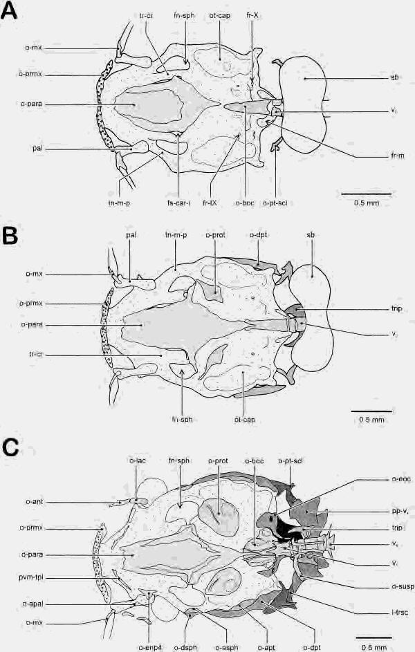
11 OSTEOCRANIUM ONTOGENY IN CLARIAS 193 closes the foramen for the truncus hyomandibularis of the nervus facialis (VII), as well as the base of the opercular process (Fig. 8B). Between the hyomandibular and quadrate bones, a gutter-like preopercular bone follows the ventrolateral border of the suspensorium, thereby partially covering the cartilaginous interhyal. The anterior ceratohyal bone is now apparent in the cleared specimen as well. The forked branches of the parurohyal bones have become extended, both in an anterior and posterior direction. Their medial tips now have fused with each other posteriorly (Fig. 4C). Ceratobranchial ossifications are observed in the cleared specimen. Both their articular facets, with the hypobranchials and epibranchials, remain unossified. Apart from an increase in number of teeth, no striking difference can be observed in the upper pharyngeal tooth plate (Fig. 7B). No more branchiostegal rays have been added since the previous stage; thus nine are present. At 11.1 mm SL, the angular bone bears a lateral gutter, which indicates the fusion with at least one splenial bone. Consequently, a complex angulo-splenio-articulo-retroarticular bone is formed. The mm SL stage (Figs. 6C, 7C, 9) Neurocranium Ventral to the ethmoid plate, the premaxillaries become more and more plate-like, as a posterior extension is noted. The membranodermal component of the frontal bone can Fig. 3. Skull of Clarias gariepinus (6.6 mm SL stage). A, dorsal view, B, lateral view, C, ventral view. (Grey indicates bone, shaded areas indicate cartilage.) c-eth, cartilago ethmoideum; c-meck, cartilago Meckeli; c- sbmx, cartilago submaxillaris; cb-i, ceratobranchiale I; cb-v, ceratobranchiale V; cd, chorda dorsalis; ch, ceratohyale; cm-sphsep, commissura spheno-septalis; cp-a, copula anterior; eb-i, epibranchiale I; eb-ii, epibranchiale II; eb-iii, epibranchiale III; fn-hyp, fenestra hypophysea; fn-sph, fenestra sphenoidea; fr-car-i, foramen arteria carotis interna; fr-i, foramen fila olfactoria; fr-x, foramen nervus vagus; hb-i, hypobranciale I; hh, hypohyale; hs, hyosymplecticum; ih, interhyale; ipb-iv, infrapharyngobranchiale IV; Im-on, lamina orbitonasalis, sensu latu; o-den-mm, os dento-mentomeckelium; o-mx, os maxillare; o-op, os operculare; o-para, os parasphenoideum; o-prmx, os praemaxillare; o-puh, os parurohyale; ot-cap, otic capsule; p-q, pars quadrata of the palatoquadratum; pal, palatinum; pc-pl, parachordal plate; pl-oc, pila occipitalis; pns-ep, pons epiphysialis; prc-op, processus opercularis; prc-pt, processus pterygoideus; prc-ra, processus retroarticularis; r-br, radius branchiostegus; sb, swimbladder; sol-n, solum nasi; su-ph-tpl, superior pharyngeal tooth plate; tn-m-p, taenia marginalis posterior; tr-cr, trabecula cranii. now be clearly distinguished from the neurodermal, gutter-like one, as it has started to cover the postpineal foramen and, consequently, the brain. Posterolaterally, the frontal bone gutter has reached that of the dermosphenotic bone. The latter bone, as well as the dermopterotic bone, has started to form the membranodermal component as well. Additionally, a perichondral ossification covers the taenia marginalis posterior at the level of these dermosphenotics, especially at the ventral side (Fig. 6C). Apparently, the autosphenotic has also formed. It is directly fused to the dermal counterpart, as is the case for the dermopterotics, and autopterotic bones. This fused bony complex of double origin is consequently referred to as the sphenotic bone ( dermosphenotic and autosphenotic bones) and the pterotic bone (dermopterotic and autopterotic bones). At the posterior corners of the chondrocranium, contacting the pterotics, lies the posttemporo-supracleithral bone, bearing a foramen for the temporal canal (Fig. 9B). The latter feature is an additional argument for stating that the posttemporal is present and part of the bony complex. From this stage on, the parasphenoid plays an important role in the reinforcement of the skull floor, as the trabecular bars have become split in two by the continued lateral expansion of the foramen for the internal carotid artery (Fig. 9A). At this stage, the parasphenoid consequently establishes the ventral connection between the anterior part of the neurocranium (anterior to the sphenoid fenestra) and the posterior part (posterior to the fenestra). The previously slender, lateral wings have consequently become broader. The basioccipital has formed two longitudinal ridges between which the posterior extension of the parasphenoid comes to lie (Fig. 6C). At this stage, the basioccipital supports the otoliths of the lagena and the sacculus, i.e., the asteriscus and the sagitta, respectively. Lateral to the basioccipital, the exoccipitals have enclosed the foramen of the nervus vagus (X). The latter bones form the lateral margins of the foramen magnum, thus enclosing the pilae occipitales, until they contact the parieto-supraoccipital bone complex dorsally. The prootics have become enlarged and ovally shaped and take part in the ossification of the border of the sphenoid fenestra (Fig. 6C). They support the otolith of the utriculus, i.e., the lapillus. Anteriorly, behind the premaxillary bones, two small,

12 Fig. 4. Ontogeny of the parurohyale in Clarias gariepinus (ventral view). A, 6.6 mm SL stage; B, 7.7 mm SL stage; C, 10.0 mm SL stage; D, 11.6 mm SL stage; E, 12.7 mm SL stage; F, 21.5 mm SL stage; G, mm SL stage. (Black indicates parurohyal bone, grey indicates ligaments, shaded areas indicate cartilage.) bb-i, basibranchiale I; bb-ii, basibranchiale II; cb-i, ceratobranchiale I; cb-ii, ceratobranchiale II; ch, ceratohyale; fr-hy, foramen hyoideum; fr-puh, foramen parurohyalis; fspuh, fissura parurohyalis; hb-i, hypobranchiale I; hb-ii, hypobranchiale II; hh, hypohyale; I-puh-hh, ligamentum parurohyalo-hypohyale; o-bb-ii, os basibranchiale II; o-bb-iii, os basibranchiale III; o-cb-i, os ceratobranchiale I; o-cb-ii, os ceratobranchiale II; o-ch-a, os ceratohyale anterior; o-hb-i, os hypobranchiale I; o-hb-ii, os hypobranchiale II; o-hh-v, os hypohyale ventrale; o-puh, os parurohyale.
13 OSTEOCRANIUM ONTOGENY IN CLARIAS 195 splint-like bones arise on the ventral face of the ethmoid plate (Fig. 6C). As can be derived from further ontogeny, these bones correspond to the prevomeral tooth plates that are formed prior to the median prevomeral bone itself. Dentition, however, could not yet be discerned. At this stage, the first signs of the dermal bones bordering the rigid skull laterally are present. At the level of the palatine, two small ossicles have formed, enclosing the anterior part of the infraorbital canal. These ossicles correspond to the neurodermal component of the antorbital bone and lacrimal bone ( os infraorbitale I) (Fig. 9B). Splanchnocranium The previously cartilaginous lower jaw has now become almost completely enclosed with bone. An anterolateral process of the angulosplenio-retroarticular complex fits between the ventral and lateral processes of the dentosplenio-mentomeckelian complex. At this interdigitating surface, the coronoid process, as well as almost the complete medial face of Meckel s cartilage is still exposed (Fig. 7C). At the level of its articulation with the orbitonasal lamina, the palatine has become ossified perichondrally, thus forming the autopalatine bone (Fig. 6B). The articular facet of the autopalatine itself is still cartilaginous, although surrounded by bone. The entopterygoid is now a small, triangular bone, bordering the autopalatine ventrally (Figs. 6C, 7C, 9B). The anterior tip of the pterygoid process of the suspensorium has also started to ossify, forming the metapterygoid bone. This perichondral bone, however, is still separated from the quadrate bone. The latter has become expanded dorsally and caudally, compared with the previous stage. Dorsally, the initiation of the membranous outgrowth of the quadrate has started. Although still separated from the quadrate, the hyomandibular bone has formed the corresponding membranous outgrowths as well, at its anterior border. The preopercular bone has become tubelike now, as the gutter has closed. This bone still follows the ventrocaudal margin of the hyosymplecticum, with its anterior part lying horizontally and its posterior part vertically. At the level of the articulation with the neurocranium, the hyomandibular bone bears a cartilaginous rim. Compared to the previous stage, the opercular bone has extended even more ventrally. In this cleared and stained specimen, three ossifications of the hyoid bar are discernable: the anterior ceratohyal in the middle, and the newly formed posterior ceratohyal and ventral hypohyal (Fig. 7C). The latter bone is penetrated by a foramen, through which passes the hyoid artery (Fig. 4D). The articulation of the nine branchiostegal rays occurs with the anterior ceratohyal bone and the cartilaginous part separating the latter from the posterior ceratohyal bone. The parurohyal bone now clearly forms one single unit, as a horizontal bony lamella has formed between the median and two lateral processes (Fig. 4D). The ligamentous connection of the parurohyal with the hyoid bar occurs at the level of the ventral hypohyals. The anterior copula bears two ossified rings at the position of the second and third basibranchial bones. At their anterolateral tips, the hypobranchials I and II have started to ossify as well (Fig. 7C). All the epibranchial bones are observed in the cleared specimen of 11.6 mm SL. The upper pharyngeal tooth plate has enlarged rather isometrically, but is still supported mainly by the unossified, fourth infrapharyngobranchial element. At 12.0 mm SL, all mandibular ossifications are present. The last to develop is the coronomeckelian bone, which is formed at the medial face of the coronoid process, between the dento-spleniomentomeckelian and the angulo-splenioarticulo-retroarticular bone complexes (Fig. 10A). At this stage, the splenial gutter of the dental bone is still open anteriorly, but gradually becomes closed off and incorporated in the bone complex (Fig. 10). The mm SL stage (Figs. 4E, 11, 12A) Neurocranium The bones of the skull roof have started to close up the unossified regions in the skull as they make contact with the surrounding bones. The laterodermethmoids follow the commissura spheno-septalis as they grow posteriorly. Serial sections of a 13.0 mm SL specimen show that the laterodermethmoids are fused to the perichondral supraethmoid, up to the level of the precerebral lamina. Posterior to that level, the supraethmoid is missing, whereas the dermal bones have extended above the posterior part of this lamina and the sphenoseptal commissures, separated from it by connective tissue. This configuration supports a possible dermal origin of these bones. The plate-like part of the frontal bone becomes extended anteriorly, medially and posteriorly. Posteriorly, the frontals have contacted the parietals (thus
14 Figure 5
15 OSTEOCRANIUM ONTOGENY IN CLARIAS 197 fused to the supraoccipital bone), and the initiation of an interdigitation can be observed. Medially, the frontals have continued to cover the pre- and postpineal fenestra, as well as enclosing the cartilaginous epiphysial bridge (Fig. 11A). Anterior to the entrance of the supraorbital canal into the frontal bone, a small, gutter-like nasal bone has differentiated. Laterocaudally, the bony skull margin is formed by the sphenotic and pterotic bone, covering the posterior part of the taenia marginalis posterior and the otic capsule. The sphenotic bone is restricted to the taenia marginalis and the anterior part of the otic capsule, whereas the pterotic bone covers about two thirds of that capsule. The perichondral part of both these bones has expanded over the otic capsules, ventrally and dorsally. Posteriorly, the two anterior processes of the posttemporo-supracleithral bone have expanded anteriorly, as the fork between them fits onto the posterior margin of the pterotic bone (Fig. 11B). The ossified transscapular ligament is a stout process, contacting the laterodorsal margin of the parapophysis of the fourth vertebra. In the skull floor, two more bones have been added. Ossification of the lateral ethmoid has started at the level of the articular facet of the lamina orbito-nasalis, for the articulation with the autopalatine (Fig. 12A). It borders the articular facet laterally, and has Fig. 5. Skull of Clarias gariepinus (7.7 mm SL stage). A, dorsal view; B, lateral view; C, ventral view. (Grey indicates bone, shaded areas indicate cartilage.) ast, asteriscus (lagenar otolith); c-eth, cartilago ethmoideum; c-meck, cartilago Meckeli; cb-i, ceratobranchiale I; cd, chorda dorsalis; ch, ceratohyale; cm-sphsep, commissura spheno-septalis; cp-a, copula anterior; cp-p, copula posterior; eb-i, epibranchiale I; fn-sph, fenestra sphenoidea; fr-car-i, foramen arteria carotis interna; fr-i, foramen fila olfactoria; fr-x, foramen nervus vagus; hb-i, hypobranchiale I; hh, hypohyale; hs, hyosymplecticum; ih, interhyale; ipb-iii, infrapharyngobranchiale III; lap, lapillus (utricular otolith); Im-on, lamina orbitonasalis, sensu latu; o-boc, os basioccipitale; o-den-mm, os dentomentomeckelium; o-eoc, os exoccipitale; o-mx, os maxillare; o-op, os operculare; o-para, os parasphenoideum; o-prmx, os praemaxillare; o-pt-scl, os posttemporosupracleithrum; o-puh, os parurohyale; ot-cap, otic capsule; p-q, pars quadrata of the palatoquadratum; pal, palatinum; pns-ep, pons epiphysialis; pp-v 4, parapophysis of vertebra 4; prc-co, processus coronoideus; prc-op, processus opercularis; prc-pt, processus pterygoideus; prc-ra, processus retroarticularis; r-br, radius branchiostegus; sag, sagitta (saccular otolith); sb, swimbladder; sn, supraneurale; su-ph-tpl, superior pharyngeal tooth plate; tn-m-p, taenia marginalis posterior; tr-cr, trabecula cranii; trip, tripus; tt-p, tectum posterius; v 2, vertebra 2; v 6, vertebra 6. covered the ventral part of the preorbital base. At the anterodorsal border, the pterosphenoid bones have appeared at the ventral part of the taenia marginalis posterior (Fig. 12A). The prevomeral tooth plates have expanded, and the first teeth can be distinguished. The parasphenoid becomes extended anteriorly, touching the prevomeral tooth plates. Posteriorly, the parasphenoid narrows and terminates in a slender process lying between the longitudinal ridges of the basioccipital. This kind of overlap allows the long and slender interdigitation, which can be observed in later stages. The basioccipital has enlarged, especially in an anterior direction, as it now forms the complete floor for the sacculus, enclosing the sagitta (Fig. 12A). The exoccipitals now border the foramen for the glossopharyngeus nerve posteriorly. At the lateral face of the skull, a second infraorbital bone has been added, posterior to the lacrimal bone. As is still the case for the anterior two canal bones, this infraorbital bone is tubular. It borders the eye ball anteroventrally (Fig. 11B). Ventral to the preopercular bone, a triangular interopercular bone is formed, close to the anterior margin of the opercular ventral tip. Serial sections of a 15.2 mm SL specimen indicate that the lateral ethmoid consists of a perichondral ossification, bearing a plate-like extension. The lamina orbitonasalis ossifies perichondrally and becomes laterally and dorsally enlarged by a bony plate. However, a separate ossification of the plate-like part could not be discerned. At 18.7 mm SL, the prevomeral tooth plates are interconnected and have fused to the median prevomeral bone. This bone already interdigitates with the parasphenoid. Three more dermal canal bones are added at this stage, covering the skull laterally: the third and fourth infraorbital bones, and the suprapreopercular one. All of them are still tubular or gutter-like. The infraorbital series is completed in this stage. The onset of perichondral ossification of the orbitosphenoid could also be discerned, covering the cartilaginous floor of the ethmo-orbital region. Splanchnocranium The bones of the hyoid arch have expanded, especially the anterior ceratohyal. It is, however, still separated from both the ventral hypohyal and posterior ceratohyal by cartilage (Fig. 11B). The interhyal is still cartilaginous and is continuous with both the hyoid arch and the hyosymplectic carti-
16 Figure 6
17 OSTEOCRANIUM ONTOGENY IN CLARIAS 199 Fig. 6. Neurocranium of Clarias gariepinus (ventral view). A, 7.7 mm SL stage; B, 10.0 mm SL stage; C, 11.6 mm SL stage. (Grey indicates bone, shaded areas indicate cartilage.) fn-sph, fenestra sphenoidea; fr-car-i, foramen arteria carotis interna; fr-ix, foramen nervus glossopharyngeus ( fenestra basicapsularis posterior); fr-x, foramen nervus vagus; fr-m, foramen magnum; I-trsc, ligamentum transcapularis; o-ant, os antorbitale; o- apal, os autopalatinium; o-apt, os autopteroticum; o- asph, os autosphenoticum; o-boc, os basioccipitale; o- dpt, os dermopteroticum; o-dsph, os dermosphenoticum; o-enp4, sesamoid os entopterygoideum type 4; o-eoc, os exoccipitale; o-lac, os lacrimale ( os infraorbitale I); o-mx, os maxillare; o-para, os parasphenoideum; o- prmx, os praemaxillare; o-prot, os prooticum; o-pt-scl, os posttemporo-supracleithrum; o-susp, os suspensorium; ot-cap, otic capsule; pal, palatinum; pp-v 4, parapophysis of vertebra 4; pvm-tpl, prevomeral tooth plate; sb, swimbladder; tn-m-p, taenia marginalis posterior; tr-cr, trabecula cranii; trip, tripus; v 1, vertebra 1; v 2, vertebra 2; v 6, vertebra 6. lage. As is the case for the previous stage, all branchiostegal rays articulate with the anterior ceratohyal and the cartilaginous part between the latter and the posterior ceratohyal. All suspensorial bones have expanded, closing in the cartilaginous parts between them. The membranous outgrowths of the quadrate and hyomandibular are more pronounced, and a distinct articular ridge between the latter and the neurocranium is present (Fig. 11B). The articulation between the suspensorium and the neurocranium is situated at the level of the posterior part of the sphenotic and the anterior part of the pterotic. Evidence of a paired origin of the parurohyal is now lost, as the anterior tips also have fused, leaving a foramen for the passage of the parurohyal artery, as can be derived from serial sections of a 46.8 mm SL specimen (Figs. 4E, 13). At 18.7 mm SL, the third infrapharyngobranchial also becomes ossified. The mm SL stage (Figs. 4F, 12B, 14) Neurocranium All bones are present. Most of them have started to close off the unossified parts almost completely. In the 21.5 mm SL specimen, the premaxillaries have expanded posteriorly, thus supporting the nasal sacs. The ethmoid plate and precerebral lamina are almost completely ossified, as the contralateral laterodermethmoid bones have fused medially. The nasal bones have become tubular, in which the branching of the supraorbital canal can already be distinguished (Adriaens et al., 97). In the cleared specimen, the lateral ethmoid ossification is now observed on the dorsal face of the skull (Fig. 14A). The frontal bones have expanded substantially in all directions. Anterolaterally, they interdigitate with the lateral ethmoid, anteriorly with the mesethmoid, posterolaterally with the sphenotics and the pterotics, caudally with the parieto-supraoccipital complex, and medially the two frontals meet at the level of the epiphysial bridge (Fig. 14A). The parieto-supraoccipital bone has started to close off the postpineal fenestra, as have the frontals. Posteriorly, the former bone complex bears a distinct, pointed supraoccipital process, which reaches up to the fourth vertebra. Rudimentary contact between the parieto-supraoccipital bone and the pterotic bone is already established. Anterior expansion of the pterotic bone has deprived the sphenotic bone from any contact with the parieto-supraoccipital bone complex. Medial expansion of the sphenotic bone, at the ventral side of the skull, has resulted in the ossification of the lateral border of the sphenoid fenestra. At its posterior margin, the posttemporo-supracleithral bone has become plate-like and borders the pterotic bone both laterocaudally and mediocaudally. The lateral-line canal, which exits this bone complex, is enclosed by a pair of small ossicles. In the skull floor, the orbitosphenoid is clearly visible (Fig. 12B). It arises as a paired ossification of the ethmoid plate and the preorbital base, lateral to the anterior part of the parasphenoid. The orbitosphenoids are still separated from the surrounding bones and border the sphenoid fenestra anteriorly. The lateral wings of the parasphenoid have expanded extensively, as they contact the ventral extension of the pterosphenoid bones. Consequently, this connection subdivides the sphenoid fenestra into an anterior foramen for the optic nerve and a posterior trigemino-facial foramen. The prootics have come into close contact with the parasphenoid, sphenotics, pterotics, basioccipital, and exoccipital bones, but no true interdigitation has occurred (Fig. 12B). Basioccipital and parasphenoid, however, already strongly interdigitate. The second infraorbital bone has extended caudally, as it also now borders the eye ventrally. The third infraorbital bone lies at the posterior margin of the eye. A gap is still present between the third and the second infraorbital bone. All these infraorbital bones are still tubular, apart from the fourth one, which lies at the lateral margin
18 Figure 7
19 OSTEOCRANIUM ONTOGENY IN CLARIAS 201 of the skull, between the frontal and sphenotic bones. Apparently, the fourth infraorbital bone arises last, although it subsequently becomes the largest one of the series, as can be derived from the later stages. The suprapreopercular bone has become platelike, where the neurodermal and membranodermal components already can be clearly distinguished. In the 46.8 mm SL specimen, a plate-like, nasal, triangular in cross section, could be observed, indicating the presence of the membranodermal component. An articulation is present between the small antorbital bone and a small cartilaginous protuberance of the anterior articular facet of the autopalatine (Fig. 15A). The antorbital bone partially encloses the base of the nasal barbel. Splanchnocranium The mandibula has become more heavily ossified, as the dento-splenio-mentomeckelian complex becomes connected more solidly to the angulo-splenio-articulo-retroarticular complex through an extensive interdigitation (Fig. 10B,C). The splenial bones have closed their gutters completely, leaving only the pores where the mandibular canal exits the bone (Adriaens et al., 97). At its medial face, the coronoid process gradually becomes enclosed as well (Fig. 10C). A well-developed articular facet for articulation with the quadrate is present, bearing a substantial retroarticular process. Fig. 7. Splanchnocranium of Clarias gariepinus (dorsal view). A, 7.7 mm SL stage; B, 10.0 mm SL stage; C, 11.6 mm SL stage. (Grey indicates bone, shaded areas indicate cartilage.) c-meck, cartilago Meckeli; cb-i, ceratobranchiale I; ch, ceratohyale; cp-a, copula anterior; cp-p, copula posterior; eb-i, epibranchiale I; eb-iv, epibranchiale IV; hb-i, hypobranchiale I; hh, hypohyale; hs, hyosymplecticum; in-ph-tpl, inferior pharyngeal toothplate; ipb-iii, infrapharyngobranchiale III; ipb-iv, infrapharyngobranchiale IV; o-ang-c, os angulo-splenioarticulo-retroarticulare complex; o-art, os articulare; o- bb-ii, os basibranchiale II; o-bb-iii, os basibranchiale III; o-cb-i, os ceratobranchiale I; o-cb-iii, os ceratobranchiale III; o-cb-iv, os ceratobranchiale IV; o-cb-v, os ceratobranchiale V; o-ch-a, os ceratohyale anterior; o- den-c, os dento-splenio-mentomeckelium complex; o-denmm, os dento-mentomeckelium; o-eb-i, os epibranchiale I; o-enp4, sesamoid os entopterygoideum type 4; o-hb-i, os hypobranchiale I; o-hh-v, os hypohyale ventrale; o- hm, os hyomandibulare; o-mp, os metapterygoideum; o-op, os operculare; o-pop, os praeoperculare; o-puh, os parurohyale; o-q, os quadratum; prc-op, processus opercularis; prc-pt, processus pterygoideus; prc-un, processus uncinatus; r-br, radius branchiostegus; su-ph-tpl, superior pharyngeal tooth plate. The autopalatine is almost completely ossified, except for its rostral and caudal tips (Fig. 12B). Anteriorly, the tip establishes an articular facet for the double-headed maxillary bone, enclosing the maxillary barbel. Although no true articulation is present with the caudal tip of the autopalatine, an ossification is lacking. The sesamoid entopterygoid bone connects to the metapterygoid bone and the prevomeral bone through a ligamentous strap. The metapterygoid bone has started to form its membranous outgrowths, as was already the case for the quadrate and the hyomandibular bone. Although more extensively ossified, the latter two bones are still separated from each other by cartilage (Fig. 14B). The preopercular bone has initiated the formation of the membranodermal component, and the branching of the preopercular canal can be recognized in the neurodermal part. The dorsal opening of that canal in the preopercular bone is positioned exactly ventral to the entrance of the canal into the suprapreopercular bone. The opercular bone is still triangular, being ligamentously connected to the interopercular bone at its ventral tip. The latter bone has become broader compared to the previous stage. The anterior ceratohyal bone now reaches the posterior one. Interdigitation between these two bones has started only at the dorsal side of the hyoid bar, whereas cartilage separates them ventrally (Fig. 14B). Anteriorly, the anterior ceratohyal bone has reached the ventral hypohyal bone, where the first signs of interdigitation are apparent (Fig. 4F). The articulation between the hyoid bar and the branchiostegal rays is still comparable to that in the previous stage, i.e., at the level of the anterior ceratohyal bone and the cartilaginous region posterior to it. At this stage, one branchiostegal ray is added, comprising a total of 10. The parurohyal bone bears a small but distinct foramen, and the three caudal processes have become elongated (Fig. 4F). Anteriorly, the double ligamentous connection to the ventral hypohyals is still present. The bones of the branchial basket have not changed substantially compared to the previous stage. Basibranchials of the second and third branchial arches are separated by cartilage on the anterior copula. Very small hypobranchials are present on the first and second branchial arch; all ceratobranchials are well ossified. Their articular facets, with the hypo- and epibranchials, however, re-
20 Figure 8
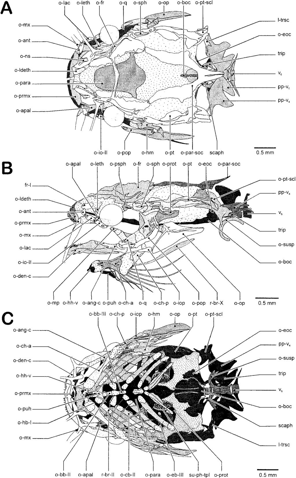
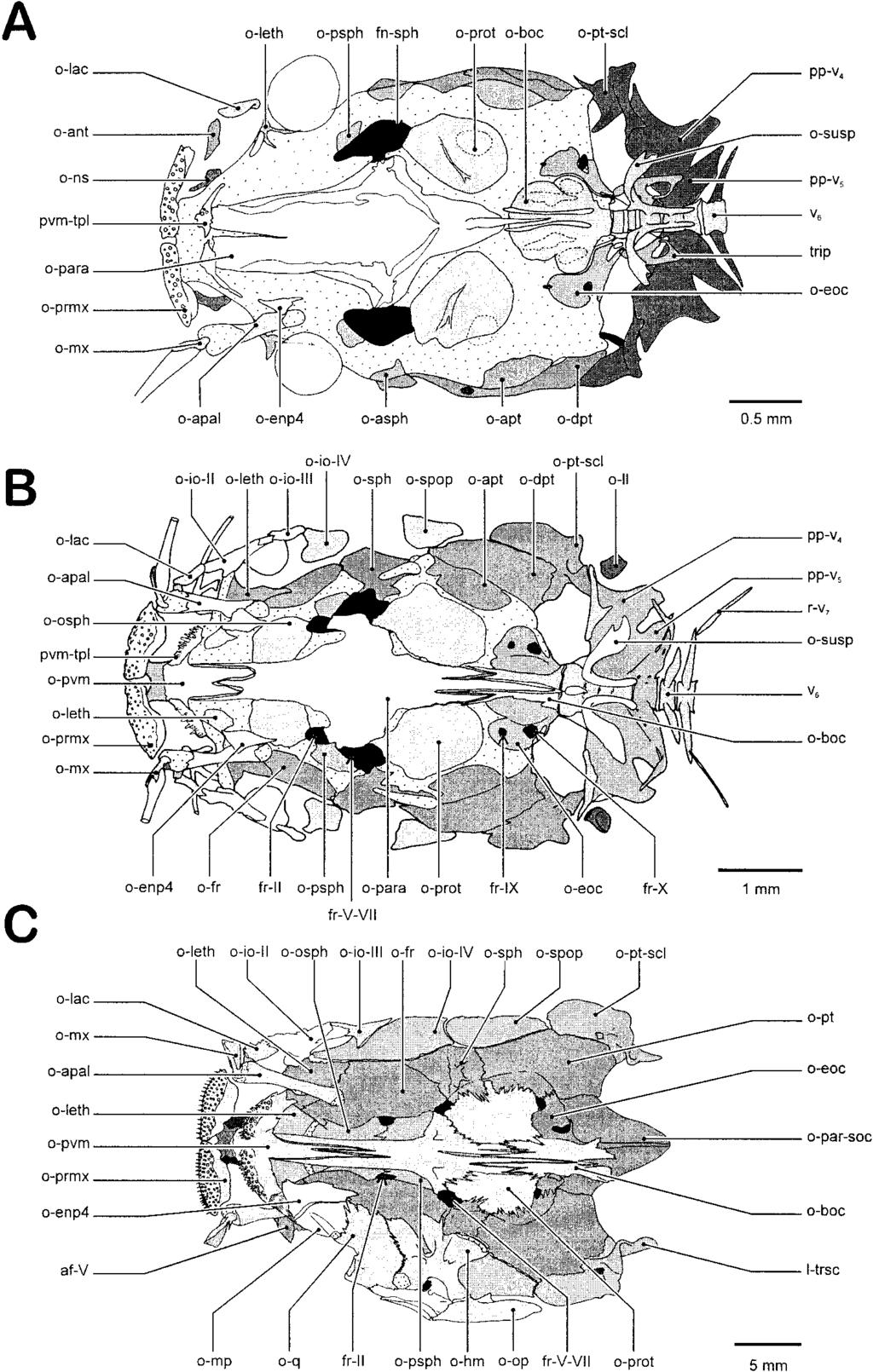
21 OSTEOCRANIUM ONTOGENY IN CLARIAS 203 mains unossified. Although no fifth epibranchial has developed, the corresponding ceratobranchial element bears an unossified head at both ends. The third epibranchial element bears a well-developed, medially directed uncinate process at its caudal margin. At 46.8 mm SL, the splanchnocranium is complete. A small, gutter-like splenial bone is present lateral to the articulation between mandibular and suspensorium. This splenial embraces the sensory canal, at the transition from the mandibular to the preopercular canal. Dorsomedially, the angulosplenio-articulo-retroarticulare complex has become extended rostrally, coming into contact with the dento-splenio-mentomeckelian bone. As a result, the cartilaginous coronoid process is enclosed by bone laterally and medially. At its dorsomedial face, the hypohyal cartilage bears a small, but distinct process, which becomes ossified at this stage. Lying at the dorsal face of the ventral hypohyal, this bone corresponds to the dorsal hypohyal. The fourth infrapharyngobranchial element also starts to ossify, bordering the cartilage dorsally, medially and ventrally. The mm SL stage (Figs. 4G, 10D, 12C, 15 23) Neurocranium This juvenile stage is to some degree a good copy of the adult configuration, although reduced in size. The mesethmoid is relatively broad, bearing two well-developed Fig. 8. Skull of Clarias gariepinus (10.0 mm SL stage). A, dorsal view; B, lateral view; C, ventral view. (Gray indicates bone, shaded areas indicate cartilage.) c-meck, cartilago Meckeli; fn-sph, fenestra sphenoidea; fr-car-i, foramen arteria carotis interna; fr-i, foramen fila olfactoria; fr-ix, foramen nervus glossopharyngeus ( fenestra basicapsularis posterior); fr-ot, foramen ramus oticus nervus facialis; I-trsc, ligamentum transcapularis; lm-on, lamina orbitonasalis, sensu latu; lm-pc, lamina praecerebralis; o-art, os articulare; o-boc, os basioccipitale; o-cb-i, os ceratobranchiale I; o-cb-ii, os ceratobranchiale II; o-cb-v, os ceratobranchiale V; o-ch-a, os ceratohyale anterior; o-den-c, os dento-splenio-mentomeckelium complex; o-dpt, os dermopteroticum; o- dsph, os dermosphenoticum; o-eoc, os exoccipitale; o-fr, os frontale; o-hm, os hyomandibulare; o-mx, os maxillare; o-op, os operculare; o-para, os parasphenoideum; o-pop, os praeoperculare; o-prmx, os praemaxillare; o- prot, os prooticum; o-pt-scl, os posttemporo-supracleithrum; o-puh, os parurohyale; o-q, os quadratum; o- susp, os suspensorium; ot-cap, otic capsule; pal, palatinum; pns-ep, pons epiphysialis; pp-v 4, parapophysis of vertebra 4; prc-co, processus coronoideus; prc-un, processus uncinatus; sb, swimbladder; su-ph-tpl, superior pharyngeal tooth plate; tn-m-p, taenia marginalis posterior; trip, tripus; v 6, vertebra 6. preethmoid processes, which correspond to the well-ossified preethmoid cornua (Adriaens and Verraes, 97c). Interdigitation occurs with the lateral ethmoids, laterally, and the frontals, posteriorly. Together with the lateral ethmoid, the laterally curved preethmoid processes enclose the nasal bone. The lateral ethmoid has become plate-like, enclosing the cartilaginous orbito-nasal lamina. At its ventral face, the initial articular facet between this lamina and the palatine can still be distinguished, as an ossification is lacking (Fig. 17A). Medially the lateral ethmoid bears a funnel through which the olfactory lobes pass. Laterally, it bears a distinct process for the articulation with the dorsal process of the second infraorbital bone (Fig. 15A). Posteriorly, the lateral ethmoid interdigitates with the frontals, but no overlapping could be observed. At its lateral margin, the lateral ethmoid is connected through a connective tissue sheet to the dorsal margin of the anterior part of the fourth infraorbital bone. The frontals are the largest, paired skull bones, which border the anterior fontanella. Compared to the previous stage, both frontals are sutured to each other posteriorly, thus subdividing the postpineal fenestra into a postepiphysial part of the anterior fontanella and the posterior fontanella (Fig. 15A). In small specimens, the anterior fontanella is bordered anteriorly by the mesethmoid. A second region of interdigitation between contralateral frontals occurs at the level of the epiphysial bridge, which has become completely enclosed by a tubular outgrowth of the dermal frontals (Fig. 17B). In the skull roof, the frontals connect through sutures to the sphenotics (posterolaterally), the pterotics (posteriorly) and the parieto-supraoccipital bone complex (posteromedially). Laterally, they connect through connective tissue to the central part of the fourth infraorbital bone. Ventrally, the frontals connect rigidly to the orbitosphenoids and the pterosphenoids (Fig. 12C). The sphenotics have become plate-like as well, enclosing the anterolateral part of the otic capsule (Fig. 12C). They are bordered by the frontals and pterotics, to which they are strongly sutured. They have lost contact with the parieto-supraoccipital bone (Fig. 15A). Ventrally, they connect to the prootic and pterosphenoid bones. At their ventrolateral margin, they enclose the anterior part of the articular facet for the suspensorium, which is preceded by a stout lateroventral process (Figs. 17C, 21E).
22 Figure 9
23 OSTEOCRANIUM ONTOGENY IN CLARIAS 205 The pterotics are large bones, enclosing the posterolateral part of the otic capsule, and bearing large, lateral, plate-like extensions. Medially, they are sutured to the parieto-supraoccipital bone. The connection with the posttemporo-supracleithral bone is restricted to the posterolateral part. Consequently, the pterotics form part of the posterior margin of the juvenile skull (Fig. 15A). Ventrally, they connect to the prootics and exoccipitals (Figs. 12C, 17D) and form the posterior part of the articular facet for articulation with the suspensorium (Fig. 21E). In a specimen of mm SL, however, the pterotic seemed to be separated partially from the parieto-supraoccipital and exoccipital bones (which may be due to insufficient staining), where a very small ossicle was found covering that region (Fig. 16). Based on its position, as well as the fact that it seems to be a perichondral bone, at the level of the posterior semicircular canal, it must correspond to the epiotic, which could not be observed in any previous stage. It is, however, possible that because of its reduced size and insufficient staining, it has been overlooked in the other specimens, as was also the case for Nawar ( 54). The parietosupraoccipital bone complex is large, with its previously paired anterior part now fused to each other medially. Apparently, a true fusion has occurred, in contrast with the Fig. 9. Skull of Clarias gariepinus (11.6 mm SL stage). A, dorsal view. B, lateral view; C, ventral view. (Grey indicates bone, shaded areas indicate cartilage.) fn-sph, fenestra sphenoidea; fr-i, foramen fila olfactoria; o-ang-c, os angulo-splenio-articulo-retroarticulare complex; o-ant, os antorbitale; o-apal, os autopalatinum; o-bb-ii, os basibranchiale II; o-bb-iii, os basibranchiale III; o-boc, os basioccipitale; o-cb-i, os ceratobranchiale I; o-cb-iv, os ceratobranchiale IV; o-ch-a, os ceratohyale anterior; o-ch-p, os ceratohyale posterior; o-den-c, os dento-splenio-mentomeckelium complex; o-dpt, os dermopteroticum; o-dsph, os dermosphenoticum; o-eb-iv, os epibranchiale IV; o-enp4, sesamoid os entopterygoideum type 4; o-eoc, os exoccipitale; o-fr, os frontale; o-hh-v, os hypohyale ventrale; o-hm, os hyomandibulare; o-iop, os interoperculare; o-lac, os lacrimale ( os infraorbitale I); o-ldeth, os latero-dermethmoideum; o-mp, os metapterygoideum; o-mx, os maxillare; o-op, os operculare; o-par-soc, os parieto-supraoccipitale; o-para, os parasphenoideum; o-pop, os praeoperculare; o-prmx, os praemaxillare; o-prot, os prooticum; o-pt-scl, os posttemporo-supracleithrum; o-puh, os parurohyale; o-q, os quadratum; o-susp, os suspensorium; pp-v 4, parapophysis of vertebra 4; pp-v 5, parapophysis of vertebra 5; pvm-tpl, prevomeral tooth plate; r-br, radius branchiostegus; scaph, scaphium; su-ph-tpl, superior pharyngeal tooth plate; trip, tripus; v 1, vertebra 1; v 6, vertebra 6; v 7, vertebra 7. posterior part of the frontals, as no signs of a suture can be observed anymore (Fig. 17E). The left-right fusion is not complete, as a posterior fontanella remains. Observations of larger adults indicate a slow, but progressive closure of both the anterior and posterior fontanella. At its ventral face, the complex bears a median ridge, which is formed in the mediosagittal septum. Caudolaterally, the bone interdigitates with the exoccipitals (Fig. 17E). The posttemporo-supracleithral bone is a plate-like complex, bordering the pterotics laterally and laterocaudally. Ventrally, it bears a mediocaudally directed process for articulation with the parapophysis of the fourth vertebra, as well as an articular facet for articulation with the cleithral bone (Fig. 17F). It does not bear any connection with the basioccipital, which is the case in many siluriform fishes (Fig. 12C). Most presumably, this process corresponds to the ossification of the transcapular ligament (see Discussion). Anteriorly, two plate-like premaxillaries articulate with the ventral face of the preethmoid processes of the mesethmoid. Premaxillaries, with the exception of the posterior margin, are completely covered with teeth. They lack any differentiations like an ascending process or maxillary process (Fig. 18A). No true articular facet, with secondary cartilage, is present between the premaxillary bone and the mesethmoid. Both connect only through connective tissue. An articulation of the premaxillary with the maxillary bone is also absent. Only a distinct string of ligamentous tissue connects the two. The maxilla is typically advanced siluriform, as it is socket-like, enclosing the base of the maxillary barbel. It bears a double-headed articular facet for articulation with the rostral tip of the palatine (Fig. 18B). As in most siluriform fish, the maxillary bone has been modified to take part in the palatine-maxillary mechanism (Adriaens and Verraes, 97a). The antorbital bone is still small and overlies the rostral tip of the palatine. Compared to the previous stage, most infraorbital bones have undergone a major transformation in shape, as they are no longer tubular. The lacrimal bone bears a serrated ventral edge, whereas the second infraorbital bone possesses a massive part for the articulation with the lateral process of the lateral ethmoid (Fig. 15A). Consequently, both these bones form the anterior border of the orbita, whereas the remaining
24 Figure 10
25 OSTEOCRANIUM ONTOGENY IN CLARIAS 207 part of the second infraorbital bone borders the orbita anteroventrally. The posteroventral and posterior border is demarcated by the third infraorbital bone (Fig. 15B). Dorsally, the slender supraorbital process of the fourth infraorbital bone forms the dorsal margin, but is elongated and much broader prosterior to the eye. In Clarias gariepinus, the fourth infraorbital bone reaches up to the suprapreopercular bone. The latter bone has become plate-like as well, filling up the gap between the fourth infraorbital bone and the posttemporo-supracleithral bone (Fig. 15B). The skull floor consists of a central, longitudinal and narrow bridge, which becomes broader posteriorly, as it forms the base of the braincase. Anteriorly, the hypoethmoid part of the mesethmoid interdigitates with the prevomeral bone. The latter has become arrow-like, bearing two well-developed tooth plates, to which it is fused. Posteriorly, the prevomeral bone interdigitates through a very slender, forked suture with the parasphenoid (Fig. 19A). Dorsally, the prevomeral bone is connected to the lateral ethmoid, more exactly to the part enclosing the olfactory lobes (Fig. 12C). The parasphenoid consists of a small anterior part, which is expanded laterally at the level of the earlier formed lateral wings, which contact the pterosphenoid bones. Posterior to these wings, the parasphenoid becomes narrow again, finally interdigitating posteriorly with the basioccipitals. The interdigitation with the latter occurs through many more sutures than is the case for the prevomeral bone (Fig. 19B). At its dorsal face, the parasphenoid connects rigidly to the orbitosphenoid (in front of the lateral wings), the pterosphenoids (at the lateral wings), and the prootics (posterior to the lateral wings) (Figs. 12C, 19B). The basioccipital no longer forms an articular surface with the first vertebra, Fig. 10. Ontogeny of the lower jaw in Clarias gariepinus (medial view). A, 12.0 mm SL stage; B, 14.8 mm SL stage; C, 19.0 mm SL stage; D, mm SL stage. (Grey indicates Meckel s cartilage, shaded areas indicate bone.) af-iii, articulatory facet of the os angulosplenio-articulo-retroarticulate with the os quadratum; c-meck, cartilago Meckeli; con-t, conical teeth; mndsym, mandibular symphysis; o-ang, os angulare; o-art, os articulare; o-com, os coronomeckelium; o-den, os dentale; o-den-spl, os spleniale (dentale); o-den-vp, ventral process of the os dento-splenio-mentomeckelium complex; o-mm, os mentomeckelium; o-rart, os retroarticulare; prc-co, processus coronoideus; vil-t, villiform teeth. but now interdigitates with the complex of vertebrae, formed in relation to the Weberian apparatus (Figs. 16, 19C). Lateroventrally, the basioccipital connects to the exoccipitals, posteriorly, and to the prootics, anteriorly, both through short sutures. At its dorsal face, the basioccipital bears a bowllike structure housing the sinus impar perilymphaticus of the Weberian complex (Fig. 16) (Chardon, 67; Radermaker et al., 89). The orbitosphenoids connect to the lateral ethmoids anteriorly, the frontals dorsally, and the parasphenoid posteriorly. They are separated from the pterosphenoids by the foramen for the optic nerve (Fig. 12C). Although the orbitosphenoids are formed as paired ossifications, they have fused to a single, gutter-like bone in the juvenile stage (Fig. 19D). The pterosphenoids, however, remain paired ossifications, which connect to the orbitosphenoids, the parasphenoid, and the sphenotics (Fig. 12C). The prootics form the anterolateral floor of the brain cavity and enclose the utriculus with its otolith (the lapillus) (Fig. 19F). The perichondral prootics are sutured to a whole set of bones: the pterosphenoid, the parasphenoid, the sphenotics, the pterotics, the exoccipitals, the basioccipital, and the parieto-supraoccipital. It takes part in the bordering of the trigemino-facial foramen (Fig. 12C). Finally, the exoccipitals form the posterolateral ossifications of the brain cavity floor and also enclose the otoliths of the sacculus and the lagena (i.e., the sagitta and asteriscus respectively) (Fig. 19G). These bones interdigitate with the basioccipitals, prootics, and the parieto-supraoccipital bone, whereas a synchondrosis seems to be present with the pterotics. The exoccipital bone encloses the foramina for the glossopharyngeus, vagus, and hypoglossus nerves (Figs. 12C, 16, 19G). Splanchnocranium The mandibula is fully ossified, although Meckel s cartilage remains partially exposed (Fig. 10D). The coronoid process is well protected by bone, with only its dorsal tip remaining uncovered. Caudally, the retroarticular process is rather short and bears two processes, both for the attachments of ligaments: a ligament running to the interopercular bone and one to the hyoid bar (Fig. 20A). The dento-splenio-mentomeckelian bone complex is provided with a large patch of teeth. Two types of teeth are observed: villiform teeth at the outer margin, and conical teeth at the inner margin (Fig. 20B). The
26 Figure 11
27 OSTEOCRANIUM ONTOGENY IN CLARIAS 209 surface for dentition seems to be expanded, as a kind of rostral extension of the bone has occurred. At its medial face, this bone complex bears a socket that encloses the anterior part of Meckel s cartilage. Caudally, a well-developed ventral and lateral process can be distinguished, between which fits the lateroventral process of the angulo-splenioarticulo-retroarticular complex. At the level of the coronoid process, a distinct coronomeckelian bone covers Meckel s cartilage dorsally, anterior to the coronoid process (Fig. 10D). The autopalatine is a rod-shape bone, bearing three unossified regions: (1) the anterior tip that enables the articulation with the maxillary bone, (2) the slender and elongated articular facet for the articulation with the lateral ethmoid, and (3) the posterior tip, which does not articulate with any bone (Fig. 20C). Between the mandibula and the suspensorium, the number of isolated splenial bones has increased to two, whereas in larger specimens three of them were present (Fig. 15B) (Adriaens et al., 97). The quadrate bone bears a solid articular facet with the mandibula and is provided with a substantial dorsal, membranous outgrowth (Fig. 21C). The quadrate connects both synchondrally and through sutures with the metapterygoid bone, anteriorly, and with the hyomandibula, posteriorly (Fig. 21A). The hyomandibula also has a membranous Fig. 11. Skull of Clarias gariepinus (12.7 mm SL stage). A, dorsal view; B, lateral view; C, dorsal view of the splanchnocranium. (Grey indicates bone, shaded areas indicate cartilage.) fr-i, foramen fila olfactoria; I-trsc, ligamentum transcapularis; o-ang-c, os angulosplenio-articulo-retroarticulare complex; o-ant, os antorbitale; o-apal, os autopalatinum; o-bb-ii, os basibranchiale II; o-bb-iii, os basibranchiale III; o-boc, os basioccipitale; o-cb-ii, os ceratobranchiale II; o-ch-a, os ceratohyale anterior; o-ch-p, os ceratohyale posterior; o-den-c, os dento-splenio-mentomeckelium complex; o-eb- III, os epibranchiale III; o-eoc, os exoccipitale; o-fr, os frontale; o-hb-i, os hypobranchiale I; o-hh-v, os hypohyale ventale; o-hm, os hyomandibulare; o-io-ii, os infraorbitale II; o-iop, os interoperculare; o-lac, os lacrimale ( os infraorbitale I); o-ideth, os latero-dermethmoideum; o-leth, os latero-ethmoideum; o-mp, os metapterygoideum; o-mx, os maxillare; o-ns, os nasale; o-op, os operculare; o-par-soc, os parieto-supraoccipitale; o-para, os parasphenoideum; o-pop, os praeoperculare; o-prmx, os praemaxillare; o-prot, os prooticum; o-psph, os pterosphenoideum; o-pt, os pteroticum; o-pt-scl, os posttemporosupracleithrum; o-puh, os parurohyale; o-q, os quadratum; o-sph, os sphenoticum; o-susp, os suspensorium; pp-v 4, parapophysis of vertebra 4; pp-v 5, parapophysis of vertebra 5; r-br, radius branchiostegus; scaph, scaphium; su-ph-tpl, superior pharyngeal tooth plate; trip, tripus; v 6, vertebra 6. plate, which connects to a similar outgrowth of the quadrate (Fig. 21A). Dorsally, a cartilaginous strip is present for articulation with the neurocranium. Anterior to this strip, the bone has a distinct dorsal process, which fits into a cavity in the sphenotic bone, additionally, a ventral process of the sphenotic fits into a cavity posterior to the hyomandibular process (Fig. 21E). Due to such a connection, the suspensorium and neurocranium are interlocked to a certain degree (see Discussion). At its posterior margin, the hyomandibula bears a substantial opercular process with a cartilaginous articular facet (Fig. 21B). At its medial face, the hyomandibula bears two foramina: (1) a small foramen just above the insertion of the ligament running to the hyoid bar, and (2) a larger one posterior and dorsal to it. This larger foramen is penetrated by the truncus hyomandibularis, whereas through the smaller foramen a blood vessel enters. Compared to the situation in previous stages, the foramen through which the truncus leaves has shifted ventrally, due to the fact that a bony crest of the hyomandibular bone has grown ventrally, thereby covering the foramen of the hyosymplectic cartilage. At this stage, the latter foramen is bordered by both the hyomandibula and the quadrate. Medially, the hyomandibula forms a ridge for the attachment of a stout ligament that runs to the hyoid bar (Fig. 21A) (Adriaens and Verraes, 94). The unossified part, separating the hyomandibula from the quadrate, corresponds to the part that in previous stages was connected to the interhyale and may be considered as an unossified symplectic. At its ventral margin, the hyomandibula interdigitates with the preopercular bone (Figs. 15B, 21A). This bone does not bear a distinct vertical and horizontal limb, as is the case in many catfish (Fig. 24A). Its caudal margin consists of the neurodermal part enclosing the preopercular canal with two of its branches (Adriaens et al., 97). At its anterodorsal side, it bears a plate-like extension for the interdigitation with the hyomandibula and the quadrate (Fig. 24A). The plate-like expansion has persisted in the metapterygoid and entopterygoid bone as well (Fig. 21D). As can be derived from the previous stages, the ossification of the metapterygoid initiates from a perichondral ossification of the pterygoid process, which can still be distinguished in this stage. In
28 Figure 12
29 OSTEOCRANIUM ONTOGENY IN CLARIAS 211 Fig. 12. Neurocranium of Clarias gariepinus (ventral view). A, 12.7 mm SL stage; B, 21.5 mm SL stage; C, mm SL stage. (Grey indicates bone, shaded areas indicate cartilage.) af-v, articulatory facet of the os lateroethmoideum with the os infraorbitale II; fn-sph, fenestra sphenoidea; fr-ii, foramen fasciculus opticus; fr-ix, foramen nervus glossopharyngeus ( fenestra basicapsularis posterior); fr-x, foramen nervus vagus; fr-v- VII, foramen of the trigemino-facial nerve complex; I- trsc, ligamentum transcapularis; o-ant, os antorbitale; o-apal, os autopalatinum; o-apt, os autopteroticum; o-asph, os autosphenoticum; o-boc, os basioccipitale; o-dpt, os dermopteroticum; o-enp4, sesamoid os entopterygoideum type 4; o-eoc, os exoccipitale; o-fr, os frontale; o-hm, os hyomandibulare; o-io-ii, os infraorbitale II; o-io-iii, os infraorbitale III; o-io-iv, os infraorbitale IV; o-lac, os lacrimale ( os infraorbitale I); o-leth, os lateroethmoideum; o-ii, lateral line ossicle; o-mp, os metapterygoideum; o-mx, os maxillare; o-ns, os nasale; o-op, os operculare; o-osph, os orbitosphenoideum; o-par-soc, os parieto-supraoccipitale; o-para, os parasphenoideum; o-prmx, os praemaxillare; o-prot, os prooticum; o-psph, os pterosphenoideum; o-pt, os pteroticum; o-pt-scl, os posttemporo-supracleithrum; o-pvm, os prevomerale; o-q, os quadratum; o-sph, os sphenoticum; o-spop, os suprapraeoperculare; o-susp, os suspensorium; pp-v 4, parapophysis of vertebra 4; pp-v 5, parapophysis of vertebra 5; pvm-tpl, prevomeral tooth plate; r-v 7, rib of vertebra 7; trip, tripus; v 6, vertebra 6. this stage, both ventrally and dorsally, this central rod bears plate-like extensions. The ventral extension corresponds to the ectopterygoid process of Arratia ( 92). Considerable discussion concerns the homology of the pterygoid bones in catfish and even in fish in general (Howes and Teugels, 89; Arratia, 90, 92). The metapterygoid is strongly connected to the quadrate through the ventral synchondrosis, but more strongly through the extensive suturation along the dorsal plates (Fig. 21A). Consequently, the metapterygoid no longer contacts the hyomandibula. Rostrally, the metapterygoid has a straight margin, connected to the entopterygoid through a ligamentous strip. The entopterygoid in Clarias gariepinus is ligamentously connected to a whole set of surrounding bones: (1) the metapterygoid, posteriorly, (2) the autopalatine, dorsally, (3) the prevomeral bone, anteriorly, and (4) the lateral ethmoid, anterolaterally. Caudally, the entopterygoid bone bears a distinct process (Fig. 21D). The hyoid bar is almost completely ossified, although cartilaginous strips seem to persist between all bones (Figs. 15C, 22A). At the medial face of the ventral hypohyal, the dorsal hypohyal remains separated by cartilage from both the ventral hypohyal and the anterior ceratohyal. The ventral hypohyal is much larger than the dorsal. Ventromedially, the latter bone forms an interdigitation with the anterior ceratohyal bone, whereas the rest is interconnected through a synchondrosis. Mediolateral to these sutures, it is attached through a stout ligament that is connected to the anterior tips of the parurohyal bone (Fig. 4G). A basihyal bone is absent. The anterior ceratohyal bone is the largest of the hyoid bar (Fig. 22C). At its medial face, it bears a shelf against which the first six branchiostegal rays articulate (Fig. 22A). The following two rays articulate with the cartilaginous strip, which separates the anterior ceratohyal bone from the posterior one ventrally. The posterior two branchiostegal rays articulate with the ventral margin of the posterior ceratohyal bone. The posterior two branchiostegal rays articulate with the ventral margin of the posterior ceratohyal bone. The posterior ceratohyal bone tapers posteriorly and terminates as a solid cone (Fig. 22D). At the dorsal face of this cone is a stout ligament that connects the hyoid bar to the hyomandibula (Fig. 21A). Another large ligament connects the lateral face with the medial process of the mandibular retroarticular process (Fig. 20A). In this stage, the interhyal is completely reduced (Adriaens and Verraes, 94). The parurohyal bone has become more solidly ossified, with three distinct, caudal processes. Although the median process is the result of a fusion of two processes, it is more slender than the lateral processes (Fig. 4G). The two heads of the paired sternohyoideus muscle fit nicely into the fork of the parurohyal bone (Adriaens and Verraes, 97d). The length of the ligaments, in relation to the size of the parurohyal, seems to have become reduced substantially, as the rostral tips of the parurohyal now partially fit into a small gap at the medial face of the ventral hypohyals. The main body of the parurohyal still bears a foramen, penetrated by a ventral branch of the ventral aorta (Fig. 13). The latter artery is here referred to as the parurohyal artery. It enters the parurohyal dorsally, where it splits into two arteries before it leaves the bone ventrally. Between the two rostral processes of the parurohyal, a vein (here referred to as the vena parurohyalis) branches off of the vena jugularis inferior and curves caudally, against the ventral side of the bone and between the two branches of the artery (Fig. 13B). The branchiostegal rays differ slightly in shape, depending on
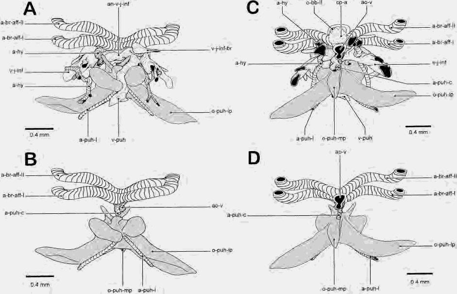
30 Figure 13
31 OSTEOCRANIUM ONTOGENY IN CLARIAS 213 their position. The anterior ray is a slender rod with a broader articular part, whereas the posterior ray has a distinct articular facet, penetrated by a foramen (Fig. 22E), with a distal, elongated, plate-like part (Fig. 22A). Ossification of the branchial basket seems to have completed, although several cartilaginous elements lack any bone (Fig. 15C). The anterior copula still bears two ossifications, corresponding to the second and third basibranchial bones (Fig. 23B C). No signs of the first basibranchial could be discerned at this stage (Fig. 23A). The posterior copula also lacks ossifications of the fourth and fifth basibranchial (Fig. 23D E). Hypobranchial bones I and II of the previous stage have expanded, as they now cover almost half of the surface of the corresponding cartilaginous elements (Figs. 15C, 23A,B). The third hypobranchial element is still unossified. The cartilaginous fourth hypobranchial is believed to remain fused to the ceratobranchial cartilage and is unossified as well (Fig. 23D) (Adriaens and Verraes, 97c). All five ceratobranchials are well ossified. They consist of the central, perichondral rod, which bears two caudal ridges for attachment of the gill rods, as well as for the housing of blood vessels and nerves. The tooth plate, covering the dorsal face of the fifth ceratobranchial bone, has become broader, especially at its anterior half (Fig. 23E). The epibranchials are more elongated, compared with the previous stage, especially epibranchials I and II (Fig. 15C). This elongation seems to be coupled to the elongation of the third infrapharyngobranchial bone. Medially, these bones converge and attach to each other through cartilage, which most probably corresponds to the unossified remains of infrapharyngobranchials I and II Fig. 13. Graphical, 3-D reconstruction of the parurohyal bone and related bloodvessels in Clarias gariepinus (46.8 mm SL stage). A, frontal view; B, frontal view with vena jugularis inferior and arteria hyoidea removed; C, caudal view; D, caudal view with vena jugularis inferior and arteria hyoidea removed. (Grey indicates bone.) a-br-aff-i, arteria branchialis afferens I; a-br-aff-ii, arteria branchialis afferens II; a-hy, arteria hyoidea; a- puh-c, arteria parurohyalis communis; a-puh-i, arteria parurohyalis lateralis; an-v-j-inf, anastomosis between left and right venae jugulares inferiores; ao-v, aorta ventralis; cp-a, copula anterior; o-bb-ii, os basibranchiale II; o-puh-lp, lateral process of the os parurohyale; o-puh-mp, medial process of the os parurohyale; v-j-inf, vena jugularis inferior; v-j-inf-br, branch of the vena jugularis inferior; v-puh, vena parurohyalis. (Adriaens and Verraes, 97c). This cartilage connects these epibranchials to the anterior tip of the third infrapharyngobranchial bone (Fig. 15C). Epibranchiale III has a welldeveloped uncinate process at its caudal margin (Fig. 23C). Medially, this epibranchial articulates with the posterior tip of the third infrapharyngobranchial and the anterior margin of the fourth one (Fig. 15C). The fourth epibranchial bone bears a horizontal, plate-like extension at its middle portion, for the support of the uncinate process (Fig. 23D). Medially, this epibranchial articulates with the posterolateral margin of the fourth infrapharyngobranchial element. As mentioned, only the third and fourth infrapharyngobranchials ossify in Clarias gariepinus (Fig. 15C). The third is rod-like, with both its articular tips nonossified (Fig. 23C). The fourth one is rather triangular, with its lateral margin nonossified (Fig. 23D). The upper pharyngeal tooth plate is enlarged and is supported dorsally by the fourth infrapharyngobranchial bone, as well as the medial part of epibranchials III and IV (Fig. 15C). The opercular series in juvenile Clarias gariepinus consists of the opercular bone, the preopercular, and interopercular bones (Fig. 15B). The subopercular bone is missing, as is the case in most siluriform fish (see Discussion). The preopercular bone attaches to the suspensorium, as mentioned above. The opercular bone is triangular and articulates with the opercular process of the hyomandibula (Fig. 15B). Consequently, at its anterior margin, the opercular bone bears a well-developed articular facet, which is in contact with a medial crest for the insertion of the levator operculi muscle and the opercular part of the hyohyoidei adductor muscles (Fig. 24B) (Adriaens and Verraes, 97b). The ventral tip of the opercular bone attaches to the interopercular bone through a ligamentous strip (Fig. 15B). This interopercular bone remains triangular, as it tapers rostrally to become elongated in a stout ligament that is attached to the lateral process of the mandibular retroarticular process (Figs. 20A, 24C). The interopercular bone also attaches through connective tissue to the lateral face of the posterior ceratohyal bone. DISCUSSION Bone in Clarias gariepinus is cellular, although the number of osteocytes is low. Ostariophysans, in general, together with some other primitive teleostean lineages (Meu-

32 Figure 14
33 OSTEOCRANIUM ONTOGENY IN CLARIAS 215 nier and Huysseune, 92), are characterized by the presence of cellular bone. According to the terminology of Patterson ( 77), all three bone types in fish are formed in C. gariepinus: cartilage bones, dermal bones, and membrane bones. In C. gariepinus, all cartilage bones ossify perichondrally. True enchondral ossification was not observed but may occur in larger specimens. Bony elements are compact, lamellar ossifications early in ontogeny, whereas most of them become spongious in the juvenile stage. The cartilage bones, lining the cartilaginous structures, are surrounded with membranous apolamellae, which frequently are formed in a radiating manner. Most dermal bones are extremely trabecular, with the exception of the prevomeral bone and parasphenoid bone. Dermal tooth plates become spongy also. At least one type of membrane bone is also present: the sesamoid bones (coronomeckelian and entopterygoid bones). The fully developed skull of C. gariepinus consists of about 150 bones (both sides included), which can be subdivided into five categories: (1) 22 anamestic dermal bones, (2) 14 dermal canal bones, (3) 37 perichondral bones, (4) two sesamoid bones and (5) three bones of a compound nature. Fig. 14. Skull of Clarias gariepinus (21.5 mm SL stage). A, dorsal view; B, lateral view; C, dorsal view of splanchnocranium. (Grey indicates bone, shaded areas indicate cartilage.) fr-i, foramen fila olfactoria; ipb-iv, infrapharyngobranchiale IV; o-ang-c, os angulo-splenioarticulo-retroarticulare complex; o-ant, os antorbitale; o-bb-ii, os basibranchiale II; o-boc, os basioccipitale; o-cb-iv, os ceratobranchiale IV; o-ch-a, os ceratohyale anterior; o-cl, os cleithrum; o-com, os coronmeckelium; o-den-c, os dento-splenio-mentomeckelium complex; o-eb-i, os epibranchiale I; o-eb-iv, os epibranchiale IV; o-enp4, sesamoid os entopterygoideum type 4; o-eoc, os exoccipitale; o-fr, os frontale; o-hb-ii, os hypobranchiale II; o-hh-v, os hypohyale ventrale; o-hm, os hyomandibulare; o-io-ii, os infraorbitale II; o-io-iii, os infraorbitale III; o-io-iv, os infraorbitale IV; o-iop, os interoperculare; o-ipb-iii, os infrapharyngobranchiale III; o-lac, os lacrimale ( os infraorbitale I); o-leth, os latero-ethmoideum; o-ii, lateral line ossicle; o-meth, os mesethmoideum; o-mp, os metapterygoideum; o-mx, os maxillare; o-ns, os nasale; o-op, os operculare; o-osph, os orbitosphenoideum; o-par-soc, os parieto-supraoccipitale; o-para, os parasphenoideum; o-pop, os praeoperculare; o-prmx, os praemaxillare; o-prot, os prooticum; o-psph, os pterosphenoideum; o-pt, os pteroticum; o-pt-scl, os posttemporo-supracleithrum; o-q, os quadratum; o-sph, os sphenoticum; o-spop, os suprapraeoperculare; o-susp, os suspensorium; pp-v 4, parapophysis of vertebra 4; pp-v 5, parapophysis of vertebra 5; prc-op, processus opercularis; r-br, radius branchiostegus; r-v 7, rib of vertebra 7; v 6, vertebra 6; v c, vertebral complex of the Weberian apparatus. Siluriform relationships of the osteocranium of Clarias gariepinus Although a comparative osteology is beyond the scope of this report, some of the features used to describe the cranial osteology of Clarias gariepinus, are briefly discussed. The paired origin of the prevomeral bone has been observed in most teleost fish (De Beer, 37; Verraes, 73). This bone is considered nonhomologous with the vomeral bone of tetrapods, although it is frequently referred to as vomeral bone in teleosts (de Beer, 37; Harrington, 55; Daget, 64). In Clarias gariepinus, it appears that the prevomeral bone itself is unpaired (whether or not a fusion occurs between paired blastemata or not could not be discerned) and becomes fused to paired tooth plates. In the closely related heteropneustid species, Heteropneustes fossilis, an unpaired prevomeral bone arises, prior to its dentition (Srinivasachar, 58). Researchers seem to come to a consensus that the toothless prevomeral bone is an unpaired, dermal bone that becomes fused to paired, autogenous toothplates, whereas the latter may be homologous with pharyngeal tooth plates of the premandibular arch (Daget, 64; Srinivasa Rao and Lakshmi, 84; Arratia, 87). The prevomeral bone in siluriform fish is generally T-shaped, bearing two patches of teeth at its lateral wings and interdigitating with the parasphenoid through very long sutures (Bhimachar, 33; Merriman, 40; Tilak, 63b, 65a; Lundberg, 82; Srinivasa Rao and Lakshmi, 84; Howes and Fumihito, 91). Both tooth patches may be fused to each other (Merriman, 40; Tilak, 61, 63b,c; Rastogi, 63; Arratia, 87; Howes and Fumihito, 91) or may even be absent (Tilak, 65a, 67; Gauba, 70; Lundberg, 82; Vignes and Garcia, 87; Ferraris, 96). Fusion between both tooth plates in adult Clariidae seems to be the general trend (Tilak, 63c; Teugels, 82). In case dentition is lacking, an unpaired origin of the prevomeral bone has been observed (Kindred, 19), with its lateral wings sometimes being absent (Lundberg, 82; Howes, 83). Heavily dentate prevomeral tooth plates are present in Pangasiidae and Siluridae, where these plates become closely aligned with autogenous palatine tooth plates, or may even fuse to them (Tilak, 61; Roberts and Vidthayanon, 91). In Chacidae, the prevomeral bone is absent as an independent ossification (Tilak, 71; Brown and Ferraris, 88).
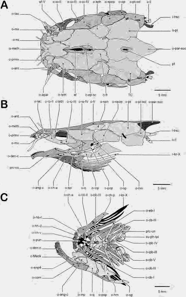
34 Figure 15
35 OSTEOCRANIUM ONTOGENY IN CLARIAS 217 Fig. 16. Skull of Clarias gariepinus (140.1 mm SL stage). Caudal view. (Small circles indicate cartilage.) cd, chorda dorsalis; fr-ix, foramen nervus glossopharyngeus ( fenestra basicapsularis posterior); fr-m, foramen magnum; fr-x, foramen nervus vagus; fr-xii, foramen nervus hypoglossus; Gr sin-imp, groove for the sinus impar perilymphaticus; I-trsc, ligamentum transcapularis; o-boc, os basioccipitale; o-eoc, os exoccipitale; o- epot, os epioticum; o-par-soc, os parieto-supraoccipitale; o-prot, os prooticum; o-pt, os pteroticum; o-pt-scl, os supracleithrum. For a discussion and in- and outgroup comparison of the parietal and supraoccipital bone complex, we refer to Fink and Fink ( 81, 96) and Arratia and Gayet ( 95). The Fig. 15. Skull of Clarias gariepinus (127.0 mm SL stage). A, dorsal view; B, lateral view; C, dorsal view of the splanchnocranium. (Grey indicates bone, shaded areas indicate cartilage.) af, anterior fontanella; af-v, articulatory facet of the os lateroethmoideum with the os infraorbitale II; c-meck, cartilago Meckeli; I-trsc, ligamentum transcapularis; o-ang-c, os angulo-splenioarticulo-retroarticulare complex; o-ant, os antorbitale; o-apal, os autopalatinum; o-bb-iii, os basibranchiale III; o-cb-i, os ceratobranchiale I; o-cb-iii, os ceratobranchiale III; o-cb-v, os ceratobranchiale V; o-ch-a, os ceratohyale anterior; o-ch-p, os ceratohyale posterior; o-com, os coronomeckelium; o-den-c, os dento-splenio-mentomeckelium complex; o-eb-i, os epibranchiale I; o-enp4, sesamoid os entopterygoideum type 4; o-ep-br, ossification of the os frontale around the epiphyseal bridge; o-fr, os frontale; o-hb-i, os hypobranchiale I; o-hb-ii, os hypobranchiale II; o-hh-d, os hypohyale dorsale; o-hh-v, os hypohyale ventrale; o-hm, os hyomandibulare; o-io-ii, os infraorbitale II; o-io-iii, os infraorbital III; o-io-iv, os infraorbitale IV; o-iop, os interoperculare; o-ipb-iii, os infrapharyngobranchiale III; o-ipb-iv, os infrapharyngobranchiale IV; o-lac, os lacrimale ( os infraorbitale I); o-leth, os latero-ethmodeum; o-ii, lateral line ossicle; o-meth, os mesethmoideum; o-mp, os metapterygoideum; o-mx, os maxillare; o-ns, os nasale; o-op, os operculare; o-par-soc, os parieto-supraoccipitale; o-pop, os praeoperculare; o-prmx, os praemaxillare; o-pt, os pteroticum; o-pt-scl, os posttemporo-supracleithrum; o-puh, os parurohyale; o-q, os quadratum; o-sph, os sphenoticum; o-spl, ossa splenialia; o-spop, os suprapraeoperculare; pf, posterior fontanella; prc-co,processus processus coronoideus; prcun, processus uncinatus; r-br, radius branchiostegus; su-phtpl, superior pharyngeal tooth plate; TC, temporal canal. presence of a separate parietal ossification, which has been observed in several siluriform taxa, is now generally accepted to explain the compound nature of this bone (Arratia, 87; Fink and Fink, 96). For a discussion on the homology of the posttemporo-supracleithral bone in Siluriformes, we refer to Lundberg ( 75), Arratia and Gayet ( 95), and Fink and Fink ( 81, 96). Concerning the transscapular ligament, several constructions seem to be present in Siluriformes. In many catfish, the posttemporo-supracleithral bone connects to the basioccipital through a bony limb, the ossified transscapular ligament, which attaches through a ligament to the lateral face of the basioccipital (Bhimachar, 33; Merriman, 40; Hubbs and Miller, 60; Tilak, 61, 63b, 65a, 67, 71; Rastogi, 63; Lundberg, 75; Poll, 77; Srinivasa Rao and Lakshmi, 84; Howes and Fumihito, 91). In others, no such a connection is observed. Then, mostly, a medial limb connects the anterior vertebrae, at the level of the vertebral center (Tilak, 63b), or contacts the parapophyses (Srinivasacher, 58; Nawar, 54; Tilak, 63c; Gauba, 70; Howes, 83; present study). Regan ( 11) mentioned that a lower limb of the posttemporal (posttemporo-supracleithral bone of present study) to the basioccipital is absent in Clariidae, Loricariidae, and Callichthyidae. This author also mentioned that the posttemporal is absent in Clariidae, whereas the su-
36 Figure 17
37 OSTEOCRANIUM ONTOGENY IN CLARIAS 219 Fig. 18. Maxillary bones of juvenile Clarias gariepinus. A, premaxillary, dorsal view (125.5 mm SL stage); B, maxillary, caudal view (174.5 mm SL stage). (Orientation cross: D, dorsal, V: Ventral.) af-x, articulatory facets of the os maxillare with the os autopalatinum; crprmx, dorsal crest of the os praemaxillare, for the articulation with the cornua prae-ethmoidea; I-mx-pal, ligamentum maxillo-palatinum; s-mx-b, socket of the maxillary barbel. Fig. 17. Skull roof bones of juvenile Clarias gariepinus (125.5 mm SL stage). A, lateral ethmoid, ventral view; B, frontal, ventral view; C, sphenotic, ventral view. D, pterotic, ventral view; E, Parieto-suproccipital, ventral view; F, posttemporo-supracleithral, ventral view. (Orientation cross: A: Anterior, P: Posterior, L: Lateral, M: Medial.) af-v, articulatory facet of the os lateroethmoideum with the os infraorbitale II; af-viii, articulatory facet of the os lateroethmoideum with the os autopalatinum; af-xi, articulatory facet of the os posttemporo-supracleithrum with the os cleithrum; cr-par-soc, medioventral crest of the posterior part of the os parietosupraoccipital; fn-pop-a, anterior part of the postpineal foramen; fn-prp, fenestra praepinealis; fr-m-b, border of the foramen magnum; fr-v-vii-b, border of the foramen of the trigemino-facial nerve complex; Gr a-sc-c, groove for the anterior semicircular canal; Gr nerv-i, groove of the fila olfactoria; intd-eoc, interdigitation with the os exoccipitale; intd-fr, interdigitation with the os frontale; intd-leth, interdigitation with the os lateroethmoideum; intd-meth, interdigitation with the os mesethmoideum; intd-osph, interdigitation with the os orbitosphenoideum; intd-par-soc, interdigitation with the os parietosupraoccipitale; intd-prot, interdigitation with the os prooticum; intd-psph, interdigitation with the os pterosphenoideum; intd-pt, interdigitation with the os pteroticum; intd-pt-scl, interdigitation with the os posttemporosupracleithrum; intd-sph, interdigitation with the os sphenoticum; IOC, infraorbital canal; I-trsc, ligamentum transcapularis; lap, lapillus (utricular otolith); o-epbr, ossification of the os frontale around the epiphyseal bridge; o-sph-sp, sphenotic spine; OTC, otic canal; pf, posterior fontanella; PMC, preoperculo-mandibular canal; sch-eoc, synchondrosis with the os exoccipitale; SOC, supraorbital canal; TC, temporal canal; TC-pt, pterotic branch of the temporal canal. pracleithral bone is connected with a posterior process firmly united to the air-bladder capsule. In Diplomystidae, Arratia ( 87) observed a ventromedial process of the posttemporo-supracleithral bone, which is bifurcated and attached to the cranium (at the level of the basioccipital) and to the parapophyses through ligaments. The question raised here is whether the process, attaching to the basioccipital in most catfish corresponds to the one connecting the Weberian apparatus in others. The connection to the basioccipital is believed to be the ossified Baudelot s ligament, which generally runs from the supracleithral bone to the basioccipital in other teleosts. Lundberg ( 75) considered the connection to the parapophyses of the vertebrae as a differentiation (ventral process) of the transscapular ligament of his supracleithrum ( posttemporo-supracleithrum of present study). A double connection of the posttemporo-supracleithral bone to the basioccipital and Weberian apparatus, as observed by Arratia ( 87) in Diplomystidae, is considered to be a feature of the Siluriphysi (present Siluriformes and Gymnotiformes) (Fink and Fink, 81: character 98). Although such a bifurcation is absent in Clarias gariepinus, it could indicate the homology of the ventromedial process of the posttemporo-supracleithral bone with a part of the transscapular ligament in other catfish groups. A dermopalatine is considered to be absent in ostariophysan fish. Tooth plates at the level of the autopalatine are then regarded as neomorphic, autogenous tooth plates (Fink and Fink, 81, 96; Arratia and Schultze, 91; Arratia, 92). The dermopala-
38 Fig. 19. Skull floor bones of juvenile Clarias gariepinus. A, prevomer, ventral view (125.5 mm SL stage); B, parasphenoid, dorsal view (174.5 mm SL stage); C, basioccipital, dorsal view (125.5 mm SL stage); D, Orbitosphenoid, dorsal view (125.5 mm SL stage); E, pterosphenoid, lateral view (125.5 mm SL stage); F, prootic, medial view (125.5 mm SL stage); G, exoccipital, lateral (left) and medial (right) view (125.5 mm SL stage). ast, asteriscus (lagenar otolith); cd, chorda dorsalis; fr-ii-b, border of the foramen fasciculus opticus; fr-ix, foramen nervus glossopharyngeus ( fenestra basicapsularis posterior); fr-m-b, border of the foramen magnum; fr-v- VII-b, border of the foramen of the trigemino-facial nerve complex; fr-x, foramen nervus vagus; fr-xii, foramen nervus hypoglossus; Gr sin-imp, groove for the sinus impair perilymphaticus; intd-boc, interdigitation with the os basioccipitale; intd-eoc, interdigitation with the os exoccipitale; intd-fr, interdigitation with the os frontale; intd-leth, interdigitation with the os lateroethmoideum; intd-meth, interdigitation with the os mesethmoideum; intd-osph, interdigitation with the os orbitosphenoideum; intd-par-soc, interdigitation with the os parieto-supraoccipitale; intd-para, interdigitation with the os parasphenoideum; intd-prot, interdigitation with the os prooticum; intd-psph, interdigitation with the os pterosphenoideum; intd-pt, interdigitation with the os pteroticum; intd-pvm, interdigitation with the os prevomerole; intd-sph, interdigitation with the os sphenoticum; intd-v c, interdigitation with the vertebral complex; o-para-lp, lateral wing of the os parasphenoideum; pvmtpl, prevomeral tooth plate; sch-pt, synchondrosis with the os pteroticum; tn-m-p, taenia marginalis posterior.
39 OSTEOCRANIUM ONTOGENY IN CLARIAS 221 Fig. 20. Lower jaw and palatine of juvenile Clarias gariepinus. A, angulo-splenio-articulo retroarticular bone complex with Meckel s cartilage, dorsal view (125.5 mm SL stage); B, dento-splenio-mentomeckelian bone complex, dorsal view (174.5 mm SL stage); C, autopalatine and maxillary, dorsal view (125.5 mm SL stage). af-iii, articulatory facet of the os angulo-splenio-articuloretroarticulare with the os quadratum; af-vii, articulatory facet of the os autopalatinum with the os lateroethmoideum; af-ix, articulatory facet of the os autopalatinum with the os maxillare; c-meck, cartilago Meckeli; con-t, conical teeth; mx-b, maxillary barbel; o-ang-lp, lateral process of the os angulo-splenio-articuloretroarticulare complex; o-apal, os autopalatinum; o-denfe, frontal extension of the os dento-splenio-mentomeckelian complex; o-den-lp, lateral process of the os dentosplenio-mentomeckelium; o-den-vp, ventral process of the os dento-splenio-mentomecklium complex; o-mx, os maxillare; prc-ang-ch, processus angulo-ceratohyalis; prc-ang-iob, processus angulo-interopercularis; s-meck, socket for Meckel s cartilage; t-re-t, tendon of the m. retractor tentaculi; vil-t, villiform teeth.
40 Figure 21
41 OSTEOCRANIUM ONTOGENY IN CLARIAS 223 tine of Vandewalle et al., ( 93), observed in Clarias gariepinus, corresponds to the sesamoid entopterygoid bone of the present study. The thin dermal plate covering the anterior end of the cartilaginous pterygoid process probably may have been confused with the perichondral metapterygoid, which bears a dorsal and ventral membranous apolamella. The cartilaginous connection between the metapterygoid and the entopterygoid, as mentioned by Vandewalle et al., ( 93), could not be observed. The dermopalatine in Galeichthys felis (Ariidae), reported by Bamford ( 48: Fig. 7B), seems to correspond to the prevomeral tooth plates, as they attach to the lateral ethmoids at the same spot as is the case in most siluriforms. The homology of the urohyal bone in osteichthyan fishes has been extensively reviewed by Arratia and Schultze ( 90). In most teleost fishes the urohyal bone is an unpaired ossification of the aponeurosis, between the contralateral sternohyoideus heads, bearing two rostral processes for the ligamentous connection with the ventral hypohyals, and a ventral, horizontal plate (de Beer, 37; Patterson, 77; Arratia and Schultze, 90). In siluriform fish, however, a perichondral ossification participates in the formation of that bone, together with a paired bone of apparent sesamoid origin. This was Fig. 21. Suspensorium of juvenile Clarias gariepinus. A, suspensorium and hyoid bar, medial view (125.5 mm SL stage); B, hyomandibula, lateral view (125.5 mm SL stage). C, quadrate, lateral view (125.5 mm SL stage); D, metapterygoid and entopterygoid, lateral view (125.5 mm SL stage); E, Locking mechanism of suspensorium and neurocranium, medio-ventral view (127.0 mm SL stage). af-i, articulatory facet of the os hyomandibulare with the os sphenoticum; af-ii, articulatory facet of the os sphenoticum with the os hyomandibulare; af-iv, articulatory facet of the os quadratum with the os angulo-splenio-articulo-retroarticulare; af-xiii, articulatory facet of the os hyomandibulare with the os operculare; fr-tr-hm, foramen truncus hyomandibularis nervus facialis; intd-hm, interdigitation with the os hyomandibulare; intd-mp, interdigitation with the os metapterygoideum; intd-q, interdigitation with the os quadratum; I-hm-ch, ligamentum hyomandibulo-ceratohyale; membpl, membranous plate of the os hyomandibulare; o-ch-a, os ceratohyale anterior; o-ch-p, os ceratohyale posterior; o-enp4, sesamoid os entopterygoideum type 4; o-hh-d, os hypohyale dorsale; o-hh-v, os hypohyale ventrale; o-hm, os hyomandibulare; o-hm-sp, hyomandibular spine; o-mp, os metapterygoideum; o-pop, os praeoperculare; o-pt, os pteroticum; o-q, os quadratum; o-sph, os sphenoticum; o-sph-sp, sphenotic spine; prc-ect, processus ectopterygoideus; prc-op, processus opercularis; prcpt, processus pterygoideus; tr-hm, truncus hyomandibularis nervus facialis. also observed in the present study, in which development of the parurohyal bone is very similar to that observed in Trichomycterus areolatus (Trichomycteridae) (Arratia and Schultze, 90). In Clarias gariepinus, a perichondral bone appeared at the ventral margin of the cartilaginous basibranchial I (8.4 mm SL), after the paired elements were observed (6.6 mm SL). At 8.4 mm SL, the perichondral part was already fused to the sesamoid one. The chondroid bone of some catfish, however, could not be observed in C. gariepinus (Arratia and Schultze, 90). Three hypotheses can be put forward: (1) the perichondral part corresponds to the endoskeletal urohyal of sarcopterygians, which becomes fused to dermal bones, the latter homologous with the sarcopterygian interclavicles (Patterson, 77), or (2) as Patterson ( 77) in a way predicted, the perichondral part corresponds to the ventral part of the anterior basibranchial bone, which becomes fused with the paired tendon bones. A final possibility is (3) the secondary invasion of the ossification centers of the tendons of the sternohyoideus muscle into the basibranchial cartilage, which thus become perichondral, as already proposed by Arratia and Schulze ( 90). However, they mentioned that the parurohyal of siluroids is a novelty within teleosts in its combination of paired tendon bones with the addition of secondary cartilage or chondroid bone. In Clarias gariepinus, no such secondary cartilage, nor chondroid bone could be observed. The cartilage involved was the basibranchial I. An ossified basibranchial I is absent in presently studied siluriforms, but could be observed in some other ostariophysans, e.g., Cyprinidae (Harrington, 55; Cubbage and Mabee, 96), Catostomiade (Cypriniformes) (Weisel, 60), Characidae (Weitzman, 62), Hemiodontidae, Parodontidae (Characiformes) (Roberts, 74). Although the parurohyal of Siluriformes is considered to be homologous with the urohyal of other ostariophysans and other teleosts (Arratia and Schultze, 90), a detailed ontogenetic study, at an ultrastructural level, of the parurohyal and basibranchial cartilage could yield conclusive result on the true nature of the parurohyal in siluriform fish. Another feature related the parurohyal bone, in contrast with the teleostean urohyal bone, is the penetration of it by an artery. Arratia and Schultze ( 90) noted the
42 Fig. 22. Hyoid bar of juvenile Clarias gariepinus. A, hyoid bar with branchiostegal rays, lateral view (125.5 mm SL stage). B, hypohyals, dorsal view (127.0 mm SL stage); C, anterior ceratohyal, medial view (125.5 mm SL stage); D, posterior ceratohyal, medial view (125.5 mm SL stage); E, detail of articular facet of a branchiostegal ray, frontal view (125.5 mm SL stage). af-xv, articulatory facet of the os ceratohyale anterior with the radii branchiostegi; af-xvi, articulatory facet of the radius branchiostegus with the os ceratohyale anterior; cr-ch-a, crest of the os ceratohyale anterior, for the articulation with the radii branchiostegi; fr-br, foramen of the radii branchiostgi; intd-ch-a, interdigitation with the os ceratohyale anterior; intd-ch-p, interdigitation with the os ceratohyale posterior; intd-hh-v, interdigitation with the os hypohyale ventrale; I-hm-ch, ligamentum hyomandibulo-ceratohyale; I-puh-hh, ligamentum parurohyalo-hypohyale; o-ch-a, os ceratohyale anterior; o-ch-p, os ceratohyale posterior; o-hh-d, os hypohyale dorsale; o-hh-v, os hypohyale ventrale; r-br, radius branchiostegus.
43 OSTEOCRANIUM ONTOGENY IN CLARIAS 225 Fig. 23. Branchial basket of juvenile Clarias gariepinus (125.5 mm SL stage). A, branchial arch I, medial view; B, branchial arch II, medial view; C, branchial arch III, medial view; D, Branchial arch IV, medial view; E, branchial arch V, medial view. brct, branchictenium; c-hembr, cartilaginous supporting rods of the hemibranchia; cr-eb-iv, crest of the os epibranchiale IV, for the support of the processus uncinatus of the os epibranchiale III; hb-iii, hypobranchiale III; hb-iv, hypobranchiale IV; in-ph-tpl, inferior pharyngeal toothplate; o-bb- II, os basibranchiale II; o-bb-iii, os basibranchiale III; o-cb-i, os ceratobranchiale I; o-cb-ii, os ceratobranchiale II; o-cb-iii, os ceratobranchiale III; o-cb-iv, os ceratobranchiale IV; o-cb-v, os ceratobranchiale V; o-eb-i, os epibranchiale I; o-eb-ii, os epibranchiale II; o-eb-iii, os epibranchiale III; o-eb-iv, os epibranchiale IV; o-hb-i, os hypobranchiale I; o-hb-ii, os hypobranchiale II; o-ipb- III, os infrapharyngobranchiale III; o-ipb-iv, os infrapharyngobranchiale IV; prc-un, processus uncinatus; su-phtpl, superior pharyngeal tooth plate.
44 226 D. ADRIAENS AND W. VERRAES Fig. 24. Opercular bones of juvenile Clarias gariepinus. A, preopercular bone, lateral view (125.5 mm SL stage); B, opercular bone, medial view (174.5 mm SL stage); C, interopercular bone, medial view (174.5 mm SL stage). af-xiv, articulatory facet of the os operculare with the os hyomandibulare; fr-tr-hm-b, border of the foramen truncus hyomandibularis nervus facialis; intdhm, interdigitation with the os hyomandibulare; intd-q, interdigitation with the os quadratum; I-an-iop,ligamentum angulo-interoperculare; PMC, preoperculo-mandibulare canal; prc-do, processus dorsalis; r-op, insertion ridge on the os operculare for the m. levator operculi. passage of the hypobranchial artery, which runs anteriorly and enters the parurohyal bone dorsally, in Trichomycterus areolatus and Noturus flavus (Ictaluridae). It then penetrates the bone, leaving it ventrally. In T. areolatus, the artery runs anteriorly, splitting into three branches at the anterior margin of the hypohyals. In N. flavus, however, the artery splits into two, immediately after it leaves the parurohyal (Arratia and Schultze, 90: Fig. 19). Each branch then gives off a lateral branch at the anterior margin of the hypohyals. The situation in Clarias gariepinus seems to differ even more (Fig. 13): a ventral branch of the ventral aorta runs to the dorsal face of the parurohyal bone and penetrates it through the foramen. Ventrally, it immediately splits off two branches, as in N. flavus, but they run posteriorly, instead of anteriorly. The terminology hypobranchial artery is not adopted in the present study, as it appears that the artery penetrating the parurohyal is not homologous with the hypobranchial artery observed in other teleosts. In teleosts, the hypobranchial artery always splits off from an efferent branchial artery, in most cases the second one (Bertin, 58; Goodrich, 58). In C. gariepinus, the hypobranchial artery splits off from the fourth efferent branchial artery
45 OSTEOCRANIUM ONTOGENY IN CLARIAS 227 (Nawar, 55). As this is not the case for the artery penetrating the parurohyal bone in C. gariepinus, it cannot be referred to as the hypobranchial artery. Consequently, the term arteria parurohyalis is applied. The absence of the subopercular bone is a synapomorphic feature of Siluriformes (Gosline, 73; Fink and Fink, 81, 96). This is also the case for Clarias gariepinus, where none of the ontogenetic stages revealed the presence of any subopercular primordium. Surprisingly, a small, rudimentary suboperculum was observed in Plotosius canius (Plotosidae) and Osteogeniosus militaris (Ariidae) (Bhimachar, 33). Sequences in bone formation as response to functional demands The sequences in bone formation appear to be related to functional demands that arise in developing larvae and juveniles. These demands tend to vary to a large extent during the transformation from larva to adult, with several vulnerable moments at which the functional demands, and structural adaptations to support these demands, differ only very little (Galis et al., 94). The initiation of cranial ossification during ontogeny in teleost fish is coupled to respiratory and feeding requirements. In Clarias gariepinus, the opercular bone is the first one to appear (4.1 mm SL), supporting the opercular skin fold (Fig. 25). Opercular ossification starts at the articular facet of the opercular process of the hyosymplecticum, as could be observed in other teleosts (Verraes, 73; Huysseune, 85). From there on, the bone becomes a posteriorly extended rod, bearing a ventral plate (Fig. 2B). Myological data indicate that some of the muscles acting on the opercular bone arise between 5.2 and 6.8 mm SL. Although the adductor and levator operculi are present at that stage, it is only the levator muscle that already inserts onto the opercular bone (Adriaens and Verraes, 97b). However, prior to a possible muscular displacement of the opercular bone, the opercular skin fold is displaced by active respiratory movements, which already seem to occur at the 5.2 mm SL stage (Surlemont et al., 89). At this stage, opening and closing of the mouth is observed, coupled to ad- and abduction of the cheek region. A similar correlation between the onset of opercular ossification and the initiation of active respiratory movements was observed in Galeichthys feliceps (Siluriformes, Ariidae) (Tilney and Hecht, 93), Catostomus commersoni (Cypriniformes, Catostomidae) (McElman and Balon, 80) and Gadus morhua (Gadiformes, Gadidae) (Hunt von Herbing et al., 96a). In Poecilia reticulata (Cyprinodontiformes, Poeciliidae) ossification already starts while the larva still lies within the egg membrane. Although no true active respiration, by means of gills, is performed, respiratory movements were already observed (Weisel, 67). It appears that the alternating mechanical load onto the opercular skin fold, early during ontogeny, may be related to the early ossification of the opercular bone, prior to mechanical load exerted by muscular contraction of the opercular muscles (Herring, 93). The formation of a substantial, horizontal rod in the opercular bone in C. gariepinus may well be related to the insertion of the levator operculi, as in the adult situation, this rod corresponds to the levator operculi crest (Fig. 24B) (Adriaens and Verraes, 97b). Other ossifications related to the onset of respiratory movements are the branchiostegal rays, as they support the branchiostegal membrane in a similar way as the opercular bone supports the opercular skin fold (Fig. 25). The initiation of branchiostegal ray ossification generally follows that of the opercular bone rather rapidly, e.g., in Clarias gariepinus, Heterobranchus longifilis (Vandewalle et al., 97), Galeichthys feliceps (Tilney and Hecht, 93), Chrysichthys auratus (Vandewalle et al., 95), Barbus barbus (Vandewalle et al., 92), Danio rerio (Cubbage and Mabee, 96), Catostomus macrocheilus (Weisel, 67), Oncorhynchus mykiss (Verraes, 73), Poecilia reticulata (Weisel, 67), Oryzias latipes (Langille and Hall, 87). In Gadus morhua, however, a time lapse is present between the two ossifications (Hunt von Herbing et al., 96a). The gradually adding of branchiostegal rays consequently covers the support of the gradually increasing branchiostegal membrane. The early differentiation of a dorsal process of the opercular bone in C. gariepinus seems to be related to the insertion of the dilatator operculi muscle. At 7.2 mm SL the insertion of a very indistinct tendon of the dilatator onto the opercular bone was overlooked in a previous study (Adriaens and Verraes, 97b), as in the serial sections, the tendon appeared to be the anterior ossification of the opercular bone. Early ossifications involve most, but not all, dentulous bones. In Clarias gariepinus the premaxilaries, dental bones, and upper

46 228 D. ADRIAENS AND W. VERRAES Figure 25
Development of the Chondrocranium in the Suckermouth Armored Catfish Ancistrus cf. triradiatus (Loricariidae, Siluriformes)
") JOURNAL OF MORPHOLOGY 266:331 355 (2005) Development of the Chondrocranium in the Suckermouth Armored Catfish Ancistrus cf. triradiatus (Loricariidae, Siluriformes) Tom Geerinckx,* Marleen Brunain, and
JOURNAL OF MORPHOLOGY 266:331 355 (2005) Development of the Chondrocranium in the Suckermouth Armored Catfish Ancistrus cf. triradiatus (Loricariidae, Siluriformes) Tom Geerinckx,* Marleen Brunain, and
Stijn Devaere 1 *, Dominique Adriaens 1, Walter Verraes 1 and Guy G. Teugels 2 INTRODUCTION
 J. Zool., Lond. (2001) 255, 235±250 # 2001 The Zoological Society of London Printed in the United Kingdom Cranial morphology of the anguilliform clariid Channallabes apus (GuÈ nther, 1873) (Teleostei:
Cranial Architecture of Tube-Snouted Gasterosteiformes (Syngnathus rostellatus and Hippocampus capensis)
") JOURNAL OF MORPHOLOGY 271:255 270 (2010) Cranial Architecture of Tube-Snouted Gasterosteiformes (Syngnathus rostellatus and Hippocampus capensis) Heleen Leysen, 1 * Philippe Jouk, 2 Marleen Brunain, 1
JOURNAL OF MORPHOLOGY 271:255 270 (2010) Cranial Architecture of Tube-Snouted Gasterosteiformes (Syngnathus rostellatus and Hippocampus capensis) Heleen Leysen, 1 * Philippe Jouk, 2 Marleen Brunain, 1
REVISION OF THE GENUS MARTINICHTHYS, MARINE FISH (TELESOSTEI, TSELFATIIFORMES) FROM THE LATE CRETACEOUS OF KANSAS (UNITED STATES)
 FROM THE LATE CRETACEOUS OF KANSAS (UNITED STATES)") 1 REVISION OF THE GENUS MARTINICHTHYS, MARINE FISH (TELESOSTEI, TSELFATIIFORMES) FROM THE LATE CRETACEOUS OF KANSAS (UNITED STATES) TAVERNE L., 2000. Revision of the genus Martinichthys, marine fish (Teleostei,
1 REVISION OF THE GENUS MARTINICHTHYS, MARINE FISH (TELESOSTEI, TSELFATIIFORMES) FROM THE LATE CRETACEOUS OF KANSAS (UNITED STATES) TAVERNE L., 2000. Revision of the genus Martinichthys, marine fish (Teleostei,
CRANIAL OSTEOLOGY OF SCHIZOTHORAICHTHYS NIGER (MECKEL) MISRA (CYPRINIDAE: SCHIZOTHORACINAE). L NEUROCRANIUM
 MISRA (CYPRINIDAE: SCHIZOTHORACINAE). L NEUROCRANIUM") CRANIAL OSTEOLOGY OF SCHIZOTHORAICHTHYS NIGER (MECKEL) MISRA (CYPRINIDAE: SCHIZOTHORACINAE). L NEUROCRANIUM A. R. YousuF, A. K. PANDIT AND A. R. KHAN Postgraduate Department of Zoology, University of Kashmir,
CRANIAL OSTEOLOGY OF SCHIZOTHORAICHTHYS NIGER (MECKEL) MISRA (CYPRINIDAE: SCHIZOTHORACINAE). L NEUROCRANIUM A. R. YousuF, A. K. PANDIT AND A. R. KHAN Postgraduate Department of Zoology, University of Kashmir,
Osteology of the Clupeiform fish, genus Hyperlophus (II)
") Bull. Kitakyushu Mas. Nat. Hist., 4: 77-102. December 31, 1982 Osteology of the Clupeiform fish, genus Hyperlophus (II) Yoshitaka Yabumoto Kitakyushu Museum of Natural History, Nishihonmachi, Yahatahigashiku,
Bull. Kitakyushu Mas. Nat. Hist., 4: 77-102. December 31, 1982 Osteology of the Clupeiform fish, genus Hyperlophus (II) Yoshitaka Yabumoto Kitakyushu Museum of Natural History, Nishihonmachi, Yahatahigashiku,
Biology 3315 Comparative Vertebrate Morphology Skulls and Visceral Skeletons
 Biology 3315 Comparative Vertebrate Morphology Skulls and Visceral Skeletons 1. Head skeleton of lamprey Cyclostomes are highly specialized in both the construction of the chondrocranium and visceral skeleton.
AMERICAN MUSEUM NOVITATES Published by
 AMERICAN MUSEUM NOVITATES Published by Number 782 THE AmzRICAN MUSEUM OF NATURAL HISTORY Feb. 20, 1935 New York City 56.81, 7 G (68) A NOTE ON THE CYNODONT, GLOCHINODONTOIDES GRACILIS HAUGHTON BY LIEUWE
Osteology and Relationships of the Eel Diastobranchus capensis (Pisces, Synaphobranchidae) I
 I") Pacific Science (1975), Vol. 29, No.2, p. 159-163 Printed in Great Britain Osteology and Relationships of the Eel Diastobranchus capensis (Pisces, Synaphobranchidae) I P. H. J. CASTLE2 ABSTRACT: An osteological
Pacific Science (1975), Vol. 29, No.2, p. 159-163 Printed in Great Britain Osteology and Relationships of the Eel Diastobranchus capensis (Pisces, Synaphobranchidae) I P. H. J. CASTLE2 ABSTRACT: An osteological
Exceptional fossil preservation demonstrates a new mode of axial skeleton elongation in early ray-finned fishes
 Supplementary Information Exceptional fossil preservation demonstrates a new mode of axial skeleton elongation in early ray-finned fishes Erin E. Maxwell, Heinz Furrer, Marcelo R. Sánchez-Villagra Supplementary
SUPPLEMENTARY ONLINE MATERIAL FOR. Nirina O. Ratsimbaholison, Ryan N. Felice, and Patrick M. O connor
 http://app.pan.pl/som/app61-ratsimbaholison_etal_som.pdf SUPPLEMENTARY ONLINE MATERIAL FOR Nirina O. Ratsimbaholison, Ryan N. Felice, and Patrick M. O connor Ontogenetic changes in the craniomandibular
Osteological description of Barbus lacerta Heckel, 1843 (Cyprinidae) from Tigris basin of Iran
 from Tigris basin of Iran") 2016; 4(4): 473-477 E-ISSN: 2320-7078 P-ISSN: 2349-6800 JEZS 2016; 4(4): 473-477 2016 JEZS Received: 18-05-2016 Accepted: 19-05-2016 Nasrin Nikmehr Soheil Eagderi Pariya Jalili Osteological description
2016; 4(4): 473-477 E-ISSN: 2320-7078 P-ISSN: 2349-6800 JEZS 2016; 4(4): 473-477 2016 JEZS Received: 18-05-2016 Accepted: 19-05-2016 Nasrin Nikmehr Soheil Eagderi Pariya Jalili Osteological description
2. Skull, total length versus length of the presacral vertebral column: (0); extremely elongated neck (e.g. Tanystropheus longobardicus).
; extremely elongated neck (e.g. Tanystropheus longobardicus).") Character list of the taxon-character data set 1. Skull and lower jaws, interdental plates: absent (0); present, but restricted to the anterior end of the dentary (1); present along the entire alveolar
Character list of the taxon-character data set 1. Skull and lower jaws, interdental plates: absent (0); present, but restricted to the anterior end of the dentary (1); present along the entire alveolar
A new species of Hsisosuchus (Mesoeucrocodylia) from Dashanpu, Zigong Municipality, Sichuan Province
 from Dashanpu, Zigong Municipality, Sichuan Province") A new species of Hsisosuchus (Mesoeucrocodylia) from Dashanpu, Zigong Municipality, Sichuan Province Yuhui Gao (Zigong Dinosaur Museum) Vertebrata PalAsiatica Volume 39, No. 3 July, 2001 pp. 177-184 Translated
A new species of Hsisosuchus (Mesoeucrocodylia) from Dashanpu, Zigong Municipality, Sichuan Province Yuhui Gao (Zigong Dinosaur Museum) Vertebrata PalAsiatica Volume 39, No. 3 July, 2001 pp. 177-184 Translated
A preliminary note on Bobasatrania groenlandica.
 A preliminary note on Bobasatrania groenlandica. BY EIGIL NIELSEN. The third part of my monograph on the Triassic fishes from East Greenland is planned to deal Mvith Bobasatrania, a genus of deep-bodied
Fig. 5. (A) Scaling of brain vault size (width measured at the level of anterior squamosal/parietal suture) relative to skull size (measured at the
 Scaling of brain vault size (width measured at the level of anterior squamosal/parietal suture) relative to skull size (measured at the") Fig. 5. (A) Scaling of brain vault size (width measured at the level of anterior squamosal/parietal suture) relative to skull size (measured at the distance between the left versus right temporomandibular
Fig. 5. (A) Scaling of brain vault size (width measured at the level of anterior squamosal/parietal suture) relative to skull size (measured at the distance between the left versus right temporomandibular
ONLINE APPENDIX 1. Morphological phylogenetic characters scored in this paper. See Poe (2004) for
 for") ONLINE APPENDIX Morphological phylogenetic characters scored in this paper. See Poe () for detailed character descriptions, citations, and justifications for states. Note that codes are changed from a
ONLINE APPENDIX Morphological phylogenetic characters scored in this paper. See Poe () for detailed character descriptions, citations, and justifications for states. Note that codes are changed from a
PUBLISHED BY THE AMERICAN MUSEUM OF NATURAL HISTORY CENTRAL PARK WEST AT 79TH STREET, NEW YORK, NY 10024
 PUBLISHED BY THE AMERICAN MUSEUM OF NATURAL HISTORY CENTRAL PARK WEST AT 79TH STREET, NEW YORK, NY 10024 Number 3383, 43 pp., 21 figures, 3 color plates December 27, 2002 Paradox Lost : Skeletal Ontogeny
Comparative Osteology of the Genus Pachytriton (Caudata: Salamandridae) from Southeastern China
 from Southeastern China") Asian Herpetological Research 2012, 3(2): 83 102 DOI: 10.3724/SP.J.1245.2012.00083 Comparative Osteology of the Genus Pachytriton (Caudata: Salamandridae) from Southeastern China Yunke WU 1, Yuezhao WANG
Asian Herpetological Research 2012, 3(2): 83 102 DOI: 10.3724/SP.J.1245.2012.00083 Comparative Osteology of the Genus Pachytriton (Caudata: Salamandridae) from Southeastern China Yunke WU 1, Yuezhao WANG
Mammalogy Lecture 8 - Evolution of Ear Ossicles
 Mammalogy Lecture 8 - Evolution of Ear Ossicles I. To begin, let s examine briefly the end point, that is, modern mammalian ears. Inner Ear The cochlea contains sensory cells for hearing and balance. -
UNIVtKSlT v C p. ILLINOIS srary AT URBANA-CHAMPAIGN L ZOLOGY
 UNIVtKSlT v C p ILLINOIS srary I AT URBANA-CHAMPAIGN L ZOLOGY CO CO /kjjuh^^i IUHMT FIELDIANA: GEOLOGY A Continuation of the GEOLOGICAL SERIES of FIELD MUSEUM OF NATURAL HISTORY VOLUME 41 FIELD MUSEUM
HONR219D Due 3/29/16 Homework VI
 Part 1: Yet More Vertebrate Anatomy!!! HONR219D Due 3/29/16 Homework VI Part 1 builds on homework V by examining the skull in even greater detail. We start with the some of the important bones (thankfully
Development of the Skull of the Hawksbill Seaturtle, Eretmochelys imbricata
 JOURNAL OF MORPHOLOGY 274:1124 1142 (2013) Development of the Skull of the Hawksbill Seaturtle, Eretmochelys imbricata Christopher A. Sheil* Department of Biology, John Carroll University, 20700 North
SUPPLEMENTARY INFORMATION
 Character 155, interdental ridges. Absence of interdental ridge (0) shown in Parasaniwa wyomingensis (Platynota). Interdental ridges (1) shown in Coniophis precedens. WWW.NATURE.COM/NATURE 1 Character
Mammalogy Laboratory 1 - Mammalian Anatomy
 Mammalogy Laboratory 1 - Mammalian Anatomy I. The Goal. The goal of the lab is to teach you skeletal anatomy of mammals. We will emphasize the skull because many of the taxonomically important characters
Development of the Skull of Dermophis mexicanus (Amphibia: Gymnophiona), With Comments on Skull Kinesis and Amphibian Relationships
, With Comments on Skull Kinesis and Amphibian Relationships") JOURNAL OF MORPHOLOGY 173:203-223 (1982) Development of the Skull of Dermophis mexicanus (Amphibia: Gymnophiona), With Comments on Skull Kinesis and Amphibian Relationships MARVALEE H. WAKE AND JAMES HANKEN
JOURNAL OF MORPHOLOGY 173:203-223 (1982) Development of the Skull of Dermophis mexicanus (Amphibia: Gymnophiona), With Comments on Skull Kinesis and Amphibian Relationships MARVALEE H. WAKE AND JAMES HANKEN
TWO THREE-DIMENSIONALLY PRESERVED TELEOST NEUROCRANIA FROM THE CORSICANA FORMATION (UPPER CRETACEOUS, MAESTRICHTIAN), BEXAR COUNTY, TEXAS, U.S.A.
, BEXAR COUNTY, TEXAS, U.S.A.") Paludicola 10(3):1.37-144 September 2015 by the Rochester Institute of Vertebrate Paleontology TWO THREE-DIMENSIONALLY PRESERVED TELEOST NEUROCRANIA FROM THE CORSICANA FORMATION (UPPER CRETACEOUS, MAESTRICHTIAN),
Paludicola 10(3):1.37-144 September 2015 by the Rochester Institute of Vertebrate Paleontology TWO THREE-DIMENSIONALLY PRESERVED TELEOST NEUROCRANIA FROM THE CORSICANA FORMATION (UPPER CRETACEOUS, MAESTRICHTIAN),
Williston, and as there are many fairly good specimens in the American
 56.81.7D :14.71.5 Article VII.- SOME POINTS IN THE STRUCTURE OF THE DIADECTID SKULL. BY R. BROOM. The skull of Diadectes has been described by Cope, Case, v. Huene, and Williston, and as there are many
VERTEBRATA PALASIATICA
 VERTEBRATA PALASIATICA ONLINE SUPPLEMENTARY MATERIAL Panxianichthys imparilis gen. et sp. nov., a new ionoscopiform (Halecomorphi) from the Middle Triassic of Guizhou Province, China XU Guang-Hui 1,2 SHEN
Cranial osteology of the African gerrhosaurid Angolosaurus skoogi (Squamata; Gerrhosauridae) HOLLY A. NANCE
 HOLLY A. NANCE") African Journal of Herpetology, 2007 56(1): 39-75. Herpetological Association of Africa Original article Cranial osteology of the African gerrhosaurid Angolosaurus skoogi (Squamata; Gerrhosauridae) HOLLY
African Journal of Herpetology, 2007 56(1): 39-75. Herpetological Association of Africa Original article Cranial osteology of the African gerrhosaurid Angolosaurus skoogi (Squamata; Gerrhosauridae) HOLLY
FURTHER STUDIES ON TWO SKELETONS OF THE BLACK RIGHT WHALE IN THE NORTH PACIFIC
 FURTHER STUDIES ON TWO SKELETONS OF THE BLACK RIGHT WHALE IN THE NORTH PACIFIC HIDEO OMURA, MASAHARU NISHIWAKI* AND TOSHIO KASUYA* ABSTRACT Two skeletons of the black right whale were studied, supplementing
YANGCHUANOSAURUS HEPINGENSIS - A NEW SPECIES OF CARNOSAUR FROM ZIGONG, SICHUAN
 Vol. 30, No. 4 VERTEBRATA PALASIATICA pp. 313-324 October 1992 [SICHUAN ZIGONG ROUSHILONG YI XIN ZHONG] figs. 1-5, pl. I-III YANGCHUANOSAURUS HEPINGENSIS - A NEW SPECIES OF CARNOSAUR FROM ZIGONG, SICHUAN
Published in "Zoological Journal of the Linnean Society 181 (3): , 2017" which should be cited to refer to this work.
: , 2017 which should be cited to refer to this work.") Published in "Zoological Journal of the Linnean Society 181 (3): 604 637, 2017" which should be cited to refer to this work. A new genus and species for the amiiform fishes previously assigned to Amiopsis
Published in "Zoological Journal of the Linnean Society 181 (3): 604 637, 2017" which should be cited to refer to this work. A new genus and species for the amiiform fishes previously assigned to Amiopsis
Cranial Osteology of the Andean Lizard Stenocercus guentheri (Squamata: Tropiduridae) and Its Postembryonic Development
 and Its Postembryonic Development") JOURNAL OF MORPHOLOGY 255:94-113 (2003) Cranial Osteology of the Andean Lizard Stenocercus guentheri (Squamata: Tropiduridae) and Its Postembryonic Development Omar Torres-Carvajal* Natural History Museum
JOURNAL OF MORPHOLOGY 255:94-113 (2003) Cranial Osteology of the Andean Lizard Stenocercus guentheri (Squamata: Tropiduridae) and Its Postembryonic Development Omar Torres-Carvajal* Natural History Museum
Descriptive anatomy of Iso rhothophilus (Ogilby), with a phylogenetic analysis of Iso and a redefinition of Isonidae (Atheriniformes)
, with a phylogenetic analysis of Iso and a redefinition of Isonidae (Atheriniformes)") aqua, Journal of Ichthyology and quatic Biology Descriptive anatomy of Iso rhothophilus (Ogilby), with a phylogenetic analysis of Iso and a redefinition of Isonidae (theriniformes) Basim Saeed, Walter
aqua, Journal of Ichthyology and quatic Biology Descriptive anatomy of Iso rhothophilus (Ogilby), with a phylogenetic analysis of Iso and a redefinition of Isonidae (theriniformes) Basim Saeed, Walter
THE family Carangidae (jacks, trevallies, pompanos,
 Copeia 2010, No. 2, 312 333 Osteology and Systematics of Parastromateus niger (Perciformes: Carangidae), with Comments on the Carangid Dorsal Gill-Arch Skeleton Eric J. Hilton 1, G. David Johnson 2, and
Bite Performance in Clariid Fishes With Hypertrophied Jaw Adductors as Deduced by Bite Modeling
 JOURNAL OF MORPHOLOGY 253:196 205 (2002) Bite Performance in Clariid Fishes With Hypertrophied Jaw Adductors as Deduced by Bite Modeling Anthony Herrel, 1 *Dominique Adriaens, 2 Walter Verraes, 2 and Peter
Bee. ioot. Surv. india, 81 : , 1983
 Bee. ioot. Surv. india, 81 : 105-235, 1983 STUDIES ON SOME ASPECTS OF CRANIAL ANATOMY OF INDIAN LEIOGNA THIDS (PERCIFORMES: LEIOGNATHIDAE). PART 1. OSTEOLOGY. By RANI SINGH* Zoological Survey of India,
A new sauropod from Dashanpu, Zigong Co. Sichuan Province (Abrosaurus dongpoensis gen. et sp. nov.)
") A new sauropod from Dashanpu, Zigong Co. Sichuan Province (Abrosaurus dongpoensis gen. et sp. nov.) by Ouyang Hui Zigong Dinosaur Museum Newsletter Number 2 1989 pp. 10-14 Translated By Will Downs Bilby
A new sauropod from Dashanpu, Zigong Co. Sichuan Province (Abrosaurus dongpoensis gen. et sp. nov.) by Ouyang Hui Zigong Dinosaur Museum Newsletter Number 2 1989 pp. 10-14 Translated By Will Downs Bilby
List of characters used in the phylogenetic analysis. Capital letters T, R, and L, refer to
 1 Supplementary data CHARACTER LIST List of characters used in the phylogenetic analysis. Capital letters T, R, and L, refer to characters used by Tchernov et al. (2000), Rieppel, et al. (2002), and Lee
SOME LITTLE-KNOWN FOSSIL LIZARDS FROM THE
 PROCEEDINGS OF THE UNITED STATES NATIONAL MUSEUM issued SWsK \ {^^m ^V ^^ SMITHSONIAN INSTITUTION U. S. NATIONAL MUSEUM Vol. 91 Washington : 1941 No. 3124 SOME LITTLE-KNOWN FOSSIL LIZARDS FROM THE OLIGOCENE
Lesson 16. References: Chapter 9: Reading for Next Lesson: Chapter 9:
 Lesson 16 Lesson Outline: Phylogeny of Skulls, and Feeding Mechanisms in Fish o Agnatha o Chondrichthyes o Osteichthyes (Teleosts) Phylogeny of Skulls and Feeding Mechanisms in Tetrapods o Temporal Fenestrations
PUBLISHED BY THE AMERICAN MUSEUM OF NATURAL HISTORY CENTRAL PARK WEST AT 79TH STREET, NEW YORK, NY 10024
 PUBLISHED BY THE AMERICAN MUSEUM OF NATURAL HISTORY CENTRAL PARK WEST AT 79TH STREET, NEW YORK, NY 10024 Number 3455, 21 pp., 10 figures, 1 table October 28, 2004 Redescription of Santanichthys diasii
AN INTERPRETATION OF THE SKULL OF BUETTNERIA, WITH SPECIAL REFERENCE TO THE CARTILAGES AND SOFT PARTS
 CONTBIBUTIONS FFt6~ THE MUSEUM OF PALEONTOLOGY UNIVERSITY OF MICHIGAN VOL VI, No. 6, pp. 71-111. (14 figs.) OCLY)BEB 1, 1941 AN INTERPRETATION OF THE SKULL OF BUETTNERIA, WITH SPECIAL REFERENCE TO THE
The cranial osteology of Belebey vegrandis (Parareptilia: Bolosauridae), from the Middle Permian of Russia, and its bearing on reptilian evolution
, from the Middle Permian of Russia, and its bearing on reptilian evolution") Blackwell Publishing LtdOxford, UKZOJZoological Journal of the Linnean Society0024-4082 2007 The Linnean Society of London? 2007 1511 191214 Original Articles RUSSIAN BOLOSAURID REPTILER. R. REISZ ET AL.
Blackwell Publishing LtdOxford, UKZOJZoological Journal of the Linnean Society0024-4082 2007 The Linnean Society of London? 2007 1511 191214 Original Articles RUSSIAN BOLOSAURID REPTILER. R. REISZ ET AL.
Department of Biology, Faculty of Science, Razi University, Kermanshah, Iran 2
 Iranian Journal of Animal Biosystematics (IJAB) Vol.13, No.2, 247-262, 2017 ISSN: 1735-434X (print); 2423-4222 (online) DOI: 10.22067/ijab.v13i2.64614 A comparative study of the skull between Trachylepis
Anatomy. Name Section. The Vertebrate Skeleton
 Name Section Anatomy The Vertebrate Skeleton Vertebrate paleontologists get most of their knowledge about past organisms from skeletal remains. Skeletons are useful for gleaning information about an organism
Skeletal System:The Skull
 kar28303_ch07.qxd 2/17/05 12:37 age 234 CHATER 7 Skeletal System:The Skull ITRODUCTIO CHODROCRAIU Embryology SLACHOCRAIU Embryology Origin of aws Types of aw Attachments DERATOCRAIU arts of the Dermatocranium
Chondrocranium and dermal bones of the Lowland Streaked Tenrec Hemicentetes semispinosus
 Vertebrate Zoology 60 (1) 2010 37 72 37 Museum für Tierkunde Dresden, ISSN 1864-5755, 18.05.2010 Chondrocranium and dermal bones of the Lowland Streaked Tenrec Hemicentetes semispinosus (Afrosoricida,
A New Genus and Species of Marine Catfishes (Siluriformes; Ariidae) From the Upper Eocene Birket Qarun Formation, Wadi El-Hitan, Egypt
 From the Upper Eocene Birket Qarun Formation, Wadi El-Hitan, Egypt") Philadelphia College of Osteopathic Medicine DigitalCommons@PCOM PCOM Scholarly Papers 1-1-2017 A New Genus and Species of Marine Catfishes (Siluriformes; Ariidae) From the Upper Eocene Birket Qarun Formation,
Philadelphia College of Osteopathic Medicine DigitalCommons@PCOM PCOM Scholarly Papers 1-1-2017 A New Genus and Species of Marine Catfishes (Siluriformes; Ariidae) From the Upper Eocene Birket Qarun Formation,
The Egyptian German Society for Zoology. The Journal of Basic & Applied Zoology.
 The Journal of Basic & Applied Zoology (2012) 65, 214 219 The Egyptian German Society for Zoology The Journal of Basic & Applied Zoology www.egsz.org www.sciencedirect.com Studies on the ontogeny of Streptopelia
New Carnivorous Dinosaurs from the Upper Cretaceous of Mongolia
 1955 Doklady, Academy of Sciences USSR 104 (5):779-783 New Carnivorous Dinosaurs from the Upper Cretaceous of Mongolia E. A. Maleev (translated by F. J. Alcock) The present article is a summary containing
OF THE TRIAS THE PHYTOSAURIA
 THE PHYTOSAURIA OF THE TRIAS MAURICE G. MEHL University of Wisconsin Some time ago the writer gave a brief notice of a new genus of phytosaurs of which Angistorhinus grandis Mehl was the type.' It is the
PALEONTOLOGY AND BIOSTRATIGRAPHY OF MONGOLIA
 PALEONTOLOGY AND BIOSTRATIGRAPHY OF MONGOLIA THE JOINT SOVIET-MONGOLIAN PALEONTOLOGICAL EXPEDITION (Transactions, vol. 3) EDITORIAL BOARD: N. N. Kramarenko (editor-in-chief) B. Luvsandansan, Yu. I. Voronin,
A NEW GENUS AND SPECIES OF AMERICAN THEROMORPHA
 A NEW GENUS AND SPECIES OF AMERICAN THEROMORPHA MYCTEROSAURUS LONGICEPS S. W. WILLISTON University of Chicago The past summer, Mr. Herman Douthitt, of the University of Chicago paleontological expedition,
Supplementary Information (ZHU and YU: A primitive fish close to the common ancestor of tetrapods and lungfish)
") 1 Supplementary Information (ZHU and YU: A primitive fish close to the common ancestor of tetrapods and lungfish) ------------------------------------------ I. List of 158 characters used for phylogenetic
1 Supplementary Information (ZHU and YU: A primitive fish close to the common ancestor of tetrapods and lungfish) ------------------------------------------ I. List of 158 characters used for phylogenetic
NEW INFORMATION ON THE CRANIUM OF BRACHYLOPHOSAURUS CANADENSIS (DINOSAURIA, HADROSAURIDAE), WITH A REVISION OF ITS PHYLOGENETIC POSITION
, WITH A REVISION OF ITS PHYLOGENETIC POSITION") Journal of Vertebrate Paleontology 25(1):144 156, March 2005 2005 by the Society of Vertebrate Paleontology NEW INFORMATION ON THE CRANIUM OF BRACHYLOPHOSAURUS CANADENSIS (DINOSAURIA, HADROSAURIDAE), WITH
Journal of Vertebrate Paleontology 25(1):144 156, March 2005 2005 by the Society of Vertebrate Paleontology NEW INFORMATION ON THE CRANIUM OF BRACHYLOPHOSAURUS CANADENSIS (DINOSAURIA, HADROSAURIDAE), WITH
Cranial osteology and phylogenetic relationships of Hamadasuchus rebouli (Crocodyliformes: Mesoeucrocodylia) from the Cretaceous of Morocco
 from the Cretaceous of Morocco") Blackwell Publishing LtdOxford, UKZOJZoological Journal of the Linnean Society0024-4082 2007 The Linnean Society of London? 2007 1494 533567 Original Articles HAMADASUCHUS REBOULIH. C. E. LARSSON and H.-D.
Blackwell Publishing LtdOxford, UKZOJZoological Journal of the Linnean Society0024-4082 2007 The Linnean Society of London? 2007 1494 533567 Original Articles HAMADASUCHUS REBOULIH. C. E. LARSSON and H.-D.
OSTEOLOGICAL NOTE OF AN ANTARCTIC SEI WHALE
 OSTEOLOGICAL NOTE OF AN ANTARCTIC SEI WHALE MASAHARU NISHIWAKI* AND TOSHIO KASUYA* ABSTRACT This is a report of measurements on the skeleton of a male se1 whale caught in the Antarctic. The skeleton of
[Accepted 8th October CONTENTS INTRODUCTION
 183 THE CRANIAL MORPHOLOGY OF A NEW GENUS AND SPECIES OF ICTIDOSAURAN BY A. W. CROMPTON S. A. Museum, Cape Town [Accepted 8th October 19571 (With 7 figures in the text) CONTENTS lntroduction..............
The Development of the Chondrocranium of Typhlonectes compressicaudus (Gymnophiona), with Comparison to Other Species
, with Comparison to Other Species") The Development of the Chondrocranium of Typhlonectes compressicaudus (Gymnophiona), with Comparison to Other Species Marvalee H. Wake; Jean-Marie Exbrayat; Michel Delsol Journal of Herpetology, Vol. 19,
The Development of the Chondrocranium of Typhlonectes compressicaudus (Gymnophiona), with Comparison to Other Species Marvalee H. Wake; Jean-Marie Exbrayat; Michel Delsol Journal of Herpetology, Vol. 19,
THE UNIVERSITY ILL OF ILLINOIS LIBRARY NATURAL HISTORY SURVEY. V. 6 cop
 THE UNIVERSITY OF ILLINOIS LIBRARY NATURAL HISTORY SURVEY 5705 ILL V. 6 cop A- Return this book on or before the Latest Date stamped below. A charge is made on all overdue books. JUL 1 3 mu. of I. Library
Friedman and Coates: Early morphological diversification of coelacanths
 Electronic Appendix A. Supplemental Morphological Data. Figure A1. Holopterygius nudus Jessen (P 7789a), latest Givetian-earliest Frasnian, Bergisch-Gladbach, Germany. Skull and pectoral girdle. (a) Photograph
A M E G H I N I A N A. Revista de la Asociación Paleontológia Argentina. Volume XV September-December 1978 Nos. 3-4
 A M E G H I N I A N A Revista de la Asociación Paleontológia Argentina Volume XV September-December 1978 Nos. 3-4 COLORADIA BREVIS N. G. ET N. SP. (SAURISCHIA, PROSAUROPODA), A PLATEOSAURID DINOSAUR FROM
SUPPLEMENTARY INFORMATION
 doi:10.1038/nature22966 TABLE OF CONTENTS PART A. MATRIX CONSTRUCTION AND CODING CHANGES PART B. PHYLOGENETIC CHARACTER LIST PART C. NEXUS SCRIPTS PART D. REFERENCES CITED IN PART A. MATRIX CONSTRUCTION
The Discovery of a Tritylodont from the Xinjiang Autonomous Region
 The Discovery of a Tritylodont from the Xinjiang Autonomous Region Ailing Sun and Guihai Cui (Institute of Vertebrate Paleontology, Paleoanthropology, Academia Sinica) Vertebrata PalAsiatica Volume XXVII,
Osteology and myology of Phrynosoma p. platyrhinos Girard and Phrynosoma d. hernandesi Girard
 Brigham Young University Science Bulletin, Biological Series Volume 9 Number 4 Article 1 6-1968 Osteology and myology of Phrynosoma p. platyrhinos Girard and Phrynosoma d. hernandesi Girard Richard L.
CRANIAL ANATOMY OF ENNATOSAURUS TECTON (SYNAPSIDA: CASEIDAE) FROM THE MIDDLE PERMIAN OF RUSSIA AND THE EVOLUTIONARY RELATIONSHIPS OF CASEIDAE
 FROM THE MIDDLE PERMIAN OF RUSSIA AND THE EVOLUTIONARY RELATIONSHIPS OF CASEIDAE") Journal of Vertebrate Paleontology 28(1):160 180, March 2008 2008 by the Society of Vertebrate Paleontology ARTICLE CRANIAL ANATOMY OF ENNATOSAURUS TECTON (SYNAPSIDA: CASEIDAE) FROM THE MIDDLE PERMIAN
Journal of Vertebrate Paleontology 28(1):160 180, March 2008 2008 by the Society of Vertebrate Paleontology ARTICLE CRANIAL ANATOMY OF ENNATOSAURUS TECTON (SYNAPSIDA: CASEIDAE) FROM THE MIDDLE PERMIAN
DEVELOPMENT OF THE HEAD AND NECK PLACODES
 DEVELOPMENT OF THE HEAD AND NECK Placodes and the development of organs of special sense L. Moss-Salentijn PLACODES Localized thickened areas of specialized ectoderm, lateral to the neural crest, at the
THE SKULLS OF ARAEOSCELIS AND CASEA, PERMIAN REPTILES
 THE SKULLS OF REOSCELIS ND CSE, PERMIN REPTILES University of Chicago There are few Permian reptiles of greater interest at the present time than the peculiar one I briefly described in this journal' three
A Short Report on the Occurrence of Dilophosaurus from Jinning County, Yunnan Province
 A Short Report on the Occurrence of Dilophosaurus from Jinning County, Yunnan Province by Hu Shaojin (Kunming Cultural Administrative Committee, Yunnan Province) Vertebrata PalAsiatica Vol. XXXI, No. 1
Postilla PEABODY MUSEUM OF NATURAL HISTORY YALE UNIVERSITY NEW HAVEN, CONNECTICUT, U.S.A.
 Postilla PEABODY MUSEUM OF NATURAL HISTORY YALE UNIVERSITY NEW HAVEN, CONNECTICUT, U.S.A. Number 117 18 March 1968 A 7DIAPSID (REPTILIA) PARIETAL FROM THE LOWER PERMIAN OF OKLAHOMA ROBERT L. CARROLL REDPATH
Gladiopycnodontidae, a new family of pycnodontiform fishes from the Late Cretaceous of Lebanon, with the description of three genera
 European Journal of Taxonomy 57: 1-30 ISSN 2118-9773 http://dx.doi.org/10.5852/ejt.2013.57 www.europeanjournaloftaxonomy.eu 2013 Taverne L. & Capasso L. This work is licensed under a Creative Commons Attribution
Mammalogy Lab 1: Skull, Teeth, and Terms
 Mammalogy Lab 1: Skull, Teeth, and Terms Be able to: Goals of today s lab Locate all structures listed on handout Define all terms on handout what they are or what they look like Give examples of mammals
AKROMYSTAX TILMACHITON GEN. ET SP. NOV., A NEW PYCNODONTID FISH FROM THE LEBANESE LATE CRETACEOUS OF HAQEL AND EN NAMMOURA
 Journal of Vertebrate Paleontology 25(1):27 45, March 2005 2005 by the Society of Vertebrate Paleontology AKROMYSTAX TILMACHITON GEN. ET SP. NOV., A NEW PYCNODONTID FISH FROM THE LEBANESE LATE CRETACEOUS
PALEONTOLOGICAL CONTRIBUTIONS'
 THE UNIVERSITY OF KANSAS PALEONTOLOGICAL CONTRIBUTIONS' August, 1965 Paper 1 NEW UPPER CRETACEOUS TELEOST FISH FROM TEXAS DAVID BARDACK Department of Biological Sciences, University of Illinois at Chicago
Osteology of the telescopefishes of the genus Gigantura (Brauer, 1901), Teleostei: Aulopiformes
, Teleostei: Aulopiformes") Zoological Journal of the Linnean Society, 2017, 179, 338 353. With 8 figures Osteology of the telescopefishes of the genus Gigantura (Brauer, 1901), Teleostei: Aulopiformes PETER KONSTANTINIDIS 1 * and
Zoological Journal of the Linnean Society, 2017, 179, 338 353. With 8 figures Osteology of the telescopefishes of the genus Gigantura (Brauer, 1901), Teleostei: Aulopiformes PETER KONSTANTINIDIS 1 * and
AMERICAN MUSEUM. Novitates PUBLISHED BY THE AMERICAN MUSEUM OF NATURAL HISTORY CENTRAL PARK WEST AT 79TH STREET
 AMERICAN MUSEUM Novitates PUBLISHED BY THE AMERICAN MUSEUM OF NATURAL HISTORY CENTRAL PARK WEST AT 79TH STREET NEW YORK, N.Y. 10024 U.S.A. NUMBER 2662 NOVEMBER 21, 1978 RONN W. COLDIRON Acroplous vorax
A NEW SPECIES OF EXTINCT TURTLE FROM THE UPPER PLIOCENE OF IDAHO
 A NEW SPECIES OF EXTINCT TURTLE FROM THE UPPER PLIOCENE OF IDAHO By Charles W. Gilmore Curator, Division of Vertebrate Paleontology United States National Museum Among the fossils obtained bj^ the Smithsonian
complex in cusp pattern. (3) The bones of the coyote skull are thinner, crests sharper and the
 The bones of the coyote skull are thinner, crests sharper and the") DISTINCTIONS BETWEEN THE SKULLS OF S AND DOGS Grover S. Krantz Archaeological sites in the United States frequently yield the bones of coyotes and domestic dogs. These two canines are very similar both
DISTINCTIONS BETWEEN THE SKULLS OF S AND DOGS Grover S. Krantz Archaeological sites in the United States frequently yield the bones of coyotes and domestic dogs. These two canines are very similar both
Osteology and systematic affinities of Palaeonotopterus greenwoodi Forey 1997 (Teleostei: Osteoglossomorpha)
") Zoological Journal of the Linnean Society (2001), 133: 25-52. With 14 figures doi:lo.1006/zjls.2000.0284, available online at httpj/~r~~.idealibrary.com on ID E C Osteology and systematic affinities of
Zoological Journal of the Linnean Society (2001), 133: 25-52. With 14 figures doi:lo.1006/zjls.2000.0284, available online at httpj/~r~~.idealibrary.com on ID E C Osteology and systematic affinities of
The Evolution of the Tetrapod Middle Ear in the Rhipidistian-Amphibian Transition
 AM. ZOOLOGIST, 6-379-397(1966). The Evolution of the Tetrapod Middle Ear in the Rhipidistian-Amphibian Transition KEITH STEWART THOMSON Department, of Biology and Penbody Museum, Yale University New Haven,
A New Ceratopsian Dinosaur from the Upper
 SMITHSONIAN MISCELLANEOUS COLLECTIONS VOLUME 63. NUMBER 3 A New Ceratopsian Dinosaur from the Upper Cretaceous of Montana, with Note on Hypacrosaurus (With Two Plates) CHARLES W. GILMORE Assistant Curator
 The following text is generated from uncorrected OCR. [Begin Page: Page 1] A NEW CERATOPSIAN DINOSAUR FROM THE UPPER CRETACEOUS OF MONTANA, WITH NOTE ON HYPACROSAURUS ' By CHARLES W. GILMORE assistant
SUPPLEMENTARY INFORMATION
 doi:10.1038/nature13086 Part I. Supplementary Notes A: Detailed Description of Cotylocara macei gen. et sp. nov. Part II. Table of Measurements for holotype of Cotylocara macei (CCNHM-101) Part III. Supplementary
A Fossil Snake (Elaphe vulpina) From A Pliocene Ash Bed In Nebraska
 From A Pliocene Ash Bed In Nebraska") University of Nebraska - Lincoln DigitalCommons@University of Nebraska - Lincoln Transactions of the Nebraska Academy of Sciences and Affiliated Societies Nebraska Academy of Sciences 198 A Fossil Snake
University of Nebraska - Lincoln DigitalCommons@University of Nebraska - Lincoln Transactions of the Nebraska Academy of Sciences and Affiliated Societies Nebraska Academy of Sciences 198 A Fossil Snake
Description of Cranial Elements and Ontogenetic Change within Tropidolaemus wagleri (Serpentes: Crotalinae).
.") East Tennessee State University Digital Commons @ East Tennessee State University Electronic Theses and Dissertations 5-2016 Description of Cranial Elements and Ontogenetic Change within Tropidolaemus
East Tennessee State University Digital Commons @ East Tennessee State University Electronic Theses and Dissertations 5-2016 Description of Cranial Elements and Ontogenetic Change within Tropidolaemus
.56 m. (22 in.). COMPSOGNATHOID DINOSAUR FROM THE. Medicine Bow, Wyoming, by the American Museum Expedition
. COMPSOGNATHOID DINOSAUR FROM THE. Medicine Bow, Wyoming, by the American Museum Expedition") Article XII.-ORNITHOLESTES HERMANNI, A NEW COMPSOGNATHOID DINOSAUR FROM THE UPPER JURASSIC. By HENRY FAIRFIELD OSBORN. The type skeleton (Amer. Mus. Coll. No. 6I9) of this remarkable animal was discovered
Article XII.-ORNITHOLESTES HERMANNI, A NEW COMPSOGNATHOID DINOSAUR FROM THE UPPER JURASSIC. By HENRY FAIRFIELD OSBORN. The type skeleton (Amer. Mus. Coll. No. 6I9) of this remarkable animal was discovered
EUGENE S. GAFFNEY' ABSTRACT. pattern characterized by a large and well-develimens INTRODUCTION
 AMERICAN MUSEUM Norntates PUBLISHED BY THE AMERICAN MUSEUM CENTRAL PARK WEST AT 79TH STREET, Number 2737, pp. 1-22, figs. 1-1 3 OF NATURAL HISTORY NEW YORK, N.Y. 10024 June 29, 1982 Cranial Morphology
Supporting Online Material for
 www.sciencemag.org/cgi/content/full/329/5998/1481/dc1 Supporting Online Material for Tyrannosaur Paleobiology: New Research on Ancient Exemplar Organisms Stephen L. Brusatte,* Mark A. Norell, Thomas D.
Cranial Anatomy of the Spade-Headed Amphisbaenian Diplometopon zarudnyi (Squamata, Amphisbaenia) Based on High-Resolution X-ray Computed Tomography
 Based on High-Resolution X-ray Computed Tomography") JOURNAL OF MORPHOLOGY 267:70 102 (2006) Cranial Anatomy of the Spade-Headed Amphisbaenian Diplometopon zarudnyi (Squamata, Amphisbaenia) Based on High-Resolution X-ray Computed Tomography Jessica Anderson
JOURNAL OF MORPHOLOGY 267:70 102 (2006) Cranial Anatomy of the Spade-Headed Amphisbaenian Diplometopon zarudnyi (Squamata, Amphisbaenia) Based on High-Resolution X-ray Computed Tomography Jessica Anderson
v:ii-ixi, 'i':;iisimvi'\>!i-:: "^ A%'''''-'^-''S.''v.--..V^'E^'-'-^"-t''gi L I E) R.ARY OF THE VERSITY U N I or ILLINOIS REMO
 R.ARY OF THE VERSITY U N I or ILLINOIS REMO") "^ A%'''''-'^-''S.''v.--..V^'E^'-'-^"-t''gi v:ii-ixi, 'i':;iisimvi'\>!i-:: L I E) R.ARY OF THE U N I VERSITY or ILLINOIS REMO Natural History Survey Librarv GEOLOGICAL SERIES OF FIELD MUSEUM OF NATURAL
"^ A%'''''-'^-''S.''v.--..V^'E^'-'-^"-t''gi v:ii-ixi, 'i':;iisimvi'\>!i-:: L I E) R.ARY OF THE U N I VERSITY or ILLINOIS REMO Natural History Survey Librarv GEOLOGICAL SERIES OF FIELD MUSEUM OF NATURAL
Marshall Digital Scholar. Marshall University. F. Robin O Keefe Marshall University,
 Marshall University Marshall Digital Scholar Biological Sciences Faculty Research Biological Sciences 2008 Cranial anatomy and taxonomy of Dolichorhynchops bonneri new combination, a polycotylid (Sauropterygia:
The Chondroeranium of Calotes versicolor (Baud.) with, a Description, of the Osteocranium
 with, a Description, of the Osteocranium") The Chondroeranium of Calotes versicolor (Baud.) with, a Description, of the Osteocranium of a just-hatched Young By L. S. Eamaswami (Department of Zoology, University of Mysore, Intermediate College,
The Chondroeranium of Calotes versicolor (Baud.) with, a Description, of the Osteocranium of a just-hatched Young By L. S. Eamaswami (Department of Zoology, University of Mysore, Intermediate College,
Development of the Dermal Skeleton in Alligator mississippiensis (Archosauria, Crocodylia) With Comments on the Homology of Osteoderms
 With Comments on the Homology of Osteoderms") JOURNAL OF MORPHOLOGY 269:398 422 (2008) Development of the Dermal Skeleton in Alligator mississippiensis (Archosauria, Crocodylia) With Comments on the Homology of Osteoderms Matthew K. Vickaryous* and
JOURNAL OF MORPHOLOGY 269:398 422 (2008) Development of the Dermal Skeleton in Alligator mississippiensis (Archosauria, Crocodylia) With Comments on the Homology of Osteoderms Matthew K. Vickaryous* and
Brigham Young University Science Bulletin, Biological Series
 Brigham Young University Science Bulletin, Biological Series Volume 11 Number 1 Article 1 6-1970 Osteological and mylogical comparisons of the head and thorax regions of Cnemidophorus tigris septentrionalis
A New Dromaeosaurid Theropod from Ukhaa Tolgod (Ömnögov, Mongolia)
") PUBLISHED BY THE AMERICAN MUSEUM OF NATURAL HISTORY CENTRAL PARK WEST AT 79TH STREET, NEW YORK, NY 10024 Number 3545, 51 pp., 25 figures, 1 table December 7, 2006 A New Dromaeosaurid Theropod from Ukhaa
PUBLISHED BY THE AMERICAN MUSEUM OF NATURAL HISTORY CENTRAL PARK WEST AT 79TH STREET, NEW YORK, NY 10024 Number 3545, 51 pp., 25 figures, 1 table December 7, 2006 A New Dromaeosaurid Theropod from Ukhaa
Biology 340 Comparative Embryology Lecture 12 Dr. Stuart Sumida. Evo-Devo Revisited. Development of the Tetrapod Limb
 Biology 340 Comparative Embryology Lecture 12 Dr. Stuart Sumida Evo-Devo Revisited Development of the Tetrapod Limb Limbs whether fins or arms/legs for only in particular regions or LIMB FIELDS. Primitively
AUSTRALIAN MUSEUM SCIENTIFIC PUBLICATIONS
 AUSTRALIAN MUSEUM SCIENTIFIC PUBLICATIONS Leighton Kesteven, H., 1931. Contributions to the cranial osteology of the fishes. No. VII. The skull of Neoceratodus forsteri: a study in phylogeny.. Records
Bulletin of Big Bend Paleo-Geo An Open Access Publication from Mosasaur Ranch Museum, Terlingua and Lajitas, Texas All rights reserved
 Bulletin of Big Bend Paleo-Geo An Open Access Publication from Mosasaur Ranch Museum, Terlingua and Lajitas, Texas All rights reserved This was a private report in 2003 on my thoughts on Platecarpus planifrons.