A peer-reviewed version of this preprint was published in PeerJ on 17 May 2016.
|
|
|
- Grace McBride
- 6 years ago
- Views:
Transcription
1 A peer-reviewed version of this preprint was published in PeerJ on 17 May View the peer-reviewed version (peerj.com/articles/2036), which is the preferred citable publication unless you specifically need to cite this preprint. Piñeiro G, Núñez Demarco P, Meneghel MD. (2016) The ontogenetic transformation of the mesosaurid tarsus: a contribution to the origin of the primitive amniotic astragalus. PeerJ 4:e2036
2 The ontogenetic transformation of the mesosaurid tarsus: a contribution to the origin of the primitive amniotic astragalus Graciela Piñeiro, Pablo Núñez Demarco, Melitta D Meneghel The hypotheses about the origin of the primitive amniotic tarsus are very speculative. Early studies argued that the origin of the astragalus, one of the largest proximal bones in the tarsus of basal amniotes, was produced by either the fusion of two, three, or even four of the original tarsal bones, the intermedium, the tibiale and the proximal centralia (c4 and c3), or that the intermedium alone transforms into the primitive astragalus. More recent studies have shown that the structure of the tarsus in Captorhinus supports the former hypothesis about a fusion of the intermedium, the tibiale, the proximal centrale (c4) and eventually c3, producing a purportedly multipartite structure of the amniotic astragalus, but the issue remained contentious. Very well preserved tarsi of the Early Permian aquatic amniote Mesosaurus tenuidens Gervais, , which represent the most complete ontogenetic succession known for a basal amniote (the other exceptional one is provided by the Late Permian diapsid Hovasaurus boulei Piveteau,1926), suggest that there is more than one ossification center for the astragalus and that these fuse during late embryonic stages or maybe early after birth. A non-hatched Mesosaurus in an advanced stage of development shows that the tarsus is represented by a single bone, most probably the astragalus, which seems to be formed by the suturing of three bones, which we interpret as being the intermedium, the tibiale, which could have already integrated the c4 in an earlier stage of the development, and the c3. An amniote-like tarsal structure is observed in very basal Carboniferous and Permian tetrapods such as Proterogyrinus, Gephyrostegus, the diadectids Diadectes and Orobates, some microsaurs like Tuditanus and Pantylus, and possibly Westlothiana, taxa that were all considered as true amniotes in their original descriptions. Therefore, the structure of the amniotic tarsus, including the configuration of the proximal series formed by the astragalus and the calcaneum, typically a pair of enlarged bones, could have been established well before the first recognized amniote walked on Earth. Accordingly, the tarsus of these taxa does not constitute specialized convergences that appeared in unrelated groups, they might be instead, part of a transformation series that involves taxa closely related to the early amniotes as some hypotheses have suggested.
3 1 The ontogenetic transformation of the mesosaurid tarsus: a 2 contribution to the origin of the primitive amniotic astragalus 3 4 Graciela Piñeiro, Pablo Núñez Demarco, Melitta D Meneghel 5 6 The hypotheses about the origin of the primitive amniotic tarsus are very speculative. 7 Early studies argued that the origin of the astragalus, one of the largest proximal bones in 8 the tarsus of basal amniotes, was produced by either the fusion of two, three, or even four 9 of the original tarsal bones, the intermedium, the tibiale and the proximal centralia (c4 10 and c3), or that the intermedium alone transforms into the primitive astragalus. More 11 recent 12 studies have shown that the structure of the tarsus in Captorhinus supports the former 13 hypothesis about a fusion of the intermedium, the tibiale, the proximal centrale (c4) and 14 eventually c3, producing a purportedly multipartite structure of the amniotic astragalus, 15 but the issue remained contentious. Very well preserved tarsi of the Early Permian 16 aquatic amniote Mesosaurus tenuidens Gervais, , which represent the most 17 complete ontogenetic succession known for a basal amniote (the other exceptional one is 18 provided 19 by the Late Permian diapsid Hovasaurus boulei Piveteau,1926), suggest that there is 20 more than one ossification center for the astragalus and that these fuse during late 21 embryonic stages or maybe early after birth. A non-hatched Mesosaurus in an advanced 22 stage of development shows that the tarsus is represented by a single bone, most probably 23 the astragalus, which seems to be formed by the suturing of three bones, which we 24 interpret as being the intermedium, the tibiale, which could have already integrated the c4 25 in an 26 earlier stage of the development, and the c3. An amniote-like tarsal structure is observed 27 in very basal Carboniferous and Permian tetrapods such as Proterogyrinus, 28 Gephyrostegus, the diadectids Diadectes and Orobates, some microsaurs like Tuditanus 29 and Pantylus, and possibly Westlothiana, taxa that were all considered as true amniotes in 30 their original descriptions. Therefore, the structure of the amniotic tarsus, including the 31 configuration of the proximal series formed by the astragalus and the calcaneum, 32 typically a pair of 33 enlarged bones, could have been established well before the first recognized amniote 34 walked on Earth. Accordingly, the tarsus of these taxa does not constitute specialized 35 convergences that appeared in unrelated groups, they might be instead, part of a 36 transformation series that involves taxa closely related to the early amniotes as some 37 hypotheses have suggested PeerJ reviewing PDF (2015:12:8383:2:0:NEW 13 Apr 2016) Manuscript to be reviewed 42
4 43 The ontogenetic transformation of the mesosaurid tarsus: a contribution to the origin of the amniotic astragalus Graciela Piñeiro, Pablo Núñez Demarco and Melitta Meneghel The hypotheses about the origin of the primitive amniotic tarsus are very speculative. Early studies argued that the origin of the astragalus, one of the largest proximal bones in the tarsus of basal amniotes, was produced by either the fusion of two, three, or even four of the original tarsal bones, the intermedium, the tibiale and the proximal centralia (c4 and c3), or that the intermedium alone transforms into the primitive astragalus. More recent studies have shown that the structure of the tarsus in Captorhinus supports the former hypothesis about a fusion of the intermedium, the tibiale, the proximal centrale (c4) and eventually c3, producing a purportedly multipartite structure of the amniotic astragalus, but the issue remained contentious. Very well preserved tarsi of the Early Permian aquatic amniote Mesosaurus tenuidens Gervais, , which represent the most complete ontogenetic succession known for a basal amniote (the other exceptional one is provided by the Late Permian diapsid Hovasaurus boulei Piveteau, 1926), suggest that there is more than one ossification center for the astragalus and that these fuse during late embryonic stages or maybe early after birth. A non-hatched Mesosaurus in an advanced stage of development shows that the tarsus is represented by a single bone, most probably the astragalus, which seems to be formed by the suturing of three bones, which we interpret as being the intermedium, the tibiale, which could have already integrated the c4 in an earlier stage of the development, and the c3. An amniote-like tarsal structure is observed in very basal Carboniferous and Permian tetrapods such as Proterogyrinus, Gephyrostegus, the diadectids Diadectes and Orobates, some microsaurs like Tuditanus and Pantylus, and possibly
5 Westlothiana, taxa that were all considered as true amniotes in their original descriptions. Therefore, the structure of the amniotic tarsus, including the configuration of the proximal series formed by the astragalus and the calcaneum, typically a pair of enlarged bones, could have been established well before the first recognized amniote walked on Earth. Accordingly, the tarsus of these taxa does not constitute specialized convergences that appeared in unrelated groups, they might be instead, part of a transformation series that involves taxa closely related to the early amniotes as some hypotheses have suggested.
6 44 The ontogenetic transformation of the mesosaurid tarsus: a contribution to the origin of the 45 amniotic astragalus Graciela Piñeiro 1, Pablo Núñez Demarco 1 and Melitta Meneghel Instituto de Ciencias Geológicas, Facultad de Ciencias. Iguá CP Montevideo, 49 Uruguay Laboratorio de Sistemática e Historia Natural de Vertebrados, IECA, Facultad de 51 Ciencias,Universidad de la República, Montevideo, Uruguay Corresponding Author: Graciela Piñeiro, Instituto de Ciencias Geológicas, Facultad de 54 Ciencias. Iguá CP Montevideo, Uruguay. fossil@fcien.edu.uy
7 INTRODUCTION The origin of the astragalus and the calcaneum in the ankle of basal amniotes has been 72 considered as an adaptation to terrestrial locomotion and a key innovation in the origin of 73 Amniota (Romer, 1956). Taking into account the elements present in the tarsus of basal 74 tetrapods, it is clear that there was a strong reduction in the number of bones that form the 75 primitive amniotic tarsus. This reduction can be explained by the fusion or loss of some tarsal 76 bones in the ancestral amniotes despite the homology of these elements not always is well 77 established. According to previous contributions, it is widely acknowledged that the calcaneum 78 is derived from the fibulare, ie. from only one of the precursor bones present in the tarsus of non- 79 amniote tetrapods. However, the origin of the astragalus, as well as the identification of the 80 ancestral bones that give origin to it, are contentious (Peabody, 1951; Rieppel, 1993; Kissel, 81 Dilkes & Reisz, 2002; Berman & Henrici, 2003; O Keefe et al., 2006; Meyer & Anderson, ). Some authors supported the classic hypothesis of a unitary origin for the astragalus, from 83 the intermedium (see Romer, 1956) or perhaps from the fusion of this bone to the tibiale (e.g. 84 Holmgren, 1933; Gegenbaur, 1864 in Schaeffer, 1941). However, Peabody, 1951, following 85 Holmgren (1933), suggested that the origin of the astragalus is produced by the fusion of three 86 bones; mainly the intermedium, one of the proximal centralia (c4) and perhaps, the tibiale 87 (Peabody, 1951, figure 2). A modification of this proposal, although supporting the composite
8 88 origin for the astragalus, was suggested by O Keefe et al. (2006) by including also the third 89 centrale as a component of the fused element (four-center hypothesis). Indeed, there is evidence 90 of a fusion between the tibiale and the proximal centrale (c4) in Gephyrostegus (Schaeffer, 1941; 91 Holmes, 1984) which possesses an amniote-like tarsus (Carroll, 1970), thus, this fusion may 92 have occurred early in the evolution of the amniotic tarsus. Peabody s (1951) hypothesis was 93 subsequently refuted by Rieppel (1993) who stated, based on embryological evidence from 94 extant reptiles, that the reptilian astragalus is a neomorph. But Rieppel s (1993) suggestion was 95 not widely accepted and the hypothesis on the multipartite structure of the reptilian astragalus 96 remains plausible. Recent reports of well-preserved tarsi from apparently young individuals of 97 several captorhinid species (Kissel, Dilkes & Reisz, 2002; Berman & Henrici, 2003; O Keefe et 98 al., 2005, 2006), which will be discussed later, demonstrate that the matter is still open. 99 Embryological studies show only two cartilaginous condensations close to the distal end 100 of the fibula in most extant reptiles, one for the astragalus and the other for the calcaneum 101 (Schaeffer, 1941; Rieppel, 1993), but the presence of additional anlagen for the tibiale, remains 102 contentious. Mainly due to this evidence, the widespread view about the origin of the astragalus 103 before Peabody s (1951) contribution was in favor of a slightly transformed intermedium as the 104 astragalus precursor. 105 Another characteristic of the primitive amniotic tarsus is the articulation of the proximal 106 tarsal elements (astragalus and calcaneum) with centralia 1 and 2, which are placed distally and 107 often fuse to each other (Peabody, 1951). The fused element (c1+c2), commonly named the 108 centrale or lateral centrale, has been suggested to form the navicular bone, characteristically 109 present in therapsid-grade synapsids and mammals (Broom, 1915; 1924, Jenkins, 1971). 110 Moreover, five distal tarsals are present, the first and the fourth commonly being the largest.
9 111 Here we investigate the origin and evolution of the amniotic astragalus by a thorough 112 study of several almost complete and some incomplete mesosaur skeletons and natural external 113 molds and casts, including well-preserved feet. Moreover, well preserved, isolated astragali and 114 calcanea of individuals in different ontogenetic stages, including the tarsus of one non-hatched 115 Mesosaurus tenuidens and hatchling individuals, were also analyzed for completing an 116 ontogenetic sequence previously unknown for any other Early Permian amniote. This amazing 117 record provides useful data for characterizing the tarsal structure in early and late juvenile stages, 118 and helps us to understand the transition towards the acquisition of the adult tarsal morphology. 119 We present a synoptic view of the evidence we found for homologizing the primitive amniotic 120 astragalus to the intermedium plus possibly the tibiale and proximal centralia, and propose that 121 the suturing of these elements occurred during the embryonic stage, producing a very specialized 122 single bone in the hatchlings. We also report the invariable presence of a navicular-like bone 123 (fusion of c1+c2?) in Mesosaurus tenuidens (contra Modesto, 1996a-b; 1999) and discuss the 124 possibility if this character is polymorphic for mesosaurs as observed in basal synapsids (Romer 125 & Price, 1940) Institutional Abbreviations: FC-DP: Fossil Vertebrates of Facultad de Ciencias, Montevideo, 128 Uruguay; GP/2E: Instituto de Geociências (section Palaeontology), São Paulo University, São 129 Paulo, Brazil; SMF-R: Senckenberg-Institut, Frankfurt, Germany, MN: Museu Nacional de Rio 130 de Janeiro, Brazil; AMNH: American Museum of Natural History, New York, USA MATERIALS AND METHODS 133
10 134 The specimens used in this study are part of several palaeontological collections and consist of 135 almost complete and well preserved Mesosaurus tenuidens individuals and partially preserved 136 skeletons that include the hind limbs, which are the subject of our study. They allow us to 137 address the structure of the mesosaur tarsus and its component bones at different stages of 138 development. All these materials plus isolated complete astragali and calcanea from juvenile and 139 mature individuals were analyzed by using a binocular microscope and different techniques of 140 photography, as well as by digital drawings. Specimens from FC-DPV, GP/2E, MN and SMF-R 141 were personally analyzed by the senior author (GP), while the specimens from the AMNH were 142 studied from photographs kindly provided by personnel of that institution Methods 145 In order to evaluate the structure and ontogenetic variation of the mesosaurid tarsus, particularly 146 that of the astragalus, we carried out an anatomical study of 50 mesosaurid specimens assigned 147 to the species Mesosaurus tenuidens. We selected 18 individuals with well-preserved tarsi, 148 including a non-hatched individual in a late stage of development, to represent an idealized 149 ontogenetic transition (Figs. 1-6) Distinction of juvenile from adult mesosaurs 152 The recognition of young, immature individuals from adult, mature ones was not easy to 153 determine in mesosaurs. Modesto (1996a, 1999, 2006, 2010) made a detailed study of the 154 characters that can be used to recognize the three monospecific genera that compose the Family 155 Mesosauridae. He concluded that the main characters (e.g. tooth morphology, head-to-neck 156 ratios, presacral vertebral counts, presence/absence of pachyostotic ribs and hemal arches) used
11 157 for taxonomic purposes are valid to separate three monospecific mesosaurid taxa. Nevetheless, 158 Piñeiro (2002, 2004, 2008) revised some of the characters that have been previously used as 159 taxonomically diagnostic and found that they could instead be ontogenetic conditions 160 distinguishing alternatively immature and mature specimens or could even represent sexual 161 dimorphism. Reliable characters that can be useful to differentiate juvenile (immature) from 162 adult (mature) mesosaurid individuals can be derived from changes in the morphology and 163 structure of the coracoid and the scapula in the shoulder girdle and the pubis in the pelvic girdle 164 (Piñeiro, 2004). These bones are simple rounded plate-like structures in very young individuals, 165 only acquiring the suchlike shape in adults; the coracoid develops into a roughly rectangular 166 bone with anterior and medial convex margins (Modesto, 1996; Piñeiro, 2004). The coracoid 167 notch pierces the bone medially but is very poorly developed in young individuals. It becomes a 168 true coracoid foramen in adults, when both bones suture and eventually fuse to form the scapulo- 169 coracoid. These bones can fuse leaving no trace of any suture between them, even in apparently 170 young adults, or the suture may remain visible even in large, adult individuals (Piñeiro, 2002), 171 evidencing perhaps intraspecific or sexual variability (Piñeiro, 2004). Similar morphological 172 changes are seen in the pubis, from being a small, plate-like rounded bone to a more kidney- 173 shaped element that develops a pubic notch or a true obturator foramen totally enclosed by bone. 174 Other aspects of the skeleton morphology will be part of a forthcoming paper, and will not, 175 therefore, be discussed here. Even though the characters reviewed above are useful as 176 complementary data to help identify the development stage in mesosaurs, the presence of well 177 ossified carpal and tarsal bones was the most useful feature for considering maturity in 178 mesosaurs. We consider here that an individual is mature when in the tarsus, the astragalus and
12 179 the calcaneum approach each other and the foramen for the perforating artery appears between 180 them. 181 Centralia and Navicular Nomenclature 182 The c1 is often named as the lateral centrale and the c2 as the medial centrale. But, when 183 only one distal tarsal is seen (it could result from the fusion of c1+ c2 or it could be just the c2), 184 it is often identified as the centrale (e.g. Schaeffer, 1941, Currie, 1981, Lewis, 1964, Reisz & 185 Fröbisch, 2014), or as the distal centrale (e.g. Carroll, 1970) or as the lateral centrale (e.g. 186 Peabody, 1952, Modesto, 1999, Reiz & Dilkes, 2003), even though these bones are always 187 placed medially in the tarsus, or even as the navicular (Schaeffer, 1941). Similarly, the c4 is 188 called the proximal centrale (e.g. Kissel, Dilkes & Reisz, 2002; Berman & Henrici, 2003) or 189 posterior centrale (Olson 1964). On the other hand, there is no stable designation for the c3 and it 190 can be mistaken for the c4 when it is called the proximal centrale (Carroll, 1970; Holmgren, ) or even considered a distal centrale (Fröbisch, 2008; Hall, 2007). This lack of consensus in 192 the literature on how to refer to specific centralia increases the confusion about the establishment 193 of evolutionary patterns for the early amniotic tarsus. Therefore, we decided to use the following 194 naming criterion: we refer to the bone (or fused bones) placed distally to the astragalus in the 195 mesosaur tarsus as the navicular, and we use the name "proximal centrale" only when it cannot 196 be determined if it is the c4 or c SYSTEMATIC PALAEONTOLOGY 199 Amniota Haeckel, Proganosauria Baur, Mesosauridae Baur, 1889
13 202 Mesosaurus tenuidens Gervais, Figures The mesosaurid tarsus (Figs. 1-9) displays a plesiomorphic construction regarding the 205 structures observed in other basal amniotes as Hylonomus lyelli, Paleothyris acadiana and 206 Petrolacosaurus kansensis (Carroll, 1964, 1969; Peabody, 1952; Reisz, 1981). It is also 207 essentially equivalent to the tarsus of basal synapsids (Romer & Price, 1940; Romer, 1956) and it 208 even mirrors the structure described for some microsaurs, particularly Tuditanus, and Pantylus, 209 the embolomere Proterogyrinus, Westlothiana and Gephyrostegus (Carroll, 1968; 1970; Carroll 210 & Baird, 1968; Holmes, 1984; Smithson, 1989, although see also Smithson et al., 1994) (Fig ) Description. All specimens from Uruguay were collected either in bituminous or non- 214 bituminous shale of the Early Permian (Artinskian) Mangrullo Formation (Piñeiro, 2004; Piñeiro 215 et al., 2012a, b); all the material coming from Brazil was collected in the correlative Iratí 216 Formation (Santos et al., 2006). Each of the constituent tarsal elements will be described for the 217 specimens representing the transition regarding their ontogenetic stage and the morphological 218 changes detected: 219 1) FC-DPV 2504 (Figs. 1-2A, 9). An almost complete and well preserved non-hatched 220 Mesosaurus tenuidens from Uruguay, which is curled as if within an egg (Piñeiro et al., 2012b). 221 It consists of an external mould of a small, still poorly ossified skeleton that suffered strong 222 dorsoventral compression during diagenesis. This is evidenced by the displacement of the ribs 223 and feet which are overlapping each other, as well as by the reduced three-dimensionality 224 (suggesting strong compression) of the delicate skeleton, which represents the smallest mesosaur
14 225 yet found (see Figs. 1-2 to better appreciate the small size of the specimen). While some of the 226 constituent bones of the feet may not be completely ossified (considering the small size and the 227 poor preservation of the manus), the extraordinary preservation of the specimen allowed us to 228 reconstruct the structure of the tarsus and to describe the bones that seem to be present (Fig. 9). 229 Both astragali are preserved, but only one of them shows the precursor bones articulated (see 230 Fig. 9); the other was probably affected by the lateral compression that the specimen suffered 231 during the early stages of fossilization, producing the separation of the bones. Neither one is 232 preserved in its original anatomical position, but they were not too much displaced. Most 233 probably, considering the curled disposition of the skeleton, the astragali dropped from their 234 original position close to the zeugopodium to near the metatarsals when the soft tissues were 235 decomposed. A similar displacement is observed in very young specimens of Hovasaurus boulei 236 as figured by Caldwell (1994). The composite astragalus is shown as if it has turned itself over 237 before reaching its final position. This was obviously favored by the presence of the enclosing 238 egg membrane that prevented long transportation and loss of such tiny bones. Considering this 239 taphonomic explanation, and following the anatomical disposition of the bones we interpreted 240 the sutured bones, to be the intermedium, the tibiale (which possibly has fused to c4) and 241 possibly the c3, confirming Peabody s (1951) and O Keefe et al. (2006) theory about the 242 presence of a composite astragalus in the tarsus of early amniotes. The c4 (and maybe also c3) 243 ossifies early in aquatic and terrestrial reptiles (Shubin & Alberch 1986; Rieppel, 1992a,b, 1993; 244 Caldwell, 1994, among others), and the former fuses to the tibiale in Proterogyrinus scheelei 245 (Holmes, 1984). On the other hand, c1 and c2 (= navicular ) may ossify very late in mesosaurs, 246 (Figs. 4-6, 8). Thus, taking into account the tarsal structure shown by early amniotes, and
15 247 considering that mesosaurids are a very basal group, our suggested tarsal arrangement for the 248 non-hatched mesosaurid tarsus is plausible. 249 The distal tarsals are no visible in the specimen. They could be still unossified judging 250 from the fact that distal tarsals ossify later than metatarsals in amniotes and at least metatarsals 251 II, III, IV and V were partially, or possibly completely ossified in FC-DPV 2504, but no 252 metatarsal I, which is apparently absent (see Sheil & Portik, 2008 and references therein). 253 Otherwise (but very improbably) due to their very small size, they would not be visible if they 254 were displaced between the overlapping metatarsals ) GP-2E 272 (Figs. 1-3B). This specimen is a well preserved very young individual from Brazil. 256 The ribs are not as pachyostotic as can be observed in other immature specimens, but apart from 257 that condition, the specimen does not show relevant anatomical differences to M. tenuidens. The 258 silhouette of part of the body can be reconstructed due to the preservation of the skin. The 259 interdigital membrane that unites the toes to the claws can be delimited as well as the robustness 260 of the leg musculature, even in such a young individual. What could have been the plantar 261 aponeurosis covers most of the tarsal bones (Fig. 3B). However, two elements (maybe 262 mineralized cartilages) placed very close to the fibula are interpreted here as a possible astragalus 263 (the largest bone) and an incipient, smaller calcaneum, which was distally displaced. It is 264 difficult to believe that, covered by the, highly resistant plantar membrane, this tarsal bone can 265 appear as displaced from its original anatomical position. But considering that in very early 266 stages of development the astragalus and the calcaneum are the only bones ossified, we 267 hypothesize that the small size of the bone and gravity combined to move it distally after the 268 decay of flesh tissues started, particularly damaging the skin and muscle insertions. Otherwise, 269 the calcaneum is covered by the aponeurosis and it is not visible or it is a very small fragmentary
16 270 bone that is observed medially to the fibula (see Fig. 3B). It is also possible to see shadow-like 271 structures that can be interpreted as some of the distal tarsals (e.g. dt4), which begin to ossify at 272 very early ontogenetic stages in extant reptiles (Caldwell, 1994; Sheil & Portik, 2008). What 273 appear to be scratch marks (according to Sedor & Costa Da-Silva, 2004) are observed close to 274 the left foot, possibly produced by the individual before its sudden death. But these structures 275 more likely are part of the muscle and skin that form the base of the tail, exquisitely preserved. 276 These taphonomic features support the hypothesis that the tarsal elements, even if still 277 cartilaginous, could have been perfectly preserved, but covered by the plantar aponeurosis, 278 which is not frequently observed in fossil tetrapods ) SMF-R 4496 (Figs. 1-3C). This specimen constitutes an external mould of a partially 280 preserved posterior trunk and tail, with associated pelvic girdle and limbs from the Iratí 281 Formation. This is the specimen that best shows the structure of the tarsus in immature, juvenile 282 mesosaurids; the preserved bones might be partially ossified. The specimen is comparatively 283 larger than the two described above; its tarsus is formed by two small roughly rounded bones, 284 which can be homologized with the astragalus (the larger one) and the calcaneum (the smaller 285 one), which do not meet, but lie one in front of the other and are positioned as in adult 286 individuals. Despite its apparent general subcircular outline, the astragalus indeed shows a 287 structure similar to that preserved in adults or sub-adult individuals, bearing thickened 288 articulating areas and some suture lines. Although it is difficult to establish with confidence 289 which of the original bones are involved, it is possible to suggest a putative arrangement based 290 on the astragalus of the non-hatched mesosaurid (see Fig. 3C) ) AMNH (Figs. 1-3D) is an articulated, very complete skeleton of a young mesosaur, 292 which bears a tarsus showing the same structure seen in SMF-R 4496 (probably because they are
17 293 individuals of equivalent age). Both the astragalus and the calcaneum can be seen close to each 294 other. Again, the astragalus shows the same structure as in the small, previously analysed 295 specimens, and what appear to be sutures between component bones can be seen on the dorsal 296 surface (see Fig. 3D) ) MN 4741 and SMF-R 4934 (Figs. 1-3E-F respectively) and SMF-R 4513 (Figs. 1-3G) from 298 Brazil are a little larger than the specimens previously described. Even though their similar still 299 small size, SMF-R 4513 is probably ontogenetically older judging for the tarsal features. We can 300 see for the first time the morphological differences between both the proximal tarsal bones in the 301 ontogenetic series, the astragalus being transformed into a more stylized and more easily 302 recognizable element (see for instance Fig. 3G). Astragalus and calcaneum are preserved close 303 to each other, and the foramen for the perforating artery is incipient but visible at approximately 304 the midpoint length between these bones (see SMF-R 4513, Figs. 1-3G). SMF-R 4513 (Figs G) is probably an adult or a subadult individual. There are three bones present; two proximal 306 tarsal elements are visible, the larger one being the astragalus which features a morphology 307 which is similar to those observed in more mature individuals (Fig.3). It is a stout bone tending 308 to reach the L-shaped outline characteristic of the basalmost amniotes and some other tetrapods 309 (see the distribution and schematic morphology of the tarsal bones in Fig. 10). The foramen for 310 the perforating artery is placed at the midlength of the lateral margin, and an intimate area of 311 contact is being generated between astragalus and calcaneum at this point (Fig 3G). A small 312 bone can be seen distal to the astragalus-calcaneum contact in SMF-R 4513, which is located 313 proximal to the distal tarsal elements, including probably the dt4. It could be the navicular 314 starting to ossify, which will be well developed later, in mature Mesosaurus specimens.
18 315 6) At later stages, these bones develop a short contact through the lateral margin of the astragalus 316 and the medial margin of the calcaneum (Figs. 4-6H to P), so, the remaining analysed specimens 317 (FC-DPV 2497, GP-2E 114, GP-2E 5610, SMF-R 4710, SMF-R 44 70, GP-2E 5816, GP-2E , GP-2E 5740 and FC-DPV 2058 (see figures 4-6H-P) represent adult individuals. Most of 319 them possess the complete series of tarsal elements: astragalus, calcaneum and navicular, as 320 well as five distal tarsals, where the first and the fourth are often the largest, although this can be 321 very variable (Fig. 6). 322 In summary, the mesosaur tarsus consists of two proximal bones identified as the 323 astragalus and the calcaneum plus a single navicular-like element and five elements in the distal 324 tarsal series (Fig. 7), resulting in 8 or 9 tarsal bones. The bones that form the navicular may be 325 the centralia 1 and 2 considering that c4 and c3 ossify very early in the ontogeny of other fossil 326 and extant sauropsids, while the former are the last to become visible (Caldwell, 1994) RESULTS AND DISCUSSION Following the evidence provided by the studied specimens, which notably includes the 331 partially preserved tarsus of a non-hatched mesosaurid in an advanced stage of development, we 332 can see the significant morphological transformation that the mesosaur astragalus experienced 333 during ontogeny. The non-hatched Mesosaurus tenuidens found in the Early Permian of Uruguay 334 (see Piñeiro et al., 2012a, b) is so exquisitely preserved that it allows us to describe the 335 morphology of what we interpret to be a composite astragalus that is one millimeter in length! It 336 possibly shows the precursors of the typical amniotic astragalus united by weak sutures (Fig. 9). 337 The following postnatal, early stages of mesosaur ontogeny are characterized by the presence of
19 338 sub-circular to roughly square small bones, mainly representing the astragalus as a single bone 339 (and the more frequently preserved), although some young specimens still show the tripartite 340 structure (Figs. 1-3 C-E) which is not easy to observe directly from photographs because of the 341 very small size of the specimens. The sutures between the precursor bones in the astragalus of 342 larger, adult individuals can often be deduced from not always well preserved features (e.g. 343 sutures, rugose surfaces and thickened margins) (Figs. 6 and 8C). 344 In the early stages of development, astragalus and calcaneum seem to have been 345 separated, as there is no evidence of contact between them. The foramen for the perforating 346 artery is not visible; we consider both these features as useful in identifying juvenile, immature 347 mesosaurids. At the following stage, the astragalus becomes more quadrangular in shape, 348 approaches the calcaneum, and an incipient foramen for the perforating artery develops. At this 349 stage, mesosaurids appear to be young adults and possibly, mature individuals, judging by the 350 further ossification of the overall skeleton. The remaining transformations are crucial for the 351 growth of the individuals for improving their capabilities for capturing prey and for their 352 reproductive traits (see Ramos, 2015; Villamil et al., 2015; Piñeiro et al., 2012a). The proximal 353 border of the astragalus in adult individuals is deep and bears an extended rectangular facet for 354 the fibula, making an almost immobile articulation between these bones, as in basal synapsids 355 (Romer & Price, 1940). The foramen for the perforating artery is well developed in large 356 (mature) individuals where the notches in both bones approach each other to form a conspicuous 357 true foramen (see Figs. 4-6 H to P). The groove for the passage of the perforating artery crosses 358 the bone medially and proximally, where a rugose area is visible (Figs. 4 and 6). Probably it 359 marks the line of suture of both of the larger bones seen in the astragalus of the non-hatched 360 mesosaurid, implicating the intermedium and the c4+tibiale complex. Considering this
20 361 hypothesis as the most probable, another line of suture located at the medial corner of the 362 astragalus of adult individuals may correspond to the delimitation of the tibiale and includes the 363 articular facet for the tibia at the medial margin (Figs. 6, and 8). This suture line is also seen to 364 be continue at the medial margin, where it runs just above the articular facet for the tibia. This 365 facet is wide and teardrop-shaped, which allows for a broad (comparatively motile) articulation 366 with the tibia (Fig. 8 A and C), considering the oblique angle and the short surface at which the 367 contact is produced. It is interesting to note that the same type of articulations (and very similarly 368 shaped facets) for the fibula and the tibia were described for the pelycosaur tarsus, as well as 369 the presence of a medio-ventral extension interpreted as a cartilaginous remnant of the tibiale 370 (Romer & Price, 1940) Limb ossification patterns In Mesosaurus a significant delay in mesopodial ossification is noted, following the pattern 375 observed in most aquatic tetrapods (Rieppel, 1992 a-b; Caldwell, 1994) such as Hovasaurus 376 boulei Currie, 1981, from which we also know an almost complete ontogenetic succession in the 377 development of the tarsus (Caldwell, 1994). Thus, long bones (propodials, epipodials and 378 metapodials) become ossified while the mesopodials are still formed of cartilage. However, 379 unlike in Hovasaurus, where the astragalus and the calcaneum of very young specimens are of 380 nearly the same size, in Mesosaurus the first is clearly larger than the latter, thus supporting the 381 hypothesis that the astragalus is the first bone to ossify in the mesosaur tarsus, arising from the 382 suturing and later fusion of at least three bones that are present in the non-hatched mesosaurid. 383 Taking into account this information, along with the evidence from Carboniferous tetrapods and
21 384 the evidence provided by the non-hatched specimen, the mesosaurid tarsal ossification proceeds 385 in the following sequence: intermedium, tibiale+centrale 4 (and c3?, see Fig. 9 and O Keefe et 386 al. 2006), calcaneum, distal tarsal four, the navicular and the remaining bones (distal tarsals and 5). The sequence of ossification of the distal tarsal bones is not clear, however. 388 Contrary to what seen in extant sauropsids, where the calcaneum is the first tarsal element 389 that ossifies (Fröbisch, 2008), the fibulare (the calcaneum homologous) ossifies much later in 390 mesosaurs and aquatic fossil diapsids; in Hovasaurus boulei it is suggested that it appears after 391 the c4 does (after Caldwell, 1994). Thus, it may be possible that it is already present in the tarsus 392 of the non-hatched mesosaurid (Fig. 9), but if so, it should have been very small. Considering the 393 presence of only two bones in juvenile individuals, identified as the astragalus and the calcaneum 394 (Figs. 1-3), it is possible that the intermedium and the tibiale (which possibly is a composite bone 395 if it already fused to c4) fuse early in ontogeny, as some previous workers have suggested (e.g. 396 Gegenbaur & Williston, in Schaeffer, 1941). Indeed, the tibiale fuses to c4 in Proterogyrinus, 397 suggesting that these bones also ossify early, and this event was proposed as the first step 398 towards the formation of the amniotic astragalus, as both these bones also fuse to the 399 intermedium later (Holmes, 1984). 400 This pattern of ossification is mostly in agreement with recent discoveries in those fields 401 of paleontology and developmental genetics looking for patterns and processes of vertebrate limb 402 evolution (Caldwell, 2002 and references therein). Moreover, it highlights, at least in basal 403 tetrapods, the potential conservatism of the underlying genetic controls of limb development 404 patterns, exceptions are related to different ecological and functional adaptations (see below) The astragalus during ontogeny
22 The astragalus is the largest bone in the mesosaurid tarsus, featuring an L-shaped outline 410 in dorsal view in mature specimens (see Figs. 4, 7). 411 The shape of the astragalus changes dramatically during ontogeny; mature individuals 412 show a stout, roughly squared bone with broad articulating facets for the crus (Fig. 8 A and C). 413 This bone also possesses a wide, shelf-like latero-distal facet for receiving the centrale or 414 navicular (Figs. 6-7), which can be totally separated from the astragalus, or partially fused so 415 that the free, unfused part of the bone can only be seen on the ventral surface (Fig. 8). 416 However, the astragalus of immature mesosaurids is a delicate, roughly rounded or 417 maybe subquadrangular bone bearing an evident thick dorso-medial border which developed into 418 very well defined articulating areas during growth, producing a slightly excavated central area in 419 the dorsal margin for the fibula and a broad, medially placed almost sub-triangular surface for 420 the tibia. These thickened margins can be seen even in very small newborn individuals (see Fig C-G). 422 In his 1993 study, Rieppel stated that the mesosaurid astragalus does not show any 423 evidence of being a fusion of the plesiomorphically separated tarsal elements; to him all the 424 suture-like structures (e.g. delicate grooves or thickenings) seen on the ventral surface 425 correspond to attachments of muscles and tendons, and the medial groove delimitates the passage 426 of the perforating artery. Even though the mesosaur astragalus of post-hatching stages does not 427 show the tripartite structure described in Captorhinus (Peabody, 1951; Fox & Bowman, 1966; 428 Kissel, Dilkes & Reisz, 2002 and references therein), it seems to have been derived from the 429 junction of at least three bones, as we can deduce from the tarsus of the non-hatched mesosaurid 430 (Fig. 9) where we interpret although with doubts, that the incipient astragalus is the only bone in
23 431 the tarsus, showing suturing for the intermedium, the tibiale and maybe both the proximal 432 centralia (c4+c3). Actually, some of the original joints remained in some specimens, but they 433 show a slightly different pattern from that described by Peabody (1951) because the mediodistal 434 Y-shaped suture for intermedium, c4 and c3 is not as evident in the studied specimens (see 435 figures 3, 6, 8) The mesosaur navicular The navicular is a bone present in both synapsid and sauropsid amniotes. In the latter, it 440 is observed at least in their basalmost representatives: a navicular is found in captorhinids, 441 basal diapsids, some Parareptilia and Mesosauridae and in all pelycosaurs (Figs. 8, and 10). 442 Later, it becomes a bone that is only characteristic of derived synapsids and living mammals and 443 it is lost in crown diapsids. In mesosaurs it ossifies at a late stage (at the same time that the 444 foramen for the perforating artery forms) and is separated from the astragalus in most individuals 445 or abuts against the distal margin of this bone, even fusing partially with it in mature individuals 446 (Figs. 6 and 8). That means that the presence of the navicular in mesosaurs is indicative of 447 maturity. 448 The presence of the navicular in Mesosaurus is a novel characteristic, as all but one 449 (Modesto, 1996a, b; 1999) of the previous workers did not mention its presence in descriptions 450 of the mesosaurid tarsus. Indeed, Modesto (1996a,b) described the presence of a lateral centrale 451 only in Stereosternum and stated that this bone is never present in Mesosaurus. We have enough 452 evidence to confirm that a transversely elongated bone is invariably present distal to the 453 astragalus in all the analysed mature specimens most frequently representing two sutured
24 454 bones identified as the centralia c1 and c2 present in pelycosaurs and other basal amniotes. 455 As these bones suture to the astragalus in very mature individuals, as also seems to occur in 456 Captorhinus aguti (Peabody, 1951), it becomes difficult to identify its presence in the tarsus, as 457 probably occurred with the specimens studied by Modesto (1996,1999) assigned to Mesosaurus 458 tenuidens. We first become aware of the presence of a navicular in Mesosaurus from an 459 isolated, relatively large astragalus where the fusion of c1 and c2 has not yet been completed (see 460 Fig. 8 for more detail of this condition). It firstly appears as two sutured (but not fused) bones 461 (Figs. 4 and 6 H-I), and there seems to be a reduction in the size of c1, which becomes a pointed 462 medial tip that is not preserved in most individuals because of the fragility of its suture to c2 (see 463 Figs. 3G; 8B-C). As a result, in Mesosaurus, the navicular strongly abuts the platform-like 464 facet on the distal margin of the astragalus (Figs. 6P, 8). 465 This variable condition concerning the fusion of centralia 1 and 2 recalls that observed in 466 pelycosaurs, in which some species show the centralia 1 and 2 as separate bones (e.g. 467 Ophiacodon), while others show them fused (e.g. Haptodus) (Romer & Price, 1940) (Fig. 10). It 468 is likely that this is an ontogenetic, perhaps heterochronic condition in mesosaurs (L. Gaetano 469 and D. Marjanović, personal communication), but this needs to be tested by analysis of more 470 than one individual of the same species at different stages of development. For instance, the 471 morphology of the c1 in mesosaurids is very similar to that of the putative medial centrale of 472 Sphenacodon ferox (according to Henrici et al., 2005), and if it is repositioned medially to the 473 lateral central we can obtain a navicular-like bone in Sphenacodon. Thus, the small size of the 474 tarsal bones of early amniotes and the possibility that they can be displaced from their original 475 positions, plus to the fact that the recognition of homologous bones seems to be a difficult 476 endeavor, make it likely that the real nature of the tarsus structure in several taxa could remain
25 477 obscure. Mesosaurs may provide a good opportunity to revisit and gain a better understanding of 478 the processes that are involved in the origin and early evolution of the amniotic tarsus Morphological changes supporting an evolutionary transition in the origin of the amniote 481 tarsus Although most previous workers (e.g. Carroll, 1964; Berman & Henrici, 2003; O Keefe 484 et al., 2006; Meyer & Anderson, 2013, and references therein) accepted the composite origin of 485 the astragalus following the contribution of Peabody (1951), the reappraisal of that condition and 486 its significance performed by Rieppel (1993) introduced controversy. This last author rejected 487 the multipartite origin of the astragalus, arguing that there was a lack of unequivocal ontogenetic 488 evidence that would show that the bones which would form the composite astragalus are present 489 in at least some stage of development. He rejected the proposed composite origin of the 490 astragalus by Peabody (1951) mainly based on the fact that this bone derives from a single 491 ossification center in extant reptiles and that, according to Sewertzoff (1908), lizards have just a 492 single block of cartilage close to the distal end of the fibula and tibia where the calcaneum and 493 the astragalus later ossifies. In Sphenodon punctatus, the astragalus originates by the 494 condensation of more than one chondrogenic element, but they fuse during the embryonic stage 495 (Rieppel, 1993), and interestingly, there are also two chondrogenic condensations distal to the 496 fibula in pleurodiran turtles (Fabrezi et al. 2009). In Podocnemis species for instance, one is the 497 intermedium and the other is an elongated element, postaxially placed, which is interpreted to be 498 the tibiale+c4 (Fabrezi et al. 2009). There is also a connective connection between c4 and the 499 intermedium in Phrynops hylarii, showing a tarsal pattern that seems to be consistent with the
26 500 basic early amniote tarsal construction as suggested by mesosaurs and other basal, non-amniote 501 taxa. 502 In lizards, the tarsal formation is not as clear as in turtles. Rieppel (1992a), considered 503 that the proximal cartilage anterior to the fibulare is the astragalus, however, there are not 504 conclusive embryological studies that show the homology of the anterior tarsal cartilages in 505 lizards (Fabrezi, Abdala & Martínez Oliver, 2007). The morphogenetic approach of Shubin and 506 Alberch (1986) seems to be useful to reconstruct the skeletal morphology in lizard limbs, and 507 then, to identify the developmental constrains that can produce deviations in some groups from 508 the otherwise apparently conservative pattern (see Fabrezi, Abdala & Martínez Oliver, 2007). 509 On the other hand, the presence of more than one cartilage condensation, apparently 510 homologous with the ancestral tetrapod tarsals, has been recently described to be present during 511 early embryonic stages in the development of six different orders of modern birds (Ossa Fuentes, 512 Mpodozis & Vargas, 2015) and also in chameleons (Diaz & Trainor, 2015). However, their 513 homology to the earliest amniote condition is difficult to establish,when the pattern is observed 514 in such very specialized groups. Indeed, in the above mentioned papers, (Ossa-Fuentes, 515 Mpodozis & Vargas, 2015; Diaz & Trainor, 2015) it is suggested that the intermedium and the 516 tibiale (although the latter is not pretty much apparent from the figures provided by Diaz & 517 Trainor, 2015) appear as independent ossifications at very early stages of the development. On 518 the other hand, Ossa-Fuentes, Mpodozis & Vargas (2015) observed that in the six groups that 519 they studied, in contrast to the most common condition in birds (i+fe), the intermedium forms a 520 separate ossification center that later fuses to the astragalus (sic) forming the ascending process 521 characteristic of dinosauromorphs. Thus, the astragalus should be the tibiale? Moreover, the 522 pattern of ossification that Ossa-Fuentes, Mpodozis & Vargas (2015) suggest, where the fibulare
27 523 is the first to ossify, followed by the putative intermedium and later by the tibiale, is very 524 different to that currently accepted to occur in basal amniotes. 525 The centralia, which are considered basic components of the astragalus structure, are 526 recognized in stem-lepidosaurs. However, these bones are not detected in dinosauromorphs and 527 in many extant diapsids (e.g., chameleons and birds). Therefore, they must have fused to a 528 different bone than the astragalus or disappeared during the evolution of modern sauropsids as 529 they are not recognizable during the ontogeny of the most advanced taxa". 530 Selective pressures to reduce the number of tarsal bones in the sense that they are an 531 extension of the epipodials, favour stability by strengthening the feet to drive the body forward. 532 Thus, the acquisition of unitary, stout structures instead of several separate, delicate bones was 533 an improvement for sustained locomotion capabilities. Therefore, we have to be cautious 534 regarding these findings, considering the high variability shown by the chameleons tarsal 535 structure, and the lack of embryological evidence in the fossil taxa for use in comparison. 536 Therefore, as we previously mentioned, the possibility that neomorphic elements are present in 537 such derived groups cannot be ruled out with the available data. 538 Indeed, there are several known examples of tetrapods, possibly stem amniotes, that 539 allow us to deduce the steps of fusion of the tarsal bones leading to the attainment of the amniote 540 condition. Thus, as the embryology of extant lizards suggests, the fusion of these elements in the 541 development of the amniote ankle is produced in the embryonic stage (Fabrezi, Abdala & 542 Martínez Oliver, 2007) and so, it is not possible to address their original ossification centers any 543 more (Gauthier et al., 1988). Rieppel (1993) observed that associations of tarsal bones are 544 common in amphibians and that, while centralia 1 and 2 can be fused or separated, c3 and c4 545 may be fused, or rather, one of them can be lost. Thus, according to Rieppel (1993) the
28 546 association between the tibiale and c4 may be casual and may not represent a condition of 547 phylogenetic relevance. However, we can see a real transition from closely related, supposedly 548 non amniote taxa (e.g. Gephyrostegus, Westlothiana, Tuditanus, Pantylus (see Ruta, Coates & 549 Quicke, 2003 and Marjanović & Laurin (2015), for the phylogenetic position of these taxa), to 550 the acquisition of the primitive amniotic tarsal configuration (see figure 10). Thus, if we consider 551 the association of the tibiale and c4 observed in some Proterogyrinus specimens (Holmes, 1984) 552 and possibly present in the tarsus of the non-hatched mesosaurid (see Fig. 9) as the first step 553 towards the development of the amniotic tarsus (Holmes, 1984), we can reconstruct the 554 succession including Gephyrostegus (see Carroll, 1970 as a reference of the tarsal structure in 555 this last taxon) where the tibiale+c4 (and c3?, see O Keefe et al., 2006) complex is associated 556 with the intermedium to form the composite amniotic astragalus, a configuration that is also 557 present in some microsaurs (e.g. Tuditanus punctulatus, Carroll & Baird, 1972; Carroll & 558 Gaskill, 1978 and Pantylus cordatus, Carroll, 1968) and possibly in Westlothiana (Smithson, but see Smithson et al. 1994). Within that transformation, the fibulare becomes the 560 calcaneum and c1 and c2 remain as the only centralia present, either as separated bones or fused 561 to form a single element, the navicular Phylogenetic context supporting the evolutionary transition On a phylogenetic point of view, even considering that there is not complete consensus 566 about the relationships of the taxa involved in the transition, their relationships seem to be 567 supported by the most recent cladistics analyses of basal tetrapods: Ruta, Coates & Quicke, ; Vallin & Laurin, 2004; Klembara, 2005; Ruta & Coates, 2007; Marjanović & Laurin,
29 , 2015 (see Fig. 11). These phylogenies show Proterogyrinus as an embolomere 570 anthracosaur, although the relationships of this taxon are contentious and were not completely 571 resolved (see Ruta, Coates & Quicke, 2003). Gephyrostegus is very close to Seymouriamorpha 572 and to microsaurs, a hypothesis supported by the Laurin & Reisz (1997) tree, which also argues 573 that lepospondyls are a monophyletic group closely related to amniotes (see also Marjanović & 574 Laurin, 2015). Otherwise, if microsaurs are paraphyletic to other lepospondyls and to the 575 amniote stem, as other workers suggest (Olori, 2015), they could have been the last phylogenetic 576 intermediaries in our evolutionary transformation series. 577 It is noteworthy that some taxa which are not classified as amniotes have an amniote-like 578 tarsus or at least developed the large proximal tarsal bones that characterize the amniotic tarsus, 579 the astragalus and the calcaneum (Fig. 10). Notable examples of this feature are the diadectids 580 earlier analysed (Romer & Byrne, 1931; Romer, 1944), although adults show the autapomorphic 581 condition of a fusion between both the proximal bones to produce an astragalocalcaneum bone. 582 Within lepospondyls, the microsaurs Pantylus (Carroll, 1968) and Tuditanus punctulatus have 583 intriguingly, an amniote-like tarsus (Carroll & Baird, 1972). Moreover, the proterogyrinid 584 Proterogyrinus scheelei, Gephyrostegus bohemicus and probably Westlothiana lizziae also have 585 an amniote-like tarsus (see Holmes, 1984; Smithson, 1989). Because mesosaurids are very basal 586 amniotes (Laurin & Reisz, 1995; Piñeiro et al., 2012b) or basal parareptiles (Modesto, 1996 a-b; 587 Modesto, 1999; Piñeiro, 2004) we explored these taxa in order to find homologies between 588 putative plesiomorphic, non-amniotic tarsi and their corresponding structure in mesosaurids 589 according to the different ontogenetic stages described for the group The status of Westlothiana and microsaurs and its role in the transition
30 Regarding the condition in Westlothiana, Smithson (1989), reconstructed the tarsus as 594 very amniote-like, including within it nine bones (see Smithson, 1989, figure 2d). There were 595 certainly nine bones in the preserved material although they were not preserved in their original 596 anatomical position. But, later, (Smithson et al., 1994) pointed out that the tarsus of Westlothiana 597 is indeed very plesiomorphic (or amphibian-like) because it included ten, rather than nine bones 598 (see figure 20A in Smithson et al., 1994). We do not find enough evidence to refute the former 599 reconstruction or for validate the latter, thus, a proposal about the tarsus structure in 600 Westlothiana would be very speculative at this stage. Moreover, the renaming of the two large, 601 proximally placed bones originally described as the astragalus and the calcaneum as an 602 intermedium and a fibulare, is also speculative because this last bone is difficult to identify from 603 the preserved specimen, where the foot bones are mostly disarticulated and obscured by the 604 caudal vertebrae (Smithson et al., 1994). Besides, according to these authors, the putative 605 intermedium is L-shaped, a characteristic very frequently found in the astragalus of early 606 amniotes. Despite Westlothiana possessing other advanced conditions that may suggest its 607 relation to the amniote clade, it also retains some plesiomorphic features in the skeleton such as a 608 prefrontal-postfrontal contact, excluding the frontal from the orbital margin (Smithson, 1989). 609 Thus, the reconstruction of the real structure of the tarsus in Westlothiana may be crucial to an 610 understanding of the evolutionary transition to the origin of the amniotic astragalus as we have 611 figured it out in this contribution. We hope that our paper will encourage new studies on this 612 taxon. 613 Concerning microsaurs, these ecologically diverse, small-bodied tetrapods are credible 614 candidates for being part of the stem leading to the emergence of the earliest amniotes. They
31 615 develop a tarsus with a very amniote-like morphology, and as was recently demonstrated they 616 even show a similar ossification pattern, with the intermedium (?astragalus) and the fibulare 617 (?calcaneum) being the first tarsal bones to ossify (see Olori, 2015). They are also the only 618 proximal elements in the tarsus as in all amniotes, and naming them as intermedium and fibulare 619 is just arbitrary at this stage, if we have no embryological information to prove their identity. We 620 have to take into account that in mesosaurids the astragalus and the calcaneum are the only 621 proximal tarsal bones in born individuals, despite the former deriving from the fusion of three or 622 four bones Diadectids Diadectids were recently considered to be amniotes (Berman, 2000), and as such, they 627 would have had an amniote tarsus. Recent discoveries of possible juvenile diadectid tarsi 628 including a putative composite astragalus formed by the intermedium, the tibiale and the 629 proximal centrale (c4, as it was identified) introduced interesting new data to the origin of the 630 amniotic astragalus (Berman & Henrici, 2003). Later, this material was assigned to the species 631 Orobates pabsti, a diadectid (Berman et al., 2004). Nevertheless, the holotype specimen of 632 Orobates described by Berman & Henrici (2003) and Berman et al. (2004: 29) as having a 633 tripartite astragalus (MNG 10181) was recently subjected to an in-deep study using micro-focus 634 computed tomography scans (Nyakatura et al., 2015), which allowed for a thoughtful anatomical 635 understanding of the specimen. The scanned image and digital reconstruction show that there are 636 seven separated bones in the tarsus of Orobates, whose morphology suggests that could be 637 homologized with immature astragalus and calcaneum plus two centralia (c1+c2) and three small
32 638 distal tarsals. Indeed, despite the very good preservation of the individual, it was apparently 639 subjected to severe diagenetic distortion; the bones were embedded in a crystalline calcite matrix 640 and there was a significant chemical substitution around their margins (cf. Nyakatura et al., ). That taphonomic feature could have produced a configuration that, under direct 642 examination, led to the interpretation of Berman & Henrici (2003) about the presence of a 643 composite astragalus in Orobates. 644 Berman & Henrici (2003) also described two associated (maybe sutured) tarsal bones 645 which they recognized as the intermedium and the fibulare of a juvenile Diadectes. However, the 646 shape of the bones, mostly subcircular, and their relative size and proportions, remind us of the 647 astragalus and calcaneum of a very young individual, taking into account the ontogenetic stages 648 described here for the very basal amniote Mesosaurus tenuidens. 649 This new configuration matches the pattern of the tarsus already known for diadectids: 650 distinct astragalus and calcaneum in young individuals, which fuse later to produce an 651 astragalocalcaneum in very mature adults. Thus, diadectids have a very amniote-like tarsus. The 652 non-diadectid diadectomorphs (Tseajaia campi) do not have a well-defined tarsus, but this can be 653 masked by the not sufficiently good preservation of the specimen feet. Even though, in Tseajaia 654 campi, three distinct bones seem to form the proximal line (Moss, 1972), some fusions tending to 655 achieve the amniote-like pattern can be hypothesized to be present: the tibiale fuses to c4 as the 656 evolutionary transition reviewed above suggests, and the intermedium, shown by Moss (1972) as 657 fusing to c4, indeed fuses to c3 (see Figs. 10, 12), supporting the putative incorporation of both 658 centralia into the amniotic astragalus as O Keefe et al have suggested and as it is shown by 659 the tarsus in the non-hatched mesosaurid (Fig.9).
33 660 Some groups like diadectids and seymouriamorphs for instance, show a high plasticity in 661 producing different patterns often correlated to a different expression of otherwise highly 662 conserved regulatory genes (Shubin, 2002). Therefore, the expression of these genes and the 663 consecutive structure of the tarsus may be regulated by the different ecological pressures to what 664 some have to adapt along the different stages of their development. Juvenile or young adult 665 Diadectes show a conservative tarsus, and distinct astragalus and calcaneum were described as 666 being present (Romer & Byrne, 1931; Romer, 1944; Berman & Henrici, 2003). However, 667 astragalocalcaneum fusion is shown to occur in very large and mature individuals, where it 668 would seem that the movement between these bones becomes very limited or null (Romer, ) Hylonomus lyelli Revising the evidence from other basal amniotes such as Hylonomus lyelli (Carroll, 1964; 674 Meyer & Anderson, 2013) we found some inconsistencies related to the identification of the 675 bones figured, perhaps as an attempt to follow the Peabody s (1951) suggestion of a tripartite 676 origin of the astragalus. Thus, Meyer & Anderson (2013), following Carroll (1964, fig. 1), 677 identified the astragalus and calcaneum from a partially disarticulated specimen where the feet 678 are completely disassociated and considered the calcaneum of Hylonomus as two times larger 679 than the astragalus. According to the information found in Carroll (1964, p. 72, fig. 8) and based 680 on the ontogenetic succession that we described here for mesosaurs, the calcaneum can 681 sometimes be equal in size to the astragalus or even a little larger, but never that much larger. 682 Thus, we could deduce both that it is an incomplete astragalus missing the intermedium, as
34 683 Meyer & Anderson proposed in the text and in figure 3 (but this would suggest that the type 684 specimen of Hylonomus lyelli belonged to a very young individual and it does not appear to be 685 the case, see figure 1 of Carroll, 1964), or that the bone identified as the calcaneum is the 686 astragalus or that the bone is neither the astragalus nor the calcaneum. We are inclined to accept 687 the last hypothesis because the overall small size of the individual suggests that these bones are 688 much too large to be tarsal bones; they seem to be elements of the pelvic girdle, possibly the 689 pubis (see figure 1 of Carroll, 1964). The well identified astragalus of Hylonomus lyelli (see 690 figure 8 of Carroll, 1964) does not show any trace of sutures Captorhinids Taking into account the previous evolutionary transition in favor of a composite origin of 695 the amniotic astragalus, which of course may also include other taxa, the interpretation of 696 Peabody (1951) and later workers of the presence of more than one ossification center in the 697 astragalus of Captorhinus and other basal amniotes seems sensible. However, other extensive 698 descriptions of Captorhinus (e.g. Fox & Bowman, 1966) do not provide more conclusive 699 evidence about the structure of the tarsus and, as Rieppel (1993) claimed, it is necessary to have 700 ontogenetic evidence (e.g. articulated skeletons of very young individuals of Captorhinus and/or 701 of related taxa) to demonstrate the homology of the bones composing the tripartite astragalus and 702 their presence in the earliest stages of development. Isolated astragali from the Lower Permian of 703 Oklahoma were described by Kissel, Dilkes & Reisz (2002) as belonging to Captorhinus 704 magnus, showing the putative tripartite structure visible only from the dorsal surface of the 705 bones. However, this feature was discussed by Rieppel (1993) who argued that the putative
35 706 unclosed sutures should be interpreted as areas of muscular attachment, or grooves for blood 707 vessel irrigation, or fractures. 708 Concerning Captorhinus, most of the isolated astragali figured by Peabody (1951) clearly 709 belong to mature animals, according to their size and structure (see Fox and Bowman, 1966, for 710 comparison); the smallest one already shows the same morphology seen in the larger ones. If the 711 astragali shown by Peabody (1951) partially represent an ontogenetic transformation series, they 712 cannot confidently demonstrate that the apparent tripartite structure is derived from the fusion of 713 three or four of the plesiomorphic tarsal bones. A feature that could not support the hypothesis of 714 the tripartite structure is that the sutural lines and groove patterns present in Captorhinus as 715 described by Peabody (1951) are only visible on the ventral surface of the bone; alternatively, it 716 suggests that the fusion started on the dorsal surface and was not completed in adult individuals. 717 The same condition can be observed in the large captorhinid Captorhinus magnus (Kissel, Dilkes 718 & Reisz, 2002). 719 Fragmentary pedes referred to juvenile and adult individuals of the giant, largest known 720 captorhinid Moradisaurus grandis from the Upper Permian of Niger, were figured and described 721 by O Keefe et al. (2005, 2006). Even though the bones were found in association and it was 722 possible to recognize the identity of some of them, they represent isolated and disarticulated 723 pedes whose referral to Moradisaurus can be possible but not accurate, at least no more, than to 724 any other basal tetrapod of the same size. Nevertheless, based on the pes assigned to a juvenile 725 captorhinid, O Keefe et al. (2006) suggested that the c3 is also a component of the multipartite 726 amniote astragalus, occupying its latero-distal corner. However, the individualization of the 727 constituent bones of the juvenile tarsus and all the possible arrangements show that there is a 728 bone, dorsal to the intermedium that does not belong to the tarsus, unless it is part of the
36 729 intermedium yet not totally ossified because the juvenile condition of the specimen. But, that 730 bone is the only that is totally isolated from the rest of the tarsus, which excepting the four distal 731 tarsals, appears as a co-ossified structure. 732 Even though our reconstruction of the non-hatched Mesosaurus tarsus is consistent with 733 the O Keefe et al. (2006) reconstruction of the Moradisaurus tarsus in the fact that the c3 may be 734 part of the astragalus, the arrangement of the bones seems to have been very different in both 735 taxa. Moreover, the putative calcaneum has a very developed notch for the perforating artery, 736 which does not match with the condition in the astragalus, including the evident individualization 737 of the constituent bones. It is also difficult to include the O Keefe et al. (2006) specimen because 738 their reconstruction does not show an evident fusion between the tibiale and the c4, and because 739 it is a unique, isolated, putatively juvenile pes of Moradisaurus, where the identity of the bones 740 is highly subjective. The other fragmentary pes, interpreted to pertain to an adult specimen 741 displays the typical amniotic tarsal structure and the astragalus shows no sign of the composite 742 origin The presumable implicit relationship between mesosaurids and basal synapsids regarding the 745 structure of their skull and tarsus Friedrich von Huene (1941) proposed for the first time a phylogenetic relationship 748 between Mesosaurus and some basal pelycosaurs. That suggestion was not generally 749 acknowledged by later authors who developed the currently accepted hypothesis that 750 mesosaurids are the basalmost sauropsids (Laurin & Reisz, 1995) or the basalmost parareptiles 751 (Modesto, 1999). More recently, Piñeiro (2004) found some evidence that she understood gave
37 752 support to von Huene s hypothesis (1941) but acknowledged that it should be tested in a 753 phylogenetic context. Moreover, the nature of the mesosaurid skull, discussed during more than a 754 hundred years, has been recently reassessed to show the presence of a synapsid-like lower 755 temporal fenestra in Mesosaurus tenuidens (Piñeiro et al., 2012c). This contribution gave credit 756 to the observations made by von Huene (1941) about the morphology of the mesosaur skull. 757 Similarly, the tarsus of mesosaurs has been studied by several authors, and here we have 758 demonstrated that its structure is almost identical to that described for basal synapsids, but also it 759 is equivalent to that of basal sauropsids, including one of the basalmost diapsid Petrolacosaurus 760 kansensis (Reisz, 1981). 761 Basal synapsids show a greater development of the calcaneum (Romer & Price, 1940), 762 which in some taxa roughly acquires the size of the astragalus. In contrast to this, the calcaneum 763 of Mesosaurus is smaller than the astragalus (although the size differences are less significant in 764 adult individuals), and develops a lateral expansion in the area of the heel, possibly for insertion 765 of extensor tendons including the Achilles tendon (Fig. 7). 766 Indeed, the tarsus in early amniotes is both structural and morphologically equivalent in 767 the two groups, except that in pelycosaurs there is no evidence for the multipartite formation of 768 the astragalus, thus generating doubts about the homology of these bones in synapsid and 769 sauropsid amniotes (Rieppel, 1993). However, the multipartite original structure can be seen just 770 in very young mesosaurs and it disappears before the achievement of the adult stage; but it seems 771 to be evident in captorhinids, being possibly an heterochronic pattern Evolutionary paths for the development of amniote tarsus: the mesosaur contribution 774
38 775 The morphological ontogenetic transformation presented here for Mesosaurus tenuidens 776 is the most complete known for a basal amniote (cf. Laurin & Reisz, 1995) and as such, it 777 constitutes a relevant database for studies of a different nature. The information provided for this 778 data base on the origin of the amniotic tarsus suggests that, as Peabody (1951) and previous 779 authors (e.g. Holmgren, 1933) have stated, the earliest astragalus originated from at least four 780 ossification centers (taking into account that the tibiale and c4 fuse together early in the 781 ontogeny), near the tibial and fibular distal margins. 782 According to our observations of the non-hatched Mesosaurus tenuidens which possesses 783 an astragalus formed by at least four bones, we can say that the mesosaurid astragalus is not a 784 neomorphic as Rieppel (1993) has suggested, unless we consider that once united in the earliest 785 stages of the development, these bones form a new element. Even the evidence taken from taxa 786 such as the embolomere Proterogyrinus scheelei Romer, 1970 can provide support for the 787 multipartite hypothesis and the identification of the bones provided in the present work (Holmes, ). 789 We made several interesting observations that support the already established homologies 790 and possible evolutionary paths towards the origin of the primitive amniotic astragalus. 791 Particularly in Proterogyrinus the intermedium has a very similar structure to that of the 792 astragalus of young mature mesosaurs, and the tibiale is clearly sutured against the medial corner 793 formed by c4 and the intermedium. The fibulare is also very similar to the calcaneum of the same 794 stage (see Figs. 1-6), so it is logical to presume that these bones are homologous, as already 795 stated. The main question is what happens to the remaining bones to obtain the mesosaurid (= 796 basal amniote) tarsus consisting of two large proximal elements plus one or two centralia and 797 five distal tarsals. We find evidence for the presence of c3 early in the ontogeny (Fig. 9); it is
39 798 possible that it fuses to c4 in the described mesosaur ontogenetic transformation after the c4 799 fuses to the tibiale. Indeed, based on the structure shown by Proterogyrinus (Holmes, 1984), 800 where apparently the tibiale fuses to c4, and taking into account that shown by the tarsus in the 801 captorhinomorph Labidosaurus (Williston, 1917) where the intermedium and the tibiale also fuse 802 to c4, we hypothesized three possibilities or combinations: A, the astragalus is just formed by the 803 intermedium+tibiale only, and c4 and c3 undergo a reduction in size until they finally disappear 804 (not plausible, given the probable presence of c4 and c3 in the tarsus of the non-hatched 805 mesosaurid); B, the astragalus is formed by intermedium+tibiale+c4, and c3 is reduced to be lost 806 (not probable given its putative presence in the tarsus of the non-hatched mesosaurid and taking 807 into account the proposal by O Keefe et al., 2006); C, the astragalus results from the fusion of all 808 bones, i+te+c4+c3 (Figs. 9 and 12A). The last possibility (C) seems to be supported by the 809 materials that we described here, and is consistent with that suggested by O Keefe et al. (2006), 810 who provided evidence for the inclusion of c3). It does not imply the loss of bones but a re- 811 patterning to produce the amniotic tarsus. Moreover, there are also two possibilities for the 812 formation of the navicular : 1, it results from fusion of c1 and c2; 2, it is formed by the c2 after 813 the reduction and loss of c1 (see Fig. 12B). We found probable evidence of some of these fusions 814 (the tibiale+?c4+ intermedium, c1+c2) in early stages of Mesosaurus tenuidens s development, 815 but not in all individuals. 816 If the hypotheses of the astragalus and the navicular formation are combined, we can 817 have the following six possibilities: A-1; A-2; B-1; B-2; C-1; C-2, but the evidence from 818 mesosaurs might support just C CONCLUSIONS
40 The changes produced in the mesosaur tarsus structure during ontogeny were established 823 based on the study of several specimens preserved in different stages of development. This 824 transformation series is the most complete known for a basal amniote as it includes even 825 embryological information. Our results allow for a better recognition of intraspecific 826 (ontogenetic) from interspecific variation in mesosaurs and provides more informative characters 827 that can be used in comparative studies of amniote relationships. 828 The mesosaur tarsus includes 8 or 9 bones: astragalus and calcaneum plus centralia 1 and (fused to form the mesosaur navicular ) and five distal tarsals. The navicular is proved to be 830 present in all subadult and adult mesosaurs, even in Mesosaurus where it fuses to the astragalus 831 in mature individuals. The early amniote astragalus is a composite bone as can be evidenced by 832 the presence of at most three sutured bones in the tarsus of a non-hatched mesosaurid in an 833 advanced stage of development. These bones seem to be the intermedium and the tibiale, and the 834 later fused to c4; and the c3. Thus, our study rejects the hypothesis that the amniotic astragalus is 835 neomorphic. 836 Regarding the analyzed ontogenetic series, we could determine that the attainment of 837 maturity in mesosaurs can be related to a determinate tarsus structure, which can be a good age 838 indicator to extrapolate to other groups of basal amniotes. Moreover, the morphological changes 839 observed in the mesosaur ontogeny could have strong implications in the recognition of until 840 now undocumented, ancestral developmental features of early amniotes. 841 Current morphological and comparative studies on the mesosaurid skeleton suggest other 842 interesting similarities between mesosaurids and basal synapsids that will be properly described 843 in a forthcoming paper. However, these features are also shared with other basal sauropsids and
41 844 taxa that are not even amniotes. For instance, mesosaurs share characters with taxa previously 845 known to be closer to Amniota (Panchen & Smithson, 1988; but see also Smithson et al., 1994) 846 but these hypotheses were not phylogenetically evaluated. These taxa are now considered as 847 stem or crown-tetrapods (Olori, 2015; Marjanović & Laurin, 2015) or their affinities are not yet 848 well defined (e.g. Westlothiana), but they still remain close to the earliest amniotes. This 849 commonly shared morphology among apparently unrelated but very basal taxa reflects the 850 primitive nature of mesosaurids, as already noted by Huene (1941) and other paleontologists. 851 The example of the similar tarsal structure observed in mesosaurids, some microsaurs, basal 852 synapsids and non-amniote tetrapods suggests that the evolution of the astragalus and calcaneum 853 as the most typical bones in the amniotic tarsus could be an acquisition obtained much earlier 854 than when the first recognized amniote appeared and walked on the planet Acknowledgements 857 We are indebted to Carl Mehling (Fossil Amphibian, Reptile, and Bird Collections, Division of 858 Paleontology of the American Museum of Natural History) who kindly provided the pictures of 859 specimens revised by Olivier Rieppel in his 1993 paper. 860 Prof. Ivone Cardoso Gonzalez and Lics. Alejandro Ramos, Marcelha Páez Landim and Igor 861 Fernando Olivera assisted us in the revision of the mesosaurid material housed in the Collection 862 of Departamento de Paleontologia do Instituto de Geociências, Universidade de São Paulo, 863 Brazil. Silvia Villar gave us a big help by allowing us to present the best SEM photographs that 864 could be taken of the non-hatched mesosaur tarsus, which, being a unique specimen preserved as 865 an external mould, could not be separated from the compacted shale that contains it, and neither 866 it could be treated with a golden cover before to be photographed. GP wishes to thank Jorge
42 867 Ferigolo for having allowed her to meet mesosaurs and for the valuable talks together and the 868 outstanding knowledge that he spread to learning and curious people; he made her understand 869 how much we can know from fossils to reconstruct the life during the past. We thank very much 870 Robin Hewison for kindly accepting to revise the English language. We also want to 871 acknowledge Leandro Gaetano and David Marjanović for their insightful comments, helpful 872 criticisms and editorial remarks that highly improved this manuscript References Baur G Palaeohatteria Credner and the Proganosauria. American Journal of Science : Berman, D. S Origin and early evolution of the amniote occiput. Journal of Paleontology : Berman DS & Henrici AC Homology of the astragalus and structure and function of the 881 tarsus of Diadectidae. Journal of Paleontology 77: Berman DS, Henrici AC, Sumida SS & Martens T Redescription of Seymouria 883 sanjuanensis (Seymouriamorpha) from the Lower Permian of Germany based on 884 complete mature specimens with a discussion of paleoecology of the Bromacker locality 885 assemblage. Journal of Vertebrate Paleontology 20(2): Berman DS, Henrici AC, Kissel RA, Sumida SS, Martens T A new diadectid 887 (Diadectomorpha), Orobates pabsti, from the Early Permian of central Germany. Bulletin 888 of Carnegie Museum of Natural History 35:1 36. doi: / (2004)35[1:anddop]2.0.co;2
43 890 Broom R On the origin of mammals. Phylosophical Transactions of the Royal Society of 891 London, Series B 206: Broom R Further evidence on the structure of the Eosuchian. Bulletin of the American 893 Museum of Natural History 51: Caldwell MW Developmental constraints and limb evolution in Permian and modern 895 lepidosauromorph diapsids. Journal of Vertebrate Paleontology 14: Caldwell MW From fins to limbs to fins: Limb evolution in fossil marine reptiles. 897 American Journal of Medical Genetics 112: doi: /ajmg Carroll RL The earliest reptiles. Zoological Journal of the Linnean Society 45: Carroll RL The postcranial skeleton of the Permian microsaur Pantylus. Canadian 900 Journal of Zoology 46: doi: /z Carroll RL A Middle Pennsylvanian captorhinomorph, and the interrelationships of 902 primitive reptiles. Journal of Paleontology 43: Carroll RL The ancestry of reptiles. Philosophical Trans-actions of the Royal Society of 904 London B 257: Carroll RL & Baird D The Carboniferous Amphibian Tuditanus [Eosauravus] and the 906 Distinction Between Microsaurs and Reptiles. American Museum Novitates 2337: Carroll RL & Baird D Carboniferous stem-reptiles of the Family Romeriidae. Bulletin of 908 the Museum of Comparative Zoology 143: Carroll RL & Gaskill P The Order Microsauria. Memoirs of the American Philosophical 910 Society 126, 211 pp. 911 Carroll RL Problems of the phylogenetic analysis of Paleozoic choanates. In Studies on 912 Early Vertebrates: VIIth International Symposium, Parc de Miguasha, Quebec (M.
44 913 Arsenault, H. Leliévre and P. Janvier (eds.), pp Bulletin du Muséum National 914 d Histoire Naturelle, Paris (série 4) 17 (C). 915 Currie Hovasaurus boulei, an aquatic eosuchian from the upper Permian of Madagascar. 916 Palaeontographica Africana 24: Diaz R E & Trainor P A Hand/Foot splitting and the re-evolution of mesopodial skeleton 918 elements during the evolution and radiation of chameleons. Evolutionary Biology 15: Dilkes D Carpus and tarsus of Temnospondyli. Vertebrate Anatomy Morphology 921 Palaeontology 1: Fabrezi M, Abdala V & Martínez-Oliver MI Developmental basis of limb homologies in 923 Lizards. The Anatomical Record 290: Fabrezi M, Manzano A, Abdala V & Zaher H Developmental basis of limb homology in 925 Pleurodiran turtles, and the identity of the hooked element in the chelonian tarsus. 926 Zoological Journal of the Linnean Society 155: Fox RC & Bowman MC Osteology and relationships of Captorhinus aguti (Cope) 929 (Reptilia: Captorhinomorpha). University of Kansas Paleontological Contributions, 930 Vertebrata 11: Fröbisch N Ossification patterns in the tetrapod limb conservation and divergence from 932 morphogenetic events. Biological Reviews 83: Gauthier JA, Kluge AG & Rowe T The early evolution of the Amniota; n Benton M J ed. 934 The Phylogeny and Classification of the Tetrapods, Vol. 1. Clarendon Press, Oxford
45 936 Gervais P Description du Mesosaurus tenuidens reptile fossile de l Afrique 937 australe. In Académie des Sciences et Lettres de Montpellier, Mémoires de la Section des 938 Sciences, Tome Sixième. Boehm et Fils, Montpellier, Gervais P Du Mesosaurus tenuidens, reptile fossile de l Afrique australe. Comptes 940 Rendus de l Académie des Sciences 60: Godfrey S J The postcranial skeletal anatomy of the Carboniferous tetrapod Greererpeton 942 burkemorani Romer, Philosophical Transactions of the Royal Society of London B: 943 Biological Sciences 323(1213): Hall BK Fins into limbs: Evolution, Development and Transformation. Chicago: 945 University of Chicago Press. 433 p. 946 Haeckel, E., Generelle Morphologie Der Organismen. Reimer, Berlin. 947 Henrici A, Berman D, Lucas SG, Heckert AB, Rinehart LF & Zeigler KE The carpus and 948 tarsus of the Early Permian synapsid Sphenacodon ferox (Eupelycosauria: 949 Sphenacodontidae). In Lucas, S.G. and Zeigler, K.E., eds. The Nonmarine Permian, New 950 Mexico. Museum of Natural History and Science Bulletin No. 30: Holmes R The Carboniferous amphibian Proterogyrinus scheelei Romer, and the early 952 evolution of tetrapods. Philosophical Transactions of the Royal Society of London, B : Holmgren N On the origin of the tetrapod limb. Acta Zoologica 14: Huene F v Osteologie und systematische Stellung von Mesosaurus. Palaeontographica, 956 Abteilung A 92: Jenkins Jr. FA The postcranial skeleton of African Cynodonts. Peabody Museum of 958 Natural History. Bulletin 36:1 216.
46 959 Kissel RA, Dilkes DW & Reisz RR Captorhinus magnus, a new captorhinid (Amniota: 960 Eureptilia) from the Lower Permian of Oklahoma, with new evidence on the homology of 961 the astragalus. Canadian Journal of Earth Sciences 39: Klembara, J A new discosauriscid seymouriamorph tetrapod from the Lower Permian of 963 Moravia, Czech Republic. Acta Palaeontologica Polonica 50(1): Laurin M The importance of global parsimony and historical bias in understanding 965 tetrapod evolution. Part I. Systematics, middle ear evolution, and jaw suspension. Annals 966 des Sciences Naturelles, Zoologie 19: Laurin M & Reisz R R A reevaluation of early amniote phylogeny. Zoological Journal 968 of the Linnean Society 113: Laurin M & Reisz RR A new perspective on tetrapod phylogeny. In: Sumida S, Martin K 970 eds. Amniote Origins Completing the Transition to Land. San Diego: Academic Press, Laurin M, Reisz RR A new study of Solenodonsaurus janenschi, and a reconsideration of amniote origins and stegocephalian evolution. Canadian Journal of 974 Earth Sciences Revue canadienne des Sciences de la Terre 36: Lewis O J The homologies of the mammalian tarsal bones. Journal of Anatomy 98: (Pt 976 2): Marjanović D & Laurin M The origin(s) of modern amphibians: a commentary. 978 Evolutionary Biology 36: DOI /s Marjanović D & Laurin M Reevaluation of the largest morphologic data matrix for 980 phylogenetic analysis of Paleozoic limbed vertebrates. PeerJ PrePrints
47 981 CC-BY 4.0 Open Access rec: 18 Dec , publ: 18 Dec Meyer TE & Anderson JS Tarsal fusion and the formation of the astragalus in Hylonomus 984 lyelli, the earliest amniote, and other early tetrapods, Journal of Vertebrate Paleontology : Modesto SP. 1996a. The anatomy, relationships, and palaeoecology of Mesosaurus tenuidens 987 and Stereosternum tumidum (Amniota: Mesosauridae) from the Lower Permian of 988 Gondwana. [Ph.D. Thesis]. Toronto: University of Toronto, p Modesto SP. 1996b. Noteosaurus africanus Broom is a nomen dubium. Journal of Vertebrate 990 Paleontology 16: Modesto SP Observations on the structure of the Early Permian reptile Stereosternum 992 tumidum Cope. Palaeontologia Africana 35: Modesto, S.P., The cranial skeleton of the Early Permian aquatic reptile Mesosaurus 994 tenuidens: implications for relationships and palaeobiology. Zoological Journal of the 995 Linnean Society 146: Modesto SP The postcranial skeleton of the aquatic parareptile Mesosaurus tenuidens 997 from the Gondwanan Permian. Journal of Vertebrate Paleontology 30(5): Moss JL The morphology and phylogenetic relationships of the Lower Permian tetrapod 999 Tseajaia campi Vaughn (Amphibia: Seymouriamorpha). University of California 1000 Publications in Geological Sciences, 98: Nyakatura JA, Allen VR, Lauströer J, Andikfar A, Danczak M, Ullrich H-J, Werner Hufenbach 1002 W, Martens T, and Fischer MS A three-dimensional skeletal reconstruction of the 1003 stem amniote Orobates pabsti (Diadectidae): analyzes of body mass, centre of mass
48 1004 position, and joint mobility. PLoS ONE 10(9): doi: /journal.pone O Keefe FR, Sidor CA, Larsson HCE, Maga A & Ide O The vertebrate fauna of the Upper 1007 Permian of Niger. III. Morphology and ontogeny of the hindlimb of Moradisaurus 1008 grandis (Reptilia: Captorhinidae). Journal of Vertebrate Paleontology 25: O Keefe FR, Sidor CA, Larsson HCE, Maga A & Ide O Evolution and homology of the 1010 astragalus in early amniotes: new fossils, new perspectives. Journal of Morphology : Olori J Skeletal morphogenesis of Microbrachis and Hyloplesion (Tetrapoda: 1013 Lepospondyli) and implications for the developmental patterns of extinct, early tetrapods PLoS ONE 10(6): e doi: /journal.pone Olson, EC The family Caseidae. Fieldiana, Geology 17: Ossa-Fuentes L, Mpodozis J & Vargas AO Bird embryos uncover homology and 1017 evolution of the dinosaur ankle. Nature Communications 6:8902 DOI: /ncomms Panchen AL & Smithson TR The relationship of the earliest tetrapods. In: Benton MJ ed The phylogeny and classification of the tetrapods. Vol. 1. Amphibians, Reptiles, Birds, Oxford: Oxford University Press Peabody FE The origin of the astragalus of reptiles. Evolution 5: Peabody FE Petrolacosaurus kansensis Lane, a Pennsylvanian reptile from Kansas University of Kansas Paleontological Contributions, Vertebrata, art. 1: Piñeiro G Paleofaunas del Pérmico-Eotriásico de Uruguay. MSc Thesis. PEDECIBA, 1026 Universidad de la República, Montevideo, Uruguay, 208 p. +28 Figs.
49 1027 Piñeiro G, Paleofaunas del Pérmico y Permo-Triásico de Uruguay. Bioestratigrafía, 1028 Paleobiogeografía y sistemática. Universidad de la República Montevideo, Uruguay, p Piñeiro, G Los mesosaurios y otros fósiles de fines del Paleozoico. In: Perea D, ed., 1031 Fósiles de Uruguay. DIRAC. Facultad de Ciencias, Piñeiro G, Ramos A, Goso C, Scarabino F, & Laurin M. 2012a. Unusual environmental 1033 conditions preserve a Permian mesosaur-bearing Konservat-Lagerstätte from Uruguay Acta Palaeontologica Polonica 57 (2): doi.org/ /app Piñeiro G, Ferigolo J, Meneghel M & Laurin M. 2012b. The oldest known amniotic embryos 1036 suggest viviparity in mesosaurs. Historical Biology 24 (6): Piñeiro G, Ferigolo J, Ramos A & Laurin M. 2012c. Cranial morphology of the Early Permian 1038 mesosaurid Mesosaurus tenuidens and the evolution of the lower temporal fenestration 1039 reassessed. Comptes Rendus Palevol 11(5): Piveteau J Paleontologie de Madagascar, XIII. Amphibiens et reptiles permiens. Annales 1041 de Paleontologic 15: Ramos, A. La dieta de los reptiles Mesosauridae (Reptilia: Proganosauria) del Pérmico 1043 Temprano de Uruguay MSc. Thesis. Universidad de la República, Montevideo, 1044 Uruguay. 33 pp Reisz RR A diapsid reptile from the Pennsylvanian of Kansas. Special Publication of the 1046 Museum of Natural History, University of Kansas 7: Reisz RR & Dilkes DW Archaeovenator hamiltonensis, a new varanopid (Synapsida: 1048 Eupelycosauria) from the Upper Carboniferous of Kansas. Canadian Journal of Earth 1049 Sciences 40(4),
50 1050 Reisz R R & Fröbisch J The oldest caseid synapsid from the Late Pennsylvanian of 1051 Kansas, and the evolution of herbivory in terrestrial vertebrates. PloS ONE 9(4): e Rieppel O. 1992a. Studies on skeleton formation in reptiles: I, the postembryonic development of 1053 the skeleton in Cyrtodactylus pubisulcus (Reptilia, Gekkonidae). Journal of Zoology of 1054 London 227: Rieppel O. 1992b. Studies on skeleton formation in reptiles.iii. Patterns of ossification in the 1056 skeleton of Lacerta vivipara Jacquin (Reptilia, Squamata). Fieldiana Zoology N. Ser : Rieppel O Studies on skeleton formation in reptiles. IV. The homology of the reptilian 1059 (amniote) astragalus revisited. Journal of Vertebrate Paleontology 13: Romer AS The Permian cotylosaur Diadectes tenuitectus. American Journal of Science : Romer AS The Osteology of the Reptiles. The University of Chicago Press, Chicago, pp 1064 Romer AS & Byrne F The pes of Diadectes: notes on the primitive tetrapod limb Palaeobiologica 4: Romer AS & Price IL Review of the Pelycosauria. Geological Society of America Special 1067 Paper 28: doi: /spe28-p Ruta M, Coates MI & Quicke DLJ Early tetrapod relationships revisited. Biological 1069 Reviews 78: Ruta M & Coates MI Dates, nodes and character conflict: addressing the lissamphibian origin problem. Journal of Systematic Palaeontology 5: DOI /S
51 1073 Santos RV, Souza PA, Alvarenga CJS, Dantas EL, Pimentel EL, Oliveira CG & Araújo LM Shrimp U Pb Zircon Dating and Palynology of Bentonitic Layers from the 1075 Permian Irati Formation, Parana Basin, Brazil. Gondwana Research 9: Schaeffer B The morphological and functional evolution of the tarsus in amphibians and 1077 reptiles. Bulletin of the American Museum of Natural History 78: Sedor FA & Costa Da-Silva R Primeiro registro de pegadas de Mesosauridae (Amniota, 1079 Sauropsida) na Formação Irati (Permiano Superior da Bacia do Paraná) do Estado de 1080 Goiás, Brasil. Revista Brasileira de Paleontologia 7(2): Sewertzoff AN Studien über die Entwicklung der Muskeln, Nerven und des Skeletts der 1082 Extremitäten der niederen Tetrapoda. Bulletin de la Société Impériale des Naturalistes de 1083 Moscou, N.S. 21: Sheil A & Portik D Formation and ossification of limb elements in Trachemys scripta and 1085 a discussion of autopodial elements in turtles. Zoological Sciences 25: Shubin NH Origin of evolutionary novelty: examples from limbs. Journal of Morphology : Shubin NH & Alberch P A morphogenetic approach to the origin and basic organization 1089 of the tetrapod limb. Evolutionary Biology 20: Smithson TR The earliest known reptile. Nature 342: Smithson RT, Carroll RL, Panchen AL & Andrews SM Westlothiana lizziae from the 1092 Viséan of East Kirkton, West Lothian, Scotland, and the amniote stem. Transactions of 1093 the Royal Society of the Edinburgh, Earth Sciences 84: doi: /s
52 1095 Vallin G, Laurin M Cranial morphology and affinities of Microbrachis, and a reappraisal 1096 of the phylogeny and lifestyle of the first amphibians. Journal of Vertebrate Paleontology : Villamil J, Meneghel M, Blanco RE, Jones W, Núñez Demarco P, Rinderknecht A, Laurin M & 1099 Piñeiro, G Optimal swimming speed estimates in the Early Permian mesosaurid 1100 Mesosaurus tenuidens (Gervais, 1865) from Uruguay. Historical Biology. Published 1101 online: 14 August, Williston S W Labidosaurus Cope, a Lower Permian cotylosaur reptile from Texas Journal of Geology 25: Figure captions Figure 1. Mesosaurus tenuidens, ontogenetic transformation in the tarsus formation. Photographs 1108 of the selected specimens preserving epipodial, mesopodial and metapodial elements. The 1109 images focussed particularly on the tarsal elements preserved in each of the specimens This figure includes the earliest stages of the ontogenetic series. A. FC-DPV 2504, close up view of the limbs preserved in a non-hatched mesosaurid. The very small composite 1112 tarsus can be seen slightly distally displaced from its natural position close to the 1113 zeugopodium. See the interpretive drawings in Figs. 2A and 7 and text for further 1114 description. B. GP-2E 272, tarsus of a very young mesosaur; the constituent elements 1115 should have already started ossification, but they are covered by the plantar aponeurosis 1116 and just shadows of astragalus and distal tarsals can be seen. See interpretive drawings in 1117 figure 2B for details, C. SMF-R 4496, well preserved tarsus of a young individual, both
53 1118 astragalus and calcaneum can be observed close to the crus. See the interpretive drawing 1119 in Fig. 2C for a more detailed anatomical description of the specimen. D. AMNH 23795, 1120 tarsus of a very young mesosaur showing the astragalus and a tiny calcaneum a little 1121 laterally displaced. The calcaneum still preserves part of the suturing of the precursor 1122 bones over its visible (probably ventral) surface. Toe number one is not completely 1123 ossified yet, suggesting a very juvenile stage of this specimen. See interpretive drawings 1124 in Fig. 2D for more detailed anatomical description of the specimen. E-G. MN 4741, 1125 SMF-R 4934, and SMF-R 4513, show the progressive growing of the individuals in the 1126 ontogenetic series and the concomitant dramatic changes in the morphology of the 1127 astragalus. According to the tarsus morphology and the further ossification of the limbs 1128 and overall skeleton, the specimen in G is considered to be a young adult or a sub-adult See text for further descriptions and interpretive drawing in Fig. 2 E-G Figure 2. Mesosaurus tenuidens, ontogenetic transformation in the tarsus formation. Interpretive 1131 drawings of the specimens in Fig. 1. See text for further descriptions of each included 1132 specimen. Scale bar: 5 mm Figure 3. Mesosaurus tenuidens, ontogenetic transformation in the tarsus formation. Detailed 1134 interpretive drawings to show the morphology of the tarsus in hatchling and juvenile 1135 mesosaurid shown in figure 1 (B-G; A, is detailed in Fig.9). Putative ancestral bones that 1136 formed the mesosaur astragalus are shown as we interpreted them based on the 1137 morphology and relationships of the tarsal bones preserved in FC-DPV 2504, the non hatched mesosaurid (see Fig. 9 and text for further descriptions of each the included 1139 specimens). Anatomical Abbreviations:?ac3, putative ancestral centrale three;?ai, 1140 putative ancestral intermedium; as, astragalus;?ate+ac4, putative ancestral tibiale plus
54 1141 ancestral centrale four;?c2, putative centrale two; ca, calcaneum;?ca, possible alternative 1142 calcaneum;?dt, putative distal tarsals;?dt4, putative distal tarsal four;?na, putative 1143 navicular; pa, plantar aponeurosis Figure 4. Mesosaurus tenuidens, ontogenetic transformation in the tarsus formation Photographs of the selected specimens preserving epipodial, mesopodial and metapodial 1146 elements. From H to P. GP-2E 5610, FC-DPV 2497, GP-2E 114, SMF-R 4710, SMF-R , GP-2E 5816, GP-2E 6576, GP-2E 5740, FC-DPV All the specimens are 1148 considered as adults; they have well ossified tarsi. The preserved bones and their 1149 morphology fit into the typical pattern for basal amniotes: 2 large proximal bones 1150 (astragalus and calcaneum), a navicular (often preserving the suture between c1 and c2) 1151 and 5 distal tarsals. See Figs. 5 and 6 for interpretive drawings of the preserved bones and 1152 their main characteristic features Figure 5. Mesosaurus tenuidens, ontogenetic transformation in the tarsus formation. Interpretive 1154 drawings of the specimens in Fig.2 (H-P) showing the adult stages in the ontogenetic 1155 sequence. See text for further descriptions of each the included specimens Figure 6. Mesosaurus tenuidens, ontogenetic transformation in the tarsus formation. Detailed 1157 interpretive drawings of the specimens in Fig. 2 showing the morphology of the tarsus in 1158 adult individuals. The formation of the navicular by the fusion of c1 and c2 is shown 1159 through the series, as well as the formation and development of the foramen for the 1160 perforating artery. Notable is the variation in size and shape of the distal tarsals observed 1161 in the analysed specimens. Anatomical abbreviations: as, astragalus; c1: centrale 1; c2, 1162 centrale 2; ca, calcaneum; na: navicular ; paf, foramen for the perforating artery; 1163 I,II,III,IV,V, distal tarsals.
55 1164 Figure 7. Photograph (A) and anatomical reconstruction (B) of the crus in an adult Mesosaurus 1165 tenuidens. Colours indicate the identity of the different elements that form the tarsus and 1166 the crus. Scale bar: 10 mm Figure 8. Ontogenetic transition of the navicular in Mesosaurus tenuidens. A, FC-DPV 1502, 1168 from left to right, photographs and interpretive drawings of isolated astragalus from a 1169 young individual, in dorsal, ventral and medial views respectively. The bone shows the 1170 typical square outline of immature individuals and the remains of sutures between the 1171 original anlagen more visible on its ventral surface, which appears to display a different 1172 morphology with respect to the dorsal one. Note that there are no traces of the navicular 1173 preserved along the distal surface of the astragalus, which bears a concave margin. B, 1174 GP-2E 5203, photograph and interpretive drawing of astragalus, calcaneum and incipient 1175 navicular of a young individual in dorsal view. Recall on that the navicular is already 1176 united to the astragalus by c2, being formed by c1 and c2 and the suture between them is 1177 still well visible. C, FC-DPV 1479, photographs and interpretive drawings of an isolated 1178 astragalus from an adult individual in dorsal, ventral and medial view. Observe that the 1179 navicular is now a single bone almost completely fused to the astragalus to produce the 1180 finally resultant adult outline. C1 has transformed into a tip-like bone and remains 1181 separated from the astragalus, but it can just be seen from the ventral view, which still 1182 features different from the dorsal one. The wide and triangular facet for articulation with 1183 the tibia can be seen from the medial view. Anatomical abbreviations: a, astragalus; ca, 1184 calcaneum; c1, centrale one; c2, centrale two; ac3, ancestral centrale three; ft, facet for 1185 the articulation of the tibia; ai, ancestral intermedium; ate+ac4, ancestral tibiale plus 1186 ancestral central four. Scale bar: 5 mm.
56 1187 Figure 9. Preserved tarsus in a Mesosaurus tenuidens non-hatched individual. A, FC.DPV 2504, 1188 a non-hatched mesosaurid in the egg, showing the two feet overlapping each other by 1189 compression. B, SEM image of the foot of FC-DPV 2504 focusing on the tarsal area. The 1190 astragali can be seen in the center of the figure, mixed between the metatarsals. The 1191 astragalus seems to be not preserved. C, Interpretive drawing of the tarsus including a 1192 possible identification of the preserved bones by regarding previous hypotheses about the 1193 origin of the amniotic astragalus. The composing elements of the astragalus were colored 1194 to favour identification of the isolated bones of the left foot. Anatomical abbreviations: 1195?i, possible intermedium;?c4, possible central four; fi, fibula; mc, metacarpals;?te, 1196 possible tibiale; t, tibia. Scale bar: 1mm Figure 10. Tarsus structure in basal tetrapods, including amniote and non-amniote taxa Schematic diagram for comparing the tarsus structure in the basal tetrapods Acheloma 1199 and Greererpeton (amphibian-like tarsus) with regard to that of embolomeres and 1200 microsaurs (amniote-like tarsus) and early amniotes. Note the similar structure and 1201 construction of the microsaur tarsus with respect to the early amniote Hylonomus. See 1202 text for more details of the evolutive significance of the selected taxa. Abbreviations: as, 1203 astragalus; i, intermedium; te, tibiale; 1,2,3,4, centralia; i,ii,iii,iv,v, distal tarsals. Taxa 1204 were redrawn from the following sources: Acheloma (Dilkes, 2015); Greererpeton 1205 (Godfrey, 1989); Proterogyrinus (Holmes, 1984); Gephyrostegus (Carroll, 1970); 1206 Seymouria (Berman et al., 2000); Westlothiana (Smithson 1989, Smithson et al.,1994); 1207 Pantylus (Carroll, 1968); Tuditanus (Carroll, 1968); Diadectomorphs (Moss, 1972; 1208 Berman & Henrici 2003;); Ophiacodon and Haptodus (Romer & Price, 1940);
57 1209 Hylonomus (Carroll, 1964); Captorhinus (Fox & Bowman, 1966); Petrolacosaurus 1210 (Peabody, 1952; Reisz, 1981) Figure 11. Schematic representation of recent phylogenetic hypotheses of early tetrapod 1212 relationships showing the position of the taxa involved in the evolutionary transition to 1213 the formation of the early amniotic astragalus (see text for the figure context). A. Ruta & 1214 Coates, 2007; B. Carroll, 1995; C. Laurin & Reisz, 1999; D. Marjanović & Laurin, Figure 12. Hypotheses about the astragalus and the navicular formation. The schematic diagram 1216 shows the steps that lead to the formation of the amniotic tarsus, regarding the series of 1217 possible transformations that could have produced the primitive astragalus (A) as well as 1218 those that prevailed into the evolution of the navicular bone (B).

58 Figure 1. Mesosaurus tenuidens, ontogenetic transformation in the tarsus formation. Photographs 1221 of the selected specimens preserving epipodial, mesopodial and metapodial elements. The 1222 images focussed particularly on the tarsal elements preserved in each of the specimens This figure includes the earliest stages of the ontogenetic series. A. FC-DPV 2504, close up view of the limbs preserved in a non-hatched mesosaurid. The very small composite
59 1225 tarsus can be seen slightly distally displaced from its natural position close to the 1226 zeugopodium. See the interpretive drawings in Figs. 2A, and 7 and text for further 1227 description. B. GP-2E 272, tarsus of a very young mesosaur; the constituent elements 1228 should have already started ossification, but they are covered by the plantar aponeurosis 1229 and just shadows of astragalus and distal tarsals can be seen. See interpretive drawings in 1230 figure 2B for details, C. SMF-R 4496, well preserved tarsus of a young individual, both 1231 astragalus and calcaneum can be observed close to the crus. See interpretive drawing in 1232 Fig. 2C for a more detailed anatomical description of the specimen. D. AMNH 23795, 1233 tarsus of a very young mesosaur showing the astragalus and a tiny calcaneum a little 1234 laterally displaced. The calcaneum still preserves part of the suturing of the precursor 1235 bones over its visible (probably ventral) surface. Toe number one is not completely 1236 ossified yet, suggesting a very juvenile stage of this specimen. See interpretive drawings 1237 in Fig. 2D for more detailed anatomical description of the specimen. E-G. MN 4741, 1238 SMF-R 4934 and SMF-R 4513 show the progressive growing of the individuals in the 1239 ontogenetic series and the concomitant dramatic changes in the morphology of the 1240 astragalus. According to the tarsus morphology and the further ossification of the limbs 1241 and overall skeleton, the specimen in G is considered to be a young adult or a sub-adult See text for further descriptions and interpretive drawing in Fig. 2 E-G.
60 Figure 2. Mesosaurus tenuidens, ontogenetic transformation in the tarsus formation. Interpretive 1245 drawings of the specimens in Fig. 1. See text for further descriptions of each included 1246 specimen. Scale bar: 5 mm.
61 Figure 3. Mesosaurus tenuidens, ontogenetic transformation in the tarsus formation. Detailed 1249 interpretive drawings to show the morphology of the tarsus in hatchling and juvenile 1250 mesosaurid shown in figure 1 (B-G; A, is detailed in Fig.9). Putative ancestral bones that 1251 formed the mesosaur astragalus are shown as we interpreted them based on the 1252 morphology and relationships of the tarsal bones preserved in FC-DPV 2504, the non hatched mesosaurid (see Fig. 9 and text for further descriptions of each the included 1254 specimens). Anatomical Abbreviations:?ac3, putative ancestral centrale three;?ai, 1255 putative ancestral intermedium; as, astragalus;?ate+ac4, putative ancestral tibiale plus 1256 ancestral centrale four;?c2, putative centrale two; ca, calcaneum;?ca, possible alternative

62 1257 calcaneum;?dt, putative distal tarsals;?dt4, putative distal tarsal four;?na, putative 1258 navicular; pa, plantar aponeurosis Figure 4. Mesosaurus tenuidens, ontogenetic transformation in the tarsus formation Photographs of the selected specimens preserving epipodial, mesopodial and metapodial 1262 elements. From H to P. GP-2E 5610, FC-DPV 2497, GP-2E 114, SMF-R 4710, SMF-R , GP-2E 5816, GP-2E 6576, GP-2E 5740, FC-DPV All the specimens are 1264 considered as adults; they have well ossified tarsi. The preserved bones and their 1265 morphology fit into the typical pattern for basal amniotes: 2 large proximal bones
63 1266 (astragalus and calcaneum), a navicular (often preserving the suture between c1 and c2) 1267 and 5 distal tarsals. See Figs. 5 and 6 for interpretive drawings of the preserved bones and 1268 their main characteristic features Figure 5. Mesosaurus tenuidens, ontogenetic transformation in the tarsus formation Interpretive drawings of the specimens in Fig.2 (H-P) showing the adult stages in the 1273 ontogenetic sequence. See text for further descriptions of each the included specimens.
64 Figure 6. Mesosaurus tenuidens, ontogenetic transformation in the tarsus formation. Detailed 1276 interpretive drawings of the specimens in Fig. 2 showing the morphology of the tarsus in 1277 adult individuals. The formation of the navicular by the fusion of c1 and c2 is shown 1278 through the series, as well as the formation and development of the foramen for the 1279 perforating artery. Notable is the variation in size and shape of the distal tarsals observed 1280 in the analysed specimens. Anatomical abbreviations: as, astragalus; c1: centrale 1; c2, 1281 centrale 2; ca, calcaneum; na: navicular ; paf, foramen for the perforating artery; 1282 I,II,III,IV,V, distal tarsals.
 and anatomical reconstruction (B) of the")

65 Figure 7. Photograph (A) and anatomical reconstruction (B) of the crus in an adult Mesosaurus 1285 tenuidens. Colours indicate the identity of the different elements that form the tarsus and 1286 the crus. Scale bar: 10 mm.
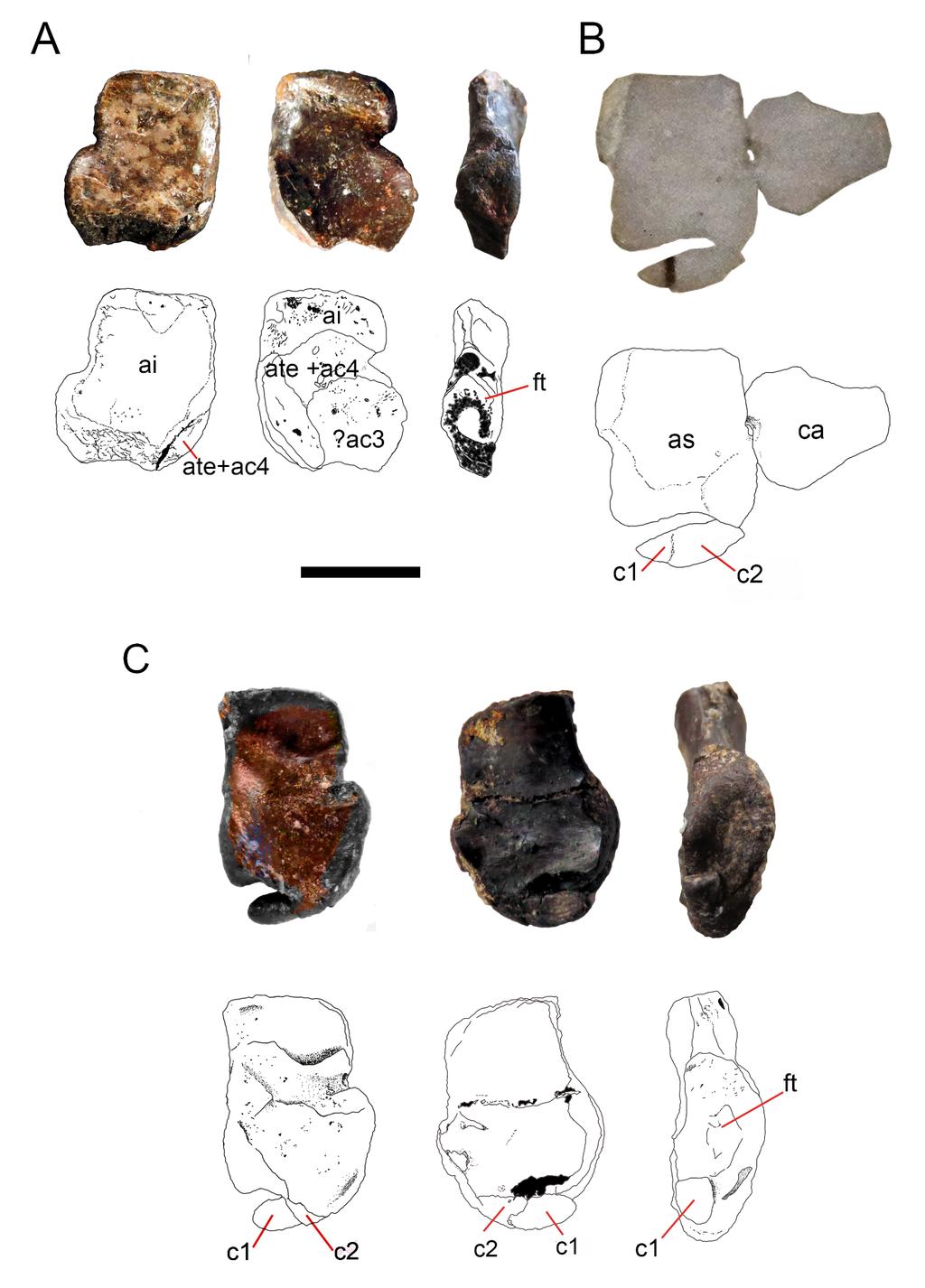
66
67 Figure 8. Ontogenetic transition of the navicular in Mesosaurus tenuidens. A, FC-DPV 1502, 1290 from left to right, photographs and interpretive drawings of isolated astragalus from a 1291 young individual, in dorsal, ventral and medial views respectively. The bone shows the 1292 typical square outline of immature individuals and the remains of sutures between the 1293 original anlagen more visible on its ventral surface, which appears to display a different 1294 morphology with respect to the dorsal one. Note that there are no traces of the navicular 1295 preserved along the distal surface of the astragalus, which bears a concave margin. B, 1296 GP-2E 5203, photograph and interpretive drawing of astragalus, calcaneum and incipient 1297 navicular of a young individual in dorsal view. Recall on that the navicular is already 1298 united to the astragalus by c2, being formed by c1 and c2 and the suture between them is 1299 still well visible. C, FC-DPV 1479, photographs and interpretive drawings of an isolated 1300 astragalus from an adult individual in dorsal, ventral and medial view. Observe that the 1301 navicular is now a single bone almost completely fused to the astragalus to produce the 1302 finally resultant adult outline. C1 has transformed into a tip-like bone and remains 1303 separated from the astragalus, but it can just be seen from the ventral view, which still 1304 features different from the dorsal one. The wide and triangular facet for articulation with 1305 the tibia can be seen from the medial view. Anatomical abbreviations: a, astragalus; ca, 1306 calcaneum; c1, centrale one; c2, centrale two; ac3, ancestral centrale three; ft, facet for 1307 the articulation of the tibia; ai, ancestral intermedium; ate+ac4, ancestral tibiale plus 1308 ancestral central four. Scale bar: 5 mm.

68

69 1310 Figure 9. Preserved tarsus in a Mesosaurus tenuidens non-hatched individual. A, FC.DPV 2504, 1311 a non-hatched mesosaurid in the egg, showing the two feet overlapping each other by 1312 compression. B, SEM image of the foot of FC-DPV 2504 focusing on the tarsal area. The 1313 astragali can be seen in the center of the figure, mixed between the metatarsals. The 1314 astragalus seems to be not preserved. C, Interpretive drawing of the tarsus including a 1315 possible identification of the preserved bones by regarding previous hypotheses about the 1316 origin of the amniotic astragalus. The composing elements of the astragalus were colored 1317 to favour identification of the isolated bones of the left foot. Anatomical abbreviations:?i, 1318 possible intermedium;?c3, possible central three; fi, fibula; mc, metacarpals;?te+c4, 1319 possible tibiale plus possible central four; t, tibia. Scale bar: 1mm. 1320
Supplementary Figure 1 Cartilaginous stages in non-avian amniotes. (a) Drawing of early ankle development of Alligator mississippiensis, as reported
 Drawing of early ankle development of Alligator mississippiensis, as reported") Supplementary Figure 1 Cartilaginous stages in non-avian amniotes. (a) Drawing of early ankle development of Alligator mississippiensis, as reported by a previous study 1. The intermedium is formed at
Supplementary Figure 1 Cartilaginous stages in non-avian amniotes. (a) Drawing of early ankle development of Alligator mississippiensis, as reported by a previous study 1. The intermedium is formed at
d a Name Vertebrate Evolution - Exam 2 1. (12) Fill in the blanks
 Fill in the blanks") Vertebrate Evolution - Exam 2 1. (12) Fill in the blanks 100 points Name f e c d a Identify the structures (for c and e, identify the entire structure, not the individual elements. b a. b. c. d. e. f.
Vertebrate Evolution - Exam 2 1. (12) Fill in the blanks 100 points Name f e c d a Identify the structures (for c and e, identify the entire structure, not the individual elements. b a. b. c. d. e. f.
Anatomy. Name Section. The Vertebrate Skeleton
 Name Section Anatomy The Vertebrate Skeleton Vertebrate paleontologists get most of their knowledge about past organisms from skeletal remains. Skeletons are useful for gleaning information about an organism
Biology 340 Comparative Embryology Lecture 12 Dr. Stuart Sumida. Evo-Devo Revisited. Development of the Tetrapod Limb
 Biology 340 Comparative Embryology Lecture 12 Dr. Stuart Sumida Evo-Devo Revisited Development of the Tetrapod Limb Limbs whether fins or arms/legs for only in particular regions or LIMB FIELDS. Primitively
1/9/2013. Divisions of the Skeleton: Topic 8: Appendicular Skeleton. Appendicular Components. Appendicular Components
 /9/203 Topic 8: Appendicular Skeleton Divisions of the Skeleton: Cranial Postcranial What makes up the appendicular skeleton? What is the pattern of serial homology of the limbs? Tetrapod front limb morphology
Postilla PEABODY MUSEUM OF NATURAL HISTORY YALE UNIVERSITY NEW HAVEN, CONNECTICUT, U.S.A.
 Postilla PEABODY MUSEUM OF NATURAL HISTORY YALE UNIVERSITY NEW HAVEN, CONNECTICUT, U.S.A. Number 117 18 March 1968 A 7DIAPSID (REPTILIA) PARIETAL FROM THE LOWER PERMIAN OF OKLAHOMA ROBERT L. CARROLL REDPATH
Title: Phylogenetic Methods and Vertebrate Phylogeny
 Title: Phylogenetic Methods and Vertebrate Phylogeny Central Question: How can evolutionary relationships be determined objectively? Sub-questions: 1. What affect does the selection of the outgroup have
From Slime to Scales: Evolution of Reptiles. Review: Disadvantages of Being an Amphibian
 From Slime to Scales: Evolution of Reptiles Review: Disadvantages of Being an Amphibian Gelatinous eggs of amphibians cannot survive out of water, so amphibians are limited in terms of the environments
These small issues are easily addressed by small changes in wording, and should in no way delay publication of this first- rate paper.
 Reviewers' comments: Reviewer #1 (Remarks to the Author): This paper reports on a highly significant discovery and associated analysis that are likely to be of broad interest to the scientific community.
Carpus and tarsus of Temnospondyli
 Vertebrate Anatomy Morphology Palaeontology 1(1):51-87 ISSN 2292-1389 Carpus and tarsus of Temnospondyli 51 David Dilkes Department of Biology & Microbiology, University of Wisconsin Oshkosh, 800 Algoma
Developmental Morphology of Limb Reduction in Hemiergis (Squamata: Scincidae): Chondrogenesis, Osteogenesis, and Heterochrony
: Chondrogenesis, Osteogenesis, and Heterochrony") JOURNAL OF MORPHOLOGY 254:211 231 (2002) Developmental Morphology of Limb Reduction in Hemiergis (Squamata: Scincidae): Chondrogenesis, Osteogenesis, and Heterochrony Michael D. Shapiro* Department of
JOURNAL OF MORPHOLOGY 254:211 231 (2002) Developmental Morphology of Limb Reduction in Hemiergis (Squamata: Scincidae): Chondrogenesis, Osteogenesis, and Heterochrony Michael D. Shapiro* Department of
Phylogeny Reconstruction
 Phylogeny Reconstruction Trees, Methods and Characters Reading: Gregory, 2008. Understanding Evolutionary Trees (Polly, 2006) Lab tomorrow Meet in Geology GY522 Bring computers if you have them (they will
First Ornithomimid (Theropoda, Ornithomimosauria) from the Upper Cretaceous Djadokhta Formation of Tögrögiin Shiree, Mongolia
 from the Upper Cretaceous Djadokhta Formation of Tögrögiin Shiree, Mongolia") First Ornithomimid (Theropoda, Ornithomimosauria) from the Upper Cretaceous Djadokhta Formation of Tögrögiin Shiree, Mongolia Tsogtbaatar Chinzorig¹, ³ *, Yoshitsugu Kobayashi², Khishigjav Tsogtbaatar³,
First Ornithomimid (Theropoda, Ornithomimosauria) from the Upper Cretaceous Djadokhta Formation of Tögrögiin Shiree, Mongolia Tsogtbaatar Chinzorig¹, ³ *, Yoshitsugu Kobayashi², Khishigjav Tsogtbaatar³,
Animal Form and Function. Amphibians. United by several distinguishing apomorphies within the Vertebrata
 Animal Form and Function Kight Amphibians Class Amphibia (amphibia = living a double life) United by several distinguishing apomorphies within the Vertebrata 1. Skin Thought Question: For whom are integumentary
Mammalogy Lecture 8 - Evolution of Ear Ossicles
 Mammalogy Lecture 8 - Evolution of Ear Ossicles I. To begin, let s examine briefly the end point, that is, modern mammalian ears. Inner Ear The cochlea contains sensory cells for hearing and balance. -
The Fossil Record of Vertebrate Transitions
 The Fossil Record of Vertebrate Transitions The Fossil Evidence of Evolution 1. Fossils show a pattern of change through geologic time of new species appearing in the fossil record that are similar to
HONR219D Due 3/29/16 Homework VI
 Part 1: Yet More Vertebrate Anatomy!!! HONR219D Due 3/29/16 Homework VI Part 1 builds on homework V by examining the skull in even greater detail. We start with the some of the important bones (thankfully
May 10, SWBAT analyze and evaluate the scientific evidence provided by the fossil record.
 May 10, 2017 Aims: SWBAT analyze and evaluate the scientific evidence provided by the fossil record. Agenda 1. Do Now 2. Class Notes 3. Guided Practice 4. Independent Practice 5. Practicing our AIMS: E.3-Examining
AMERICAN MUSEUM NOVITATES
 AMERICAN MUSEUM NOVITATES Number 1103 Published by THE AMERICAN MUSEUM OF NATURAL HISTORY New York City THE PES OF BAURIA CYNOPS BROOM BY BOBB SCHAEFFER1 In several respects the bauriamorphs are farther
Lab 2 Skeletons and Locomotion
 Lab 2 Skeletons and Locomotion Objectives The objectives of this and next week's labs are to introduce you to the comparative skeletal anatomy of vertebrates. As you examine the skeleton of each lineage,
LABORATORY EXERCISE 6: CLADISTICS I
 Biology 4415/5415 Evolution LABORATORY EXERCISE 6: CLADISTICS I Take a group of organisms. Let s use five: a lungfish, a frog, a crocodile, a flamingo, and a human. How to reconstruct their relationships?
Red Eared Slider Secrets. Although Most Red-Eared Sliders Can Live Up to Years, Most WILL NOT Survive Two Years!
 Although Most Red-Eared Sliders Can Live Up to 45-60 Years, Most WILL NOT Survive Two Years! Chris Johnson 2014 2 Red Eared Slider Secrets Although Most Red-Eared Sliders Can Live Up to 45-60 Years, Most
Video Assignments. Microraptor PBS The Four-winged Dinosaur Mark Davis SUNY Cortland Library Online
 Video Assignments Microraptor PBS The Four-winged Dinosaur Mark Davis SUNY Cortland Library Online Radiolab Apocalyptical http://www.youtube.com/watch?v=k52vd4wbdlw&feature=youtu.be Minute 13 through minute
First reptile appeared in the Carboniferous
 1 2 Tetrapod four-legged vertebrate Reptile tetrapod with scaly skin that reproduces with an amniotic egg Thus can lay eggs on land More solid vertebrate and more powerful limbs than amphibians Biggest
Mammalogy Laboratory 1 - Mammalian Anatomy
 Mammalogy Laboratory 1 - Mammalian Anatomy I. The Goal. The goal of the lab is to teach you skeletal anatomy of mammals. We will emphasize the skull because many of the taxonomically important characters
LABORATORY EXERCISE 7: CLADISTICS I
 Biology 4415/5415 Evolution LABORATORY EXERCISE 7: CLADISTICS I Take a group of organisms. Let s use five: a lungfish, a frog, a crocodile, a flamingo, and a human. How to reconstruct their relationships?
8/19/2013. Topic 4: The Origin of Tetrapods. Topic 4: The Origin of Tetrapods. The geological time scale. The geological time scale.
 Topic 4: The Origin of Tetrapods Next two lectures will deal with: Origin of Tetrapods, transition from water to land. Origin of Amniotes, transition to dry habitats. Topic 4: The Origin of Tetrapods What
Fish 2/26/13. Chordates 2. Sharks and Rays (about 470 species) Sharks etc Bony fish. Tetrapods. Osteichthans Lobe fins and lungfish
 Sharks etc Bony fish. Tetrapods. Osteichthans Lobe fins and lungfish") Chordates 2 Sharks etc Bony fish Osteichthans Lobe fins and lungfish Tetrapods ns Reptiles Birds Feb 27, 2013 Chordates ANCESTRAL DEUTEROSTOME Notochord Common ancestor of chordates Head Vertebral column
Chordates 2 Sharks etc Bony fish Osteichthans Lobe fins and lungfish Tetrapods ns Reptiles Birds Feb 27, 2013 Chordates ANCESTRAL DEUTEROSTOME Notochord Common ancestor of chordates Head Vertebral column
What are taxonomy, classification, and systematics?
 Topic 2: Comparative Method o Taxonomy, classification, systematics o Importance of phylogenies o A closer look at systematics o Some key concepts o Parts of a cladogram o Groups and characters o Homology
A new species of sauropod, Mamenchisaurus anyuensis sp. nov.
 A new species of sauropod, Mamenchisaurus anyuensis sp. nov. by Xinlu He, Suihua Yang, Kaiji Cai, Kui Li, and Zongwen Liu Chengdu University of Technology Papers on Geosciences Contributed to the 30th
Natural Sciences 360 Legacy of Life Lecture 3 Dr. Stuart S. Sumida. Phylogeny (and Its Rules) Biogeography
 Biogeography") Natural Sciences 360 Legacy of Life Lecture 3 Dr. Stuart S. Sumida Phylogeny (and Its Rules) Biogeography So, what is all the fuss about phylogeny? PHYLOGENETIC SYSTEMATICS allows us both define groups
Natural Sciences 360 Legacy of Life Lecture 3 Dr. Stuart S. Sumida Phylogeny (and Its Rules) Biogeography So, what is all the fuss about phylogeny? PHYLOGENETIC SYSTEMATICS allows us both define groups
SUPPLEMENTARY INFORMATION
 In comparison to Proganochelys (Gaffney, 1990), Odontochelys semitestacea is a small turtle. The adult status of the specimen is documented not only by the generally well-ossified appendicular skeleton
VERTEBRATE READING. Fishes
 VERTEBRATE READING Fishes The first vertebrates to become a widespread, predominant life form on earth were fishes. Prior to this, only invertebrates, such as mollusks, worms and squid-like animals, would
Williston, and as there are many fairly good specimens in the American
 56.81.7D :14.71.5 Article VII.- SOME POINTS IN THE STRUCTURE OF THE DIADECTID SKULL. BY R. BROOM. The skull of Diadectes has been described by Cope, Case, v. Huene, and Williston, and as there are many
Exceptional fossil preservation demonstrates a new mode of axial skeleton elongation in early ray-finned fishes
 Supplementary Information Exceptional fossil preservation demonstrates a new mode of axial skeleton elongation in early ray-finned fishes Erin E. Maxwell, Heinz Furrer, Marcelo R. Sánchez-Villagra Supplementary
New Carnivorous Dinosaurs from the Upper Cretaceous of Mongolia
 1955 Doklady, Academy of Sciences USSR 104 (5):779-783 New Carnivorous Dinosaurs from the Upper Cretaceous of Mongolia E. A. Maleev (translated by F. J. Alcock) The present article is a summary containing
Test one stats. Mean Max 101
 Test one stats Mean 71.5 Median 72 Max 101 Min 38 30 40 50 60 70 80 90 100 1 4 13 23 23 19 9 1 Sarcopterygii Step Out Text, Ch. 6 pp. 119-125; Text Ch. 9; pp. 196-210 Tetrapod Evolution The tetrapods arose
What is evolution? Transitional fossils: evidence for evolution. In its broadest sense, evolution is simply the change in life through time.
 Transitional fossils: evidence for evolution http://domain- of- darwin.deviantart.com/art/no- Transitional- Fossils- 52231284 Western MA Atheists and Secular Humanists 28 May 2016 What is evolution? In
Amniote Relationships. Reptilian Ancestor. Reptilia. Mesosuarus freshwater dwelling reptile
 Amniote Relationships mammals Synapsida turtles lizards,? Anapsida snakes, birds, crocs Diapsida Reptilia Amniota Reptilian Ancestor Mesosuarus freshwater dwelling reptile Reptilia General characteristics
'Rain' of dead birds on central NJ lawns explained; Federal culling program killed up to 5,000 Associated Press, January 27, 2009
 'Rain' of dead birds on central NJ lawns explained; Federal culling program killed up to 5,000 Associated Press, January 27, 2009 Study May Give Hope That Ivory-billed Woodpeckers Still Around Science
Introduction and methods will follow the same guidelines as for the draft
 Locomotion Paper Guidelines Entire paper will be 5-7 double spaced pages (12 pt font, Times New Roman, 1 inch margins) without figures (but I still want you to include them, they just don t count towards
LEIDY, SHOWING THE BONES OF THE FEET 'AND LIMBS
 CQNTEUBUTIONS FBOM THE MUSEUM OF PALEONTOLOGY (Confindion of Con&&&m froin UB Muaercm of Gcologg) UNIVERSITY OF ' MICHIGAN VOL V, No. 6, pp. 6W3 (e ph.) DEAXMBER 31,1036 A SPECIMEN OF STYLEMYS NEBRASCENSIS
On the Discovery of the earliest fossil bird in China (Sinosauropteryx gen. nov.) and the origin of birds
 and the origin of birds") On the Discovery of the earliest fossil bird in China (Sinosauropteryx gen. nov.) and the origin of birds by Qiang Ji and Shu an Ji Chinese Geological Museum, Beijing Chinese Geology Volume 233 1996 pp.
On the Discovery of the earliest fossil bird in China (Sinosauropteryx gen. nov.) and the origin of birds by Qiang Ji and Shu an Ji Chinese Geological Museum, Beijing Chinese Geology Volume 233 1996 pp.
Modern Evolutionary Classification. Lesson Overview. Lesson Overview Modern Evolutionary Classification
 Lesson Overview 18.2 Modern Evolutionary Classification THINK ABOUT IT Darwin s ideas about a tree of life suggested a new way to classify organisms not just based on similarities and differences, but
Origin and Evolution of Birds. Read: Chapters 1-3 in Gill but limited review of systematics
 Origin and Evolution of Birds Read: Chapters 1-3 in Gill but limited review of systematics Review of Taxonomy Kingdom: Animalia Phylum: Chordata Subphylum: Vertebrata Class: Aves Characteristics: wings,
DISCOVERY OF A TETRAPOD BODY FOSSIL IN THE LOWER PERMIAN YESO GROUP, CENTRAL NEW MEXICO
 Lucas, S.G. and Sullivan, R.M., eds., 2018, Fossil Record 6. New Mexico Museum of Natural History and Science Bulletin 79. DISCOVERY OF A TETRAPOD BODY FOSSIL IN THE LOWER PERMIAN YESO GROUP, CENTRAL NEW
8/19/2013. Topic 5: The Origin of Amniotes. What are some stem Amniotes? What are some stem Amniotes? The Amniotic Egg. What is an Amniote?
 Topic 5: The Origin of Amniotes Where do amniotes fall out on the vertebrate phylogeny? What are some stem Amniotes? What is an Amniote? What changes were involved with the transition to dry habitats?
Autopodial Development in the Sea Turtles Chelonia mydas and Caretta caretta
 ZOOLOGICAL SCIENCE 24: 257 263 (2007) 2007 Zoological Society of Japan Autopodial Development in the Sea Turtles Chelonia mydas and Caretta caretta Marcelo R. Sánchez-Villagra 1 *, Christian Mitgutsch
Comparative Vertebrate Anatomy
 Comparative Vertebrate Anatomy Presented by BIOBUGS: Biology Inquiry and Outreach with Boston University Graduate Students In association with LERNet and The BU Biology Teaching Laboratory Designed and
The cranial skeleton of the Early Permian aquatic reptile Mesosaurus tenuidens: implications for relationships and palaeobiology
 Blackwell Publishing LtdOxford, UKZOJZoological Journal of the Linnean Society0024-4082The Linnean Society of London, 2006? 2006 146? 345368 Original Article THE CRANIAL SKELETON OF MESOSAURUS TENUIDENSS.
muscles (enhancing biting strength). Possible states: none, one, or two.
. Possible states: none, one, or two.") Reconstructing Evolutionary Relationships S-1 Practice Exercise: Phylogeny of Terrestrial Vertebrates In this example we will construct a phylogenetic hypothesis of the relationships between seven taxa
Reconstructing Evolutionary Relationships S-1 Practice Exercise: Phylogeny of Terrestrial Vertebrates In this example we will construct a phylogenetic hypothesis of the relationships between seven taxa
Diapsida. BIO2135 Animal Form and Function. Page 1. Diapsida (Reptilia, Sauropsida) Amniote eggs. Amniote egg. Temporal fenestra.
 Amniote eggs. Amniote egg. Temporal fenestra.") Diapsida (Reptilia, Sauropsida) Vertebrate phylogeny Mixini Chondrichthyes Sarcopterygii Mammalia Pteromyzontida Actinopterygii Amphibia Reptilia! 1! Amniota (autapomorphies) Costal ventilation Amniote
Diapsida (Reptilia, Sauropsida) Vertebrate phylogeny Mixini Chondrichthyes Sarcopterygii Mammalia Pteromyzontida Actinopterygii Amphibia Reptilia! 1! Amniota (autapomorphies) Costal ventilation Amniote
A reevaluation of early amniote phylogeny
 Zoological Journal of the Linnean Society (1995), 113: 165 223. With 9 figures A reevaluation of early amniote phylogeny MICHEL LAURIN AND ROBERT R. REISZ* Department of Zoology, Erindale Campus, University
Diapsida. BIO2135 Animal Form and Function. Page 1. Diapsida (Reptilia, Sauropsida) Amniote egg. Membranes. Vertebrate phylogeny
 Amniote egg. Membranes. Vertebrate phylogeny") Diapsida (Reptilia, Sauropsida) 1 Vertebrate phylogeny Mixini Chondrichthyes Sarcopterygii Mammalia Pteromyzontida Actinopterygii Amphibia Reptilia!! Amniota (autapomorphies) Costal ventilation Amniote
Diapsida (Reptilia, Sauropsida) 1 Vertebrate phylogeny Mixini Chondrichthyes Sarcopterygii Mammalia Pteromyzontida Actinopterygii Amphibia Reptilia!! Amniota (autapomorphies) Costal ventilation Amniote
ONTOGENY OF THE SUPERNUMERARY SESAMOIDS IN THE LEG MUSCLES OF THE RING-NECKED PHEASANT. GEORG E. HUDSON, SY YING CI-IEIxl WANG, AND ERNEST E.
 ONTOGENY OF THE SUPERNUMERARY SESAMOIDS IN THE LEG MUSCLES OF THE RING-NECKED PHEASANT GEORG E. HUDSON, SY YING CI-IEIxl WANG, AND ERNEST E. PROVOST Ix has long been known to hunters, game managers, gourmets,
Vertebrate Structure and Function
 Vertebrate Structure and Function Part 1 - Comparing Structure and Function Classification of Vertebrates a. Phylum: Chordata Common Characteristics: Notochord, pharyngeal gill slits, hollow dorsal nerve
A NEW GENUS AND SPECIES OF AMERICAN THEROMORPHA
 A NEW GENUS AND SPECIES OF AMERICAN THEROMORPHA MYCTEROSAURUS LONGICEPS S. W. WILLISTON University of Chicago The past summer, Mr. Herman Douthitt, of the University of Chicago paleontological expedition,
FURTHER STUDIES ON TWO SKELETONS OF THE BLACK RIGHT WHALE IN THE NORTH PACIFIC
 FURTHER STUDIES ON TWO SKELETONS OF THE BLACK RIGHT WHALE IN THE NORTH PACIFIC HIDEO OMURA, MASAHARU NISHIWAKI* AND TOSHIO KASUYA* ABSTRACT Two skeletons of the black right whale were studied, supplementing
Field Trip: Harvard Museum of Natural History (HMNH)
") Field Trip: Harvard Museum of Natural History (HMNH) Objectives To observe the diversity of animals. To compare and contrast the various adaptations, body plans, etc. of the animals found at the HMNH.
Field Trip: Harvard Museum of Natural History (HMNH) Objectives To observe the diversity of animals. To compare and contrast the various adaptations, body plans, etc. of the animals found at the HMNH.
Origin and Evolution of Birds. Read: Chapters 1-3 in Gill but limited review of systematics
 Origin and Evolution of Birds Read: Chapters 1-3 in Gill but limited review of systematics Review of Taxonomy Kingdom: Animalia Phylum: Chordata Subphylum: Vertebrata Class: Aves Characteristics: wings,
Are Evolutionary Transitional Forms Possible?
 What Fossils Can t Tell Us Are Evolutionary Transitional Forms Possible? Dr. Raúl Esperante Geoscience Research Institute Darwin and the Fossil Record Darwin and other evolutionists before suggested that
1) Explain why the skeleton plays an important role in the overall shape of animal and human being.
 Explain why the skeleton plays an important role in the overall shape of animal and human being.") 1) Explain why the skeleton plays an important role in the overall shape of animal and human being. 2) Substantiate the differences in animal and human skeleton, with the human skeleton built in such a
1) Explain why the skeleton plays an important role in the overall shape of animal and human being. 2) Substantiate the differences in animal and human skeleton, with the human skeleton built in such a
Fishes, Amphibians, Reptiles
 Fishes, Amphibians, Reptiles Section 1: What is a Vertebrate? Characteristics of CHORDATES Most are Vertebrates (have a spinal cord) Some point in life cycle all chordates have: Notochord Nerve cord that
UN? RSITYOF. ILLIiwiS LIBRARY AT URBANA-CHAMPAIGN NATURAL HIST. SURVEY
 UN? RSITYOF ILLIiwiS LIBRARY AT URBANA-CHAMPAIGN NATURAL HIST. SURVEY FIELDIANA GEOLOGY Published by CHICAGO NATURAL HISTORY MUSEUM Volume 10 July 29, 1954 No. 17 FAUNA OF THE VALE AND CHOZA: 7 PELYCOSAURIA:
Bio 1B Lecture Outline (please print and bring along) Fall, 2006
 Fall, 2006") Bio 1B Lecture Outline (please print and bring along) Fall, 2006 B.D. Mishler, Dept. of Integrative Biology 2-6810, bmishler@berkeley.edu Evolution lecture #4 -- Phylogenetic Analysis (Cladistics) -- Oct.
Bio 1B Lecture Outline (please print and bring along) Fall, 2006 B.D. Mishler, Dept. of Integrative Biology 2-6810, bmishler@berkeley.edu Evolution lecture #4 -- Phylogenetic Analysis (Cladistics) -- Oct.
THE SKULLS OF ARAEOSCELIS AND CASEA, PERMIAN REPTILES
 THE SKULLS OF REOSCELIS ND CSE, PERMIN REPTILES University of Chicago There are few Permian reptiles of greater interest at the present time than the peculiar one I briefly described in this journal' three
1 Describe the anatomy and function of the turtle shell. 2 Describe respiration in turtles. How does the shell affect respiration?
 GVZ 2017 Practice Questions Set 1 Test 3 1 Describe the anatomy and function of the turtle shell. 2 Describe respiration in turtles. How does the shell affect respiration? 3 According to the most recent
.56 m. (22 in.). COMPSOGNATHOID DINOSAUR FROM THE. Medicine Bow, Wyoming, by the American Museum Expedition
. COMPSOGNATHOID DINOSAUR FROM THE. Medicine Bow, Wyoming, by the American Museum Expedition") Article XII.-ORNITHOLESTES HERMANNI, A NEW COMPSOGNATHOID DINOSAUR FROM THE UPPER JURASSIC. By HENRY FAIRFIELD OSBORN. The type skeleton (Amer. Mus. Coll. No. 6I9) of this remarkable animal was discovered
Article XII.-ORNITHOLESTES HERMANNI, A NEW COMPSOGNATHOID DINOSAUR FROM THE UPPER JURASSIC. By HENRY FAIRFIELD OSBORN. The type skeleton (Amer. Mus. Coll. No. 6I9) of this remarkable animal was discovered
Evolution as Fact. The figure below shows transitional fossils in the whale lineage.
 Evolution as Fact Evolution is a fact. Organisms descend from others with modification. Phylogeny, the lineage of ancestors and descendants, is the scientific term to Darwin's phrase "descent with modification."
Redpalh Museum, McGill University, Montreal, P.Q, Canada, HJA 2K6.
 143 Palaeont. afr., 21, 143-159 (1978) PERMO-TRIASSIC "LIZARDS" FROM THE KAROO SYSTEM PART II A GLIDING REPTILE FROM THE UPPER PERMIAN OF MADAGASCAR by Robert L. Carroll Redpalh Museum, McGill University,
Animal Diversity wrap-up Lecture 9 Winter 2014
 Animal Diversity wrap-up Lecture 9 Winter 2014 1 Animal phylogeny based on morphology & development Fig. 32.10 2 Animal phylogeny based on molecular data Fig. 32.11 New Clades 3 Lophotrochozoa Lophophore:
AMERICAN MUSEUM NOVITATES Published by
 AMERICAN MUSEUM NOVITATES Published by Number 782 THE AmzRICAN MUSEUM OF NATURAL HISTORY Feb. 20, 1935 New York City 56.81, 7 G (68) A NOTE ON THE CYNODONT, GLOCHINODONTOIDES GRACILIS HAUGHTON BY LIEUWE
AMERICAN NATURALIST. Vol. IX. -DECEMBER, No. 12. OR BIRDS WITH TEETH.1 OI)ONTORNITHES,
ONTORNITHES,") AMERICAN NATURALIST. Vol. IX. -DECEMBER, 1875.-No. 12. OI)ONTORNITHES, OR BIRDS WITH TEETH.1 BY PROFESSOR 0. C. MARSH. REMAINS of birds are amono the rarest of fossils, and few have been discovered except
AMERICAN NATURALIST. Vol. IX. -DECEMBER, 1875.-No. 12. OI)ONTORNITHES, OR BIRDS WITH TEETH.1 BY PROFESSOR 0. C. MARSH. REMAINS of birds are amono the rarest of fossils, and few have been discovered except
Biology Slide 1 of 50
 Biology 1 of 50 2 of 50 What Is a Reptile? What are the characteristics of reptiles? 3 of 50 What Is a Reptile? What Is a Reptile? A reptile is a vertebrate that has dry, scaly skin, lungs, and terrestrial
The Origin of Birds. Technical name for birds is Aves, and avian means of or concerning birds.
 The Origin of Birds Technical name for birds is Aves, and avian means of or concerning birds. Birds have many unusual synapomorphies among modern animals: [ Synapomorphies (shared derived characters),
The skeleton of a juvenile Lanthanotus (Varanoidea) Olivier Rieppel
 Olivier Rieppel") The skeleton of a juvenile Lanthanotus (Varanoidea) Olivier Rieppel Dept. of Geology, Field Muscum of Natural History, Roosevelt Road at Lake Shore Drive, Chicago, IL 60605-2496, USA Abstract. The cleared
The skeleton of a juvenile Lanthanotus (Varanoidea) Olivier Rieppel Dept. of Geology, Field Muscum of Natural History, Roosevelt Road at Lake Shore Drive, Chicago, IL 60605-2496, USA Abstract. The cleared
Evolution of Tetrapods
 Evolution of Tetrapods Amphibian-like creatures: The earliest tracks of a four-legged animal were found in Poland in 2010; they are Middle Devonian in age. Amphibians arose from sarcopterygians sometime
AUSTRALIAN MUSEUM SCIENTIFIC PUBLICATIONS
 AUSTRALIAN MUSEUM SCIENTIFIC PUBLICATIONS Riek, E. F., 1964. Merostomoidea (Arthropoda, Trilobitomorpha) from the Australian Middle Triassic. Records of the Australian Museum 26(13): 327 332, plate 35.
6. The lifetime Darwinian fitness of one organism is greater than that of another organism if: A. it lives longer than the other B. it is able to outc
 1. The money in the kingdom of Florin consists of bills with the value written on the front, and pictures of members of the royal family on the back. To test the hypothesis that all of the Florinese $5
By HENRY FAIRFIELD OSBORN.
 Article XI.-FORE AND HINI) LIMBS OF CARNIVOR- OUS AND HERBIVOROUS DINOSAURS FROM THE JURASSIC OF WYOMING. DINOSAUR CONTRIBU- TIONS, NO. 3. By HENRY FAIRFIELD OSBORN. In the Bone Cabin Quarry, opened by
Stuart S. Sumida Biology 342. Simplified Phylogeny of Squamate Reptiles
 Stuart S. Sumida Biology 342 Simplified Phylogeny of Squamate Reptiles Amphibia Amniota Seymouriamorpha Diadectomorpha Synapsida Parareptilia Captorhinidae Diapsida Archosauromorpha Reptilia Amniota Amphibia
290 SHUFELDT, Remains of Hesperornis.
 290 SHUFELDT, Remains of Hesperornis. [ Auk [July THE FOSSIL REMAINS OF A SPECIES OF HESPERORNIS FOUND IN MONTANA. BY R. W. SHUFELD% M.D. Plate XI7III. ExR,¾ in November, 1914, Mr. Charles W. Gihnore,
Modern taxonomy. Building family trees 10/10/2011. Knowing a lot about lots of creatures. Tom Hartman. Systematics includes: 1.
 Modern taxonomy Building family trees Tom Hartman www.tuatara9.co.uk Classification has moved away from the simple grouping of organisms according to their similarities (phenetics) and has become the study
A new basal sauropodiform dinosaur from the Lower Jurassic of Yunnan Province, China
 SUPPLEMENTARY INFORMATION A new basal sauropodiform dinosaur from the Lower Jurassic of Yunnan Province, China Ya-Ming Wang 1, Hai-Lu You 2,3 *, Tao Wang 4 1 School of Earth Sciences and Resources, China
CLADISTICS Student Packet SUMMARY Phylogeny Phylogenetic trees/cladograms
 CLADISTICS Student Packet SUMMARY PHYLOGENETIC TREES AND CLADOGRAMS ARE MODELS OF EVOLUTIONARY HISTORY THAT CAN BE TESTED Phylogeny is the history of descent of organisms from their common ancestor. Phylogenetic
A Pterodactylus with Remains of Flight Membrane. by F. Broili (with 3 plates). Read at the Conference on 7th February 1925.
. Read at the Conference on 7th February 1925.") Broili, F. (1925) Ein Pterodactylus mit Resten der Flughaut. Sitzungsberichte der Bayerischen Königlichen Akademie der Wissenschaften, Mathematischen-Physicalischen Classe, 1925, 23-32. A Pterodactylus
Broili, F. (1925) Ein Pterodactylus mit Resten der Flughaut. Sitzungsberichte der Bayerischen Königlichen Akademie der Wissenschaften, Mathematischen-Physicalischen Classe, 1925, 23-32. A Pterodactylus
The Triassic Transition
 The Triassic Transition The Age of Reptiles Begins As the Paleozoic drew to a close through the Carboniferous and Permian several important processes were at work. Assembly of Pangea Evolutionary radiation
The earliest reptiles
 J. Linn. SOC. (Zool), 45, no. 304, p. 61 With 14 tezt-figures Printed in Great Britain The earliest reptiles BY ROBERT L. CARROLL (Accepted for publication December 1963) Communicated by Errol I. White,
What is the evidence for evolution?
 What is the evidence for evolution? 1. Geographic Distribution 2. Fossil Evidence & Transitional Species 3. Comparative Anatomy 1. Homologous Structures 2. Analogous Structures 3. Vestigial Structures
Do the traits of organisms provide evidence for evolution?
 PhyloStrat Tutorial Do the traits of organisms provide evidence for evolution? Consider two hypotheses about where Earth s organisms came from. The first hypothesis is from John Ray, an influential British
Are the dinosauromorph femora from the Upper Triassic of Hayden Quarry (New Mexico) three stages in a growth series of a single taxon?
 three stages in a growth series of a single taxon?") Anais da Academia Brasileira de Ciências (2017) 89(2): 835-839 (Annals of the Brazilian Academy of Sciences) Printed version ISSN 0001-3765 / Online version ISSN 1678-2690 http://dx.doi.org/10.1590/0001-3765201720160583
Anais da Academia Brasileira de Ciências (2017) 89(2): 835-839 (Annals of the Brazilian Academy of Sciences) Printed version ISSN 0001-3765 / Online version ISSN 1678-2690 http://dx.doi.org/10.1590/0001-3765201720160583
Biology 1B Evolution Lecture 11 (March 19, 2010), Insights from the Fossil Record and Evo-Devo
, Insights from the Fossil Record and Evo-Devo") Biology 1B Evolution Lecture 11 (March 19, 2010), Insights from the Fossil Record and Evo-Devo Extinction Important points on extinction rates: Background rate of extinctions per million species per year:
Biology 1B Evolution Lecture 11 (March 19, 2010), Insights from the Fossil Record and Evo-Devo Extinction Important points on extinction rates: Background rate of extinctions per million species per year:
Major cranial changes during Triceratops ontogeny John R. Horner 1, * and Mark B. Goodwin 2
 273, 2757 2761 doi:10.1098/rspb.2006.3643 Published online 1 August 2006 Major cranial changes during Triceratops ontogeny John R. Horner 1, * and Mark B. Goodwin 2 1 Museum of the Rockies, Montana State
Characteristics of a Reptile. Vertebrate animals Lungs Scaly skin Amniotic egg
 Reptiles Characteristics of a Reptile Vertebrate animals Lungs Scaly skin Amniotic egg Characteristics of Reptiles Adaptations to life on land More efficient lungs and a better circulator system were develope
UNIVERSITY OF ILLINOIS LIBRARY AT URBANA-CHAMPAIGN BIOLOGY. Hi 01^995
 UBRARY IttBMmXHALL f^bo 71995 UNIVERSITY OF ILLINOIS LIBRARY AT URBANA-CHAMPAIGN BIOLOGY Hi 01^995 590.5 FI n.s. No. 68 BIX.CM)I( ^v V > ' of Lacerta i Olivier Kic^'ikI Pubiitation 1437 PUBLJSHI-!)
A R T I C L E S STRATIGRAPHIC DISTRIBUTION OF VERTEBRATE FOSSIL FOOTPRINTS COMPARED WITH BODY FOSSILS
 A R T I C L E S STRATIGRAPHIC DISTRIBUTION OF VERTEBRATE FOSSIL FOOTPRINTS COMPARED WITH BODY FOSSILS Leonard Brand & James Florence Department of Biology Loma Linda University WHAT THIS ARTICLE IS ABOUT
oxfitates }Ji2zercanAuseum The Triassic Dinosaur Genera Podokesaurus and Coelophysis BY EDWIN H. COLBERT'
 }Ji2zercanAuseum oxfitates PUBLISHED BY THE AMERICAN MUSEUM OF NATURAL HISTORY CENTRAL PARK WEST AT 79TH STREET, NEW YORK 24, N.Y. NUMBER 2I68 FEBRUARY 21, I964 The Triassic Dinosaur Genera Podokesaurus
Lecture 11 Wednesday, September 19, 2012
 Lecture 11 Wednesday, September 19, 2012 Phylogenetic tree (phylogeny) Darwin and classification: In the Origin, Darwin said that descent from a common ancestral species could explain why the Linnaean
Introduction to Cladistic Analysis
 3.0 Copyright 2008 by Department of Integrative Biology, University of California-Berkeley Introduction to Cladistic Analysis tunicate lamprey Cladoselache trout lungfish frog four jaws swimbladder or
SUPPLEMENTARY ONLINE MATERIAL FOR. Nirina O. Ratsimbaholison, Ryan N. Felice, and Patrick M. O connor
 http://app.pan.pl/som/app61-ratsimbaholison_etal_som.pdf SUPPLEMENTARY ONLINE MATERIAL FOR Nirina O. Ratsimbaholison, Ryan N. Felice, and Patrick M. O connor Ontogenetic changes in the craniomandibular