Systematic revision of the Miocene long-snouted dolphin Eurhinodelphis longirostris DU Bus, 1872 (Cetacea, Odontoceti, Eurhinodelphinidae)
|
|
|
- Oscar Lynch
- 6 years ago
- Views:
Transcription
 by O livier LA M BERT L a m b e rt, O., 2004. - Systematic revision o f the Miocene long-snouted dolphin Eurhinodelphis longirostris d u B us, 1872 (Cetacea, Odontoceti, Eurhinodelphinidae).")
 is revised.")
1 BULLETIN DE L INSTITUT ROYAL DES SCIENCES NATURELLES DE BELGIQUE SCIENCES DE LA TERRE, 74: , 2004 BULLETIN VAN HET KONINKLIJK BELGISCH INSTITUUT VOOR NATUURWETENSCHAPPEN AARDWETENSCHAPPEN, 74: , 2004 Systematic revision of the Miocene long-snouted dolphin Eurhinodelphis longirostris DU Bus, 1872 (Cetacea, Odontoceti, Eurhinodelphinidae) by O livier LA M BERT L a m b e rt, O., Systematic revision o f the Miocene long-snouted dolphin Eurhinodelphis longirostris d u B us, 1872 (Cetacea, Odontoceti, Eurhinodelphinidae). Bulletin de l Institut royal des Sciences naturelles de Belgique, Sciences de la Terre, 74: ,5 pis, 12figs, 2 tables; Bruxelles-Brussel, March 31, ISSN Abstract On the basis o f the redescription o f Miocene Belgian specimens, the systematic status o f the long-snouted dolphin genus Schizodelphis (Cetacea, Odontoceti, Eurhinodelphinidae) is revised. The only Belgian species previously recognized, S. longirostris, from the late early to middle Miocene o f Antwerp (north o f Belgium, southern margin of the North Sea basin), is divided here in two taxa. Some specimens are kept in that species, but re-establishing the combination Eurhinodelphis longirostris. The content o f the genus Eurhinodelphis is then investigated from several Miocene localities, essentially the Calvert Formation (Virginia and Maryland, east coast o f USA) and the Belluno Sandstones from north-eastern Italy. The only recognized species are E. cocheteuxi and E. longirostris, both o f them only found in the Belgian Miocene. Other previously described species are placed in an unnamed new genus, in Mycteriacetus n. gen., and in Ziphiodelphis. The other Belgian specimens are maintained in Schizodelphis, with the prioritary species name morckhoviensis. The species S. morckhoviensis is also identified in the Calvert Formation, while a restricted S. barnesi is tentatively diagnosed from American specimens. Key words: Eurhinodelphinidae, taxonomy, Miocene, Belgium, Schizodelphis, Eurhinodelphis. Résumé Sur base de la redescription de spécimens du Miocène de Belgique, le statut systématique du dauphin longirostre Schizodelphis (Cetacea, Odontoceti, Eurhinodelphinidae) est révisé. La seule espèce belge préalablement décrite, S. longirostris, de la fin du Miocène inférieur- Miocène moyen d Anvers (nord de la Belgique, bord sud du bassin de la Mer du Nord), est divisée en deux taxa. Une partie des spécimens est maintenue dans cette espèce, mais en rétablissant la combinaison Eurhinodelphis longirostris. Le contenu du genre Eurhinodelphis est ensuite investigué dans plusieurs localités du Miocène, particulièrement la Formation Calvert (Virginie et Maryland, côte est des Etats- Unis) et les Sables de Belluno (nord-est de l Italie). Les seules espèces reconnues sont E. cocheteuxi et E. longirostris, et cela uniquement dans le Miocène belge. Les autres espèces précédemment décrites sont placées dans un nouveau genre non nommé, dans Mycteriacetus n. gen., et dans Ziphiodelphis. Une seconde partie des spécimens belges est maintenue dans le genre Schizodelphis, avec le nom d espèce prioritaire morckhoviensis. Cette espèce S. morckhoviensis est également identifiée dans la Formation Calvert, de même que l espèce S. barnesi brièvement redéfinie. Mots-clefs: Eurhinodelphinidae, taxinomie, Miocène, Belgique, Schizodelphis, Eurhinodelphis. Introduction du Bus (1872) shortly described several species of longsnouted dolphins from the Miocene of Antwerp (North of Belgium), which he included in the genera Eurhinodelphis DU Bus, 1868 and Priscodelphinus (L eidy, 1851). A bel (1902) included in the same species E. longirostris individuals of the species Eurhinodelphis longirostris, E. ambiguus, Priscodelphinus morckhoviensis, P. elegans, and P. pulvinatus sensu du Bus, In his unpublished revision of the eurhinodelphinids from the Calvert Formation, east coast of the USA, Myrick (1979) noticed the presence of the species E. longirostris in this area, which he referred to the genus Rhabdosteus by comparison with the holotype of the type-species of the genus {R. latiradix (C ope, 1868)}, a partial rostrum also from the Calvert Formation. However, this specimen was estimated by M uizon (1988a) as too fragmentary, and regarded as an incertae sedis. M uizon referred the species E. longirostris to the genus Schizodelphis G ervais, 1861, as well as all the Rhabdosteus species recognized by M yrick (1979) in the Calvert Formation (excluding R. latiradix). M uizo n s conclusions were based on the study of the holotype of Schizodelphis sulcatus G ervais, 1853 (Miocene of France), the type-species of the genus. M uizon did not recognize S. longirostris in the Calvert Formation, where he only identified one species, S. barnesi, including the individuals from the species Rhabdosteus longirostris, R. barnesi and R. hruschkai sensu M yrick, A detailed observation o f the B elgian specim ens o f Schizodelphis longirostris sensu M uizon, 1988a allow s the reco gn itio n o f tw o genera, Eurhinodelphis and Schizodelphis, for w hich an em ended diagnosis an d a red e scription are presented here. Specimens from the Calvert Formation (M yrick, 1979), from the Belluno Sandstones (early Miocene of north-eastern Italy, P illeri, 1985), and from several other localities, previously reported to the genus Eurhinodelphis, are also briefly discussed.
. Additional specimens from the IRSNB, USNM, CMM, and MGPD are more briefly discussed. Though M y r ic k s Ph. D.")

2 148 Olivier LAMBERT M aterial and methods Most of the specimens used in this study are housed in the IRSNB. The main specimens are two well preserved skulls, IRSNB 3249-M.342 and IRSNB 3235-M.343, already described by A b el (1902). Additional specimens from the IRSNB, USNM, CMM, and MGPD are more briefly discussed. Though M y r ic k s Ph. D. thesis ( ) was not published, I use it as a starting point for the systematic revision of the Calvert eurhinodelphinids. The species Eurhinodelphis cristatus sensu d u B u s, and E. bossi sensu K e l l o g g, 1925 are referred to a new eurhinodelphinid genus that will be diagnosed in a paper in preparation; those two species are cited here as E. cristatus and E.' bossi. The Italian species E. bellunensis sensu P il l e r i, 1985, included by its author in the genus Eurhinodelphis, is referred to a new genus, Mycteriacetus n. gen., diagnosed below. Terminology. The terminology for cranial and ear bones anatomy is mainly taken from: F o r d y c e (1983 and 1994); K a s u y a (1973); M u iz o n (1984, 1987 and 1988a). The orientations of the tympanic bulla and periotic are simplified in the following descriptions, relatively to the anatomical position on the basicranium. The long axis of the tympanic is considered as anteroposterior, with ventral surfaces of inner and outer posterior prominences indicating the horizontal plane. The anterior direction of the periotic is given by the longitudinal axis of the anterior process, and the horizontal ventral plane by the surface contacting the most ventral points of pars cochlearis and anterior process. Abbreviations. CMM: Calvert Marine Museum, Solomons, Maryland, USA; IRSNB: Institut Royal des Sciences Naturelles de Belgique, Brussels; M: Fossil mammals collection of types and figured specimens from the IRSNB; MGPD: Museum of Geology and Palaeontology of Padova, Italy; MNHN: Muséum National d Flistoire Naturelle, Paris, France; USNM: United States National Museum, Smithsonian Institution, Washington D.C., USA. Explanations o f the measurements. Fig. 1. System atic palaeontology Order Cetacea B risson, 1762 Suborder Odontoceti Flow er, 1867 Superfamily Eurhinodelphinoidea M uizon, 1988a Family Eurhinodelphinidae A bel, 1901 Type-genus. Eurhinodelphis du Bus, 1867 Included genera. Eurhinodelphis, Schizodelphis G ervais, 1861, Ziphiodelphis D al Piaz, 1908, Argyrocetus L ydekker, 1894, Macrodelphinus W ilson, 1935, and Mycteriacetus n. gen. Emended diagnosis. Family of long-snouted odontocetes differing from all the other families by an edentulous premaxillary anterior part of the rostrum, longer than the mandible. Additionally, the family differs from the probably closely related family Eoplatanisidae by: a more inclined dorsomedial portion of the supraoccipital shield, a generally lower temporal fossa, the presence of a marked median groove on the tympanic, a longer anterior process of the periotic; and from the Squalodontidae, Waipatiidae and other more primitive odontocetes by: an homodont dentition with single-rooted teeth, premaxillae widened at the level of the posterior margin of the bony nares, a higher vertex. Eurhinodelphis d u Bus, 1867 Type species. E. cocheteuxi d u Bus, 1867 Included species. E. cocheteuxi and E. longirostris DU Bus, The species E. cocheteuxi was redescribed in a previous paper ( L a m b e r t, in press). Diagnosis. The genus Eurhinodelphis differs from the genera Schizodelphis and Ziphiodelphis in: maxillary part of the rostrum relatively shorter (ratio between bizygomatic width of the skull and length of the maxillary part of the rostrum > 0.5); vertical medial plate of the maxilla along the vertex antero-dorsally developed; flat to convex supraoccipital shield (shield concave in the two other genera); more elevated and narrower paroccipital process of the exoccipital with occipital condyles more highly positioned (ventral margin of the condyles nearly reaching the level of the floor of the temporal fossa); less excavated premaxillary sac fossae, which are roughly flat; zygomatic process of the squamosal relatively higher in lateral view and narrower in ventral view; absence of fossa for the postorbital lobe of the pterygoid sinus on the ventral surface of the supraorbital process (that fossa is sometimes very short but nearly always present in Schizodelphis and Ziphiodelphis). The last character is probably in a primitive state, but it clearly separates the genera. It differs from Macrodelphinus by: more longitudinally telescoped and more elevated vertex with frontals shorter than the nasals; flat to convex supraoccipital shield. It differs from Argyrocetus (provisionally only including the species A. patagonicus) in: a more elevated vertex and flat to convex more vertical supraoccipital shield. It differs from Mycteriacetus n. gen. in: a relatively wider and shorter face (ratio between bizygomatic width and length of the face from the antorbital notch to the occipital condyle > 0.95); a more elevated vertex with shorter frontals and wider nasals; flat to convex and more vertical supraoccipital shield. Eurhinodelphis longirostris DU Bus, 1872 * 1872 Eurhinodelphis longirostris DU Bus, p v Eurhinodelphis longirostris V an B en ed en & G erv a is, p. 493, pi. 58, fig. 2. v Eurhinodelphis longirostris A b e l, pi. 11, 12 and 13, pi. 17, fig. 1. v. 1988a Schizodelphis longirostris M u iz o n, p. 40. Emended diagnosis. This species differs from Eurhinodelphis cocheteuxi by: the distinctly smaller size o f the


3 Systematic revision of Eurhinodelphis longirostris Fig. 1 - Description of the measurements on the skull of eurhinodelphinids, outlines of the holotype of Eurhinodelphis cocheteuxi IRSNB 3252-M.294. A. left lateral view. B. dorsal view. C. posterior view. Explanation of the measurements on Tables 1-2.
', relatively longer and more slender rostrum (ratio between postorbital width of the skull and length of the rostrum < 0.")
. Holotype. IRSNB 3249-M.")
4 150 Olivier LAMBERT cranial skull (the general dimensions of the cranial skull o f the holotype of E. longirostris vary between 70 and 80 % of the dimensions of the holotype of E. cocheteuxi)', relatively longer and more slender rostrum (ratio between postorbital width of the skull and length of the rostrum < 0.25), especially the premaxillary apical part of the rostrum; narrower base o f the rostrum; relatively narrower vertex with a strong compression of the frontals between the supraoccipital and the nasals (a contact between nasals and supraoccipital is present on two specimens o f E. longirostris). Holotype. IRSNB 3249-M.342, a well preserved skull, only lacking teeth and ear bones, fragments of the basicranium and of the rostrum (found in January 1862, individual 1 of the species Eurhinodelphis longirostris in A bel, 1902, figured in pi. 11, 12 and 13). Referred specimens. IRSNB 3250-M.1858, a partial skull including a part of the rostrum, the two supraorbital processes, a portion of the vertex and the basicranium (individual 3 of the species Eurhinodelphis longirostris in A bel, 1902); IRSNB 3251, a part of the rostrum with the posterior part of the left maxilla (individual 7 of the species E. longirostris in A bel, 1902). No skull from the Calvert Formation could be referred to this species. Comments on the other specimens referred to E. longirostris b y A bel (1902) T he fragm entary rostrum IRSNB 3245, individual 17 o f the species Eurhinodelphis longirostris sensu A bel, 1902, show s very flattened and w ide prem axillae at the base o f the rostrum, and should then be reported to E. cristatus (pap er in prep.). The left side of a face IRSNB 3495 (Eurhinodelphis ambiguus sensu d u Bus, 1872 and individual 15 of the species E. longirostris sensu A b e l, 1902) is probably a part of a juvenile specimen of E. bossi (L a m b e rt, in press). The partial skull IRSNB 3238-M.344 (individual 2 of E. longirostris sensu A bel, 1902, pi. 18, fig. 1, figured here in PI. 1, Fig. 2, including the base of the rostrum, the supraorbital processes, a portion of the vertex with the nasals, a fragment of the supraoccipital, the two squamosals and the paroccipital process of the left exoccipital) shows some features that place it in the genus Eurhinodelphis: slightly excavated premaxillary sac fossa, flat posterior portion of the maxilla laterally to the vertex, elevated and narrow paroccipital process of the exoccipital, and absence of fossa for the postorbital lobe of the pterygoid sinus. A striking characteristic of this specimen is the strong development of the transverse premaxillary crests that are wide and thick. The measurements of the skull (see Table 1) are similar to the measured specimens of E. longirostris. However, it differs from these specimens in the smaller nasals less posteriorly displaced, and the more dorso-ventrally flattened zygomatic process of the squamosal. Because of those differences, and because it is fragmentary, this specimen IRSNB 3238-M.344 is referred to Eurhinodelphis cf. longirostris. The partial rostrum IRSNB 3225 (individual 8 of the species Eurhinodelphis longirostris sensu A bel, 1902) has size and proportions similarities with E. longirostris but no diagnostic character is observable and this fragment is referred to Eurhinodelphinidae incertae sedis. The isolated fragments of mandible IRSNB M.347 (figured by A bel, 1902, plate 17, fig. 4) probably belong to an eurhinodelphinid, with proportions roughly similar to Eurhinodelphis bossi USNM None of the individuals of E. longirostris is associated with a mandible, and the lack of diagnostic features on this mandible precludes its attribution to any eurhinodelphinid species. It is placed in Odontoceti aff. Eurhinodelphinidae. The periotics associated with the specimen IRSNB 3447-M.351 (Eurhinodelphis ambiguus sensu DU Bus, 1872, figured in A b e l, 1902, p. 122, fig. 19 and plate 17, figs ) were already clearly recognized as belonging to a physeterid ( K e llo g g, 1927). The erroneously associated symphyseal portion of mandible (figured by Abei, 1902, plate 17, fig. 6) is regarded as an Odontoceti aff. Eurhinodelphinidae. The specimen IRSNB 3244-M.346 (holotype of Priscodelphinus elegans sensu du B us, 1872) is a hypothetical association of a partial small basicranium and a vertex (figured by A bel, 1902, pi. 17, fig. 2), but without bony contact between them. The squamosal shows similarities with that of Eurhinodelphis longirostris, with a zygomatic process high in lateral view and narrow in ventral view. However, the vertex is close to that of several specimens of Rhabdosteus hruschkai sensu M yrick (1979), e.g. USNM , with an anterolateral projection o f the prominent nasal along the external nare. As most of the specimens of R. hruschkai sensu M yrick (1979) are probably referrable to the genus Schizodelphis, the association basicranium-vertex of IRSNB 3244-M.346 is regarded as doubtful and those fragments are considered as Eurhinodelphinidae incertae sedis. Locus typicus. The holotype o f Eurhinodelphis longirostris was found in Antwerp, in January 1862, and the locality cited by A bel (1902) is 4L Section'. This locality corresponds to the south-eastern portion of the city wall around Antwerp, built during fortification works in the 1860 s (see V an den B roeck, 1878), in Berchem. This section matches the south-eastern part of the present motorway R l, around the city. Stratum typicum. No precise stratigraphie data are available for the holotype and referred specimens. However, the preservation and colour of those specimens are very similar to that of the skulls of Eurhinodelphis cocheteuxi, known from the Antwerp Sands. This strongly suggests an origin in the same member, dated from late early to middle Miocene (see Louw ye et al. 2000). Redescription o f the species Eurhinodelphis longirostris (PI. 1, Fig. 1; PI. 2, Fig. 1; Figs. 2-4) General morphology. Eurhinodelphis longirostris has a relatively small braincase and a very long rostrum (see measurements, Table 1), which is nearly completely preserved on the holotype. The rostrum constitutes more than 80 % of the total length of the skull, with more than 50 % of its length made by the premaxillae.
 indicates estim ate, *+ nearly com plete, and no data. Measurements on the skulls o f E. longirostris Holotype IRSNB 3249- M.342 IRSNB 3250- M.")

5 Systematic revision o f Eurhinodelphis longirostris 151 Table 1 - M easurem ents on the skulls o f E urhinodelphis longirostris. M easurem ents are in m illim etres, (e) indicates estim ate, *+ nearly com plete, and no data. Measurements on the skulls o f E. longirostris Holotype IRSNB M.342 IRSNB M.1858 IRSNB M.344 E. aff. longirostris 1. total length skull length base rostrum-anterior maxilla length anterior orbit-posterior skull length anterior supraoccipital-anterior orbit e length orbit length temporal fossa e width rostrum anterior maxillae width base rostrum 103 e96 e width premaxillae base rostrum 67 - e width skull postorbital processes 199 el width skull zygomatic processes width bony nares width nasals maximal posterior premaxillary width 97 - e minimal posterior distance between maxillae width medio-ventral margins exoccipitals width lateral margins occipital condyles width inner margins occipital condyles height cranium height rostrum at anterior maxillae height base rostrum 57 e height temporal fossa e height ventral margin occipital condyles height occipital condyles Dorsal view. The dorsal surface of the premaxilla is convex and prominent until the base of the rostrum, with a slight narrowing and lowering just anterior to that level. The triangular elongated surface antero-medial to the premaxillary foramen is nearly smooth and partially lower than the thick and rounded lateral part of the premaxilla at that level. The premaxillary sac fossa is thick, roughly flat and progressively raising towards the vertex, lacking the deeper concavity and the more abrupt posterior elevation of Schizodelphis morckhoviensis (see below). The posterior extremity of the premaxilla contacts the antero-lateral angle of the nasal and is longitudinally incised by the erected median edge of the maxilla. The contact between premaxilla and frontal is probably absent on the holotype but it is present on IRSNB 3250-M.1858, depending upon the shape and position of the nasals on the vertex. The lateral margin of the maxilla exhibits a very weak swelling about 100 mm anterior to the antorbital notch. Several dorsal infraorbital foramina pierce the maxilla along its suture with the premaxilla, at the level and anterior to the shallow and antero-laterally open antorbital notch. The preorbital process is narrow in dorsal view and its lateral margin diverges posteriorly. A distinct elevation of the maxilla is present above the orbit. It is separated from the premaxillary sac fossa by a narrow longitudinal depression. The posterior portion of the maxilla is roughly flat and slopes antero-laterally. It is only slightly concave at the level of its overhanging median edge along the vertex. The posterior margin of the maxilla extends posteriorly 20 mm beyond the anteromedian margin of the supraoccipital. The shape of the nasals strongly varies between the two skulls on which it is preserved. On the holotype, those bones are somewhat eroded. They are wider than long and exhibit a wide contact with the supraoccipital. The frontals are reduced on the vertex to three small triangles between the nasals and the supraoccipital. This special morphology was correctly recognized by A b e l (1902), but K ello g g (1932) erroneously identified the two wide and short bones as the frontals. On IRSNB 3250-M.1858, with only the right part of the vertex preserved, the nasal

.")

6 152 Olivier LAMBERT erected median edge of maxilla postero-lateral sulcus premaxillary foramen antero-median sulcus supraoccipital temporal fossa median longitudinal depression lateral groove antorhital note mesethmoid premaxilla vomer dorsal infraorbital foramina frontai maxilla occipital condyle frontal premaxillary sac fossa floor of temporal bony narè worn mesethmoid nakai squamosal fossa Fig. 2 - Schematic drawing of the dorsal view of the skull of Eurhinodelphis longirostris IRSNB 3249-M.342 (holotype), from Antwerp,? Antwerp Sands, late early to middle Miocene. also contacts the supraoccipital on most of its width. However, the nasal is narrower than on the holotype, and a wide part of the frontal is dorsally exposed, lateral to the nasal (see Fig. 3a). The trend to a posterior shift of the nasals towards the supraocipital is present on both specimens, but variably modelling the bones o f the vertex. The sagittal section of the vertex of the skull IRSNB 3250-M allows the observation of the posterior part o f the mesethmoid. This bone deeply penetrates the frontal posteriorly below the nasal, nearly reaching the suture with the supraoccipital (see Fig. 3b). The supraoccipital shield is convex, only hollowed by a median longitudinal depression ending 15 mm before its anterior margin. This rounded shield is regularly sloping posteriorly, with a mean slope of ca. 35. Posterior view. The paroccipital process of the exoccipital is high and narrow. As a consequence, the occipital condyles are elevated, with a ventral margin nearly reaching the level of the floor of the temporal fossa. The basioccipital crests are sharp and ventrally shorter than the exoccipitals. Lateral view. The lateral groove of the rostrum starts 180 mm anteriorly to the antorbital notch. It is deep and widens over 100 mm forwardly. Then it progressively shallows and disappears more than 100 mm posteriorly to the apex of the rostrum. The maxilla-premaxilla suture leaves the floor of the groove 250 mm anteriorly to the antorbital notch, 140 mm posteriorly to the apex of the maxilla. The roof of the orbit is long and lower than the top of the temporal fossa. The frontal is roughly as thick as the maxilla. The lacrymal-jugal complex is visible in lateral view for a short length antero-ventral to the preorbital process of the frontal. The elevated zygomatic process of the squamosal is stronger than the rounded postglenoid process. Ventral view. The premaxillary part of the rostrum does not bear alveoli; the alveolar groove of the maxilla extends in the premaxilla as a thin groove with rectilinear edges precluding the presence of teeth inserted in the bone. The maxillary alveoli are eroded on the holotype, but are preserved on the proximal part of the rostrum of IRSNB 3250-M The first alveolus is 30 mm anterior to the antorbital notch. Forty-two deep alveoli are present on the first 243 mm of the right side of the rostrum and 40 on the left side. The average diameter is 4-5 mm and the septa are less than 3 mm thick. Considering the length of the maxilla on the rostrum of the holotype, the


7 Systematic revision of Eurhinodelphis longirostris 153 total number of alveoli on each side should be around 60. On the right alveolar row of IRSNB 3250, the 14th alveolus is distinctly shifted laterally and the 15th, medially, as if the posterior part of the row was pushed against the anterior part (see PI. 2, Fig. le). This might indicate a trend towards an increase in the number of maxillary teeth, or more simply a pathology. The palatines are short; their apex extends until 10 mm beyond the level of the antorbital notches. The thin lateral plate o f the palatine contacts the large infraorbital foramen. This part of the palatine was erroneously identified as part of the pterygoid by A b e l ( 1902). The condition of Eurhinodelphis longirostris is similar to that observed in E. cocheteuxi (see L a m b e r t, in press). As in this species, the palatine plate is crossed by a longitudinal crest, which disappears anteriorly before reaching the palatine-maxilla suture. Only small fragments of the pteiygoid are preserved anteriorly. The anterior pterygoid fossa clearly excavates the palatine anterior to the choana, on a short distance (10 to 25 mm). The jugal-lacrymal complex is only preserved as a small eroded knob in the bottom of the antorbital notch. The ventral face of the roof of the orbit is hollowed by a shallow sulcus which runs from the large infraorbital foramen towards the antorbital notch. There is no fossa for the postorbital lobe of the pterygoid sinus, as in Eurhinodelphis cocheteuxi, and contrary to Schizodelphis morckhoviensis (see below). The zygomatic process of the squamosal is anteriorly pointed, with a ventral apical projection for the contact with the missing jugal. The ventral apex of the postglenoid process is transversely flattened. The tympanosquamosal recess is deep, laterally extending for a short distance dorsal to the glenoid surface. The recess does not have a clear anterior limit, extending on the medial side of the zygomatic process. The falciform process of the squamosal is not completely preserved. On the holotype, it takes an anterior direction, and is interrupted by a transverse canal, very likely the path for the mandibular nerve V3, exiting in the temporal fossa through the foramen pseudo-ovale (see F o r d y c e, 1994). As the falciform process is incomplete, the presence or absence of a lateral lamina of the pterygoid can not be asserted. On the skull IRSNB 3250-M. 1858, the anterior part of the falciform process is more clearly antero-laterally deflected, along a well developed pterygoid sinus fossa on the alisphenoid. This condition, observed on every skull of Eurhinodelphis cocheteuxi, is probably related to the loss or the important reduction of the lateral lamina of the pterygoid ( L a m b e r t, in press). The foramina of the basicranium are poorly preserved. On the holotype, only the carotid foramen and the anterior margin of the foramen ovale can be observed. On the skull IRSNB M.1858, the posterior lacerate foramen has an elongated shape, with a maximal length of 18 mm and a small median constriction. There is no posterior sinus fossa, supraoccipital suture maxilla frontal. olfactory foramen mesethmoid frontal premaxilla remaxilla-maxilla suture mark Fig. 3 - Schematic drawings of the right portion of the face of Eurhinodelphis longirostris IRSNB 3250-M. 1858, from Antwerp,? Antwerp Sands, late early to middle Miocene, showing the posterior shift of the nasal and mesethmoid, respectively above and through the frontal. A. dorsal view. B. detail of the sagittal section in medial view.


 is the species Eurhinodelphis bossi K e l l o g g, 1925. In his unpublished thesis, M y r ic k ( 1979) also recognized the species E.")
8 154 Olivier LAMBERT path mandibular zygomatic process external auditory meatus \ Sp ny process / foramen ovale fragment of pterygoid infraorbital foramen worn alveolar groove maxilla plalatine pterygoid vomer vomer shallow sulcus-' longitudinal crest' pterygoid sinus fossa' choaná foramen 'pseudo-ovale' falciform process' glenoid surface' tympanosquamosal recess asioccipital crest* paroccipital process of exoccipital postglenoid process Fig. 4 - Schem atic drawing o f the ventral v iew o f the skull o f E urhinodelphis longirostris IR SN B 3249-M.342 (holotype), from Antwerp,? Antwerp Sands, late early to m iddle M iocene. and the surface dorso-medial to the spiny process is smooth and unexcavated. Comments on specimens from the Calvert Formation referred to Eurhinodelphis by M y r ic k (1979, unpublished thesis) The only previously published eurhinodelphinid from the Calvert Formation (early to middle Miocene of Maryland and Virginia) is the species Eurhinodelphis bossi K e l l o g g, In his unpublished thesis, M y r ic k ( 1979) also recognized the species E. cristatus in the Calvert Formation, and described four additional new species: E. vaughni, E. ashbyi, E. whitmorei and E. morrisi. It is outside the scope of the present study to carry out a complete systematic revision of the high number of eurhinodelphinid specimens from the Calvert Formation, but the different species of Eurhinodelphis erected by M y r ic k (1979) are briefly discussed here. After E. cristatus, Eurhinodelphis vaughni sensu M yrick, 1979 is the most common species of the genus, with seven skulls identified from the Calvert Formation. In the diagnosis of the species, M yrick (1979, p. 222) gave few characters differentiating it from E. cristatus, E. whitmorei and E. ashbyi: lack of fold on the posterior margin of the maxilla along the transverse crest, premaxillae not mesially sloping at the level o f the antorbital notches and supraorbital processes slightly elevated. Those characters roughly consist in the main features of E. bossi as defined relatively to 'E.' cristatus. And M y r ic k (1979) could not give differences with E. bossi, probably because o f the great disparity in morphologic features and morphometries among the four specimens (of E. boss i f. Actually, the measurements on the skulls of E. vaughni are globally close to E. bossi, and no clear separation could be found for any measurement. The large width of the rostrum at its base suggested by M y r ic k (1979) is only measured in one specimen that slighly exceeds in this respect the largest E. bossi. Furthermore, the morphology of the face is very similar in both groups. I suggest therefore to include the specimens identified by M y r ic k (1979) as Eurhinodelphis vaughni in E. bossi. It should be noticed that the four specimens identified as E. bossi by K e l l o g g (1925) were found in three different stratigraphie levels of the Calvert Formation, the beds 3, 5 and 10 of S h a t t u c k (1904), and that the
 suggested an average time of 120.")

9 Systematic revision o f Eurhinodelphis longirostris 155 seven specimens of 'E.' vaugni sensu M y r ic k 1979 come from the beds 12 (six of them) and 13. Supposing a highly speculative uniform rate of deposition along the two million years duration of the Calvert Formation, M y r ic k (1979) suggested an average time of years for the deposition of each of the 15 beds of the formation. The morphological variability among E. bossi as defined here might then be partially explained by, on one side, the difference of age between the beds containing the different specimens of E. bossi sensu K e l l o g g, 1925, and on the other side the younger age of the beds providing the specimens of E. vaughni sensu M y r i c k, The species Eurhinodelphis ashbyi sensu M y r i c k, 1979 is based on two partial skulls USNM and USNM It is diagnosed by M y r ic k (1979, p. 249) as somewhat similar to E. cristatus, with the following differences: probably smaller maximum adult size; pentagonal frontals with apex pointed forward between nasals; larger nasals; more pronounced overhanging of the maxillary plate by the supraoccipital crest; supraorbital processes thick but not protuberant or abruptly elevated. However, the morphology of the vertex is only observable in one of the specimens and it could easily be explained by individual variation. Actually, the shape of the frontals and nasals is close to the Belgian E. cristatus IRSNB 3237 for instance. The longitudinal telescoping of the face is also variable, giving a more or less pronounced elevation o f the transverse supraoccipital crest. The elevation of the supraorbital protuberance was shown to be variable in E. cristatus (e.g. A b e l, 1905, p. 118). Furthermore, the size of the face, even if smaller than the average for E. cristatus fits the smaller specimens of the species, including the Belgian ones. As most of the diagnostic characters of E. cristatus are observed in the two specimens of E. ashbyi sensu M y r i c k, 1979, I propose to synonymize this species with E. ' cristatus. Eurhinodelphis ' whitmorei sensu M y r ic k, 1979, based on the skull USNM 25666, was diagnosed in M y r ic k (1979, p. 254) by: a smaller maximum size relatively to E. cristatus, thicker nasals, premaxillae not mesially sloping at the level of the antorbital notches and anterolateral curve of the dorsal margin of the maxilla not as marked. The skull USNM presents all the features differentiating E. cristatus from E. bossi, except its face which is longer relatively to its width than on specimens of E. cristatus. That peculiarity, also present on some specimens of E. bossi does not seem sufficient to create a new species. Because the characters given by M y r ic k (1979) are variable within E. cristatus, USNM is referred here to that species. The last species of Eurhinodelphis described by M y r ic k (1979), E. morrisi, is also based on a single specimen, USNM The diagnosis given by M y r ic k (1979, p. 270) mainly differentiates it from E. cristatus. This seems correct as the derived characters o f E. cristatus are absent on the skull. But here again, there is no comparison with E. bossi. The dimensions of the skull are globally at the lower limit o f the interval for E. bossi (including E. vaughni sensu M y r i c k, 1979), and are very close to the skull USNM (= E. vaughni sensu M y r i c k, 1979). Several differences with E. bossi appear: lower and flatter supraorbital process; more concave and erected medial plate of the maxilla along the vertex; absence of medio-anterior point of the frontals on the vertex. It seems, however, difficult to build a new species on so few characteristics, observed in only one specimen. Therefore, the holotype of 'E. morrisi sensu M y r i c k, 1979 is provisionally referred to E. bossi. To summarize, the list of species from the genus Eurhinodelphis proposed by M y r ic k (1979) is restricted to two species: E. bossi (including E. vaughni and E. morrisi sensu M y r i c k, 1979) and E. cristatus (including E. ashbyi and E. whitmorei sensu M y r i c k, 1979). Because those two species are referred here to a new genus described in work in progress, no species of the genus Eurhinodelphis - restricted to the species E. cocheteuxi and E. longirostris - is recognized in the Calvert Formation. Comments about specimens from the Belluno Sandstones (north eastern Italy) referred to Eurhinodelphis by P illeri (1985) In 1985, P il l e r i described Eurhinodelphis sigmoideus on the basis of a well preserved skull MGPD from the Belluno Sandstones (lower Miocene of northeastern Italy), lacking the apex of the rostrum, the mandible and teeth, but with one tympanic in situ and associated with five cervical vertebrae and two thoracics. P i l l e r i gave the following short justification for the attribution of the species to Eurhinodelphis: Taxonomically speaking, this is a new species, which in view o f the essential morphological features o f the skull and the spinal column belongs to the genus Eurhinodelphis.... First, it should be noticed here that the schematic drawing of the dorsal view of the skull of Eurhinodelphis sigmoideus sensu P i l l e r i, 1985 presented by P il l e r i (1985, fig. 21) bears some important mistakes: the posterior apex o f the premaxillae is much too long and wide, the nasals are too nodulous, narrower than in reality, and the nasals are too short on the vertex (see corrected drawing, Fig. 5). When considering the genus Eurhinodelphis only including the species E. cocheteuxi and E. longirostris, E. sigmoideus sensu P i l l e r i, 1985 is closer to members of the genus Schizodelphis than to members of the genus Eurhinodelphis, with a stronger longitudinal compression of the vertex correlated to a more erected supraoccipital shield. But the most striking similarities are in fact observed when comparing E. sigmoideus sensu P i l l e r i, 1985 with Ziphiodelphis abeli, as suggested by B ia n u c c i & L a n d in ii (2002). Those two species share the following characters, absent in Eurhinodelphis and Schizodelphis-. - Wide and flattened surface of the premaxilla at the level of the antorbital notches with a median portion regularly laterally sloping. In Eurhinodelphis and Schizodelphis, this area medial to the antero-medial sulcus is narrower, thicker and less medially elevated, with a more regular triangular shape.
, from Belluno, north-eastern Italy, Belluno Sandstones, early Miocene.")

10 156 Olivier LAMBERT premaxilla ^ mesethmoid Fig. 5 - Corrected schematic drawing of the dorsal view of the skull of Ziphiodelphis sigmoideus ( P il l e r i, 1985), from Belluno, north-eastern Italy, Belluno Sandstones, early Miocene. Thickened antero-dorsal portion o f the nasals constituting the highest surface of the vertex. - Medial plate of the maxilla against the vertex keeping an elevated dorsal margin in a postero-lateral direction, giving the postero-dorsal comer of the skull a more angular aspect in lateral view. - The ventral view of the right tympanic preserved in situ on the basicranium of the type of E. sigmoideus sensu P il l e r i, 1985 (see P il l e r i, 1985, Plate 45) has proportions and size close to the tympanic of the holotype of Ziphiodelphis abeli MGPD (see D a l P ia z, 1977, Plate 3, Fig. 9), anteriorly wider than the more pointed tympanic of Eurhinodelphis cocheteuxi IRSNB M.1856 (see L a m b e r t, in press), E.' bossi USNM and Schizodelphis barnesi USNM (both figured in M u i z o n, 1988a, Fig. 6). The holotype o f Eurhinodelphis sigmoideus sensu P il l e r i, 1985 is clearly smaller than the holotype o f Ziphiodelphis abeli, with a general size of the facial skull close to the smallest individuals of Eurhinodelphis ' cristatus. Its face is relatively longer than that of Z. abeli. The vertex is slightly more elevated. A small fossa for the postorbital lobe of the pterygoid sinus nearly reaches the ventral face of the roof of the orbit in E. sigmoideus sensu P i l l e r i, 1985, while it is dorsally shorter in the holotype o f Ziphiodelphis abeli. The most striking difference is the dorsal elevation of the premaxillae, forming an elongated bulge with a maximal height of 28 mm at a level mm anteriorly to the antorbital notches. This median prominence gives the base of the rostrum a sigmoid profile (inspiring the species name sigmoideus to P i l l e r i, 1985). Those differences seem sufficient to exclude Eurhinodelphis sigmoideus sensu P i l l e r i, 1985 from the species Ziphiodelphis abeli, and I suggest to place it in the same genus, as Ziphiodelphis sigmoideus. An additional observation can be made on that skull: the descent of the suture between premaxilla and maxilla on the lateral surface of the rostrum far before the apex, characteristic o f at least the genera Eurhinodelphis, Schizodelphis, and the species E. cristatus, E. bossi, and Ziphiodelphis abeli, is visible on the type of Z. sigmoideus. The maximal length of the maxilla on the rostrum is estimated to 510 mm. The ratio between the width of the skull at the level of the zygomatic processes and that length is close to the ratio calculated for an undescribed Belgian skull of E.' cristatus, and smaller than the ratio for E. cocheteuxi and E. longirostris. The maxillary part o f the rostrum is then relatively longer in Eurhinodelphis when compared to E. cristatus and Ziphiodelphis. The second Eurhinodelphis species from the Belluno Sandstones, E. bellunensis sensu P i l l e r i, 1985, is based on the skull MGPD 26404, only lacking the very apical portion of the rostrum, with the two tympanies in situ, several teeth, and associated with its roughly complete mandible. Here again, the attribution by P il l e r i (1985) to the genus Eurhinodelphis is not supported by characters. The most striking differences between E. bellunensis sensu P i l l e r i, 1985 and the species of the revisited genus Eurhinodelphis are given here. First, the face of E. bellunensis sensu P i l l e r i, 1985 is relatively longer than that of E. longirostris and clearly longer than that o f E. cocheteuxi, with a ratio between bizygomatic width of the skull and length of the face from the antorbital notch to the occipital condyle < 0.9. The maximal width o f the premaxillae on the face is smaller, but with closer median margins just anteriorly to the external nares. The posterolateral surface o f the maxilla is much less laterally inclined, with a posterior portion narrower. The nasals are narrower (relatively to their length) and the frontals are longer. The occipital shield is strongly concave, while it is roughly flat in E. cocheteuxi and slightly convex in E. longirostris. The ventral margin of the occipital condyles is relatively lower, because of the lower paroccipital process of the exoccipital.


11 Systematic revision of Eurhinodelphis longirostris 157 Taken separately, some of the differences here above are found in Ziphiodelphis: e.g. the shape o f the premaxillae anteriorly to the external nares, the concavity of the supraoccipital shield, or the low paroccipital process of the exoccipital. But the face of Eurhinodelphis bellunensis sensu P illeri, 1985 is proportionally longer than that of Ziphiodelphis abeli and Z. sigmoideus, and the proportions of the nasals and frontals are very different on its lower vertex. In his discussion of the genus Dalpiazina, M uizon (1988a, p. 73) briefly suggested that the holotype of Eurhinodelphis bellunensis sensu P illeri, 1985 probably belongs to the genus Argyrocetus. It is also probably to that specimen that C ozzuol (1996) referred when he identified a species of the genus Argyrocetus from northern Italy. When restricting the genus Argyrocetus to its fragmentary known Argentinian type-species A. patagonicus, some similarities appear between this species and the holotype of Eurhinodelphis bellunensis sensu P illeri, 1985: a low vertex, with the nasals higher than the frontals; a weakly erected concave supraoccipital shield; a general lateral view of the mandible roughly similar; most of the measurements relatively close. However, a part of those features are linked to the low rate of longitudinal telescoping of the skull. And this is clearly a primitive character, placing E. bellunensis sensu P illeri, 1985 and Argyrocetus patagonicus in a basal position in the phylogenetic tree of the eurhinodelphinids. Actually, the only portion of the skull of A. patagonicus which can be more precisely compared with Eurhinodelphis bellunensis sensu P illeri, 1985 is the vertex. On this area, the nasals are more dorso-anteriorly elevated and relatively wider in Argyrocetus patagonicus, and the frontals shorter. The anterior portion of the face seems also relatively shorter in A. patagonicus. Actually, this type-species of the genus Argyrocetus seems too fragm entaria known to allow the inclusion of other species. Because the holotype skull of Eurhinodelphis bellunensis sensu Pilleri, 1985 is nearly complete, I suggest its inclusion in a new genus, Mycteriacetus n. gen. This name is chosen in reference to Mycteria ibis, the African yellowbilled stork, characterized by a long and robust beak similar in lateral view to the rostrum of the eurhinodelphinids. Mycteriacetus n. gen. is diagnosed by: longer and narrower supraorbital process of the maxilla, lower vertex and less erected supraoccipital shield relatively to Eurhinodelphis, Schizodelphis, and Ziphiodelphis', longer anterior part of the face, vertex more elevated, narrower nodular nasals not antero-dorsally projecting, and longer frontals on that vertex relatively to Argyrocetus; smaller size, more excavated premaxillary sac fossae, longer nasals and shorter frontals on the vertex relatively to Macrodelphinus. Comments about other specimens referred to Eurhinodelphis T he b rie f review o f Fordyce (1983) is discussed here, in addition to com m ents about m ore recently described specim ens. The fragment of rostrum constituting the holotype of the Miocene Sardinian species Eurhinodelphis sassariensis sensu C a pellini, 1887 is undiagnostic as suggested by A bel ( 1931 ), M yrick ( 1979), and B ianucci et al. ( 1994). The palate seems flatter than in eurhinodelphinids, with a rostrum relatively wider at its base and a faster anterior narrowing. The holotype of E. pacificus sensu M atsum oto, 1926, Middle Miocene of Japan, is an anterior fragment of rostrum with the corresponding mandible in situ. The author justified the attribution to the genus Eurhinodelphis by the fact that the premaxillae are longer than the maxillae and do not bear teeth. The anterior lowering of the maxilla in lateral view is much stronger than in Eurhinodelphis and other eurhinodelphinids for which this area is known. This feature gives the ventral margin of the maxilla on the rostrum a very convex shape, and allows to suggest that the premaxilla was not much longer than the maxilla. Actually, the mandible fragment might have been anteriorly shifted relatively to the rostrum, giving the impression of premaxillae much longer anteriorly. This undiagnostic fragment shows more similarities with delphinoids than with eurhinodelphinids, according to the opinion of A bel (1931) who excluded it from the genus Eurhinodelphis. The holotype of E. salentinus Z ei, 1950, from the Miocene of Pietra leccese (Apulia, Italy), is a skull too fragmentary at the level of the face to give a generic attribution. Z ei (1950) described the maxillae as occupying 3/5 of the length of the rostrum. This character is sufficient to place the specimen in the family Eurhinodelphinidae. It was placed by B ianucci & Landini (2002) in cf. Argyrocetus salentinus but no common diagnostic feature could be noticed from the figures of Z ei ( 1950) with the typespecies of the genus Argyrocetus, A. patagonicus. Another partial skull from the Pietra leccese was identified by B ianucci et al. (1994) as Eurhinodelphis cristatus sensu A bel, This skull lacks the thickening of the maxilla on the roof of the orbit and the forwards indentation of the supraoccipital and frontal on the posterior edge of the maxilla laterally to the vertex, both characters defining the species E. cristatus. This skull shows actually more similarities with E. bossi and its measurements fit well with the variability observed among the individuals of that species. However, 'E.' bossi is quite difficult to diagnose relatively to E. cristatus, as no clearly derived characters are isolated for the first species. Furthermore the skull from Pietra leccese is not well preserved and no information is available on the details of the basicranium. The strong flattening of the face might also hide characteristics of E. cristatus. Therefore, the Pietra leccese specimen is referred to E. aff. bossi. The partial odontocete skull from the late Miocene of Portugal reported by D a M ata (1963) as Eurhinodelphis cf. cristatus sensu A bel, 1902 lacks all the diagnostic characteristics of the species, contradicting M yrick (1979, p. 13). Even its attribution to the family Eurhinodelphinidae is denied here. The morphology o f the vertex,
.")

12 158 Olivier LAMBERT with a strong transversal pinching of the frontals behind wider nasals and the loss of contact between the posterior apex of the premaxillae and the frontals might indicate affinities with some kentriodontids, e.g. Liolithax pappus (see K e l l o g g, 1955; B a r n e s, 1978). As suggested by F o r d y c e (1983), the periotic from the Miocene faluns of Touraine and Anjou (France) identified by G in s b u r g & J a n v i e r (1971) as Eurhinodelphis sp. lacks several features present in the family Eurhinodelphinidae, for example the well excavated anterior bullar facet. It was actually compared by G i n s b u r g & J a n v ie r (1971) to the physeterid periotic from Antwerp erroneously reported by A b e l (1902, pi. 17, figs ) to Eurhinodelphis longirostris. The periotic of the faluns shows similarities with kentriodontids such as Liolithax pappus ( K e l l o g g, 1955) (see B a r n e s, 1978, figs. lj-2j). The holotype o f Eurhinodelphis minoensis sensu O k a z a k i, 1976 from the early to middle Miocene o f Japan is a partial mandible associated to vertebrae, ribs and detached teeth. Those fragments are not diagnostic at a generic level and no character allows a strict attribution to the family Eurhinodelphinidae. Eurhinodelphis minoensis sensu O k a z a k i, 1976 should therefore be considered as Odontoceti incertae sedis. From the same formation, O k a z a k i (1976) described a partial skull with a periotic and placed it as Eurhinodelphis sp. [erroneously discussed by F o r d y c e (1983) as a specimen of Eurhinodelphis minoensis]. The periotic was compared by F o r d y c e (1983) to kentriodontids. The skull is very incomplete and the reconstruction of the vertex by O k a z a k i (1976, fig. 4) is doubtful. From plate 2, figure 3, there are no contradictions to the kentriodontid affinities of the periotic, excluding the specimen from the family Eurhinodelphinidae. An additional isolated periotic identified as Eurhinodelphis sp. by O k a z a k i (1976, pi. 2, fig. 1) might also belong to a kentriodontid. It is referred here, as the first one, to the superfamily Delphinoidea sensu M u iz o n (1988b). The cervical vertebra from the early Miocene of Catalonia, Spain, identified by P il l e r i (1988) as Eurhinodelphis sp. (cf. E. sigmoideus) is probably not diagnostic at the generic level, as already suggested by B ia n u c c i & L a n d in i (2002) who considered it as Eurhinodelphinidae indet. From the systematic revision of the genus Eurhinodelphis, I only recognize the species E. cocheteuxi and E. longirostris, for which no associated cervical vertebra are known. Systematic discussion M u iz o n (1988a, p ) differentiated the Belgian specimens from the American specimens of Schizodelphis longirostris, contradicting M y r ic k (1979), by a list of cranial characters: shape and position of the nasals, height of the mesethmoid, excavation of the premaxillary sac fossae and their elevation towards the vertex, morphology of the base of the rostrum. From the observation of the Belgian specimens, most of those differences are present in the holotype o f Eurhinodelphis longirostris IRSNB 3249-M.342 (see description above), but they are absent in the second most complete specimen of E. longirostris sensu A b e l, 1902, IRSNB 3235-M.343: - While the nasals of the holotype of E. longirostris are in contact with the supraoccipital, 15 mm separate those bones from the supraoccipital on IRSNB M The mesethmoid of IRSNB 3235-M.343 reaches the antero-dorsal margin of the nasals, but this character is also observed in some American specimens. - The elevation of the premaxillae towards the vertex of IRSNB 3235-M.343 begins more posteriorly and is more abrupt than in the holotype of E. longirostris, as is the case in the American specimens. - The premaxillary sac fossae of IRSNB 3235-M.343 are distinctly more concave than in the holotype of E. longirostris, as is the case in the American specimens. - The left side of the rostrum of IRSNB 3235-M.343 is interrupted 140 mm anteriorly to its base, and its slightly medially compressed right side shows a slight transverse swelling, probably homologous to the swelling described by M u i z o n (1988a) for the American specimens. In fact, it seems that, in his comparison of the Belgian and American specimens, M y r i c k (1979) referred more to the skull IRSNB 3235-M.343, well figured in dorsal and ventral view by A b e l (1902, pi. 14, figs. 1-2), than to the holotype o f E. longirostris IRSNB 3249-M.342. IRSNB 3235-M.343 should be excluded from E. longirostris, and referred to the same genus as the American specimens, Schizodelphis. This skull IRSNB 3235-M.343 was first described by DU Bus (1872) as the only specimen of the species Priscodelphinus morckhoviensis (and thus the holotype). It was not figured by d u B u s (1872), but the fact that it is identifiable from his description, and that it is well preserved and associated with a periotic and a fragment of tympanic bulla, leads to recognize IRSNB 3235-M.343 as the holotype o f Schizodelphis morckhoviensis (see below). Schizodelphis G e r v a i s, 1861 Type species. Schizodelphis sulcatus ( G e r v a i s, 1853) Included species. S. sulcatus,? S. barnesi M u i z o n, 1988a, and S. morckhoviensis ( d u B u s, 1872) Diagnosis. This genus differs from: - Eurhinodelphis in: maxillary part of the rostrum relatively shorter; vertical medial plate of the maxilla along the vertex less antero-dorsally developed; concave supraoccipital shield; less elevated and wider paroccipital process of the exoccipital, with lower occipital condyles (ventral margin of the condyles much lower than the level of the floor of the temporal fossa); more excavated premaxillary sac fossa; zygomatic process of the squamosal lower in lateral view and wider in ventral view; presence of a small fossa for the postorbital lobe of the pterygoid sinus on the ventral surface o f the supraorbital process.


13 Systematic revision of Eurhinodelphis longirostris Ziphiodelphis in: narrower and thicker triangular surface of the premaxilla medially to the premaxillary foramen lacking the more regular flatness and lateral slope seen in Ziphiodelphis', vertical medial plate of the maxilla along the vertex less postero-dorsally extended, giving the postero-dorsal outline of the skull a more rounded aspect in lateral view; narrower vertex with narrower nasals lacking the antero-dorsal projection characterizing Ziphiodelphis. - Argyrocetus and Macrodelphinus in: more elevated and more transversely compressed vertex with narrower nasals; more erected supraoccipital shield close to the vertical. - Mycteriacetus n. gen. by: relatively wider and shorter face; more elevated vertex with frontals shorter than the nasals; more vertical supraoccipital shield. Schizodelphis morckhoviensis (du B us, 1872) * 1872 P riscodelphinus m orckhoviensis DU Bus, p v P riscodelphinus pulvin atu s DU Bus, p v P riscodelphinus m orckhoviensis V a n B e n e d e n & G e r v a is, p v E urhinodelphis longirostris A b e l, pi. 14, figs. 1-2, pi. 17, fig. 5, pi. 18, fig. 2. v R habdosteus longirostris M y r ic k, pi. 19, figs. b-d, pi. 20, figs. a, c and d, pi. 21, fig. b, pi. 22, fig. b and fig. 10 (unpublished), v. 1988a S chizodelphis longirostris M u iz o n, p. 45, figs. 7a and 8a. Diagnosis. Schizodelphis morckhoviensis differs from the type-species S. sulcatus in its rostrum being higher at the level of its base. Apart from that feature, no clear diagnostic character could be isolated, mainly because of the incompleteness of the holotype of S. sulcatus (see comparison below). S. morckhoviensis differs from the possibly valid species S. barnesi in: a less transversely compressed vertex with relatively wider frontals; nasals wider than long; the median margin of the maxilla along the vertex distinctly more lateral than the lateral margin of the bony nare (those two margins are roughly at the same level in S. barnesi). Holotype. IRSNB 3235-M.343, a well preserved skull, associated with the left periotic (figured by M uizon, 1988a, p. 45, figs 7-8) and a fragment of left tympanic, lacking the anterior part of the rostrum, the teeth and fragments of the basicranium (found June the 4th 1861, holotype of Priscodelphinus morckhoviensis sensu DU Bus, 1872, individual 4 of Eurhinodelphis longirostris in A bel, 1902, figured in pi. 14, figs 1-2 and 17, fig. 1). Referred specimens. IRSNB 3239-M.345, a partial skull (holotype of Priscodelphinus pulvinatus sensu DU Bus, 1872 and individual 6 o f Eurhinodelphis longirostris sensu A bel, 1902); IRSNB M.1859, a left tympanic bulla associated with a malleus {found by R. Marquet in June 1996, in Antwerp, on the excavations for a car park near the Keyzerlei (under the Rex cinema)}; and at least the individuals USNM 21291, USNM , USNM , from the east coast of the USA, identified by M yrick (1979) as Rhabdosteus longirostris. Discussion. The partial skull IRSNB 8343Z-M.1860 (PI. 5, Figs a-b, found in Kessel, 18 km south-east of Antwerp, January the 30st 1913, lacking the apical portion of the rostrum and the squamosals) shares characters with members of the genus Schizodelphis: very low occipital condyles and probably concave dorso-median surface of the supraoccipital. However, it seems to lack a fossa for the postorbital lobe of the pterygoid sinus, and the postero-median plate of the maxilla might be less concave than in that genus. Nevertheless, the preservation state - numerous small plates of bone separated by intervals filled with sediment, very different from the previously described specimens from Antwerp, precludes good estimations of the three dimensional morphology. It seems therefore more conservative to place IRSNB 8343Z- M.1860 in Eurhinodelphinidae aff. Schizodelphis. Locus typicus. The holotype was found on June 4th 1861, in Antwerp, and the locality cited by A bel ( 1902) is l3e Section'. This section is situated north-east to the 4e section where the holotype o f Eurhinodelphis longirostris was found, also along the present motorway around the city. Stratum typicum. No data are avalaible for the holotype. The tympanic bulla IRSNB M.1859 was found in the Antwerp Sands, late early to middle Miocene (L ouw ye et al. 2000). The specimens USNM 21291, USNM and USNM all come from the Calvert Formation, and more precisely from the beds 3, 11 and 12 respectively, as defined by Shattuck (1904) (see M yrick, 1979). Those beds are late early to middle Miocene o f age (V erteuil & N orris, 1996, fig. 4). Redescription o f the holotype o f S. morckhoviensis IRSNB 3235-M.343 Skull (PI. 2, Figs 2a-b ; PI. 3; Figs. 6-9) The following parts of the skull are missing: apical part of the rostrum, right preorbital process, fragments of the maxillae on the face, fragments of the supraoccipital and of the parietals, the right zygomatic process, and a major part of the pterygoids. The teeth are lost, as on most of the eurhinodelphinids from Antwerp. The right periotic and the fragmentary right tympanic were detached from the skull after the description of A bel (1902). The main measurements are given hereafter (Table 2). This skull is slightly smaller than the holotype of Eurhinodelphis longirostris. Dorsal view. The rostrum is preserved for only 200 mm. The flattening of the premaxilla towards the base of the rostrum is located on the lateral part o f that bone, which has a dorsal level roughly the same as the bordering maxilla at the level of the antorbital notch. Medially, the elongated and raised rugous triangular plate of the premaxilla, limited by the antero-median sulcus, reaches the dorsal level of the preorbital surfaces of the maxillae. The premaxillary foramen, slightly more posterior than the antorbital notch, is followed by a marked postero-lateral sulcus and a shallow postero-median sul-
 in the possibly valid S. barnesi.")


14 160 Olivier LAMBERT Table 2 - Measurements on skulls of Schizodelphis morckhoviensis and? S. barnesi. Measurements are in millimetres, (e) indicates estimate, + nearly complete, and no data. The four first specimens are placed in Schizodelphis morckhoviensis, and the three last (USNM , and ) in the possibly valid S. barnesi. Measurements on the skulls of Schizodelphis morckhoviensis and IS. barnesi IRSNB M.343 USNM USNM USNM IS. barnesi USNM IS. barnesi USNM IS. barnesi USNM length anterior orbit-posterior skull length anterior supraoccipital-anterior orbit length orbit e56 e53 8. width base rostrum width premaxillae base rostrum width skull postorbital processes 185 e202 - el width skull zygomatic processes el width bony nares e width nasals maximal posterior premaxillary width minimal posterior distance between maxillae width medio-ventral margins exoccipitals width lateral margins occipital condyles width inner margins occipital condyles height cranium height base rostrum e e height temporal fossa height ventral margin occipital condyles e height occipital condyles bony nare mesorostral groove premaxillary sac fossa postero-lateral sulcus postero-median sulcus premaxillary foramen antero-median sulcus mesethmoid nasal frontals lateral groove infraorbital foramina premaxilla' jugal' antorbital no'tch preorbital process frontal maxilla supraoccipital squamosal protuberance for maxillary foramen J m - sem ispinalis Fig. 6 - Schematic drawing of the dorsal view of the skull of Schizodelphis morckhoviensis IRSNB 3235-M.343 (holotype) from Antwerp,? Antwerp Sands, late early to middle Miocene.


15 Systematic revision o f Eurhinodelphis longirostris 161 cus. The premaxillary sac fossa is relatively short, concave, with a laterally sloping median portion partially covering the mesethmoid in front of the bony nares. The elevation of the premaxilla towards the vertex is accentuated on the last centimetres. The posterior apex of the premaxilla extends at least farther than mid-length of the nasal, and exhibits a wide contact with the frontal. The shape of the suture between premaxilla and frontal is probably less clearly defined than suggested by the figure of A b e l (1902, pi. 14, fig. 1). The antorbital notch is short and wide. The narrow concave medial plate of the maxilla along the lateral edge of the vertex is abrupt. The posterior margin of the bone is also elevated against the supraoccipital shield, forming a thick postero-laterally directed crest. This crest extends posteriorly farther than the anterior margin of the supraoccipital. The nasals are wider than long, higher than the frontals, with a smoother dorsal surface, slightly sloping anteriorly. They are anteriorly margined by the posterior plate of the mesethmoid, only partially preserved. However, fragments applied on the anterior face of the nasals show that the plate was reaching the level of the antero-dorsal edge of the nasals. Without those small and thin fragments, not connected to the more ventral part of the plate, it would have been concluded that the mesethmoid is lower than the nasals. On the postero-lateral comer of the bony nare, at the junction between mesethmoid and maxilla under the level of the premaxilla, is a thin lamina of the maxilla medially limiting a small rounded fossa (PI. 2, Fig. 2a ; Fig. 7). This fossa is antero-ventrally followed by a short sulcus along the mesethmoid. This hollowed space of the maxilla inside the bony nare is too lateral relatively to the terminal nerve foramina observed in for instance Tursiops ( R o m m e l, , f ig. 2) to be directly correlated to an olfactory function. Its position seems to be homologous to that of a small foramen observed in several odontocetes, ventral ly exiting on the orbit roof, in the posterior portion of the large infraorbital foramen (observed in Mesoplodon), or just posterior to that foramen (in Tursiops or Delphinus). It would then correspond to the additional dorsal exit from the infraorbital complex described by R o m m e l (1990, p. 36) on the lateral aspect of the internal bony nares of Tursiops, and considered as an arterial foramen, probably joined to a branch of the infraorbital nerve. A small foramen is indeed localised on the ventral surface of the orbit roof of IRSNB 3235-M.343, 5 mm posteriorly to the large infraorbital foramen. The dorso-medial part of the supraoccipital shield is strongly concave with a vertical wall against the frontals for more or less 10 mm. The ventral two thirds of the shield are globally convex towards the occipital condyles, with a sagittal groove. Well developed circular protuberances are present on the dorsolateral areas of the supraoccipital shield for the insertion o f the muscle semispinalis. Lateral view. The suture between maxilla and premaxilla on the rostrum is hollowed by a deep longitudinal groove anteriorly following a dorsal infraorbital foramen piercing the maxilla mm anteriorly to the antorbital notches. The rostrum is too incompletely preserved to estimate the apical shape of the suture - and therefore the relative length of the maxillae and premaxillae. The frontal part of the preorbital process is moderately thickened, while the maxilla is very thin in that region. The roof of the temporal fossa is slightly higher than the roof of the orbit. The zygomatic process of the squamosal is thick in lateral view, stronger than the narrow lobe o f the postmesethmoid prem axillary sac fossa. m axilla / 1 ^Y^vniesethmoii / 7 I slioit sulcus bony nare / lam ina medially lim iting I a small fossa partially broken dorsal opening o f the fossa Fig. 7 - Schematic drawing of the bony nares of Schizodelphis morckhoviensis IRSNB 3235-M.343 (holotype), from Antwerp,? Antwerp Sands, late early to middle Miocene, in right dorso-lateral view, with the detail of a small fossa on the left side.

, from Antwerp,? Antwerp Sands, late early to middle Miocene.")
, slightly shorter ventrally than the thick basioccipital crests.")
16 162 Olivier LAMBERT tympanosquamosal recess contact with jugal paroccipital process optic canal \ Pteryg >d fossa for postorbital lobe o f pterygoid sinus o f exoccipital? fossa for preorbital lobe ventral exit for arterial foramen in bony nare antorbital notch. jugal fossa for antenor sinu' C» o O «o maxilla palatine vomer alveoli vomer? area for muscle insertion infraorbital foramen rx fossa for pterygoid sinus choana path for mandibular nerve V3 foramen ovale basioccipital crest external auditory meatus falciform process posterior lacerate foramen Fig. 8 - Schematic drawing of the ventral view of the skull of Schizodelphis morckhoviensis IRSNB 3235-M.343 (holotype), from Antwerp,? Antwerp Sands, late early to middle Miocene. glenoid process. The occipital condyles are strongly protuberant, with a long distinct condylar pedicle. Posterior view. The dorsal margin of the supraoccipital shield is regularly rounded. The paroccipital process of the exoccipital is relatively low and wide (when compared for example with Eurhinodelphis longirostris), slightly shorter ventrally than the thick basioccipital crests. The occipital condyles are very low, with a ventral margin much lower (ca. 30 mm) than the floor of the temporal fossa. Ventral view. The first small alveolus appears at 75 mm o f the antorbital notch. It has a diameter of 3 mm and is separated from the next one by a septum of 3 mm. On the right side, 16 alveoli are present on a length of 118 mm. The last one has a diameter of 5 mm, and is separated from the previous one by a septum of 4.5 mm. The vomer is ventrally visible through a fenestra between the maxillae, with a maximal width of 7 mm. The palatines are long and narrow on the base of the rostrum, with a pointed apex 55 mm anterior to the antorbital notches. The median additional curve drawn on the palate of the specimen to limit a smooth and slightly excavated surface (see A b e l, 1902, pi. 14, fig. 2) probably corresponds to an area of insertion of muscles (and not to the insertion of a sinus, as proposed by A b e l, 1902, pi. 18, fig. 2). The finger-like lateral curve corresponds to the limits of a fossa for the anterior sinus. This well defined narrow fossa medially borders the base of the jugal, with the apex 30 mm anterior to the antorbital notches. This fossa has a shape and position similar to the anterior sinus of delphinids such as Tursiops or Delphinus ( F r a se r & P u r v e s, 1960, fig. 25 and plates 44-46), but with a more limited extension. It should be noticed that the anterior sinuses o f Tursiops and Delphinus are not always positioned in well defined and deep fossae as those described on the specimen IRSNB M.343. The fossa for the pterygoid sinus on the palatine reaches anteriorly the level of the antorbital notches. The pterygoids are lost in that region of the skull. The base of the jugal is antero-medial to the antorbital notch. The lacrymal is partially lacking and the lacrymalmaxilla suture is not visible. The optic canal is posteriorly bordered for its most medial part by a deep fossa, at the junction between the lateral wall of the cranial cavity and the roof of the orbit. This fossa, laterally limited by a crest, is homologous to the fossa for the postorbital lobe o f the pterygoid sinus observed in Eurhinodelphis cristatus. Its shorter lateral development on the specimen IRSNB 3235-M.343 is considered as primitive relatively to 'E.' cristatus, but more derived than in Eurhinodelphis (as defined here). A slightly concave and smooth surface, anterior to the optic canal and at the same transverse level than that fossa, might be a shallow fossa for the preorbital lobe of the pterygoid sinus. The ventral surface of the zygomatic process is wide and flat, with the exception of a protuberance indicating the contact with the jugal. The glenoid surface is wide, pointed towards the apex of the postglenoid process. The tympanosquamosal recess is well excavated and ante-

, from Antwerp,?")
, this morphology clearly indicates a contact with a complete lateral lamina o f the pterygoid.")
17 Systematic revision of Eurhinodelphis longirostris 163 basioccipital crest basioccipital shallow depression posterior lacerate foramen exoccipital parietal? area of contact with ear bones external auditory. meatus postglenoid process glenoid fossa squamosal carotid foramen pterygoid alisphenoid pterygoid sinus fossa foramen ovale path for mandibular nerve V3 falciform process foramen 'pseudo-ovale' tympanosquamosal recess zygomatic process Fig. 9 - D etail o f the left side o f basicranium o f S chizodelphis m orckhoviensis IR SN B 3235-M.343 (holotype), from Antwerp,? Antwerp Sands, late early to m iddle M iocene in ventro-lateral view. riorly limited to the anterior margin of the roof of the temporal fossa. The falciform process of the squamosal is high and antero-medially developed. By comparison with better preserved specimens of Schizodelphis from the Calvert Formation (e.g. USNM ), this morphology clearly indicates a contact with a complete lateral lamina o f the pterygoid. On the alisphenoid, the small foramen ovale (diameter of 4-5 mm) is followed latero-anteriorly by a sulcus (path for mandibular nerve V3 sensu F o r d y c e, 1994) that pierces the dorsal surface of the alisphenoid after 8-9 mm. It emerges in a small cavity dorso-median to the falciform process of the squamosal, pierces the lateral wall of that cavity, and reaches the roof of the temporal fossa dorsally to the falciform process (= foramen pseudo-ovale ). The cavity is probably a dorso-posterior extension of the pterygoid sinus fossa in the alisphenoid, which is not preserved here. The carotid foramen, located on the lateral face of the basioccipital crest at the longitudinal level of the foramen ovale, is surrounded by a slightly depressed and smooth area. The posterior lacerate foramen might be more or less completely divided in a smaller posterior and a larger anterior portion by a transverse septum. Before removal by Muizon (pers. comm.), the left periotic and tympanic were firmly fixed to the basicranium (see A b el, 1902, plate 14, fig. 2). However, the position of the ear bones at that time was already the fact of a replacement, as a number written by Abei or an older author appears on the dorsal face of the periotic. Nevertheless, the breaks on the basicranium and ear bones suggest that the attachment was made by the posterior processes of the periotic and tympanic at the level of the posterior meatal crest and post-tympanic process of the squamosal. No depression excavates the squamosal dorsally to the spiny process or the anterior surface of the paroccipital process o f the exoccipital. Ear bones (PI. 4; Figs ) Periotic. The complete left periotic of IRSNB M.343, figured by M u iz o n (1988a, figs. 7a-8a), has a total length of 35 mm. The slender and long anterior process is hollowed in ventral face by a very long and deep anterior bullar facet. This groove is occupied on more than the two thirds of its length by a fragment of the processus tubarius of the tympanic, indicating a firm contact of the two bones at that level. The elongated accessory ossicle is also preserved, medially to the facet. In lateral view, the anterior process is pointed, with a base slightly widened by a small tubercle that follows posteriorly the low dorsal crest. The moderate sized lateral tuberosity has an angulated lateral margin, and is as separated from the anterior process as for example in Eurhinodelphis cocheteuxi. The mallear fossa is well individualized. The hiatus epitympanicus is wide and shallow, nearly continuous with the posterior bullar facet surface. That ventral surface of the posterior process is wide, medio-ventrally and postero-latero-ventrally curved, elongated in a ventrally to ventro-lateral direction. It is separated from the pars cochlearis by a wide space including the facial sulcus and the stapedial muscle


 on Waipatia. The pars cochlearis is relatively small, regularly rounded and medio-laterally flattened in ventral view.")
18 164 Olivier LAMBERT A aperture for cochlear aqueduct fenestra rotunda stapedial muscle fossa facial sulcus, dorsal keel. pars cochlearis fenestra ovalis ventral foramen of facial canal accessory ossicle anterior bullar facet internal auditory meatus thin septum foramen singulare facia canal tractus spiralis foraminosus aperture for endolymphatic duct dorsal fragment of posterior bullar facet I mallear fossa\ processus tubarius fossa incudis lateral tuberosity med hiatus epitympanicus antenor process dorsal crest articular rim small tubercule lateral tuberosity posterior process med posterior process postero-lateral surface articular facet involucrum inner posterior prominence Fig. IO - Schematic drawings of the left periotic and tympanic of Schizodelphis morckhoviensis IRSNB 3235-M.343 (holotype), from Antwerp,? Antwerp Sands, late early to middle Miocene. A-B. left periotic. A. ventral view. B. dorsal view. C. left tympanic in median view. fossa. The small fossa incudis, located on the anterior apex of the posterior bullar fossa, is antero-ventrally oriented. The dorsal face of the posterior process bears an acute keel, progressively lowering and widening towards the internal auditory meatus. The keel delimits, with the median margin of the posterior bullar facet, a wide and concave median surface of the process. A low ridge on the ventro-lateral edge of the posterior process, just posterior to the hiatus epitympanicus, probably corresponds to the articular rim discussed by M u iz o n (1987) on platanistids and squalodelphinids, and by F o r d y c e (1994) on Waipatia. The pars cochlearis is relatively small, regularly rounded and medio-laterally flattened in ventral view. The fenestra rotunda is roughly circular, with a slight medial elongation and a very shallow groove towards the aperture of the cochlear aqueduct. The latter is large, located on the medio-posterior area of the pars cochlearis, with an opening dorsally oriented. The aperture for the endolymphatic aqueduct is small and circular, medial to the anterior extremity of the dorsal keel of the posterior process, at the transverse level of the tractus spiralis foraminosus. The latter is included in the antero-laterally elongated internal auditory meatus. The meatus nearly reaches the pars cochlearis-anterior process contact. In the meatus, the small foramen singulare and the more anterior facial canal are clearly separated from the tractus spiralis foraminosus by a thin longitudinal septum. Tympanic bulla. The only preserved parts of the left tympanic bulla of IRSNB 3235-M.343 are the median half of the bone and the posterior process. At least two levels of break between the involucrum and the posterior process were previously approximately repaired; the relative orientations of those two parts could therefore not be described. The total length of the bone (without the posterior process) is more than 33 mm, with a maximal height of the involucrum of 14 mm. The inner posterior prominence is narrow in ventral view, laterally margined by a well marked groove, probably ending at 22 mm from the posterior limit of the bone. The dorsal margin of the involucrum is high and parallel to the ventral margin for 13 mm. More anteriorly, the involucrum strongly narrows transversely and the dorsal margin descends progressively ventrally, without indentation. The posterior pro


19 Systematic revision of Eurhinodelphis longirostris 165 cess is rectangular and convex in postero-medial view. Irregular surfaces on the postero-lateral and anterolateral surfaces and a relatively long process indicate a more or less strong attachment to the basicranium. A second left tympanic IRSNB M.1859 (PI. 4, Figs. 2a-d), found in situ in the Antwerp Sands, is associated with the corresponding malleus (PI. 4, Figs. 2e-f; Fig. 11). The involucrum of this tympanic is roughly identical to that of the holotype of Schizodelphis morckhoviensis. The tympanic, only lacking the posterior process, has a total length of 38 mm and a width of 22.5 mm. The median groove is deep and antero-laterally deflected. The inner posterior prominence is distinctly shorter and narrower than the outer one. There is no anterior spine and antero-lateral concavity. The dorsal margin of the involucrum is flat for 16 mm, then it descends progressively anteriorly, without indentation, until a 4-5 mm long thin lamina. The sigmoid process is regularly transversely oriented, with a rounded posteroventral margin. The lateral furrow is long, narrow and deep. The similarities with S. morckhoviensis IRSNB 3235-M.343 at the level of the involucrum and inner posterior prominence are also present on the isolated tympanic of 5. cf. sulcatus figured by M uizon (1988a, fig. 6c-d). No tympanic or periotic is known for Eurhinodelphis longirostris, precluding a comparison with this roughly similarly sized species. The associated malleus has a robust processus gracilis, with a long contact to the base of the sigmoid process of the tympanic. The malleus has a total length of 5.3 mm in postero-medial view, including 3.7 mm for the articular facets. The tuberculum is then relatively short, and more pointed than in Eurhinodelphis cocheteuxi, E. cristatus and E. bossi. The manubrium is slightly higher than the processus muscularis. This malleus is close to the ones referred by M uizon (1988a, figs. 5a-b) to Schizodelphis sulcatus (MNHN RL11) and S. cf. sulcatus (none of them is articulated to a skull). Comparison with the holotype o f Schizodelphis sulcatus As suggested by M uizon (1988a), the holotype of Schizodelphis sulcatus MNHN RL 12 has a relatively flattened rostrum base, with a height of 46 mm at the level of the antorbital notches. This is less than the 60 mm measured on the skull IRSNB 3235-M.343, with a similar general size. This character is very variable among the American specimens of Schizodelphis. For example, in the group F of the species Rhabdosteus (= Schizodelphis) hruschkai sensu M yrick (1979), one skull has a height of the rostrum at the level of the antorbital notches of 47 mm, and the other one of 63 mm, for a general size of the skull differing very little. The second difference between S. sulcatus and the American and Belgian Schizodelphis sensu M uizon (1988a) is, according to M uizon (1988a), the more slender and less thick zygomatic process of the squamosal. This difference is clearly present between S. sulcatus and the Belgian Eurhinodelphis longirostris as revised here. However, the squamosal of Schizodelphis morckhoviensis IRSNB 3235-M.343 has a zygomatic process very similar to that of S. sulcatus, flattened in lateral view and wide in ventral view. Additional measurements taken on both the holotype of S. sulcatus and IRSNB 3235-M.343 give very similar size and proportions. Actually, the size differences for the face and basicranium between those two specimens almost never exceed 6-7 mm, with the exception of the height of the base o f the rostrum. Among other differences, the fossa for the postorbital lobe of the pterygoid sinus is deeper in IRSNB 3235-M.343, and the depression for the preorbital lobe is less individualized. The absence of informations about the vertex and the dorsal part of the A B processus muscularis manubrium tuberculum processus gracilis broken contact with tympanic bulla articular facets Fig Schematic drawings of the detached left malleus of Schizodelphis morckhoviensis IRSNB M.1859, from Antwerp, Antwerp Sands, late early to middle Miocene. A. postero-median view. B. postero-ventral view.


 longirostris by M y r ic k")
20 166 Olivier LAMBERT supraoccipital shield of S. sulcatus precludes deeper comparison. I assume those two specimens to be similar enough to be placed in the same species, but the fragmentary state of the holotype of S. sulcatus and its lower rostrum base lead us to retain IRSNB 3235-M.343 in a distinct species o f the same genus, S. morckhoviensis. Comparison with the American Schizodelphis Considering the transfer of some Belgian specimens to the genus Eurhinodelphis, the restricted species Schizodelphis morckhoviensis is compared to the skulls from the Calvert Formation placed in Rhabdosteus (= Schizodelphis sensu M u i z o n, 1988a) longirostris by M y r ic k (1979). The holotype of Schizodelphis morckhoviensis IRSNB 3235-M.343 has skull measurements very close to those of the American specimens USNM 21291, USNM and USNM , all of them identified as Rhabdosteus (= Schizodelphis) longirostris by M y r ic k (1979) (Table 2). The general morphology of the skull is similar, with a concave and short premaxillary sac fossa, a thin maxilla on the preorbital process, a vertical and concave medial plate of the maxilla along the vertex, depressed frontals with an irregular surface on the vertex, a dorso-ventrally flattened zygomatic process of the squamosal, a short fossa for the postorbital lobe of the pterygoid sinus on the ventral surface of the roof of the orbit, and a low and wide paroccipital process of the exoccipital with low occipital condyles. The only main difference is the size of the nasals; they are wider in IRSNB 3235-M.343 than in any of the three USNM specimens. The very thin dorsal part of the posterior plate of the mesethmoid is not well enough preserved in those specimens to predict the height it reaches in front of the nasals. Nevertheless, skulls like CMM-V-886, from the Calvert Formation, clearly congeneric with the USNM specimens, have a mesethmoid plate nearly as high as the nasals. The similarities are convincing enough to place the specimens USNM 21291, USNM and USNM in the species Schizodelphis morckhoviensis. Those newly referred specimens add information about the species at the level o f the rostrum and the basicranium. The rostrum of USNM is nearly completely preserved, with a total length of at least 659 mm. This length is slightly smaller (relatively to the width of the face) than in Eurhinodelphis bossi, E. cristatus and E. longirostris, but close to E. cocheteuxi. However, the number of specimens of the same species with a roughly complete rostrum is low, and the relative length should only be considered as indicative. The posterior part of the suture between maxilla and premaxilla on the rostrum is visible on USNM mm anteriorly to the antorbital notch, the maxilla still has a height of 8 mm. The maxillary part of the rostrum is therefore relatively longer than in Eurhinodelphis longirostris, for which the maxilla ends at 390 mm of the antorbital notches. The basicranium of the specimen USNM is finely preserved, with a complete lateral lamina of the pterygoid in contact with the falciform process of the squamosal. A high and antero-medially directed falciform process, as observed on Schizodelphis morckhoviensis IRSNB 3235-M.343, seems to be a good indicator of the presence of a lateral lamina of the pterygoid, a fragile structure never preserved in Belgian specimens. Besides the species Rhabdosteus latiradix C o p e, 1868 (Odontoceti incertae sedis following M u i z o n, 1988a) and R. longirostris, M y r ic k (1979) described two additional species of the genus in the Calvert Formation : R. barnesi and R. hruschkai. The diagnoses given by M y r ic k (1979) provide only few differences between the species, mainly based on the size of the skull, the shape of the nasals and the proportions of the vertex. Inside the species R. hruschkai, he also isolated seven morphological groups (A, A l, B, C, D, E and F), mainly differentiated by the shape of the nasals ( M y r i c k, 1979, p. 121). When comparing the specimens two by two, strong differences quickly appear, that could easily be interpreted as diagnostic at a specific level. However, when groups of more than three or four specimens must be isolated by a series of characters, strict limits are hardly found. For example, for the shape of the nasals, it is possible to find a series of morphological intermediates between completely different conditions. In that way, it seems difficult to give a clear definition of R. hruschkai frontal- maxilla nasal -premaxilla, bony nare- A Fig Schem atic drawings o f the vertex o f Schizodelphis specim ens in dorsal view, show ing the difference in the degree o f transverse com pression betw een the species S. m orckhoviensis and S. barnesi. A. S. m orckhoviensis U S N M B. S. b arn esi U SN M Both o f them com e from M aryland, U S A, Calvert Form ation, late early to m iddle M iocene.
.")

21 Systematic revision o f Eurhinodelphis longirostris 167 sensu M yrick, 1979, w ith a com bination o f characters found in all the specim ens o f the species. For R. barnesi sensu M yrick 1979, with a lower number of specimens, the measurements on the skulls and the morphology are more homogeneous (three skulls on Table 2). The four specimens USNM , USNM , USNM , and USNM , all of them found in the bed 12 of the Calvert Formation (sensu Shattuck, 1904), are characterized by nasals longer than wide on a vertex very transversely compressed. This character can be visualized by the median margin of the maxilla along the vertex being roughly at the same transverse level than the lateral margin of the bony nare (see Fig. 12). This seems sufficient to define an additional species of Schizodelphis, but only because it represents one extremity of the range of morphologies for the vertex in the American Schizodelphis. To sum m arize, the possibility exists that, contradicting the hypothesis o f M uizon (1988a), m ore than one species of the genus Schizodelphis (sensu M uizon, 1988a) is present on the Calvert Formation, but because of the high variability observed for the shape of the vertex, the definition of supplementary species does not provide much additional systematic information. The only species of Schizodelphis from the Calvert Formation also present in Antwerp is S. morckhoviensis. Acknowledgem ents P. Bultynck gave the author the opportunity to study the odontocetes from the IRSNB collections. D.J. Bohaska (Washington D.C.), S.J. Godfrey (Solomons, Maryland), C. de Muizon (Paris), Mariagabriella Fomasiero and Letizia Del Favero (Padoua) kindly provided access to the fossils in their care. R. Marquet presented the ear bones IRSNB M.1859 and indications about their stratigraphie position. Discussions with C. de Muizon and G. Bianucci were very constructive. W. Miseur did part o f the photographic work and J. Cillis took the SEM pictures. References A b el, O., Les dauphins longirostres du Boldérien (Miocène supérieur) des environs d Anvers. I. Mémoires du Musée royal d'histoire naturelle de Belgique, 1: A b el, O., Les dauphins longirostres du Boldérien (Miocène supérieur) des environs d Anvers. II. Mémoires du Musée royal d'histoire naturelle de Belgique, 2: A b e l, O., Das Skelett der Eurhinodelphiden aus d e m Oberen Miozän von Antwerpen. Mémoires du Musée royal d Histoire naturelle de Belgique, 48: B a r n e s, L. G., Outline of eastera North Pacific fossil cetacean assemblages. Systematic Zoology, 25 (4): B a r n e s, L. G., A review of Lophocetus and Liolithax and their relationships to the delphinoid family Kentriodontidae (Cetacea : Odontoceti). Science Bulletin Natural History Museum Los Angeles County, 27: B ia n u c c i, G., L a n d in i, W. & V a r ó l a, A., New remains of Cetacea Odontoceti from the Pietra leccese (Apulia, Italy). Bolletino della Société Paleontológica Italiana, 33 (2): B ia n u c c i, G. & L a n d in i, W., Change in diversity, ecological significance and biogeographical relationships of the Mediterranean Miocene toothed whale fauna. Geobios, 35, mém. spéc. 24: C a p e l l in i, G., Delfinorinco fossile di dintomi di Sassari. Memorie della r. Accademia di Scienze dell 'Istituto di Bologna, (4), 8: C o pe, E. D., Description of Eschrichtius cephalus, Rhabdosteus latiradix, Squalodon atlanticus and S. mento. Proceedings o f the Academy o f natural Sciences o f Philadelphia, 19: C o z z u o l, M. A., The record of Aquatic Mammals in Southern South America. Münchner Geowissenschaftliehe Abhandlungen, 30(A): D a l P ia z, G., Gli Odontoceti del Miocene Bellunese. Parti Quinta-decima Cyrtodelphis, Acrodelphis, P rodelphinus, Ziphiodelphis, Scaldicetus. Memorie dell 'Istituto geológico delia Université di Padova, publ. Dal Prof. G. Dal Piaz, Allegato al vol. IV (1916): D a M a t a, C. R., Nota preliminar sobre um delfmideo {Eurhinodelphis cf. cristatus) do Miocénio do Penedo, a Norte do Cabo Espichel. Boletim do Museu e Laboratorio Mineralógico e Geológico da Faculdade de Ciéncias, Universidade de Lisboa, 9 (2): d u Bus, B. A. L., Sur quelques Mammifères du Crag d Anvers. Bulletin de l Académie royale des Sciences de Belgique, 24: d u Bus, B. A. L., Mammifères nouveaux du Crag d Anvers. Bulletin de l Académie royale des Sciences de Belgique, 34: F o r d y c e, R. E., Rhabdosteid dolphins (Mammalia : Cetacea) from the middle Miocene, Lake Frame area, South Australia. Alcheringa, 7: F o r d y c e, R. E., Waipatia maerewhenua, new genus and new species (Waipatiidae, new family), an archaic late Oligocène dolphin from New Zealand. In: A. B e r t a & T. A. D e m é r é (eds.). Contributions in marine mammal paleontology honoring Frank C. Whitmore, Jr. Proceedings o f the San Diego Society of Natural History, 29: F r a s e r, F. C. & P u r v e s, P. E., Hearing in cetaceans: Evolution of the accessory air sacs and the structure of the outer and middle ear in recent cetaceans. Bulletin o f the British Museum (Natural History), Zoology, 1: G e r v a is, P., Description de quelques espèces de phoques et de cétacés. Annales des Sciences naturelles (Zoologie), 20: G e r v a is, P Sur différentes espèces de vertébrés fossiles observées pour la plupart dans le Midi de la France. Mémoires de l'académie des Sciences et des Lettres de Montpellier, 16: G in s b u r g, L. & J a n v ie r, P., Les mammifères marins des faluns miocènes de la Touraine et de l Anjou. Bulletin du Muséum national d'histoire naturelle, Paris, 22:


22 168 Olivier LAMBERT K a s u y a, T Systematic consideration of recent toothed whales based on the morphology of tympano-periotic bone. Scientific Reports o f the IVhales Research Institute, Tokyo, 2 5 : K e l l o g g, R., On the occurrence of remains of fossil porpoises of the genus Eurhinodelphis in North America. Proceedings of the United States National Museum, 66 (26): K e l l o g g, R., A Miocene long-beaked porpoise from California. Smithsonian Miscellaneous collections, 87 (2): K e l l o g g, R., Three Miocene porpoises from the Calvert Cliffs, Maryland. Proceedings of the United States National Museum, 105: L a m b e r t, O., in press. Phylogenetic affinities of the longsnouted dolphin Eurhinodelphis (Cetacea, Odontoceti) from the Miocene of Antwerp. Palaeontology. L o u w y e, S., D e C o n in c k, J. & V e r n ie r s, I, Shallow marine Lower and Middle Miocene deposits at the southern margin of the North Sea Basin (northern Belgium): dinoflagellate cyst biostratigraphy and depositional history. Geological Magazine, 137 (4): L e id y, J., Proceedings o f the Academy o f Natural Sciences o f Philadelphia, 5: L y d ek jcer, R., Contribution to the knowledge of the fossil vertebrates of Argentina. Part II. Cetacean skulls from Patagonia. Anales del Museo de La Plata, 1893: M a t s u m o t o, H., On some fossil Cetaceans of Japan. The Science Reports o f the Tôhoku imperial University, Sendai, Japan, (2) (Geology), 10 (1): M u iz o n, C. d e, Les Vertébrés de la Formation Pisco (Pérou). Deuxième partie: Les Odontocètes (Cetacea, Mammalia) du Pliocène inférieur du Sud-Sacaco. Travaux de l'institut français d'etudes andines, 27 : In: Recherches Sur Civilisations, Mémoire 50, ADPF Paris. M u iz o n, C. d e, The affinities of Notocetus vanbenedeni, an Early Miocene platanistoid (Cetacea Mammalia) from Patagonia, southern Argentina. American Museum Novitates, 2904: M u iz o n, C. d e, 1988a. Le polyphylétisme des Acrodelphidae, odontocètes longirostres du Miocène européen. Bulletin du Muséum national d'histoire naturelle, Paris, (4), 10, C, (1): M u iz o n, C. d e, 1988b. Les relations phylogénétiques des Delphinida. Annales de Paléontologie, 74 (4): M y r ic k, A. C., Variation, taphonomy and adaptation of the Rhabdosteidae (= Eurhinodelphidae) (Odontoceti, Mammalia) from the Calvert Formation o f Maryland and Virginia. Ph. D. Dissertation, University of California, Los Angeles, 1-347, University Microfilms International Ann Arbor, Michigan. O k a z a k i, Y., Miocene long-snouted porpoises from the Mizunami Group, Central Japan. Bulletin of the Mizunami Fossil Museum, 3: P il l e r i, G., The Miocene Cetacea of the Belluno sandstones (Eastern Southern Alps). Memorie di Scienze Geologiche, 36: P il l e r i, G., Record of Eurhinodelphis (Cetacea : Rhabdosteidae) from the Miocene of Catalonia, Spain. In: G. P il l e r i (ed.), Contributions to the Paleontology of some Tethyan Cetacea and Sirenia, Brain Anatomy Institute, Ostermunigen, pp R o m m e l, S., Osteology of the Bottlenose Dolphin. In: S. L e a t h e r w o o d & R. R. R e e v e s (eds), The Bottlenose Dolphin. Academic Press, San Diego, pp S h a t t u c k, G. B., Geological and paleontological relations, with a review of earlier investigations. In: W. B. C l a r k (ed.), Maryland Geological Survey, Miocene. Johns Hopkins Press, Baltimore. 543 p. V a n B e n e d e n, P. J. & G e r v a is, P., Ostéographie des cétacés vivants et fossiles. Paris, Arthus Bertrand. 634 p + atlas 64 pis. V a n d e n B r o e c k, E., Esquisse géologique et paléontologique des dépôts pliocènes des environs d Anvers, 1ère partie, Les Sables Moyens et les Sables Supérieurs d Anvers. G. Mayolez, Bruxelles. 296 p. V e r t e u il, L. D e & N o r r is, G., Miocene dinoflagellate stratigraphy and systematics of Maryland and Virginia. Micropaleontology, suppl. 42 : W il s o n, L. E., Miocene marine mammals from the Bakersfield region, California. Bulletin o f the Peabody Museum of natural History, 4 : Z e i, M. M., Sopra una nuova specie di Eurhinodelphis della Pietra leccese. Rendiconto dell'accademia delle Scienze fisiche e matematiche, Société nazionale di Scienze, Lettere ed Arti in Napoli, (4), 17: Olivier L a m b ert (boursier F.R.I.A.) Department of Palaeontology Institut Royal des Sciences Naturelles de Belgique Rue Vautier 29, B-1000 Brussels Belgium Ohvier.Lambert@naturalsciences.be Typescript submitted: October 1, Revised typescript received: January 8, 2004.
23 Systematic revision of Eurhinodelphis longirostris 169 Explanation of plates Plate 1 Fig. 1 Skull of Eurhinodelphis longirostris IRSNB 3249-M.342 (holotype), from Antwerp,? Antwerp Sands, late early to middle Miocene. A. left lateral view. Scale bar = 150 mm. B. dorsal view. C. posterior view. D. ventral view. Scale bar for B -D = 50 mm. Labels on the skull were written at the time of the work of Abei (see comments on the text for some of them wrongly identified), as: alisphenoid; bo: basioccipital; bs: basisphenoid; eo: exoccipital; me: mesethmoid; pi: palatine; pmx: premaxilla; pt: pterygoid; smx: maxilla; so: supraoccipital; v: vomer. Fig. 2 Skull of Eurhinodelphis cf. longirostris IRSNB 3238-M.344, from Antwerp,? Antwerp Sands, late early to middle Miocene, in dorsal view. Scale bar = 100 mm. P l a t e 2 Fig. 1 Eurhinodelphis longirostris IRSNB 3250-M. 1858, from Antwerp,? Antwerp Sands, late early to middle Miocene. A. Dorsal view of the right part of the face. Scale bar = 30 mm. B. Sagittal section giving a medial view of the right half of the vertex. Scale bar = 20 mm. C. Ventral view of the palate showing the right alveolar row. Scale bar = 30 mm. Fig. 2 Skull of Schizodelphis morckhoviensis IRSNB 3235-M.343 (holotype), from Antwerp,? Antwerp Sands, late early to middle Miocene. A. bony nares in right dorso-lateral view with the detail of a small fossa on the left side. Scale bar = 20 mm. B. posterior view. Scale bar = 50 mm. Labels on that skull were written at the time of the work of Abei. eo: exoccipital; me: mesethmoid; n: nasal; pmx: premaxilla; so: supraoccipital. P l a t e 3 Skull of Schizodelphis morckhoviensis IRSNB 3235-M.343 (holotype), from Antwerp,? Antwerp Sands, late early to middle Miocene. A. dorsal view. B. left lateral view. C. ventral view. Scale bar for A-C = 5 0 mm. D. detail of the left side of basicranium in ventro-lateral view. Scale bar = 20 mm. Labels on the skull were written at the time of the work of Abei. as: alisphenoid; bo: basioccipital; bs: basisphenoid; f: frontal; me: mesethmoid; n: nasal; pi: palatine; pmx: premaxilla; pt: pterygoid; smx: maxilla; so: supraoccipital; v: vomer. P l a t e 4 Fig. 1 Ear bones of Schizodelphis morckhoviensis IRSNB 3235-M.343 (holotype), from Antwerp,? Antwerp Sands, late early to middle Miocene. A-D. left periotic. A. ventral view. B. medial view. C. dorsal view. D. lateral view. Scale bar for A-D = 10 mm. E. partial left tympanic in medial view. The contact between posterior process and rest of the bone is broken at several levels, precluding checking of the correct relative position of the two parts. Scale bar = 10 mm. Fig. 2 Ear bones of Schizodelphis morckhoviensis IRSNB M. 1859, from Antwerp, Antwerp Sands, late early to middle Miocene. A-D. left tympanic. A. medial view. B. lateral view. C. ventral view. D. dorsal view. Same scale bar than IE. E-F. SEM pictures of left malleus. E. postero-median view. F. postero-ventral view P l a t e 5 Eurhinodelphinidae aff. Schizodelphis IRSNB 8343Z-M.1860, from Kessel, Antwerp Sands, late early to middle Miocene. A. dorsal view. B. posterior view. Scale bar = 100 mm.


24 170 Olivier LAMBERT P l a t e 1


25 Systematic revision o f Eurhinodelphis longirostris alveoli slightly out o f the row P late 2 171


26 172 Olivier LAMBERT P l a t e 3


27 Systematic revision of Eurhinodelphis longirostris 173 P l a t e 4


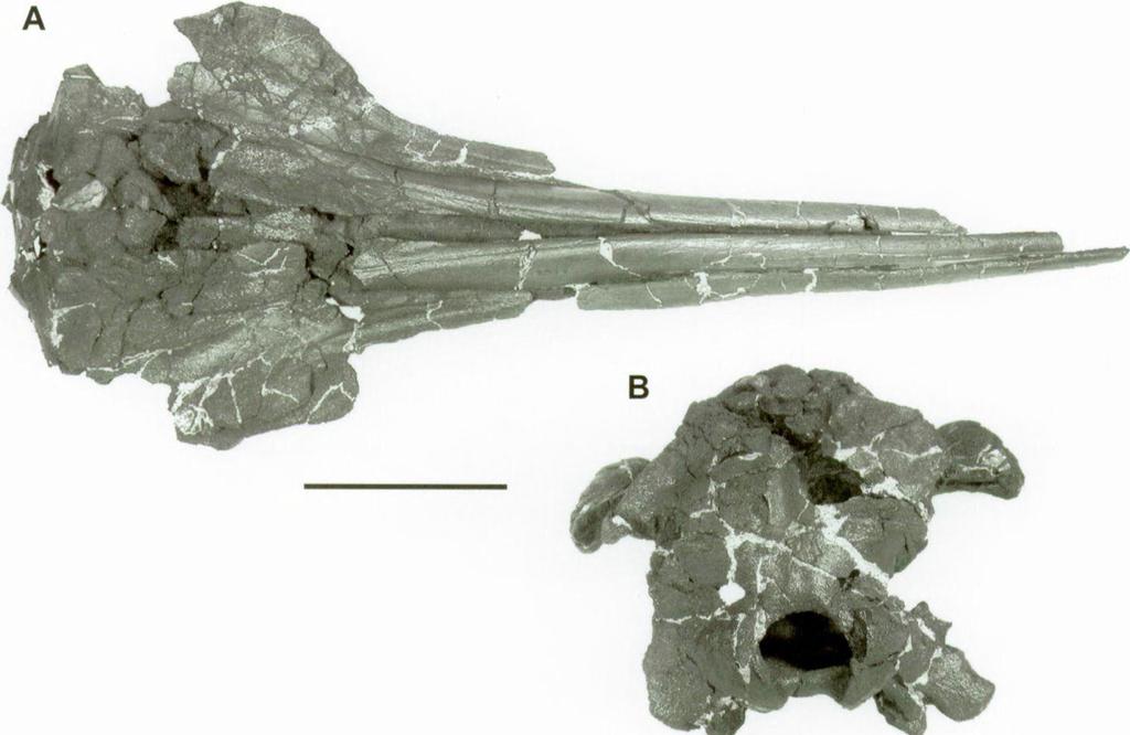
28 174 Olivier LAMBERT P l a t e 5
Review of the Miocene long-snouted dolphin Priscodelphinus cristatus. eurhinodelphinids. by Olivier LAMBERT
 BULLETIN DE L INSTITUT ROYAL DES SCIENCES NATURELLES DE BELGIQUE SCIENCES DE LA TERRE, 75: 211-235, 2005 BULLETIN VAN HET KONINKLIJK BELGISCH INSTITUUT VOOR NATUURWETENSCHAPPEN AARDWETENSCHAPPEN, 75: 211-235,
AMERICAN MUSEUM NOVITATES Published by
 AMERICAN MUSEUM NOVITATES Published by Number 782 THE AmzRICAN MUSEUM OF NATURAL HISTORY Feb. 20, 1935 New York City 56.81, 7 G (68) A NOTE ON THE CYNODONT, GLOCHINODONTOIDES GRACILIS HAUGHTON BY LIEUWE
A new archaic homodont toothed cetacean (Mammalia, Cetacea, Odontoceti) from the early Miocene of Peru
 from the early Miocene of Peru") A new archaic homodont toothed cetacean (Mammalia, Cetacea, Odontoceti) from the early Miocene of Peru Olivier LAMBERT Institut royal des Sciences naturelles de Belgique, D.O. Terre et Histoire de la Vie,
A new archaic homodont toothed cetacean (Mammalia, Cetacea, Odontoceti) from the early Miocene of Peru Olivier LAMBERT Institut royal des Sciences naturelles de Belgique, D.O. Terre et Histoire de la Vie,
Exceptional fossil preservation demonstrates a new mode of axial skeleton elongation in early ray-finned fishes
 Supplementary Information Exceptional fossil preservation demonstrates a new mode of axial skeleton elongation in early ray-finned fishes Erin E. Maxwell, Heinz Furrer, Marcelo R. Sánchez-Villagra Supplementary
First record of a platanistid (Cetacea, Odontoceti) in the North Sea Basin: a review of Cyrtodelphis Abel, 1899 from the Miocene of Belgium
 in the North Sea Basin: a review of Cyrtodelphis Abel, 1899 from the Miocene of Belgium") First record of a platanistid (Cetacea, Odontoceti) in the North Sea Basin: a review of Cyrtodelphis Abel, 1899 from the Miocene of Belgium Olivier Lambert 1 1 Institut royal des Sciences naturelles de
First record of a platanistid (Cetacea, Odontoceti) in the North Sea Basin: a review of Cyrtodelphis Abel, 1899 from the Miocene of Belgium Olivier Lambert 1 1 Institut royal des Sciences naturelles de
Mammalogy Lab 1: Skull, Teeth, and Terms
 Mammalogy Lab 1: Skull, Teeth, and Terms Be able to: Goals of today s lab Locate all structures listed on handout Define all terms on handout what they are or what they look like Give examples of mammals
2. Skull, total length versus length of the presacral vertebral column: (0); extremely elongated neck (e.g. Tanystropheus longobardicus).
; extremely elongated neck (e.g. Tanystropheus longobardicus).") Character list of the taxon-character data set 1. Skull and lower jaws, interdental plates: absent (0); present, but restricted to the anterior end of the dentary (1); present along the entire alveolar
Character list of the taxon-character data set 1. Skull and lower jaws, interdental plates: absent (0); present, but restricted to the anterior end of the dentary (1); present along the entire alveolar
ONLINE APPENDIX 1. Morphological phylogenetic characters scored in this paper. See Poe (2004) for
 for") ONLINE APPENDIX Morphological phylogenetic characters scored in this paper. See Poe () for detailed character descriptions, citations, and justifications for states. Note that codes are changed from a
ONLINE APPENDIX Morphological phylogenetic characters scored in this paper. See Poe () for detailed character descriptions, citations, and justifications for states. Note that codes are changed from a
A new species of Hsisosuchus (Mesoeucrocodylia) from Dashanpu, Zigong Municipality, Sichuan Province
 from Dashanpu, Zigong Municipality, Sichuan Province") A new species of Hsisosuchus (Mesoeucrocodylia) from Dashanpu, Zigong Municipality, Sichuan Province Yuhui Gao (Zigong Dinosaur Museum) Vertebrata PalAsiatica Volume 39, No. 3 July, 2001 pp. 177-184 Translated
A new species of Hsisosuchus (Mesoeucrocodylia) from Dashanpu, Zigong Municipality, Sichuan Province Yuhui Gao (Zigong Dinosaur Museum) Vertebrata PalAsiatica Volume 39, No. 3 July, 2001 pp. 177-184 Translated
SOME LITTLE-KNOWN FOSSIL LIZARDS FROM THE
 PROCEEDINGS OF THE UNITED STATES NATIONAL MUSEUM issued SWsK \ {^^m ^V ^^ SMITHSONIAN INSTITUTION U. S. NATIONAL MUSEUM Vol. 91 Washington : 1941 No. 3124 SOME LITTLE-KNOWN FOSSIL LIZARDS FROM THE OLIGOCENE
SUPPLEMENTARY OBSERVATIONS ON THE SKULL OF
 SUPPLEMENTARY OBSERVATIONS ON THE SKULL OF THE FOSSIL PORPOISE ZARHACHIS FLAGELLATOR COPE By Remington Kellogg Of the Bureau of Biological Survey, United States Department of Agriculture During the past
Williston, and as there are many fairly good specimens in the American
 56.81.7D :14.71.5 Article VII.- SOME POINTS IN THE STRUCTURE OF THE DIADECTID SKULL. BY R. BROOM. The skull of Diadectes has been described by Cope, Case, v. Huene, and Williston, and as there are many
A NEW GENUS AND SPECIES OF AMERICAN THEROMORPHA
 A NEW GENUS AND SPECIES OF AMERICAN THEROMORPHA MYCTEROSAURUS LONGICEPS S. W. WILLISTON University of Chicago The past summer, Mr. Herman Douthitt, of the University of Chicago paleontological expedition,
complex in cusp pattern. (3) The bones of the coyote skull are thinner, crests sharper and the
 The bones of the coyote skull are thinner, crests sharper and the") DISTINCTIONS BETWEEN THE SKULLS OF S AND DOGS Grover S. Krantz Archaeological sites in the United States frequently yield the bones of coyotes and domestic dogs. These two canines are very similar both
DISTINCTIONS BETWEEN THE SKULLS OF S AND DOGS Grover S. Krantz Archaeological sites in the United States frequently yield the bones of coyotes and domestic dogs. These two canines are very similar both
Mammalogy Laboratory 1 - Mammalian Anatomy
 Mammalogy Laboratory 1 - Mammalian Anatomy I. The Goal. The goal of the lab is to teach you skeletal anatomy of mammals. We will emphasize the skull because many of the taxonomically important characters
Fig. 5. (A) Scaling of brain vault size (width measured at the level of anterior squamosal/parietal suture) relative to skull size (measured at the
 Scaling of brain vault size (width measured at the level of anterior squamosal/parietal suture) relative to skull size (measured at the") Fig. 5. (A) Scaling of brain vault size (width measured at the level of anterior squamosal/parietal suture) relative to skull size (measured at the distance between the left versus right temporomandibular
Fig. 5. (A) Scaling of brain vault size (width measured at the level of anterior squamosal/parietal suture) relative to skull size (measured at the distance between the left versus right temporomandibular
SUPPLEMENTARY INFORMATION
 doi:10.1038/nature13086 Part I. Supplementary Notes A: Detailed Description of Cotylocara macei gen. et sp. nov. Part II. Table of Measurements for holotype of Cotylocara macei (CCNHM-101) Part III. Supplementary
New Carnivorous Dinosaurs from the Upper Cretaceous of Mongolia
 1955 Doklady, Academy of Sciences USSR 104 (5):779-783 New Carnivorous Dinosaurs from the Upper Cretaceous of Mongolia E. A. Maleev (translated by F. J. Alcock) The present article is a summary containing
FURTHER STUDIES ON TWO SKELETONS OF THE BLACK RIGHT WHALE IN THE NORTH PACIFIC
 FURTHER STUDIES ON TWO SKELETONS OF THE BLACK RIGHT WHALE IN THE NORTH PACIFIC HIDEO OMURA, MASAHARU NISHIWAKI* AND TOSHIO KASUYA* ABSTRACT Two skeletons of the black right whale were studied, supplementing
A new sauropod from Dashanpu, Zigong Co. Sichuan Province (Abrosaurus dongpoensis gen. et sp. nov.)
") A new sauropod from Dashanpu, Zigong Co. Sichuan Province (Abrosaurus dongpoensis gen. et sp. nov.) by Ouyang Hui Zigong Dinosaur Museum Newsletter Number 2 1989 pp. 10-14 Translated By Will Downs Bilby
A new sauropod from Dashanpu, Zigong Co. Sichuan Province (Abrosaurus dongpoensis gen. et sp. nov.) by Ouyang Hui Zigong Dinosaur Museum Newsletter Number 2 1989 pp. 10-14 Translated By Will Downs Bilby
v:ii-ixi, 'i':;iisimvi'\>!i-:: "^ A%'''''-'^-''S.''v.--..V^'E^'-'-^"-t''gi L I E) R.ARY OF THE VERSITY U N I or ILLINOIS REMO
 R.ARY OF THE VERSITY U N I or ILLINOIS REMO") "^ A%'''''-'^-''S.''v.--..V^'E^'-'-^"-t''gi v:ii-ixi, 'i':;iisimvi'\>!i-:: L I E) R.ARY OF THE U N I VERSITY or ILLINOIS REMO Natural History Survey Librarv GEOLOGICAL SERIES OF FIELD MUSEUM OF NATURAL
"^ A%'''''-'^-''S.''v.--..V^'E^'-'-^"-t''gi v:ii-ixi, 'i':;iisimvi'\>!i-:: L I E) R.ARY OF THE U N I VERSITY or ILLINOIS REMO Natural History Survey Librarv GEOLOGICAL SERIES OF FIELD MUSEUM OF NATURAL
A NEW AUSTROSQUILLA (STOMATOPODA) FROM THE
 FROM THE") A NEW AUSTROSQUILLA (STOMATOPODA) FROM THE MARQUESAS ISLANDS BY ALAIN MICHEL Centre O.R.S.T.O.M., Noumea, New Caledonia and RAYMOND B. MANNING Smithsonian Institution, Washington, U.S.A. The At s,tstrosqzlilla
A NEW AUSTROSQUILLA (STOMATOPODA) FROM THE MARQUESAS ISLANDS BY ALAIN MICHEL Centre O.R.S.T.O.M., Noumea, New Caledonia and RAYMOND B. MANNING Smithsonian Institution, Washington, U.S.A. The At s,tstrosqzlilla
OSTEOLOGICAL NOTE OF AN ANTARCTIC SEI WHALE
 OSTEOLOGICAL NOTE OF AN ANTARCTIC SEI WHALE MASAHARU NISHIWAKI* AND TOSHIO KASUYA* ABSTRACT This is a report of measurements on the skeleton of a male se1 whale caught in the Antarctic. The skeleton of
UPOGEBIA LINCOLNI SP. NOV. (DECAPODA, THALASSINIDEA, UPOGEBIIDAE) FROM JAVA, INDONESIA
 FROM JAVA, INDONESIA") NOTES AND NEWS UPOGEBIA LINCOLNI SP. NOV. (DECAPODA, THALASSINIDEA, UPOGEBIIDAE) FROM JAVA, INDONESIA BY NGUYEN NGOC-HO i) Faculty of Science, University of Saigon, Vietnam Among material recently collected
NOTES AND NEWS UPOGEBIA LINCOLNI SP. NOV. (DECAPODA, THALASSINIDEA, UPOGEBIIDAE) FROM JAVA, INDONESIA BY NGUYEN NGOC-HO i) Faculty of Science, University of Saigon, Vietnam Among material recently collected
WHxVLEBONE WHALE FROM THE CALVERT CLIFFS, MARYLAND.
 DESCRIPTION OF A NEW GENUS AND SPECIES OF WHxVLEBONE WHALE FROM THE CALVERT CLIFFS, MARYLAND. By Remington Kellogg, Of the Bureau of Biological Survey, United States Departm'ent of Agriculture. In the
YANGCHUANOSAURUS HEPINGENSIS - A NEW SPECIES OF CARNOSAUR FROM ZIGONG, SICHUAN
 Vol. 30, No. 4 VERTEBRATA PALASIATICA pp. 313-324 October 1992 [SICHUAN ZIGONG ROUSHILONG YI XIN ZHONG] figs. 1-5, pl. I-III YANGCHUANOSAURUS HEPINGENSIS - A NEW SPECIES OF CARNOSAUR FROM ZIGONG, SICHUAN
Biology 3315 Comparative Vertebrate Morphology Skulls and Visceral Skeletons
 Biology 3315 Comparative Vertebrate Morphology Skulls and Visceral Skeletons 1. Head skeleton of lamprey Cyclostomes are highly specialized in both the construction of the chondrocranium and visceral skeleton.
Reprinted from: CRUSTACEANA, Vol. 32, Part 2, 1977 LEIDEN E. J. BRILL
 Reprinted from: CRUSTACEANA, Vol. 32, Part 2, 1977 LEIDEN E. J. BRILL NOTES AND NEWS 207 ALPHE0PS1S SHEARMII (ALCOCK & ANDERSON): A NEW COMBINATION WITH A REDESCRIPTION OF THE HOLOTYPE (DECAPODA, ALPHEIDAE)
CENE RUMINANTS OF THE GENERA OVIBOS AND
 DESCRIPTIONS OF TWO NEW SPECIES OF PLEISTO- CENE RUMINANTS OF THE GENERA OVIBOS AND BOOTHERIUM, WITH NOTES ON THE LATTER GENUS. By James Williams Gidley, Of the United States National Museum. Two interesting
List of characters used in the phylogenetic analysis. Capital letters T, R, and L, refer to
 1 Supplementary data CHARACTER LIST List of characters used in the phylogenetic analysis. Capital letters T, R, and L, refer to characters used by Tchernov et al. (2000), Rieppel, et al. (2002), and Lee
An Early Miocene long-snouted marine platanistid dolphin (Mammalia, Cetacea, Odontoceti) from the Korneuburg Basin (Austria)
 from the Korneuburg Basin (Austria)") Beitr. Paläont., 27: 407-418, Wien 2002 An Early Miocene long-snouted marine platanistid dolphin (Mammalia, Cetacea, Odontoceti) from the Korneuburg Basin (Austria) by Lawrence G. Barnes* B arnes, L. G.
Beitr. Paläont., 27: 407-418, Wien 2002 An Early Miocene long-snouted marine platanistid dolphin (Mammalia, Cetacea, Odontoceti) from the Korneuburg Basin (Austria) by Lawrence G. Barnes* B arnes, L. G.
Temporal lines. More forwardfacing. tubular orbits than in the African forms 3. Orbits larger relative to skull size than in the other genera 2.
 Asian lorises More forwardfacing and tubular orbits than in the African forms 3. Characterized by a marked extension of the ectotympanic into a tubular meatus and a more angular auditory bulla than in
Vol. XIV, No. 1, March, The Larva and Pupa of Brontispa namorikia Maulik (Coleoptera: Chrysomelidae: Hispinae) By S.
 By S.") Vol. XIV, No. 1, March, 1950 167 The Larva and Pupa of Brontispa namorikia Maulik (Coleoptera: Chrysomelidae: Hispinae) By S. MAULIK BRITISH MUSEUM (NATURAL HISTORY) (Presented by Mr. Van Zwaluwenburg
Vol. XIV, No. 1, March, 1950 167 The Larva and Pupa of Brontispa namorikia Maulik (Coleoptera: Chrysomelidae: Hispinae) By S. MAULIK BRITISH MUSEUM (NATURAL HISTORY) (Presented by Mr. Van Zwaluwenburg
THE GORGONOPSIAN GENUS, HIPPOSAURUS, AND THE FAMILY ICTIDORHINIDAE * Dr. L.D. Boonstra. Paleontologist, South African Museum, Cape Town
 THE GORGONOPSIAN GENUS, HIPPOSAURUS, AND THE FAMILY ICTIDORHINIDAE * by Dr. L.D. Boonstra Paleontologist, South African Museum, Cape Town In 1928 I dug up the complete skeleton of a smallish gorgonopsian
A skull without mandihle, from the Hunterian Collection (no.
 4 MR. G. A. BOULENGER ON CHELONIAN REMAINS. [Jan. 6, 2. On some Chelonian Remains preserved in the Museum of the Eojal College of Surgeons. By G. A. Boulenger. [Eeceived December 8, 1890.] In the course
Museo di Storia Naturale del Mediterraneo, Via Roma 234, 57125, Livorno, Italy
 bs_bs_banner Zoological Journal of the Linnean Society, 2012, 166, 876 911. With 22 figures Comparative osteology and phylogenetic relationships of Miocaperea pulchra, the first fossil pygmy right whale
OF THE TRIAS THE PHYTOSAURIA
 THE PHYTOSAURIA OF THE TRIAS MAURICE G. MEHL University of Wisconsin Some time ago the writer gave a brief notice of a new genus of phytosaurs of which Angistorhinus grandis Mehl was the type.' It is the
PALEONTOLOGICAL CONTRIBUTIONS
 THE UNIVERSITY OF KANSAS PALEONTOLOGICAL CONTRIBUTIONS August, 1965 Paper 2 A NEW WYOMING PHYTOSAUR By THEODORE H. EATON, JR. [Museum of Natural History, University of Kansas I ABSTRACT The skull of a
Anatomical description and phylogenetic analysis of Miocene beaked whale from the East African Rift Valley, Kenya
 Southern Methodist University SMU Scholar Collection of Engaged Learning Engaged Learning 4-15-2013 Anatomical description and phylogenetic analysis of Miocene beaked whale from the East African Rift Valley,
A NEW SPECIES OF EXTINCT TURTLE FROM THE UPPER PLIOCENE OF IDAHO
 A NEW SPECIES OF EXTINCT TURTLE FROM THE UPPER PLIOCENE OF IDAHO By Charles W. Gilmore Curator, Division of Vertebrate Paleontology United States National Museum Among the fossils obtained bj^ the Smithsonian
SUPPLEMENTARY INFORMATION
 Character 155, interdental ridges. Absence of interdental ridge (0) shown in Parasaniwa wyomingensis (Platynota). Interdental ridges (1) shown in Coniophis precedens. WWW.NATURE.COM/NATURE 1 Character
A NEW SALTICID SPIDER FROM VICTORIA By R. A. Dunn
 Dunn, R. A. 1947. A new salticid spider from Victoria. Memoirs of the National Museum of Victoria 15: 82 85. All text not included in the original document is highlighted in red. Mem. Nat. Mus. Vict.,
SOME NEW AMERICAN PYCNODONT FISHES.
 SOME NEW AMERICAN PYCNODONT FISHES. By James Williams Gidley, Assistant Curator of Fossil Mammals, United States National Museum. In the United States National Museum are several specimens representing
HONR219D Due 3/29/16 Homework VI
 Part 1: Yet More Vertebrate Anatomy!!! HONR219D Due 3/29/16 Homework VI Part 1 builds on homework V by examining the skull in even greater detail. We start with the some of the important bones (thankfully
SUPPLEMENTARY ONLINE MATERIAL FOR. Nirina O. Ratsimbaholison, Ryan N. Felice, and Patrick M. O connor
 http://app.pan.pl/som/app61-ratsimbaholison_etal_som.pdf SUPPLEMENTARY ONLINE MATERIAL FOR Nirina O. Ratsimbaholison, Ryan N. Felice, and Patrick M. O connor Ontogenetic changes in the craniomandibular
Cranial osteology of the African gerrhosaurid Angolosaurus skoogi (Squamata; Gerrhosauridae) HOLLY A. NANCE
 HOLLY A. NANCE") African Journal of Herpetology, 2007 56(1): 39-75. Herpetological Association of Africa Original article Cranial osteology of the African gerrhosaurid Angolosaurus skoogi (Squamata; Gerrhosauridae) HOLLY
African Journal of Herpetology, 2007 56(1): 39-75. Herpetological Association of Africa Original article Cranial osteology of the African gerrhosaurid Angolosaurus skoogi (Squamata; Gerrhosauridae) HOLLY
A New Ceratopsian Dinosaur from the Upper
 SMITHSONIAN MISCELLANEOUS COLLECTIONS VOLUME 63. NUMBER 3 A New Ceratopsian Dinosaur from the Upper Cretaceous of Montana, with Note on Hypacrosaurus (With Two Plates) CHARLES W. GILMORE Assistant Curator
Notes on Ceratopsians and Ankylosaurs at the Royal Ontario Museum
 Notes on Ceratopsians and Ankylosaurs at the Royal Ontario Museum Andrew A. Farke, Ph.D. Raymond M. Alf Museum of Paleontology 1175 West Baseline Road Claremont, CA 91711 email: afarke@webb.org Introduction
DISCOVERY OF GENUS PLATOLENES (COLEOP TERA : TENEBRIONIDAE) FROM INDIA WITH DESCRIPTION OF TWO NEW SPECIES G. N. SABA
 FROM INDIA WITH DESCRIPTION OF TWO NEW SPECIES G. N. SABA") Rec. zool. Surv. India, 85(3) : 433-437,1988 DISCOVERY OF GENUS PLATOLENES (COLEOP TERA : TENEBRIONIDAE) FROM INDIA WITH DESCRIPTION OF TWO NEW SPECIES By G. N. SABA Zoological Survey of India M-Block,
Rec. zool. Surv. India, 85(3) : 433-437,1988 DISCOVERY OF GENUS PLATOLENES (COLEOP TERA : TENEBRIONIDAE) FROM INDIA WITH DESCRIPTION OF TWO NEW SPECIES By G. N. SABA Zoological Survey of India M-Block,
A M E G H I N I A N A. Revista de la Asociación Paleontológia Argentina. Volume XV September-December 1978 Nos. 3-4
 A M E G H I N I A N A Revista de la Asociación Paleontológia Argentina Volume XV September-December 1978 Nos. 3-4 COLORADIA BREVIS N. G. ET N. SP. (SAURISCHIA, PROSAUROPODA), A PLATEOSAURID DINOSAUR FROM
A new species of Antinia PASCOE from Burma (Coleoptera: Curculionidae: Entiminae)
") Genus Vol. 14 (3): 413-418 Wroc³aw, 15 X 2003 A new species of Antinia PASCOE from Burma (Coleoptera: Curculionidae: Entiminae) JAROS AW KANIA Zoological Institute, University of Wroc³aw, Sienkiewicza
Genus Vol. 14 (3): 413-418 Wroc³aw, 15 X 2003 A new species of Antinia PASCOE from Burma (Coleoptera: Curculionidae: Entiminae) JAROS AW KANIA Zoological Institute, University of Wroc³aw, Sienkiewicza
 The following text is generated from uncorrected OCR. [Begin Page: Page 1] A NEW CERATOPSIAN DINOSAUR FROM THE UPPER CRETACEOUS OF MONTANA, WITH NOTE ON HYPACROSAURUS ' By CHARLES W. GILMORE assistant
FIRST RECORD OF MESOPLODON DENSIROSTRIS FROM FORMOSA
 FIRST RECORD OF MESOPLODON DENSIROSTRIS FROM FORMOSA TOSHIO KASUYA* AND MASAHARU NISHIWAKI* ABSTRACT Two records of female Mesoplodon densirostris are reported. Comments on the external character, skull
TRACHEMYS SCULPTA. A nearly complete articulated carapace and plastron of an Emjdd A NEAKLY COMPLETE SHELL OF THE EXTINCT TURTLE,
 A NEAKLY COMPLETE SHELL OF THE EXTINCT TURTLE, TRACHEMYS SCULPTA By Charles W. Gilmore Curator of Vertebrate Paleontology, United States National Museum INTRODUCTION A nearly complete articulated carapace
MIOCENE DIATOMACEOUS EARTH OF LOMPOC, CALI- FORNIA.i
 DESCRIPTION OF THE SKULL OF MEGAPTERA MIO- CAENA, A FOSSIL HUMPBACK WHALE FROM THE MIOCENE DIATOMACEOUS EARTH OF LOMPOC, CALI- FORNIA.i By Remington Kellogg. Of the Bureau of Biological Survey, Department
Title: Phylogenetic Methods and Vertebrate Phylogeny
 Title: Phylogenetic Methods and Vertebrate Phylogeny Central Question: How can evolutionary relationships be determined objectively? Sub-questions: 1. What affect does the selection of the outgroup have
THE SKULLS OF ARAEOSCELIS AND CASEA, PERMIAN REPTILES
 THE SKULLS OF REOSCELIS ND CSE, PERMIN REPTILES University of Chicago There are few Permian reptiles of greater interest at the present time than the peculiar one I briefly described in this journal' three
NOTE XXXVIII. Three new species of the genus Helota DESCRIBED BY. C. Ritsema+Cz. is very. friend René Oberthür who received. Biet.
 Subshining; HELOTA MARIAE. 249 NOTE XXXVIII. Three new species of the genus Helota DESCRIBED BY C. Ritsema+Cz. The first of these species is very interesting as it belongs to the same section as the recently
Diurus, Pascoe. sp. 1). declivity of the elytra, but distinguished. Length (the rostrum and tails 26 included) mm. Deep. exception
. declivity of the elytra, but distinguished. Length (the rostrum and tails 26 included) mm. Deep. exception") 210 DIURUS ERYTIIROPUS. NOTE XXVI. Three new species of the Brenthid genus Diurus, Pascoe DESCRIBED BY C. Ritsema+Cz. 1. Diurus erythropus, n. sp. 1). Allied to D. furcillatus Gylh. ²) by the short head,
210 DIURUS ERYTIIROPUS. NOTE XXVI. Three new species of the Brenthid genus Diurus, Pascoe DESCRIBED BY C. Ritsema+Cz. 1. Diurus erythropus, n. sp. 1). Allied to D. furcillatus Gylh. ²) by the short head,
FIELDIANA GEOLOGY NEW SALAMANDERS OF THE FAMILY SIRENIDAE FROM THE CRETACEOUS OF NORTH AMERICA
 FIELDIANA GEOLOGY Published by CHICAGO NATURAL HISTORY MUSEUM Volume 10 Sbftember 22, 1968 No. 88 NEW SALAMANDERS OF THE FAMILY SIRENIDAE FROM THE CRETACEOUS OF NORTH AMERICA Coleman J. Coin AND Walter
ZOOLOGISCHE MEDEDELINGEN
 MINISTERIE VAN ONDERWIJS, KUNSTEN EN WETENSCHAPPEN ZOOLOGISCHE MEDEDELINGEN UITGEGEVEN DOOR HET RIJKSMUSEUM VAN NATUURLIJKE HISTORIE TE LEIDEN DEEL XXXVII, No. 10 10 juli 1961 THE FOSSIL HIPPOPOTAMUS FROM
Supporting Online Material for
 www.sciencemag.org/cgi/content/full/329/5998/1481/dc1 Supporting Online Material for Tyrannosaur Paleobiology: New Research on Ancient Exemplar Organisms Stephen L. Brusatte,* Mark A. Norell, Thomas D.
The cranial osteology of Belebey vegrandis (Parareptilia: Bolosauridae), from the Middle Permian of Russia, and its bearing on reptilian evolution
, from the Middle Permian of Russia, and its bearing on reptilian evolution") Blackwell Publishing LtdOxford, UKZOJZoological Journal of the Linnean Society0024-4082 2007 The Linnean Society of London? 2007 1511 191214 Original Articles RUSSIAN BOLOSAURID REPTILER. R. REISZ ET AL.
Blackwell Publishing LtdOxford, UKZOJZoological Journal of the Linnean Society0024-4082 2007 The Linnean Society of London? 2007 1511 191214 Original Articles RUSSIAN BOLOSAURID REPTILER. R. REISZ ET AL.
PSYCHE A NEW GENUS AND SPECIES OF SALDIDAE FROM SOUTH AMERICA (HEMIPTERA) BY CARL J. DRAKE AND LUDVIK HOBERLANDT. Iowa State College, Ames
 BY CARL J. DRAKE AND LUDVIK HOBERLANDT. Iowa State College, Ames") PSYCHE Vol. 59 September, 1952 No. 3 A NEW GENUS AND SPECIES OF SALDIDAE FROM SOUTH AMERICA (HEMIPTERA) BY CARL J. DRAKE AND LUDVIK HOBERLANDT Iowa State College, Ames Through the kindness of Dr. P. J.
PSYCHE Vol. 59 September, 1952 No. 3 A NEW GENUS AND SPECIES OF SALDIDAE FROM SOUTH AMERICA (HEMIPTERA) BY CARL J. DRAKE AND LUDVIK HOBERLANDT Iowa State College, Ames Through the kindness of Dr. P. J.
NORTH AMERICA. ON A NEW GENUS AND SPECIES OF COLUBRINE SNAKES FROM. The necessity of recognizing tlie two species treated of in this paper
 ON A NEW GENUS AND SPECIES OF COLUBRINE SNAKES FROM NORTH AMERICA. BY Leonhard Stejneger, and Batrachians. Curator of the Department of Reptiles The necessity of recognizing tlie two species treated of
CRANIAL OSTEOLOGY OF SCHIZOTHORAICHTHYS NIGER (MECKEL) MISRA (CYPRINIDAE: SCHIZOTHORACINAE). L NEUROCRANIUM
 MISRA (CYPRINIDAE: SCHIZOTHORACINAE). L NEUROCRANIUM") CRANIAL OSTEOLOGY OF SCHIZOTHORAICHTHYS NIGER (MECKEL) MISRA (CYPRINIDAE: SCHIZOTHORACINAE). L NEUROCRANIUM A. R. YousuF, A. K. PANDIT AND A. R. KHAN Postgraduate Department of Zoology, University of Kashmir,
CRANIAL OSTEOLOGY OF SCHIZOTHORAICHTHYS NIGER (MECKEL) MISRA (CYPRINIDAE: SCHIZOTHORACINAE). L NEUROCRANIUM A. R. YousuF, A. K. PANDIT AND A. R. KHAN Postgraduate Department of Zoology, University of Kashmir,
NAUSHONIA PAN AMEN SIS, NEW SPECIES (DECAPODA: THALASSINIDEA: LAOMEDIIDAE) FROM THE PACIFIC COAST OF PANAMA, WITH NOTES ON THE GENUS
 FROM THE PACIFIC COAST OF PANAMA, WITH NOTES ON THE GENUS") 5 October 1982 PROC. BIOL. SOC. WASH. 95(3), 1982, pp. 478-483 NAUSHONIA PAN AMEN SIS, NEW SPECIES (DECAPODA: THALASSINIDEA: LAOMEDIIDAE) FROM THE PACIFIC COAST OF PANAMA, WITH NOTES ON THE GENUS Joel
5 October 1982 PROC. BIOL. SOC. WASH. 95(3), 1982, pp. 478-483 NAUSHONIA PAN AMEN SIS, NEW SPECIES (DECAPODA: THALASSINIDEA: LAOMEDIIDAE) FROM THE PACIFIC COAST OF PANAMA, WITH NOTES ON THE GENUS Joel
MUNIDOPSIS ALBATROSSAB, A NEW SPECIES OF DEEP-SEA GALATHEIDAE (DECAPODA, ANOMURA) FROM THE EASTERN PACIFIC OCEAN
 FROM THE EASTERN PACIFIC OCEAN") MUNIDOPSIS ALBATROSSAB, A NEW SPECIES OF DEEP-SEA GALATHEIDAE (DECAPODA, ANOMURA) FROM THE EASTERN PACIFIC OCEAN BY WILLIS E. PEQUEGNAT and LINDA H. PEQUEGNAT Department of Oceanography, Texas A & M University,
MUNIDOPSIS ALBATROSSAB, A NEW SPECIES OF DEEP-SEA GALATHEIDAE (DECAPODA, ANOMURA) FROM THE EASTERN PACIFIC OCEAN BY WILLIS E. PEQUEGNAT and LINDA H. PEQUEGNAT Department of Oceanography, Texas A & M University,
SECOND RECORD OF SPECTACLED PORPOISE FROM SUBANTARCTIC SOUTHWEST PACIFIC
 SECOND RECORD OF SPECTACLED PORPOISE FROM SUBANTARCTIC SOUTHWEST PACIFIC R. EWAN FORDYCE Department of Geology, University of Otago, P, 0. Box 56, Dunedin, New Zealand ROBERT H. MATTLIN Fisheries Research
Cranial osteology and phylogenetic relationships of Hamadasuchus rebouli (Crocodyliformes: Mesoeucrocodylia) from the Cretaceous of Morocco
 from the Cretaceous of Morocco") Blackwell Publishing LtdOxford, UKZOJZoological Journal of the Linnean Society0024-4082 2007 The Linnean Society of London? 2007 1494 533567 Original Articles HAMADASUCHUS REBOULIH. C. E. LARSSON and H.-D.
Blackwell Publishing LtdOxford, UKZOJZoological Journal of the Linnean Society0024-4082 2007 The Linnean Society of London? 2007 1494 533567 Original Articles HAMADASUCHUS REBOULIH. C. E. LARSSON and H.-D.
A new kentriodontid (Cetacea: Delphinoidea) from the Middle Miocene of Hungary
 from the Middle Miocene of Hungary") Mitt. Mus. Nat.kd. Berl., Geowiss. Reihe 8 (2005), 53 73 / DOI 10.1002/mmng.200410004 A new kentriodontid (Cetacea: Delphinoidea) from the Middle Miocene of Hungary Emese Kazár* Geological Institute of
Mitt. Mus. Nat.kd. Berl., Geowiss. Reihe 8 (2005), 53 73 / DOI 10.1002/mmng.200410004 A new kentriodontid (Cetacea: Delphinoidea) from the Middle Miocene of Hungary Emese Kazár* Geological Institute of
BREVIORA LEUCOLEPIDOPA SUNDA GEN. NOV., SP. NOV. (DECAPODA: ALBUNEIDAE), A NEW INDO-PACIFIC SAND CRAB. Ian E. Efford 1
, A NEW INDO-PACIFIC SAND CRAB. Ian E. Efford 1") ac lc BREVIORA CAMBRIDGE, MASS. 30 APRIL, 1969 NUMBER 318 LEUCOLEPIDOPA SUNDA GEN. NOV., SP. NOV. (DECAPODA: ALBUNEIDAE), A NEW INDO-PACIFIC SAND CRAB Ian E. Efford 1 ABSTRACT. Leucolepidopa gen. nov.
ac lc BREVIORA CAMBRIDGE, MASS. 30 APRIL, 1969 NUMBER 318 LEUCOLEPIDOPA SUNDA GEN. NOV., SP. NOV. (DECAPODA: ALBUNEIDAE), A NEW INDO-PACIFIC SAND CRAB Ian E. Efford 1 ABSTRACT. Leucolepidopa gen. nov.
VERTEBRATA PALASIATICA
 41 2 2003 2 VERTEBRATA PALASIATICA pp. 147 156 figs. 1 5 1) ( 100044), ( Parakannemeyeria brevirostris),,, : ( Xiyukannemeyeria),,, Q915. 864 60 Turfania (,1973), Dicynodon (, 1973 ; Lucas, 1998), (Lystrosaurus)
A REDESCRIPTION OF THE HOLOTYPE OF CALLIANASSA MUCRONATA STRAHL, 1861 (DECAPODA, THALASSINIDEA)
") Crustaceana 52 (1) 1977, E. J. Brill, Leiden A REDESCRIPTION OF THE HOLOTYPE OF CALLIANASSA MUCRONATA STRAHL, 1861 (DECAPODA, THALASSINIDEA) BY NASIMA M. TIRMIZI Department of Zoology, University of Karachi,
Crustaceana 52 (1) 1977, E. J. Brill, Leiden A REDESCRIPTION OF THE HOLOTYPE OF CALLIANASSA MUCRONATA STRAHL, 1861 (DECAPODA, THALASSINIDEA) BY NASIMA M. TIRMIZI Department of Zoology, University of Karachi,
A NEW SPECIES OF A USTROLIBINIA FROM THE SOUTH CHINA SEA AND INDONESIA (CRUSTACEA: BRACHYURA: MAJIDAE)
") 69 C O a g r ^ j^a RAFFLES BULLETIN OF ZOOLOGY 1992 40(1): 69-73 A NEW SPECIES OF A USTROLIBINIA FROM THE SOUTH CHINA SEA AND INDONESIA (CRUSTACEA: BRACHYURA: MAJIDAE) H P Waener SMITHSONIAN INSTITUTE
69 C O a g r ^ j^a RAFFLES BULLETIN OF ZOOLOGY 1992 40(1): 69-73 A NEW SPECIES OF A USTROLIBINIA FROM THE SOUTH CHINA SEA AND INDONESIA (CRUSTACEA: BRACHYURA: MAJIDAE) H P Waener SMITHSONIAN INSTITUTE
A Fossil Snake (Elaphe vulpina) From A Pliocene Ash Bed In Nebraska
 From A Pliocene Ash Bed In Nebraska") University of Nebraska - Lincoln DigitalCommons@University of Nebraska - Lincoln Transactions of the Nebraska Academy of Sciences and Affiliated Societies Nebraska Academy of Sciences 198 A Fossil Snake
University of Nebraska - Lincoln DigitalCommons@University of Nebraska - Lincoln Transactions of the Nebraska Academy of Sciences and Affiliated Societies Nebraska Academy of Sciences 198 A Fossil Snake
A NEW ANKYLOSAUR FROM THE UPPER CRETACEOUS OF MONGOLIA E.A. Maleev Doklady Akademii Nauk, SSSR 87:
 translated by Dr. Tamara and F. Jeletzky, 1956 A NEW ANKYLOSAUR FROM THE UPPER CRETACEOUS OF MONGOLIA E.A. Maleev 1952. Doklady Akademii Nauk, SSSR 87:273-276 Armored dinosaurs make a considerable part
CRANIAL ANATOMY OF ENNATOSAURUS TECTON (SYNAPSIDA: CASEIDAE) FROM THE MIDDLE PERMIAN OF RUSSIA AND THE EVOLUTIONARY RELATIONSHIPS OF CASEIDAE
 FROM THE MIDDLE PERMIAN OF RUSSIA AND THE EVOLUTIONARY RELATIONSHIPS OF CASEIDAE") Journal of Vertebrate Paleontology 28(1):160 180, March 2008 2008 by the Society of Vertebrate Paleontology ARTICLE CRANIAL ANATOMY OF ENNATOSAURUS TECTON (SYNAPSIDA: CASEIDAE) FROM THE MIDDLE PERMIAN
Journal of Vertebrate Paleontology 28(1):160 180, March 2008 2008 by the Society of Vertebrate Paleontology ARTICLE CRANIAL ANATOMY OF ENNATOSAURUS TECTON (SYNAPSIDA: CASEIDAE) FROM THE MIDDLE PERMIAN
Three new species of Microctenochira SPAETH from Brazil and Panama (Coleoptera: Chrysomelidae: Cassidinae)
") Genus Vol. 10 (1): 109-116 Wroc³aw, 31 III 1999 Three new species of Microctenochira SPAETH from Brazil and Panama (Coleoptera: Chrysomelidae: Cassidinae) JOLANTA ŒWIÊTOJAÑSKA and LECH BOROWIEC Zoological
Genus Vol. 10 (1): 109-116 Wroc³aw, 31 III 1999 Three new species of Microctenochira SPAETH from Brazil and Panama (Coleoptera: Chrysomelidae: Cassidinae) JOLANTA ŒWIÊTOJAÑSKA and LECH BOROWIEC Zoological
Florida, Gainesville, Florida, 32611, U.S.A. b Smithsonian Tropical Research Institute, Ancon, Republic of Panama,
 This article was downloaded by: [78.22.97.164] On: 04 May 2013, At: 14:02 Publisher: Taylor & Francis Informa Ltd Registered in England and Wales Registered Number: 1072954 Registered office: Mortimer
Craniodental anatomy of late Oligocene archaeohyracids (Notoungulata, Mammalia) from Bolivia and Argentina and new phylogenetic hypotheses
 from Bolivia and Argentina and new phylogenetic hypotheses") Zoological Journal of the Linnean Society, 2009, 155, 458 509. With 27 figures Craniodental anatomy of late Oligocene archaeohyracids (Notoungulata, Mammalia) from Bolivia and Argentina and new phylogenetic
Zoological Journal of the Linnean Society, 2009, 155, 458 509. With 27 figures Craniodental anatomy of late Oligocene archaeohyracids (Notoungulata, Mammalia) from Bolivia and Argentina and new phylogenetic
PALEONTOLOGY AND BIOSTRATIGRAPHY OF MONGOLIA
 PALEONTOLOGY AND BIOSTRATIGRAPHY OF MONGOLIA THE JOINT SOVIET-MONGOLIAN PALEONTOLOGICAL EXPEDITION (Transactions, vol. 3) EDITORIAL BOARD: N. N. Kramarenko (editor-in-chief) B. Luvsandansan, Yu. I. Voronin,
Anatomy. Name Section. The Vertebrate Skeleton
 Name Section Anatomy The Vertebrate Skeleton Vertebrate paleontologists get most of their knowledge about past organisms from skeletal remains. Skeletons are useful for gleaning information about an organism
A new carnosaur from Yongchuan County, Sichuan Province
 A new carnosaur from Yongchuan County, Sichuan Province by Dong Zhiming Institute of Vertebrate Palaeontology and Palaeoanthropology, Academia Sinica Zhang Yihong, Li Xuanmin, and Zhou Shiwu Chongqing
SUBFAMILY THYMOPINAE Holthuis, 1974
 click for previous page 29 Remarks : The taxonomy of the species is not clear. It is possible that 2 forms may have to be distinguished: A. sublevis Wood-Mason, 1891 (with a synonym A. opipara Burukovsky
Key to sub families of ants in Hawaii
 Key to sub families of ants in Hawaii 1 2-segmented petiole, very large bulging eyes (1a)..... Pseudomyrmecinae (Pseudomyrmex gracilis) 2-segmented petiole (1b), eyes normal, reduced or absent.... 5 Myrmicinae
ZOOLOGISCHE MEDEDELINGEN
 "f ~- >D noitnwz, tito ZOOLOGISCHE MEDEDELINGEN UITGEGEVEN DOOR HET RIJKSMUSEUM VAN NATUURLIJKE HISTORIE TE LEIDEN (MINISTERIE VAN CULTUUR, RECREATIE EN MAATSCHAPPELIJK WERK) Deel 48 no. 25 25 maart 1975
A Short Report on the Occurrence of Dilophosaurus from Jinning County, Yunnan Province
 A Short Report on the Occurrence of Dilophosaurus from Jinning County, Yunnan Province by Hu Shaojin (Kunming Cultural Administrative Committee, Yunnan Province) Vertebrata PalAsiatica Vol. XXXI, No. 1
Descriptions of New North American Fulgoridae
 The Ohio State University Knowledge Bank kb.osu.edu Ohio Journal of Science (Ohio Academy of Science) Ohio Journal of Science: Volume 5, Issue 8 (June, 1905) 1905-06 Descriptions of New North American
CI-Standard N 343 / / GB. ITALIAN CORSO DOG (Cane Corso Italiano)
") CI-Standard N 343 / 06. 06. 2007/ GB ITALIAN CORSO DOG (Cane Corso Italiano) 2 TRANSLATION : Dr. Antonio Morsiani, Dr. J.-M. Paschoud and Prof. R. Triquet. ORIGIN : Italy. DATE OF PUBLICATION OF THE ORIGINAL
CI-Standard N 343 / 06. 06. 2007/ GB ITALIAN CORSO DOG (Cane Corso Italiano) 2 TRANSLATION : Dr. Antonio Morsiani, Dr. J.-M. Paschoud and Prof. R. Triquet. ORIGIN : Italy. DATE OF PUBLICATION OF THE ORIGINAL
MORPHOMETRIC ANALYSIS OF INFRA ORBITAL FORAMEN IN HUMAN DRY SKULLS
 Original Research Article MORPHOMETRIC ANALYSIS OF INFRA ORBITAL FORAMEN IN HUMAN DRY SKULLS K. Rajeswari * 1, M. Rohinidevi 2, V. Vimala 3, D. Megala 4. ABSTRACT International Journal of Anatomy and Research,
Bulletin of Big Bend Paleo-Geo An Open Access Publication from Mosasaur Ranch Museum, Terlingua and Lajitas, Texas All rights reserved
 Bulletin of Big Bend Paleo-Geo An Open Access Publication from Mosasaur Ranch Museum, Terlingua and Lajitas, Texas All rights reserved This was a private report in 2003 on my thoughts on Platecarpus planifrons.
AMERICAN MUSEUM NOVITATES
 AMERICAN MUSEUM NOVITATES Published by Number 749 THE AMERICAN MuewmoF NATURAL HISTORY Oct. 8, 1934 56.9, 72 R (1183: 54) A NEW RHINOCEROS FROM THE SIWALIK BEDS OF INDIA BY EDWIN H. COLBERT INTRODUCTION
Scorpionyssus heterometrus gen. n., sp. n. (Acari, Laelapidae) parasitic on a scorpion from Sri Lanka
 parasitic on a scorpion from Sri Lanka") Entomol. Mitt. zool. Mus. Hamburg Bd. 9 (1988) Nr. 132 Scorpionyssus heterometrus gen. n., sp. n. (Acari, Laelapidae) parasitic on a scorpion from Sri Lanka Alex Fain and Gisela Rack (With 18 figures)
Entomol. Mitt. zool. Mus. Hamburg Bd. 9 (1988) Nr. 132 Scorpionyssus heterometrus gen. n., sp. n. (Acari, Laelapidae) parasitic on a scorpion from Sri Lanka Alex Fain and Gisela Rack (With 18 figures)
A New Pterosaur from the Middle Jurassic of Dashanpu, Zigong, Sichuan
 A New Pterosaur from the Middle Jurassic of Dashanpu, Zigong, Sichuan by Xinlu He (Chengdu College of Geology) Daihuan Yang (Chungking Natural History Museum, Sichuan Province) Chunkang Su (Zigong Historical
[Accepted 8th October CONTENTS INTRODUCTION
 183 THE CRANIAL MORPHOLOGY OF A NEW GENUS AND SPECIES OF ICTIDOSAURAN BY A. W. CROMPTON S. A. Museum, Cape Town [Accepted 8th October 19571 (With 7 figures in the text) CONTENTS lntroduction..............
Man s Best Friend? Using Animal Bones to Solve an Archaeological Mystery*
 Man s Best Friend? Using Animal Bones to Solve an Archaeological Mystery* by Elizabeth A. Scharf Department of Anthropology University of North Dakota Part I Too Good To Be True? May 28, 2018 As a specialist
Reprintedfrom: CRUSTACEANA 72,7 1999
 Reprintedfrom: CRUSTACEANA 72,7 1999 Brill - P.O. Box 9000-2300 PA Leiden The Netherlands NOTES AND NEWS ROSTRAL VARIATION IN PALAEMON CONCINNUS DANA, 1852 (DECAPODA, PALAEMONIDAE) ') S. DE GRAVE^) Department
Cranial morphology and taxonomy of South African Tapinocephalidae (Therapsida: Dinocephalia): the case of Avenantia and Riebeeckosaurus
: the case of Avenantia and Riebeeckosaurus") Cranial morphology and taxonomy of South African Tapinocephalidae (Therapsida: Dinocephalia): the case of Avenantia and Riebeeckosaurus Saniye Güven*, Bruce S. Rubidge & Fernando Abdala Evolutionary Studies
Cranial morphology and taxonomy of South African Tapinocephalidae (Therapsida: Dinocephalia): the case of Avenantia and Riebeeckosaurus Saniye Güven*, Bruce S. Rubidge & Fernando Abdala Evolutionary Studies
AN OSTEOLOGICAL STUDY OF THE CUVIER'S BEAKED WHALE,,ZIPHIUS CAVIROSTRIS, IN THE NORTHWEST PACIFIC
 AN OSTEOLOGICAL STUDY OF THE CUVIER'S BEAKED WHALE,,ZIPHIUS CAVIROSTRIS, IN THE NORTHWEST PACIFIC HIDEO OMURA ABSTRACT Skulls and postcranial skeletons of :(,iphius cavirostris were studied for eleven