SECTION 3 PARASITES REPTILES. Page 515
|
|
|
- Roderick Glenn
- 5 years ago
- Views:
Transcription
1 SECTION 3 PARASITES OF REPTILES Page 515
2 Introduction The parasites of reptiles are poorly known in South Africa. Dr. Stephan Hering- Hagenbeck came to this country to initially do the helminths of nyalas, but I convinced him to rather do the helminths of reptiles. He sought, and got, permission to collect from a number of geographical regions, and recorded a whole host of new species. I was one of his co-promoters, and some of the funding was borne by my laboratory. What follows is a selection of the papers that were published from his PhD thesis on snakes and lizards. As stated earlier, Dr Junker arrived in South Africa to do the pentastomid parasites of fish and crocodiles. Again, a selection of papers produced as result of her research on pentastomids and the results of some routine identifications or crocodile helminths are presented here. The section is arranged in two chapters, the helminths and pentastomes, and within each chapter the papers are listed firstly by the species descriptions in chronological order and then by the helminth communities, also chronologically. HELMINTH PARASITESS OF REPTILES (P 519) HERING-HAGENBECK, S.F.B.N & BOOMKER, J Spauligodon timbavatiensis n. sp. (Nematoda: Pharyngodonidae) from Pachydactylus turneri (Sauria: Gekkonidae) in the Northern Province, South Africa. Onderstepoort Journal of Veterinary Research, 65, HERING-HAGENBECK, S., BOOMKER, J., PETIT, G., KILLICK-KENDRICK, M. & BAIN, O Description of Madathamugadia hiepei n. sp. (Nematoda: Splendidofilariinae), a parasite of a South African gecko, and its development in laboratory bred Phlebotomus dubosqi (Diptera: Psychodidae). Systematic Parasitology, 47, HERING-HAGENBECK, S.F.B.N., PETTER, A.J. & BOOMKER, J Redesription of some Spauligodon spp. and of Skrjabinodon mabuyae (Sandground, 1936) Inglis, 1968 (Pharyngodonidae: Oxyuroidea) from insectivorous South African lizards. Onderstepoort Journal of Veterinary Research, 69, HERING-HAGENBECK, S.F.B.N., PETTER, A.J. & BOOMKER, J Redescription of some Thelandros and Tachygonetria spp. (Pharyngodonidae: Oxyuroidea) from the omnivorous plated lizard, Gerrhosaurus validus validus A. Smith, 1849 in South Africa. Onderstepoort Journal of Veterinary Research, 69, Page 517
3 JUNKER, K., WALLACE, K., LESLIE, A.J. & BOOMKER, J Gastric nematodes of Nile crocodiles, Crocodilus niloticus Laurenti, 1768, from the Okavango River, Botswana. Onderstepoort Journal of Veterinary Research, 73, JUNKER, K., BAIN, O. & BOOMKER, J Eustrongylides sp. (Nematoda: Dioctophymatoidea) from the stomach of a Nile crocodile, Crocodylus niloticus Laurenti, 1768, in Botswana. Onderstepoort Journal of Veterinary Research, 73, HERING-HAGENBECK, S.F.B.N. & BOOMKER, J., A check list of the nematode parasites of South African Serpentes (snakes) and Sauria (lizards). Onderstepoort Journal of Veterinary Research, 67, PENTASTOMID PARASITESS OF REPTILES (P 599) JUNKER, KERSTIN, BOOMKER, J., BOLTON, LORNA A Pentastomid infections in Nile crocodiles (Crocodylus niloticus) in the Kruger National Park, South Africa, with a description of the males of Alofia simpsoni. Onderstepoort Journal of Veterinary Research, 66, JUNKER, K., BOOMKER, J., SWANEPOEL, D. & TARASCHEWSKI, H Leiperia cincinnalis Sambon, 1922 (Pentastomida) from Nile crocodiles Crocodylus niloticus in the Kruger National Park, South Africa, with a description of the male. Systematic Parasitology, 47, JUNKER, K. & BOOMKER, J Description of Pelonia africana n.g., n. sp. (Pentastomida: Sebekidae) from the lungs of Pelomedusa subrufa and Pelusios siniatus (Chelonia) in South Africa. Onderstepoort Journal of Veterinary Research, 69, JUNKER, K., RILEY, J & BOOMKER, J Redescription of of Diesingia megastoma (Diesing, 1836) Sambon, 1922, a pentastomid parasite from the South American terrapin Hydromedusa tectifera Cope. Systematic Parasitology, 56, JUNKER, K. & BOOMKER, J A check-list of the pentastomid parasites of crocodilians and freshwater chelonians. Onderstepoort Journal of Veterinary Research, 73, Page 518
4 CHAPTER 1 Helminth parasites of reptiles Page 519
5 Page 521
6 Page 522
7 Page 523
8 Page 524
9 Page 525
10 Page 526
11 Page 527
12 Page 528
13 Page 529
14 Page 530
15 Page 531
16 Page 532
17 Page 533
18 Page 534
19 Page 535
20 Page 536
21 Page 537
22 Page 538
23 Page 539
24 Page 540
25 Page 541
26 Page 542
27 Page 543
28 Page 544
29 Page 545
30 Page 546
31 Page 547
32 Page 548
33 Page 549
34 Page 550
35 Page 551
36 Page 552
37 Page 553
38 Page 554
39 Page 555
40 Page 556
41 Page 557
42 Page 558
43 Page 559
44 Page 560
45 Page 561
46 Page 562
47 Page 563
48 Page 564
49 Page 565
50 Page 566
51 Page 567
52 Page 568
53 Page 569
54 Page 570
55 Page 571
56 Page 572
57 Page 573
58 Page 574
59 Page 575
60 Page 576
61 Page 577
62 Onderstepoort Journal of Veterinary Research, 73: (2006) Gastric nematodes of Nile crocodiles, Crocodylus niloticus Laurenti, 1768, in the Okavango River, Botswana K. JUNKER 1, K. WALLACE 2, A.J. LESLIE 2 and J. BOOMKER 1* ABSTRACT JUNKER, K., WALLACE, K., LESLIE, A.J. & BOOMKER, J Gastric nematodes of Nile crocodiles, Crocodylus niloticus Laurenti, 1768, from the Okavango River, Botswana. Onderstepoort Journal of Veterinary Research, 73: The ascaridoid nematodes Dujardinascaris madagascariensis Chabaud & Caballero, 1966, Dujardinascaris dujardini (Travassos, 1920), Gedoelstascaris vandenbrandeni (Baylis, 1929) Sprent, 1978 and Multicaecum agile (Wedl, 1861) Baylis, 1923 were recovered from the stomach contents of Crocodylus niloticus Laurenti, 1768 from the Okavango River, Botswana, together with Eustrongylides sp., a dioctophymatoid nematode usually parasitizing piscivorous birds. Dujardinascaris madagascariensis was present in most of the infected hosts, while the remaining species were mostly represented in single collections in one to three hosts. All four ascaridoid nematodes represent new geographic records. Keywords: Ascaridoidea, crocodilians, Crocodylus niloticus, nematodes, Nile crocodiles INTRODUCTION A number of gastrointestinal nematodes from crocodilian hosts have been reported in the literature (Baker 1983). Amongst these the ascaridoid nematodes belonging to the subfamily Heterocheilinae and Anisakinae are some of the most prominent spe cies (Sprent 1977, 1978, 1979a, b). Eight of the 11 genera are included in the subfamily Heterocheilinae parasitize crocodilians, namely Brevimulti caecum Mozgovoy, in Skrjabin, Shikhobalova & Mozgovoy, 1952, Dujardinascaris Baylis, 1947, Gedoelstascaris Sprent, 1978, Hartwichia Chabaud & * Author to whom correspondence is to be directed. jboomker@op.up.ac.za 1 Department of Veterinary Tropical Diseases, University of Pretoria, Private Bag X04, Onderstepoort, 0110 South Africa 2 Department of Conservation Ecology, University of Stellenbosch, Private Bag X1, Matieland, 7602 South Africa Accepted for publication 16 March 2006 Editor Page 578 Bain, 1966, Multicaecum Baylis, 1923, Ortleppascaris Sprent, 1978, Trispiculascaris Skrjabin, 1916 and Typhlophorus Von Linstow, 1906 (Sprent 1983). The genus Terranova Leiper & Atkinson, 1914 is included in the subfamily Anisakinae (Sprent 1979a). The genera Hartwichia and Trispiculascaris have as yet only been recorded from the African continent, while Brevimulticaecum occurs in South and North American crocodilians, and Typhlophorus seems exclusive to India. Gedoelstascaris and Multicaecum have both been found in Africa and Australasia, whereas Ortleppascaris is known from African as well as South and North American hosts. To date, Terranova and Dujardinascaris are the only ascaridoid genera occurring throughout the entire range of the crocodilians geographic distribution, with representatives in the Neotropics, Africa and Aus tralasia (Sprent 1977, 1978, 1979a, b, 1983). Even genera with a wide geographic distribution are generally characterized by strict species separation with respect to the various geographic areas. Multi- 111
63 Gastric nematodes of Nile crocodiles, Crocodylus niloticus Laurenti, 1768, from Botswana caecum agile (Wedl, 1861) Baylis, 1923 and Ter ranova crocodilii (Taylor, 1924) Hartwich, 1957, in fact, are the only two species that have been listed from Africa as well as Australia. In this paper we report on some nematodes recovered from the stomach contents of Nile crocodiles, Crocodylus niloticus Laurenti, 1768, in Botswana. MATERIAL AND METHODS During August 2003 to August 2005 a study was conducted by one of us (K. Wallace) on the composition of the diet of Nile crocodiles from the Okavango River, Botswana. The stomach contents of the crocodiles were pumped into separate containers and macroscopically examined. Nematodes present in these contents were collected and fixed in 70 % ethanol. They were subsequently cleared in lactophenol and identified using the descriptions of the authors listed below. Nematodes were recovered from 57 crocodiles. The results compiled herein are not based on a complete collection of the helminth parasites, for which the hosts would have had to be sacrificed, but represent incidental findings from the stomach contents of the various crocodile hosts. RESULTS The ascaridoids Dujardinascaris madagascariensis Chabaud & Caballero, 1966, Dujardinascaris dujardini (Travassos, 1920), M. agile and Gedoelstascaris vandenbrandeni (Baylis, 1929) Sprent, 1978 were re covered from the crocodiles. One male and one female specimen of the dioctophymatoid genus Eustrongylides Jägerskiöld, 1909 were present in a single host. Helminth diversity was low in the Nile crocodiles examined, with the genus Dujardinascaris being the most commonly encountered. Dujardinascaris madagascariensis was recovered from most of the infected hosts, while the remaining species, D. dujardini, G. vandenbrandeni and M. agile, only occurred in a few (1 3) of the crocodiles. The majority of the crocodiles (46) were only parasitized by one ascaridoid species, usually Dujardi nascaris madagascariensis. Two of the ascaridoid species were present at the same time in only six hosts. Multiple infections with more than two species per host were not encountered. DISCUSSION While it is difficult to distinguish between some of the females of the various Dujardinascaris spp., D. madagascariensis is distinct from the other four African species in that the vagina opens through a distinct papilla between the lips of the vulva. In some of our specimens remains of copulatory cement could still be observed on the papilla. The majority of the male specimens were assigned to D. madagasca r i- ensis on the basis of the length of their spicules. The spicules of D. dujardini are distinctly longer than those of D. madagascariensis, whereas the spicules of both Dujardinascaris gedoelsti Sprent, 1977 and Du jardinascaris puylaerti Sprent, 1977 are considerably shorter (Sprent 1977). Dujardinascaris petterae Sprent, McKeown & Cremin, 1998 has short, unequal spicules (Sprent, McKeown & Cremin 1998). A single male specimen possessed the typical trifurcate gubernaculum of D. dujardini and a single female with a sinuous vagina, but without a vaginal papilla was assigned to the same species. All the parasites reported in this study have previously been reported from crocodiles. Dujardinascaris dujardini has been recorded from C. niloticus and Crocodylus cataphractus from Afri ca, as well as from Crocodylus porosus in India (Ya ma - guti 1961). Sprent (1977) lists crocodile as its type host and the Nile crocodile as additional host from Zambia and the Democratic Republic of the Congo. More recently, D. dujardini was reported from C. niloticus from Egypt (El-Dien Mahmoud 1999). Dujar dina sceris madagascariensis is listed from C. niloticus and C. cataphractus in Madagascar, Angola and the Democratic Republic of the Congo (Sprent 1977). However, the recovery of D. dujardini and D. madagascariensis from crocodiles in Botswana represents a new geographic record for these parasites. Three additional representatives of the genus Dujardinascaris have been reported from the African continent. Sprent et al. (1998) described D. petterae from Osteolaemus tetraspis in the Congo. Dujardinascaris gedoelsti Sprent, 1977 and D. puylaerti Sprent, 1977 were collected from C. niloticus in the Republic of the Congo (Sprent 1977). The latter species was also present in Zambia (Sprent 1977). None of the above three species was recovered from crocodiles in Botswana. Gedoelstascaris vandenbrandeni is one of two species that Sprent (1978) removed from the genus Dujardinascaris and placed in a new genus, namely Ge doelstascaris. Gedoelstascaris vandenbrandeni 112 Page 579
64 K. JUNKER et al. occurs only in African crocodiles and has been recorded from C. niloticus and C. cataphractus in Angola, Zambia and the Democratic Republic of the Congo. Its Australian counterpart, Gedoelstascaris australiensis (Baylis 1931) Sprent, 1978, parasitizes Crocodylus johnstoni and C. porosus and has been found in hosts from Australia as well as the Solomon Islands (Sprent 1978). There are no previous records of G. vandenbrandeni in Botswana. Of the four ascaridoid nematodes found in this study, M. agile is the only one with a geographic distribution extending beyond the African continent and utilizing hosts other than African crocodilians. Its type host is C. niloticus from Egypt, but it has also been recovered from C. cataphractus and was recorded from the Republic of the Congo, Zambia and Zim babwe. Hosts from the Australasian region are C. palus tris, C. johnstoni and Gavialis gangeticus. India and Australia are listed as localities (Sprent 1979b). Botswana constitutes a new geographic record for M. agile. Literature regarding the prevalence and intensity of gastric nematode infections in crocodiles and alligators is scant and the data on both are somewhat variable (Cherry & Ager 1982; Ladds & Sims 1990; Goldberg, Bursey & Aquino-Shuster 1991). The latter might be explained by the fact that not many concise studies regarding the gastric nematode fauna of crocodilians have been conducted and findings often represent the data from few or single hosts. Ladds & Sims (1990) report a prevalence of 41 % for Dujardinascaris mawsonae Sprent, 1977 in young crocodiles belonging to two species, C. porosus and Crocodylus novaeguineae, in Papua New Guinea. The range of intensity of infection is given as 1 20, but as many as 60 and 100 worms were recovered from two crocodiles in good condition. Histological examination of the gastric wall revealed the presence of Capillaria sp. in 60 % of the hosts. Dujardinascaris waltoni Sprent, 1977 was the only nematode parasite present in Alligator mississippiensis in South Florida. It was collected from 93 % of the hosts and the mean intensity of infection was high (89 %), with a maximum burden of 413 specimens per alligator (Cherry & Ager 1982). Contrary to our findings, Dujardinascaris was the least prevalent ascaridoid genus in Caiman yacare in Paraguay, but was nevertheless the one with the highest mean intensity of infection. Brevimulticaecum baylisi Travassos, 1933 had the highest prevalence followed by Ortleppascaris alata Baylis, 1947 (Goldberg et al. 1991). The genus Brevimulticaecum is exclusive to caimans and alligators in the New World (Sprent 1979). The genus Ortleppascaris is represented in Africa by a single species, Ortleppascaris nigra Gedoelst, 1916 from C. niloticus and C. cataphractus (Sprent 1978), but was not present in the crocodiles in Botswana. No clear picture regarding the occurrence of multiple infections with ascaridoid nematodes emerges from the literature. Some authors report single species infections (Cherry & Ager 1982; Ladds & Sims 1990), while others list three or four species, without, however, specifying how many of these were recovered per individual host (Goldberg et al. 1991; Scott, Simcik & Craig 1997). Scott et al. (1997) examined the helminth fauna of 50 American alligators and came to the conclusion that the infracommunity structure was depauperate when compared to homoiothermic hosts, a statement which complies with the well documented fact that the helminth diversity of reptiles, in general, is less pronounced than that of mammalian and avian hosts (Hering- Hagenbeck & Boomker 2000). With the exception of Eustrongylides sp., all the parasites were collected from their typical predilection site in the host, but D. dujardini, D. gedoelsti and G. vandenbrandeni have also been reported from the intestine (Sprent 1977; Sprent et al. 1998). The genus Eustrongylides occurs in the wall of the proventriculus of its piscivorous avian final hosts and utilizes fish as intermediate hosts (Measures 1987). Little is known about the life-cycle of any of the parasites found during this study, but fishes seem to play an important role as intermediate hosts of all the species (Sprent 1977, 1978, 1979a, b). Studies on the stomach contents of Nile crocodiles reveal a significant change in their feeding habits as the individuals grow larger. Despite this ontogenetic food-shift, fish remain one of the most important dietary items throughout the crocodiles lifespan. Fish were found in the stomachs of 60 % of crocodiles ranging from m in total length, and fish were still recovered from nearly 40 % of specimens > 4.5 m, (Ross 1989; Alderton 1992). As one of the main prey items, fish would appear to be the intermediate host of choice to ensure the successful completion of the life-cycle of these gastric nematodes. ACKNOWLEDGEMENTS This study was made possible by a grant to A.J. Leslie by the National Research Foundation and the 113 Page 580
65 Gastric nematodes of Nile crocodiles, Crocodylus niloticus Laurenti, 1768, from Botswana Earthwatch Institute, USA. We thank the Department of Wildlife and National Parks, Botswana and the Ministry of Environment, Wildlife and Tourism, Botswa na. Our thanks also go to Pertec International and the entire Okavango Crocodile Project field team. REFERENCES ALDERTON, D Crocodiles and alligators of the world. London: Cassel plc. BAKER, M.R Nematode parasitism in amphibians and reptiles. Canadian Journal of Zoology, 62: CHERRY, R.H. & AGER, A.L Parasites of American alligators (Alligator mississippiensis) in South Florida. Journal of Parasitology, 68: EL-DIEN MAHMOUD, N.E Crocodiles as a source of nematode larval infestation among fish species in Lake Naser, Egypt (with 9 figures). Assiut Veterinary Medical Journal, 40: GOLDBERG, S.R., BURSEY, C.R. & AQUINO-SHUSTER, A.L Gastric nematodes of the Paraguayan caiman, Caiman ya care (Alligatoridae). Journal of Parasitology, 77: HERING-HAGENBECK, S.F.B.N. & BOOMKER, J A check-list of the nematode parasites of South African Serpentes (snakes) and Sauria (lizards). Onderstepoort Journal of Veterinary Research, 67:1 13. LADDS, P.W. & SIMS, L.D Diseases of young captive crocodiles in Papua New Guinea. Australian Veterinary Journal, 67: MEASURES, L.N Revision of the genus Eustrongylides Jägerskiöld, 1909 (Nematoda: Dioctophymatoidea) of piscivorous birds. Canadian Journal of Zoology, 66: ROSS, C.A. (Ed.) Crocodiles and alligators. London: Meerehurst Press. SCOTT, T.P., SIMCIK, S.R. & CRAIG, T.M Endohelminths of American alligators (Alligator mississippiensis) from southeast Texas. Journal of the Helminthological Society of Washington, 64: SPRENT, J.F.A Ascaridoid nematodes of amphibians and reptiles: Dujardinascaris. Journal of Helminthology, 51: SPRENT, J.F.A Ascaridoid nematodes of amphibians and reptiles: Gedoelstascaris n.g. and Ortleppascaris n.g. Journal of Helminthology, 52: SPRENT, J.F.A. 1979a. Ascaridoid nematodes of amphibians and reptiles: Terranova. Journal of Helminthology, 53: SPRENT, J.F.A. 1979b. Ascaridoid nematodes of amphibians and reptiles: Multicaecum and Brevimulticaecum. Journal of Helminthology, 53: SPRENT, J.F.A Ascaridoid nematodes of amphibians and reptiles: Typhlophorus, Hartwichia and Trispiculascaris. Journal of Helminthology, 57: SPRENT, J.F.A., MCKEOWN, E.A. & CREMIN, M Dujardinascaris spp. (Nematoda: Ascaridoidea) in Old World crocodilians. Systematic Parasitology, 39: YAMAGUTI, S The nematodes of vertebrates. Volume III, Part I & II, in Systema Helminthum. New York: Interscience Publishers. 114 Page 581
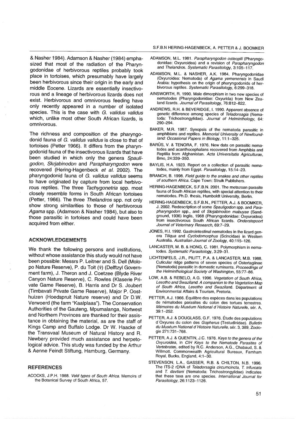
66 Onderstepoort Journal of Veterinary Research, 73: (2006) RESEARCH COMMUNICATION Eustrongylides sp. (Nematoda: Dioctophymatoidea) from the stomach of a Nile crocodile, Crocodylus niloticus Laurenti, 1768, in Botswana K. JUNKER 1, O. BAIN 2 and J. BOOMKER 1* ABSTRACT JUNKER, K., BAIN, O. & BOOMKER, J Eustrongylides sp. (Nematoda: Dioctophymatoidea) from the stomach of a Nile crocodile, Crocodylus niloticus Laurenti, 1768, in Botswana. Onderstepoort Journal of Veterinary Research, 73: During a study conducted between 2003 and 2005 on the diet of Nile crocodiles in Botswana, two young adult nematodes, one male and one female, belonging to the genus Eustrongylides Jägerskiöld, 1909 were recovered from the stomach contents of one of these animals. The caudal bursa of the male is present and the ejaculatory duct could be identified, but the spicule could not be seen. The vulva of the female has opened and the anus is situated on a terminal protruberance. Measurements and drawings of these specimens are provided, together with some data on the occurrence and lifecycles of members of the genus Eustrongylides in crocodilians world-wide and in African hosts in particular. Piscivorous birds are the usual final hosts of these nematodes. It is probable that the specimens described herein had developed in a paratenic fish host, and that the latter had been eaten by the crocodile. Keywords: Botswana, Crocodylus niloticus, Eustrongylides sp., Nile crocodile??? During a study conducted between 2003 and 2005 on the composition of the diet of Nile crocodiles, Crocodylus niloticus Laurenti, 1768, in Botswana, nematode parasites were collected from their stomach contents and identified (Junker, Wallace, Leslie & Boomker 2006). Two large young adult specimens of the genus Eustrongylides Jägerskiöld, 1909, one male and one female, were recovered from a single crocodile only (Fig. 1). The male is 110 mm long, 650 μm wide; posterior part with subterminal constriction; ventral precloacal 1 Department of Veterinary Tropical Diseases, University of Pretoria, Private Bag X04, Onderstepoort, 0110 South Africa 2 Muséum National d Histoire Naturelle, Parasitologie comparée et Modèles expérimentaux, USM 307, CP52, 61 rue Buffon, Paris, Cedex 05 France * Author to whom correspondence is to be directed. joop.boomker@up.ac.za Accepted for publication 18 May 2006 Editor Page 582 sucker; caudal bursa with peripheral cuticular ornamentation, similar to that described for the genus (Karmanova 1968; Measures 1988a); ejaculatory duct identified but not the spicule. The female is 122 mm long, μm wide; vulva opened, close to anus; anus on a flattened terminal protruberance, which is 35 μm high. The specimens are deposited at the Muséum National d Histoire Naturelle Paris, Access number 169 JW. This is a somewhat unusual finding since the genus Eustrongylides usually occurs in the wall of the proventriculus of its piscivorous avian final hosts and utilizes oligochaetes as intermediate hosts, in which it reaches the third larval stage (Anderson 2001). Fish subsequently serve as paratenic hosts in which the parasites reach the fourth stage and continue to grow. At this stage the reproductive system is highly developed (Measures 1988b; Anderson 2000). Coy- 315
67 Eustrongylides sp. (Nematoda: Dioctophymatoidea) from Crocodylus niloticus Laurenti, 1768, in Botswana A D C B E FIG. 1 Eustrongylides sp.. Caudal extremities of immature adults. A C Male. A: Ventral view. B: Left lateral view. C: Internal peripheral ornamentation of the caudal sucker. D E Female. D: Subventral view. E: Right lateral view. Note that the anus is situated on a terminal protruberance. Scales in μm: A, B = 125; C = 20; D, E = 75 ner, Spalding & Forrester (2002) found that fish could serve as intermediate as well as paratenic hosts for Eustrongylides ignotus Jägerskiöld, Only a few publications refer to Eustrongylides sp. from crocodilian hosts. Ladds & Sims (1990) found immature Eustrongylides sp., mm in length, free in the abdomen of two of 54 crocodiles in Papua New Guinea and Goldberg, Bursey & Aquino-Shuster (1991) report Eustrongylides sp. from the stomach contents of three of 115 wild-caught Caiman yacare (Daudin, 1802) in Paraguay. However, they could not determine whether the specimens of Eustrongylides they collected were recently released from intermediate host cysts and were likely to die, or whether they may have survived, with C. yacare becoming a paratenic host. Similarly, our data is insufficient to decide whether the presence of Eustrongylides sp. in the stomach of the crocodile is accidental or represents an unusual life-cycle. Without the benefit of further life-cycle studies, it would appear as if the crocodile had ingested an infected fish and that the parasites were released from their host during the digestive process or had actively started migrating upon the death of their host. Considering that the specimens were adults, albeit immature, one could also speculate that the crocodile had ingested an infected waterbird. However, no remains of feathers were found in the stomach contents of the crocodile (Kevin Wallace, personal communication date?). A substantial portion of the diet of Nile crocodiles, ranging from very small to large in size, is made up of fish, with aquatic birds also forming part of their diet (Ross 1989; Alderton 1999). It is thus not too surprising to find fish or bird parasites in a crocodile s stomach. Little information is available on the prevalence of Eustrongylides in African paratenic as well as final hosts, and despite Measures (1988a) revision of the genus, many taxonomic problems remain unsolved. She only confirmed three species, namely Eustrongylides tubifex (Nitzsch in Rudolphi, 1819) Jägerskiöld, 1909, Eustrongylides excisus Jägerskiöld, 1909 and 316 Page 583
68 K. JUNKER, O. BAIN & J. BOOMKER E. ignotus and declared nine species as species inquirendae. A 26.5 % prevalence of larvae of Eustrongylides africanus Jägerskiöld, 1909, one of the species considered doubtful by Measures (1988a), and which is only known by the female, has been reported in catfish, Clarias gariepinus (Burchell, 1822) as well as Cla rias anguillaris (Linnaeus, 1758), from the Bida floodplain of Nigeria (Ibiwoye, Balogun, Ogunsusi & Agbontale 2004). The latter authors indicate that fish can serve as intermediate, reservoir and as final hosts. Eustrongylides africanus has also been recorded as part of the parasite fauna of two of six marabou storks, Leptoptilus crumeniferus (Lesson, 1831), in Uganda (Moriearty, Pomeroy & Wanjala 1972) as well as in Ardea goliath Cretzschmar, 1829, Pelecanus rufescens Gmelin, 1789, and Anhinga melano gas ter (Daudin, 1802) in the Sudan (Measures 1988a). Eustrongylides sp. has been recovered from fish at Lake Tana, Ethiopia (Eshetu & Enyew 2003). Asanji (1990) examined C. gariepinus in Cameroon and found an overall prevalence of infection of 68.9 %. The latter author reports that cysts containing various larval stages were present in the muscles and visceral organs. While the above indicates that Eustrongylides sp. is indeed quite common and wide-spread on the African continent, it equally emphasizes the paucity of data available on this parasite in Africa. It is to be hoped that an effort will be made to rectify this and to clarify the uncertain systematic status of, amongst others, E. africanus. ACKNOWLEDGEMENTS The senior author is supported by a Claude Leon Foundation Post Doctoral Fellowship grant. REFERENCES ALDERTON, D Crocodiles and alligators of the world. London: Cassel plc. ANDERSON, R.C Nematode parasites of vertebrates, their development and transmission, 2 nd ed. Wallingford and New York: CABI Publishing. ASANJI, M.F First record of the prevalence and intensity of Eustrongylides spp. (Nematoda: Dioctophymatoidea) in the African catfish, Clarias gariepinus (Family Claridae), in Mezam Division, Cameroon. Bulletin of Animal Health and Production in Africa, 38: COYNER, D.F., SPALDING, M.G. & FORRESTER, D.J Epizootiology of Eustrongylides ignotus in Florida: distribution, density and natural infections in intermediate hosts. Journal of Wildlife Diseases, 38: ESHETU, Y. & ENYEW, M Parasites of fish at Lake Tana, Ethiopia. Sinet, Ethiopian Journal of Science, 26: GOLDBERG, S.R., BURSEY, C.R. & AQUINO-SHUSTER, A.L Gastric nematodes of the Paraguayan caiman, Caiman yacare (Alligatoridae). Journal of Parasitology, 77: IBIWOYE, T.I.I., BALOGUN, A.M., OGUNSUSI, R.A. & AGBON- TALE, J.J Determination of the infection densities of mudfish Eustrongylides in Clarias gariepinus and Clarias anguillaris from Bida floodplain of Nigeria. Journal of Applied Sci ence & Environmental Management, 8: JUNKER, K., WALLACE, K., LESLIE, A.J. & BOOMKER, J Gastric nematodes of Nile crocodiles, Crocodylus niloticus Lau renti, 1768, from the Okavango River, Botswana. Onderste poort Journal of Veterinary Research, 73: KARMANOVA, E.M [Principles of Nematology, edited by K.I. Skrjabin, Vol. XX. Dioctophymata of animals and man and the diseases caused by them] Moscow: Izdatelstvo Nauka. LADDS, P.W. & SIMS, L.D Diseases of young captive croc odiles in Papua New Guinea. Australian Veterinary Journal, 67: MEASURES, L.N. 1988a. Revision of the genus Eustrongylides Jägerskiöld, 1909 (Nematoda: Dioctophymatoidea) of piscivorous birds. Canadian Journal of Zoology, 66: MEASURES, L.N. 1988b. Epizootiology, pathology, and description of Eustrongylides tubifex (Nematoda: Dioctophymatoidea) in fish. Canadian Journal of Zoology, 66, MORIEARTY, P.L., POMEROY, D.E. & WANJALA, B Para sites of the marabou stork [(Leptoptilos crumeniferus (Lesson)] in Queen Elizabeth National Park, Uganda. East African Wildlife Journal, 10: ROSS, C.A. (Ed.) Crocodiles and alligators. London: Meere hurst Press. 317 Page 584
69 Page 585
70 Page 586
71 Page 587
72 Page 588
73 Page 589
74 Page 590
75 Page 591
76 Page 592
77 Page 593
78 Page 594
79 Page 595
80 Page 596
81 Page 597
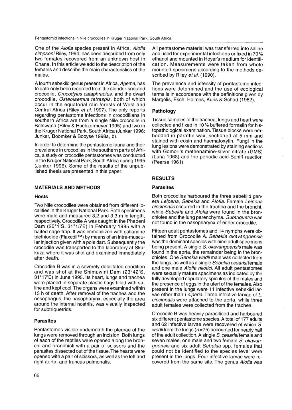
82 CHAPTER 2 Pentastomid parasites of reptiles Page 599
83 Page 601
84 Page 602
85 Page 603
86 Page 604
87 Page 605
88 Page 606
89 Page 607
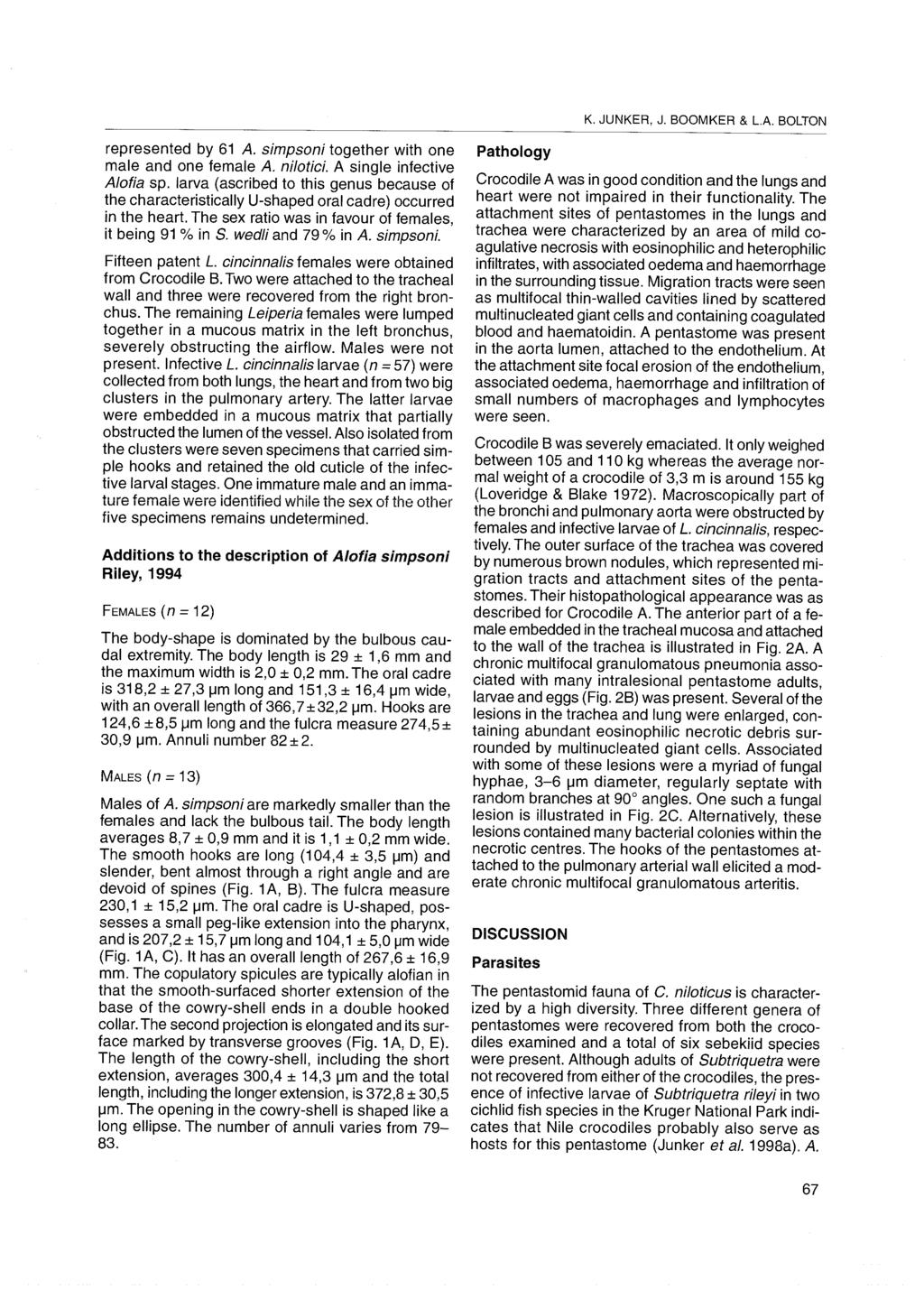
90 Systematic Parasitology 47: 29 41, Kluwer Academic Publishers. Printed in the Netherlands. 29 Leiperia cincinnalis Sambon, 1922 (Pentastomida) from Nile crocodiles Crocodylus niloticus in the Kruger National Park, South Africa, with a description of the male K. Junker 1,J.Boomker 2, D. Swanepoel 3 & H. Taraschewski 4 1 Zoologisches Institut der Universität Karlsruhe (TH), Abt. Ökologie/Parasitologie, Kaiserstrasse 12, Karlsruhe, Germany. Present address: Department of Veterinary Tropical Diseases, University of Pretoria, Private Bag X04, Onderstepoort, 0110 South Africa 2 Department of Veterinary Tropical Diseases, University of Pretoria, Private Bag X04, Onderstepoort, 0110 South Africa 3 South African National Parks, Private Bag X402, Skukuza, 1350 South Africa 4 Zoologisches Institut der Universität Karlsruhe (TH), Abt. Ökologie/Parasitologie, Kaiserstrasse 12, Karlsruhe, Germany Accepted for publication 7th September, 1999 Abstract A single male and several adult females of the pentastomid Leiperia cincinnalis were recovered from the trachea of five of six Nile crocodiles examined in 1995 and Infective larvae, pre-adult males and females, as well as mature males, occurred in clusters in the pulmonary artery but infective larvae and pre-adult females were also occasionally taken from the lungs. Irrespective of the developmental stage, the intensity of infection was 3, 6, 48, 72 and 79. Sixty-four percent of eggs recovered from the posterior part of the uterus of a patent L. cincinnalis female contained fully-developed primary larvae and these were used to infect 24 Mozambique bream Oreochromis mossambicus. Within a week of infection all the fish died and hatched primary larvae were recovered from the stomach and anterior part of the intestine. Eggs that had not hatched were found to be unsegmented. The total primary larval count in seven fish was 18, 12, 1, 25, 16, >40 and >50. Descriptions with detailed measurements are given of the females, the males, the eggs, the primary larvae and the infective larvae of L. cincinnalis. Introduction Leiperia cincinnalis Sambon, 1922 (syn. Reighardia cincinnalis Vaney & Sambon, 1910) (Pentastomida) is common in Nile crocodiles Crocodylus niloticus Laurenti on the African continent (Sambon, 1922; Heymons, 1940; Fain, 1961; Junker, 1996). Although known for a long time, little information was available on the life-cycle of L. cincinnalis and the descriptions given were often inadequate. Riley & Huchzermeyer (1996) re-assessed the genus Leiperia Sambon, 1922 and re-examined material of L. cincinnalis collected by various authors. None of the collections included mature males, but the morphology of the females, Author to whom correspondence is to be directed. nymphs and pre-adults were described in considerable detail (Riley & Huchzermeyer, 1996). In 1995 and 1998 experimental and field studies were conducted on pentastome infections of fish and crocodiles in the Kruger National Park (KNP) in South Africa (Junker, 1996; Junker, Boomker & Booyse, 1998a,b). Although the pentastome genera Alofia Giglioli, 1922 and Sebekia Sambon, 1922 were recovered from the crocodiles, in this paper we describe our findings and present additional data on the measurements and morphology of the females and the infective larvae of L. cincinnalis. The eggs and primary larvae are also described, and observations on the life-cycle are provided. The morphology of the males of L. cincinnalis is described for the first time. Page 608
91 30 Table 1. Collection data of Nile crocodiles from the Kruger National Park. Host number Date collected Locality Sex Length (m) Condition A/95 22/2/95 Phabeni Dam (25 1 S, E) M 3.2 good B/95 27/6/95 Shimuwini Dam (23 42 S, E) M 3.3 emaciated 1/98 10/6/98 Silwervis Dam (23 13 S, E) F 2.8 good 2/98 10/6/98 Silwervis Dam (23 13 S, E) M 2.4 good 3/98 10/6/98 Silwervis Dam (23 13 S, E) F 3.2 good 4/98 10/6/98 Silwervis Dam (23 13 S, E) F 2.7 good F, Female; M, Male. Materials and methods The collection data of the crocodiles examined are listed in Table 1. The crocodiles from the Phabeni and Silwervis Dams were caught in a baited cage and immobilised with gallamine triethiodide (Flaxedil TM ) before they were shot. The specimen from the Shimuwini Dam was killed with a single shot through the brain. The nasopharynx, trachea, lungs, heart and aorta of each crocodile were examined. After removal of the trachea and the oesophagus, the nasopharynx, especially the area just above the internal nostrils, was visually checked for pentastomids belonging to the genus Subtriquetra Sambon, The trachea was opened lengthwise and all Leiperia removed. The lungs were cut open along the bronchi and bronchioles using a pair of scissors and placed into trays containing phosphate buffered saline (PBS). Pentastomes were either removed directly from the lung tissue or collected from the PBS after migrating out of the lungs. The heart and its blood vessels were cut open and the chambers and inner surface of the blood vessels examined. For morphological studies pentastomes were fixed and stored in cold 70% ethyl alcohol, and later mounted and cleared in Hoyer s medium. Those pentastomes used for infection of the intermediate fish hosts were kept in PBS. Twenty-four Mozambique bream Oreochromis mossambicus Peters (50 70 mm long, obtained from a local breeder) were infected with eggs of L. cincinnalis from the crocodile from the Phabeni Dam. Eggs were collected from the posterior part of the uterus and concentrated in regular tap-water. In order to check the viability of the eggs, a drop of the egg-suspension was heated to 30 C and the number of hatched larvae was estimated. Groups of four bream each were placed into onelitre beakers supplied with air stones and filled with 600 ml of water. To each beaker a drop of the egg-suspension was added. The fish were infected overnight and all died after 6 8 d. Within a few hours of death the abdominal cavity of each fish was opened by ventral incision and rinsed over a 38 µm sieve. The stomach, intestine and swim-bladder of each were placed between two perspex slides exerting light pressure. These, as well as the residue on the sieve, were examined under a stereoscopic microscope. Results Leiperia cincinnalis is a common pentastome in the Nile crocodile and five of six crocodiles examined in this study harboured this parasite (Table 2 summarises the numbers and developmental stages recovered from the respective hosts). Three adult L. cincinnalis females were attached to the trachea and three infective larvae recovered from the pulmonary artery of Crocodile A/95 while Crocodile B/95 harboured 15 patent females in the trachea and bronchi. A total of 64 immature instars were recovered from the lungs, the heart and mainly the aorta pulmonalis, where the immatures occurred in two clusters. A single infective larva of L. cincinnalis was found in the lungs of Crocodile 1/98, and a cluster of 47 specimens, representing different developmental stages together with cast cuticles, were recovered from the aorta pulmonalis. Six of the Leiperia were fullydeveloped males. In two other specimens well sclerotised cirrus tips were visible, but the single hooks, the oral cadre and the copulatory spicules were hardly chitinised. Eleven of the parasites were infective nymphs carrying double hooks. Many of the specimens with Page 609

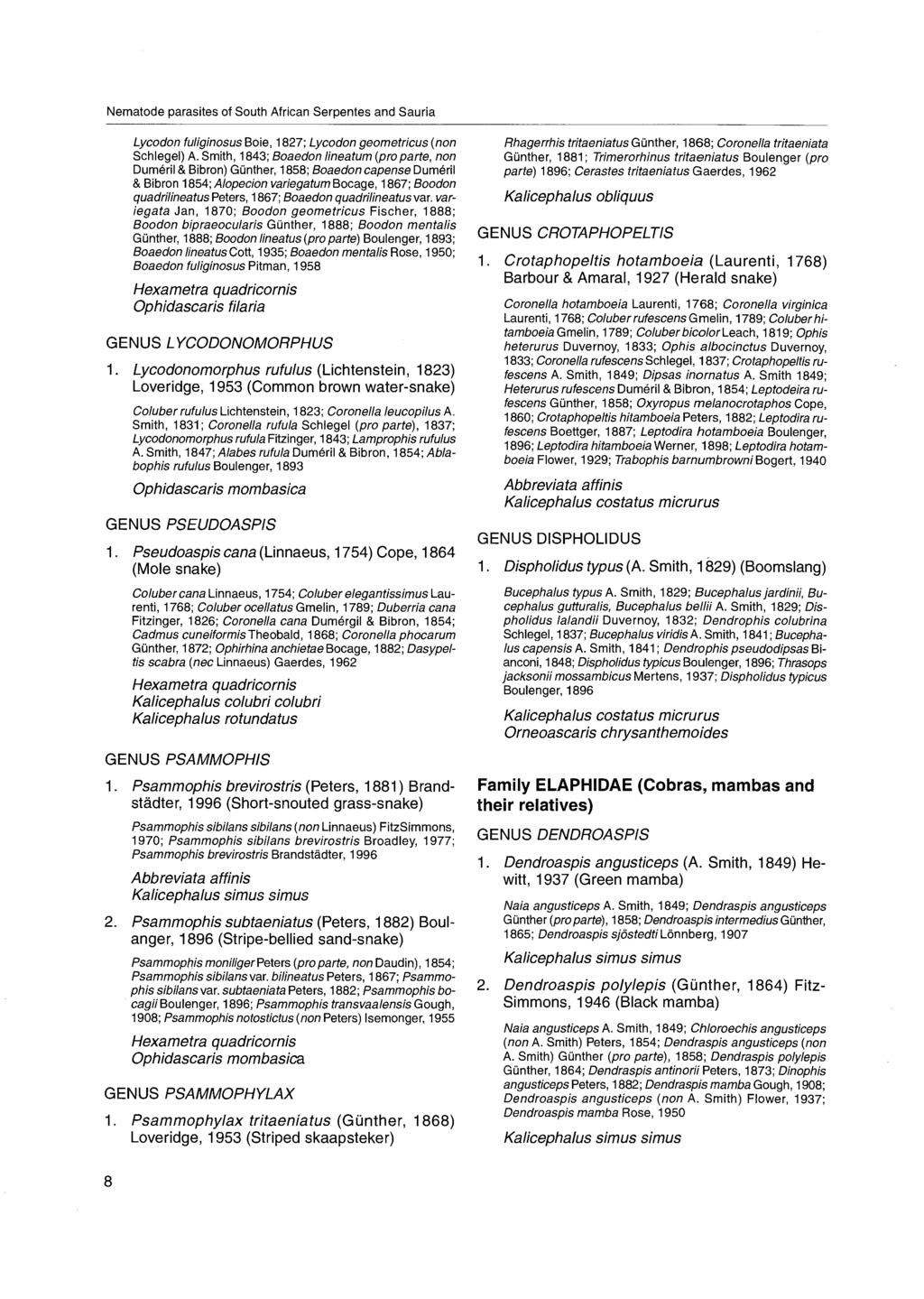
92 31 Figure 1. A. Mature females attached to a bronchus of the crocodile from the Shimuwini Dam, Kruger National Park. B. The right anterior hook and oral cadre of a mature female recovered from the same crocodile. The hook carries a distinct dorsal notch. C. Detail of the left hooks of another mature female from the Shimuwini crocodile, possessing a prominent dorsal notch. D. The anterior part of a male (No. 4.18/98) showing the hooks, the oral cadre and the copulatory spicules. The elaborate cirrus tips as well as the chitinised armoured tubes that form part of the cirri are visible furthest right. E. Detail of the left posterior and anterior hook of the male recovered from the trachea (No. 2.3/98). The hooks are flat-topped with only a slight indent in the dorsal margin. The anterior apodemes are lobe-like and permeated by pores (arrows). F. Oral cadre of specimen No. 2.3/98 showing the numerous pores around the pharynx as well as the large anterior flanges (arrows). Scale-bars: B,D, 400 µm; C, 200 µm; E, 100 µm; F, 50 µm. Page 610
 and accompanied by a strongly-chitinised scoop-like structure (lower arrow). B.")
93 32 Figure 2. A. Right copulatory spicule (No. 4.19/98). The collar around the spatulate extension is heavily chitinised and carries rings of chitin. The cirrus tip is flared (upper arrow) and accompanied by a strongly-chitinised scoop-like structure (lower arrow). B. The anterior part of an infective larva, showing the large double-hooks and the comparatively small oral cadre. C. Eggs of L. cincinnalis and two hatching primary larvae. D. Detail of a primary larva and split inner egg membrane. E. Posterior end of female (No. 2.1/98) found in copula showing the eversed vaginal lips (arrow). Part of the ruptured cirrus, visible as a transparent thread (left), is next to the abdomen. F. Cirrus tip of the male located in the vagina of No.2.1/98. Note the prominent chloride cell pore caps on the cuticula of the female. Scale-bars: A,C,F, 100 µm; B,E, 500 µm; D, 50 µm. Page 611
94 33 Table 2. The numbers and developmental stages of Leiperia cincinnalis recovered from Nile crocodiles in the Kruger National Park. Host number Developmental stage Total Females Males Pre-adults Infective larvae A/ B/ / / / / simple hooks were still surrounded by the old cuticula of the infective larva. Crocodile 2/98 harboured two female L. cincinnalis in the trachea and a single male was found attached next to one of the females. The cirrus tips were absent and the anterior part of the spatulate extension of both the copulatory spicules projected through the anterior genital opening. One of the cirrus tips was seen in the anterior third of the vagina of female No. 2.1/98 (Figure 2F) and the vaginal lips of the latter specimen were still everted (Figure 2E). The female was 4.5 cm long and 1 mm wide, and the abdomen was only just beginning to coil. No Leiperia were found in the pulmonary artery or the lungs. No L. cincinnalis were recovered from Crocodile 3/98, but Crocodile 4/98 harboured 72 specimens. Eleven females, ranging in body length from 4.5 to 10.5 cm, were attached to the trachea and the uteri of four, all of them less than 6 cm long, contained no eggs, but the latter were present in the remainder. Two prepatent Leiperia females were found in the lungs but were damaged in the recovery process. The remaining 59 specimens were collected from the pulmonary artery, where they formed a cluster similar to that found in Crocodile 1/98. A total of 15 males were isolated from this cluster; 10 of these were fully mature, but in the remainder the copulatory spicules were not yet fully developed. One of the mature males was still within the cuticula of the infective larva and the simple hooks of another preceding instar were visible. The sex of another 11 L. cincinnalis, with simple hooks, remained undetermined. In addition, 33 infective larvae were present. Six days after the experimental infection, two O. mossambicus were found dead and during the following two days all the fish died. Upon dissection, empty egg shells or the primary larvae themselves were found in the stomach and anterior part of the intestine. We also recovered intact pentastomid eggs, the majority of which were unsegmented. The primary larval count in seven fish was 18, 12, 1, 25, 16, >40 and >50. Leiperia cincinnalis Sambon, 1922 Description Females (Table 3) The general morphology of the females of L. cincinnalis examined in this study conformed largely to that described by Riley & Huchzermeyer (1996). Additional and comparative data are presented in Table 3. Every fully-mature, gravid female examined (n = 6) possessed hooks with a prominent dorsal notch (Figure 1B,C). Two females recovered from the trachea (No. 2.1/98 and No. 4.7/98, 5.5 cm long) had hooks marked by only a slight indentation in the dorsal surface. These specimens were considerably shorter and had not reached patency. The hooks, as well as measurements of the oral cadre of one specimen (No. 4.7/98), were slightly smaller than those of the patent females. An immature female from the aorta pulmonalis carried flat-topped hooks, while those of another specimen (No. 2.1/98) could not be measured. Males (Table 4) Well-fixed male specimens possessed a straight, cylindrical abdomen, tapering slightly to a rounded end. The male recovered from the trachea was 2.0 cm long while the body length of 3 males taken from the aorta ranged from 1.8 to 2.0 cm. Heavily chitinised fulcra supported the prominent hooks (Figure 1E). In comparison to the length of the hooks, the fulcra appeared rather broad and compact. Page 612
95 34 Table 3. The main characteristics of female Leiperia cincinnalis recovered from Nile crocodiles in the Kruger National Park in 1995 and For comparative purposes data of Riley & Huchzermeyer (1996) and Junker (1996) are included. All measurements are in micrometres unless otherwise indicated. Source Specimen number Number of annuli Body length (mm) Mouth dimensions Hook dimensions Overall length Cadre length Width Hook length Base length Hook depth Fulcrum length This paper 4.1/98 NM NM NM NM NM 4.3/98 NM NM NM 4.7/98 a NM /98 NM 95 NM NM NM /95 NM NM NM NM NM NM 2.2/95 NM NM NM NM NM NM Junker (1996) CWT2 NM 131 NM NM NM 354 NM NM 645 CWT NM NM 651 Lei NM 355 NM NM 703 Riley & Huchzermeyer (1996) NM NM NM NM NM NM NM NM NM NM NM NM NM 85? NM 640 a Uterus devoid of eggs. NM, Not measured. The term base length in this paper refers to the same structure as the term hook depth used by Riley & Huchzermeyer (1996). Page 613
96 Page 614 Table 4. The main characteristics of the males of Leiperia cincinnalis recovered from Nile crocodiles in the Kruger National Park. All specimens were recovered from the pulmonary artery (AP), excepting 2.3/98 from the trachea of Crocodile 2/98. For comparative purposes data of three immature males (1927.III ) examined by Riley & Huchzermeyer (1996) are included. All measurements are in micrometres. Specimen number Mouth dimensions Hook dimensions Copulatory spicules Overall length Cadre length Width Hook length Base length Hook depth Fulcrum length Total length Cowry shell length Width Length of armoured tube Number of grooves 4.18/ b 150b 90b 420b NM / NM NM NM NM NM / a 166a 87a 437a a a 4.21/ a 163a 90a 426b NM 115a / NM 240b 166b 78b 447b a 124a / a 161a NM 431c NM 115a / b 153b NM NM / a 145a NM 423c NM 133a / NM NM NM 432b NM / b 174b NM 407a NM 133a / NM NM NM 428b NM 637 NM 110a NM 1.17/98 NM NM NM 216a 152a 90a NM 791a 607 NM 104 NM 1.18/98 NM NM NM 255a 182a 90a 437a 805a 649a NM NM NM 1.19/ c 171c 87c NM NM / b 158b 93b NM 787a 616a NM 101a NM 1.6/ c 169c 82c 453a NM Average (AP) SD (AP) NM / c 81c NM NM NM 13a 1927.III d NM NM NM 290 NM NM NM NM NM NM NM NM NM 280 NM NM NM NM NM NM NM NM NM NM NM NM NM a Only one feature was measured. b Only two features were measured. c Only three features were measured. d After Riley & Huchzermeyer (1996). 35
97 36 The anterior apodeme of the hooks widened into a lobe-like structure permeated by numerous pores (Figure 1E). A certain degree of variability characterised the hook morphology of male L. cincinnalis (n = 17). In some specimens the hooks were flat-topped, in others only slight depressions were visible and in yet others, hooks had a distinct dorsal notch. Flat-topped hooks and hooks with a dorsal notch could at times be seen in the same specimen. The hooks and fulcra of the males were considerably shorter than those of the females and the measurements were actually nearer to those of the infective larvae (Junker, 1996). However, their gross morphology closely resembled that of females. This was true of the oral cadre with its characteristic Leiperia shape (Figure 1F). Starting in the posterior third of the oral cadre, the lateral prongs of the oral cadre merged gradually, giving it a V- shaped profile (Riley & Huchzermeyer, 1996). Both the large anterior flanges, as well as the area between the sclerotized supports of the pharynx, possessed the same pores as females (Riley & Huchzermeyer, 1996) (Figure 1B,F). The paired copulatory spicules were strongly chitinised and the spatulate anterior extension carried an average of 13 rings of chitin folds while the collar around the neck of the latter extension was marked by heavy chitinous rugosities (Figures 1D, 2A, 3A,B). The cirrus tip was a flattened trumpet of longitudinally-striated chitin (Riley & Huchzermeyer, 1996) (Figures 2A, 3A,C) and the whole structure was demarcated from the remaining unmodified section of the cirrus by a short armoured tube supported by rings of chitin, as described for Leiperia australiensis Riley & Huchzermeyer, 1996 by the latter authors. Parallel to the trumpet, and in close association, a second chitinous structure was visible (Figures 2A, 3C). This structure gave the impression of a scoop with serrated sides rolled up towards the mid-line. It was very similar in shape to the gubernaculum of Sambonia lohrmanni (Sambon 1910) Noc & Giglioli, 1922 (see Fain, 1961). While often separated under cover-slip pressure, in some specimens the trumpet ran through the scoop and the latter served as a support. Infective larvae (Table 5) The distinctly elongate and slender body, together with the rounded shape of the head, gave the infective larvae of L. cincinnalis a characteristic appearance, which sets it apart from other sebekiid larvae (Junker et al., 1998a). As detailed descriptions of the morphology of the infective larvae of L. cincinnalis are available (Junker, 1996; Riley & Huchzermeyer, 1996), we present only the main measurements of infective larvae examined in this study. Primary larvae The primary larvae (Figures 2C,D, 3D) have a distinct penetration apparatus located in front of the U-shaped mouth ring, in the anterior part of the body. It consisted of a lancet-shaped median stylet and 2 lateral stylets. The latter were split into a Y-shaped tip with 2 blades. Two pairs of stubby limbs were double-hooked and the tip of the tail carried 2 minute chitinous terminal thorns. Curled larvae measured 101 ± 7 µm (n= 11), but reached 122 ± 9 µm in length when the doublehooked tail was extended. The width of the primary larvae taken midway between the front and the hind pair of limbs averaged 60 ± 5 µm. Eggs The eggs of L. cincinnalis (Figure 2C,D) consisted of 2 membranes: a spherical outer shell, 233 ± 14 µmin diameter which surrounded a hyaline substance; and an ovoid inner membrane, 103±13 µm long and 80± 7 µm wide, which enclosed the primary larva. Of the eggs recovered from the posterior part of the uterus, 64% contained fully-developed primary larvae, of which 19% hatched when warmed to 30 C. When placed into a hypertonic sugar solution the eggs lost their spherical shape and shrivelled up. Depending on the time of exposure, eggs would swell up again when transferred back into regular tap-water. Primary larvae hatched in 2 stages. Using its penetration apparatus and the claws, the primary larva started tugging at the inner membrane until it broke. Subsequently, the larva stretched to its full length, unfolding its ventrally curved tail, and started random movements which split the outer egg membrane lengthwise, setting the larva free. Movements during the migration through host tissue were stereotyped, and fore and hind extremities were used alternately. While the anterior pair of limbs pulled towards the body, the posterior limbs moved away from the body in a downward motion. Subsequently, the hind limbs were brought back to the ventral side and the anterior limbs were spread widely. At the same time the tail alternately stretched and curled ventrally. Page 615
98 37 Figure 3. A. Right copulatory spicule and cirrus tip (No. 4.18/98). B. Left copulatory spicule (No. 2.3/98). C. Detail of cirrus tip (No. 4.27/98). D. Primary larva showing the penetration apparatus, the U-shaped oral cadre, the clawed limbs and the tail carrying two minute terminal thorns. Scale-bars: A-C, 100 µm; D, 25 µm. Page 616
99 38 Table 5. The main characteristics of the infective larvae of Leiperia cincinnalis recovered from Nile crocodiles from the Shimuwini Dam and from cichlids from the Phabeni Dam in the Kruger National Park in 1995 (LM19, LM20). For comparison additional data are included (Riley & Huchzermeyer, 1996; Junker, Boomker & Booyse, 1998a). All measurements are in micrometres unless otherwise indicated. Source Specimen number Number of annuli Body length (mm) Mouth dimensions Hook dimensions Overall length Cadre length Width Hook length Fulcrum length This paper CW CWA CWA NM NM NM CWA CWA CWA CWA CWH Average SD Junker, Boomker & Booyse (1998a) LM LM20 NM Riley & Huchzermeyer (1996) F F NM NM NM F NM, not measured. Page 617
100 39 Discussion Leiperia cincinnalis was the dominant pentastome species in the three crocodiles from the Silwervis Dam, but the intensity of infection was variable. Infective larvae and pre-adults recovered from the pulmonary artery accounted for most of these infections (Table 2). The development of L. cincinnalis in its final hosts has been largely speculative. Rodhain & Vuylsteke (1932) described tufts of double- as well as singlehooked larvae attached to the aorta of a crocodile from the Congo. Their reference to the genital apparatus being more developed in the single-hooked specimens suggested the presence of males, but, unfortunately, these authors did not describe them. Rodhain & Vuylsteke (1932) discussed the possibility of an obligatory developmental phase in the circulatory system, but thought it exceptional rather than the rule. That it is indeed a specific pattern of development within the genus of Leiperia has since been confirmed in L. australiensis and by this study. We recovered infective larvae, as well as young females, from the lungs. Thus the lungs are a route of migration for the infective larvae on their way to the cardio-vascular system and for the adults on their way to their attachment sites in the trachea. Considering the high numbers of infective larvae and young adults often encountered in the pulmonary artery, the question arises why comparatively few adults are found in the trachea. This is especially true for the males, which occur in substantial numbers in the cardio-vascular system, but hardly ever in the trachea or lung. The relatively shorter life-span of male versus female pentastomes may partly explain this (Riley & Self, 1980; Riley, 1986; Junker et al., 1998b). Whether the majority of pre-adults get lost during migration, or whether an immune reaction from the host reduces their number, will remain speculative until more extensive studies can be undertaken. A male and a female L. cincinnalis, recovered from one crocodile were attached next to each other in the trachea and probably had been in copula but were separated during recovery. We conclude that L. cincinnalis copulate in the trachea and not in the circulatory system; Riley & Huchzermeyer (1996) had already stated their doubts as to the likelihood of such a difficult procedure taking place in the narrow surroundings of the pulmonary artery. We have found males with single hooks and completely developed copulatory spicules still encased in the cuticle of the infective larva in the pulmonary artery and this supports the speculation of Riley & Huchzermeyer (1996) that only one moult is necessary for the infective larva to develop into a fully adult male. However, another fully mature male surrounded by both the cuticula of the infective larva and the cuticula of a preceding single-hooked stage indicates that the males of L. cincinnalis undergo at least one additional moult after having reached sexual maturity. Unlike moulting in females, moulting in the males does not seem to be related to any further growth. Specimen No. 4.18/98, in addition to the male recovered from the trachea, was well within the range of the measurements of the other males (Table 4). Once again, these data emphasise the variability of structures encountered when dealing with pentastomes as already stated by Riley & Huchzermeyer (1995). Riley & Huchzermeyer (1996) compared the hook morphology of females of L. cincinnalis from Uganda and the Congo. While both Ugandan specimens had flat-topped hooks, the hooks of the two Congo specimens, one of them described by Fain (1961), were characterised by a dorsal notch. Riley & Huchzermeyer (1996) speculated that the difference in hook morphology might indicate geographical variation, pointing out that the data set was too small to draw any final conclusions. Our collection comprises male and female Leiperia, some of which carry flat-topped hooks and some of which possess hooks with a dorsal notch; sometimes both hook types occur in the same specimen. Visibility of the dorsal notch in the hooks can be influenced by the orientation of the hooks under cover-slip pressure and, especially in females, it becomes more prominent as specimens mature and grow. The dorsal notch is generally more distinct in the females than in the males. Measurements of the males and females were relatively uniform, especially the dimensions of the copulatory spicules, and gave no indication of the presence of two species. Our findings suggest that all the specimens belong to L. cincinnalis. This, however, does not exclude the existence of a second Leiperia species in Africa, especially in view of the fact that the flat-topped hooks of the Ugandan specimens were considerably larger than the notched hooks of specimens collected from South Africa. The size of the hosts, as well as the intensity of infection, may influence the final size reached by the parasites, but our data do not support this speculation. Morphometric analysis of mature females taken from the heavily infected crocodile from the Shimuwini Dam, from the moderately infected Crocodile 4/98, and Page 618
101 40 from the lightly infected Crocodile 2/98 did not differ substantially. Unfortunately, the oral cadres of the Leiperia from Uganda could not be measured and no male specimens were available for comparison. In terms of gross morphology, the copulatory spicules of the L. cincinnalis males examined during this study and those of L. australiensis are very similar (Riley & Huchzermeyer, 1996). The cirrus tip in particular is very distinctive. The collar around the spatulate extension of the copulatory spicules of L. cincinnalis does not extend as far as the double collar found in L. australiensis. While the copulatory spicules of L. cincinnalis are larger than the ones of L. australiensis, the hook and mouth dimensions are quite similar. Fish have long been known to be the intermediate hosts for L. cincinnalis (Fain, 1961) and field studies indicate that the infection with pentastomes has no adverse effect on the development of the intermediate host (Junker et al., 1998a). We believe that two factors are responsible for the high mortality of the experimentally infected O. mossambicus. Thesefishhada fungal infection that was aggravated by stress experienced during the experimental infection. Furthermore, we are convinced that the damage caused by the tissue migration of the hatched primary larvae contributed substantially to the subsequent death of the fish. This was especially true where 10 or more primary larvae were present. It has been reported that porocephalid females shed a relatively high number of eggs per day, all of which contain fully-developed primary larvae (Riley, 1981, 1986). Junker et al. (1998b) recovered 3,400 eggs, 70% of which contained fully-developed, active primary larvae, from a single female Sebekia wedli Giglioli, 1922 that had been placed into phosphate buffered saline for one hour. While this figure is impressive, we are of the opinion that our experimental fish were exposed to a concentration of eggs that they would not normally encounter. Once the eggs have been ingested by a suitable host, hatching of primary larvae seems to be a highly successful process, as most of the eggs recovered intact from fish did not contain fully-developed larvae. The eggs of L. cincinnalis offer maximum protection for the primary larvae during the transition between the final and the intermediate host. Eggs are passed into the water via the faeces or sputum and depend entirely on chance ingestion by a bottom-feeding fish. The inner vitelline membrane is tough and it takes a considerable effort on the part of the primary larva to rupture it. Our findings suggest that the hyaline substance surrounding the inner shell protects the larva; however, the egg-shells are too thin to offer lasting protection from desiccation once removed from their natural aquatic environment. Several authors have shown that pentastome eggs are in fact very resistant to environmental influences. Salazar (1965) found a high resistance towards acids and preservatives. Bosch (1987) reported that eggs maintained in physiological solution at 4 8 C remained infective for more than three months and assumed that high temperatures and dehydration were the main factors in killing larvae. The eggs of L. cincinnalis began to hatch under cover-slip pressure when warmed to 30 Cand needed no additional stimuli. Increase in temperature has the same effect in the pentastomid genera Elenia Heymons, 1932 (Porocephalida) and Raillietiella Sambon, 1910 (Cephalobaenida) (Bosch, 1987), but in Reighardia sternae (Diesing, 1864) Ward, 1899 (Cephalobaenida) hatching occurred only at 40 C(Banaja, James & Riley, 1975). These data reflect the physiology of their respective hosts. R. sternae has a direct life-cycle in homoiothermic seabirds (Banaja et al., 1975), which maintain a constant bodytemperature of C (Eckert, 1993), whereas the intermediate hosts of Leiperia, Elenia and Raillietiella are heterothermic. The eggs of L. cincinnalis, as well as the hatching of the primary larva and subsequent movements during tissue migration, conform largely to those of Porocephalus crotali (Humboldt, 1808) Humboldt, 1811 of the Louisiana muskrat (Penn, 1942). It appears that the primary larvae of pentastomes are capable of generalised, very basic and stereotyped movements that are nevertheless sufficient for simple tissue migration from the alimentary tract into tissues. Acknowledgements We wish to thank the Board of Trustees, South African National Parks Board, for placing the animals at our disposal. This work was supported by a grant to the senior author from the Landesgraduiertenförderung, Baden-Württemberg, Germany. Page 619
102 41 References Banaja, A.A., James, J.L. & Riley, J. (1975) An experimental investigation of a direct life-cycle in Reighardia sternae (Diesing, 1864), a pentastomid parasite of the herring gull (Larus argentatus). Parasitology, 71, Bosch, H. (1987) Vergleichende experimentelle Untersuchungen zur Biologie der Pentastomidengattungen Raillietiella (Cephalobaenida) und Elenia (Porocephalida) unter Berücksichtigung der Erforschungsgeschichte der Pentastomiden. PhD Thesis, Faculty of Biology, University of Hohenheim. Eckert, R. (1993) Tierphysiologie. Stuttgart: Georg Thieme Verlag, Fain, A. (1961) Les Pentastomides de l Afrique Centrale. Annales de Musée Royale de l Afrique Centrale, Série8,92, Heymons, R. (1940) Beiträge zur Systematik der Pentastomiden. III. Pentastomiden mit spiralig gekrümmten Körperformen. Zeitschrift für Parasitenkunde, 2, Junker, K. (1996) Untersuchungen zur Biologie und Lebensweise der in Crocodilia parasitierenden Pentastomida in Südafrika. MSc Thesis, University Fridericiana of Karlsruhe. Junker, K., Boomker, J. & Booyse, D.G. (1998a) Pentastomid infections in cichlid fishes in the Kruger National Park and the description of the infective larva of Subtriquetra rileyi n. sp. Onderstepoort Journal of Veterinary Research, 65, Junker, K., Boomker, J. & Booyse, D.G. (1998b) Experimental studies on the life-cycle of Sebekia wedli (Pentastomida: Sebekidae). Onderstepoort Journal of Veterinary Research, 65, Penn, G.H. (1942) The life history of Porocephalus crotali, apar- asite of the Louisiana muskrat. Journal of Parasitology, 28, Riley, J. (1981) Some observations on the development of Porocephalus crotali (Pentastomida: Porocephalida) in the western diamondback rattlesnake (Crotalus atrox). International Journal for Parasitology, 11, Riley, J. (1986) The biology of pentastomids. Advances in Parasitology, 25, Riley, J. & Huchzermeyer, F.W. (1995) Description of four species of pentastomid parasites belonging to the genera Alofia Giglioli, 1922 and Sebekia Sambon, 1922, from a single Nile crocodile Crocodylus niloticus from Botswana. Systematic Parasitology, 31, Riley, J. & Huchzermeyer, F.W. (1996) A reassessment of the pentastomid genus Leiperia Sambon, 1922, with a description of a new species from both the Indopacific crocodile Crocodylus porosus and Johnston s crocodile C. johnsoni in Australia. Systematic Parasitology, 34, Riley, J. & Self, J.T. (1980) On the systematics and life-cycle of the pentastomid genus Kiricephalus Sambon, 1922 with descriptions of three new species. Systematic Parasitology, 1, Rodhain, J. & Vuylsteke, C. (1932) Contribution à l étude des Porocéphales des Crocodiles Africains. Révue de Zoologie et de Botanique Africaines, 23, Salazar, N.P. (1965) Human pentastomiasis in the Philippines, with notes on a Taiwan species. Acta Medica Philippina, Series 2, 1, Sambon, L.W. (1922) A synopsis of the family Linguatulidae. Journal of Tropical Medicine and Hygiene, 25, ; Page 620
103 Onderstepoort Journal of Veterinary Research, 69:53 59 Description of Pelonia africana n. g., n. sp. (Pentastomida: Sebekidae) from the lungs of Pelomedusa subrufa and Pelusios sinuatus (Chelonia) in South Africa K. JUNKER 1 and J. BOOMKER 2 * ABSTRACT JUNKER, K. & BOOMKER, J Description of Pelonia africana n. g., n. sp. (Pentastomida: Sebekidae) from the lungs of Pelomedusa subrufa and Pelusios sinuatus (Chelonia) in South Africa. Onderstepoort Journal of Veterinary Research, 69:53 59 The terrapins Pelomedusa subrufa and Pelusios sinuatus taken from a water reservoir in the Northern Province, South Africa, were examined for pentastome infections. Two immature specimens, a patent female and a mature male, were obtained from the lungs of four hosts, each of which harboured a single specimen. Based on the morphology of the specimens the new monospecific genus, Pelonia africana n. g., n. sp., is described. It is characterized by smooth, dorsally convex hooks with sharply bent blades. The oral cadre is more or less U-shaped. Delicate chitinous fibres, which can be difficult to see, connect the lateral prongs anteriorly. In this, as well as the morphology of the copulatory spicules, it is most like Sebekia wedli. The latter, however, possesses spinous hooks, which are absent in P. africana. The hooks are slightly and the copulatory spicules markedly larger in P. africana than in S. wedli. The lack of a double-hooked collar at the terminal end of the cowryshell shaped base of each copulatory spicule and the absence of a peg-like extension of the oral cadre into the oesophagus, distinguishes P. africana from members of the genus Alofia. The oral cadre of the South American species Diesingia megastoma, from aquatic chelonians, is more than twice the size than that of Pelonia and there is a distinct difference in shape. The hooks of the genus Diesingia are flat-topped, and both the anterior as well as the posterior fulcra carry cowllike extensions. The number of annuli, in D. megastoma and approximately 30 in P. africana, further separates the two genera. The most striking feature of Diesingia which sets it apart from Pelonia and the other genera of the family Sebekidae, is the configuration of its copulatory spicules. Pelonia and Diesingia share morphological features with all the other sebekiids, but it is the unique combination of diagnostic characters that separates the two genera from those, as well as from each other. Keywords: Chelonia, Pelomedusa, Pelonia africana, Pelusios, pentastomes, terrapins * Author to whom correspondence is to be directed 1 Zoologisches Institut der Universität Karlsruhe (TH), Abt. Ökologie/Parasitologie, Kaiserstrasse 12, Karlsruhe, Germany Present address: Department of Veterinary Tropical Diseases, University of Pretoria, Private Bag X04, Onderstepoort, 0110 South Africa 2 Department of Veterinary Tropical Diseases, University of Pretoria, Private Bag X04, Onderstepoort, 0110 South Africa Accepted for publication 23 January 2002 Editor INTRODUCTION The majority of the six genera comprising the family Sebekidae Fain, 1961 occur exclusively in crocodilians. However, it has been speculated that a single species of the genus Sebekia Sambon, 1922 may also reach maturity in piscivorous turtles (Dukes, Shealy & Rogers 1971). Until now, only the South American genus Diesingia Sambon, 1922 has been known to be exclusive to a chelonian definitive host (Sambon 1922; Heymons 1941; Over- 53 Page 621
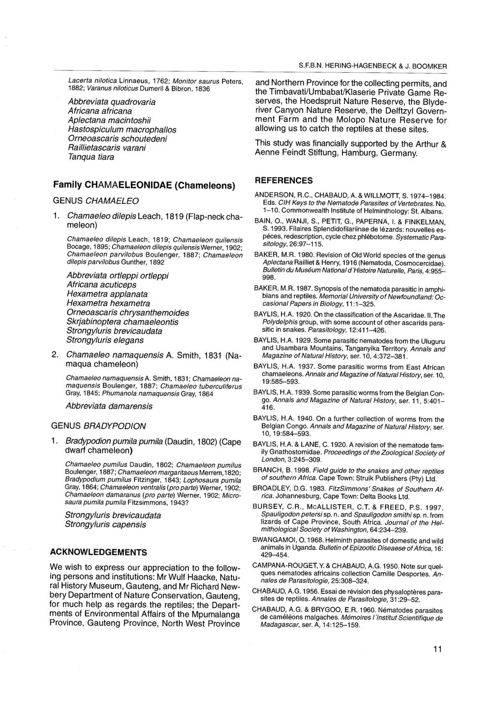
104 Pelonia africana n. g., n. sp. (Pentastomida: Sebekidae) in South Africa street, Self & Vliet 1985; Riley 1994) and, generally speaking, information on the pentastome fauna of tortoises, terrapins and turtles is scarce. Fain (1961) refers to a nymphal pentastome found encysted in the liver of Kachuga lineata, a semiaquatic oriental tortoise, which Hett (1924) assumed to be the infective larva of Subtriquetra megacephala (Baird, 1853) Sambon, The latter genus belongs to the family Subtriquetridae Fain, 1961, which is also believed to be exclusive to crocodilians (Riley 1986; Winch & Riley 1986; Junker, Boomker & Booyse 1998a). In addition, some five genera of terrapins from North America have been reported to harbour nymphs of Sebekia mississippiensis Overstreet, Self & Vliet, 1985, a crocodilian pentastome described from the American alligator, Alligator mississippiensis (Dukes et al. 1971; Boyce 1985; Overstreet et al. 1985). Significantly, all the literature cited above pertains to nymphal developmental stages of pentastomes. It would appear that Diesingia megastoma (Diesing, 1836) (Sambon, 1922) is currently the only pentastome of which mature specimens have been recovered from the chelonian hosts, Hydromedusa tectifera and Phrynops geoffroanus (= Hydraspis geoffroyana) from Brazil (Diesing 1836; Heymons 1941; Self & Rego 1985; Da Fonseca & Ruiz 1956). The authors are not aware of any publications dealing with adult pentastomid parasites of chelonians from any other continent. In this paper a pentastome from the lungs of two South African terrapins, Pelomedusa subrufa and Pelusios sinuatus, is described. P. subrufa occurs in pans, marshy areas and slow moving waters throughout southern Africa, and P. sinuatus inhabits large rivers and pans in the north-east of southern Africa. Both terrapins are omnivorous and fish form part of their diet (Patterson 1991). Pelonia africana n. g, n. sp. shares morphological similarities with all the other genera of the family Sebekidae, but nevertheless possesses a unique combination of diagnostic criteria. Slide mounted specimens of D. megastoma were re-examined and found to be distinctly different from the pentastomes recovered from the South African terrapins. We thus consider it appropriate to erect a new genus to accommodate these specimens. MATERIAL AND METHODS In 2000 five P. sinuatus (host numbers Psin1 5) and a single P. subrufa (host number Psub1), with carapace lengths varying from cm, were obtained from pans or marshy areas near the Arabie Dam, Northern Province. This dam is fed mainly by the Olifants River, but the Elands, Moses and Motsiphiri Rivers also feed into it (A. Hoffman, personal communication 2000). Terrapins were either killed by intraperitoneal injection with sodium pentobarbitone (Eutha-naze ) or decapitated. The plastron and carapace were removed and the trachea, as well as the nasopharynx, were examined for pentastomes. The liver and heart were transferred into separate vials containing tap water, and the soft and delicate lungs were placed into a Petri dish, also containing tap water. Pentastomes were either dissected from the organs or collected from the tap water after they had migrated out of the organs. For morphological studies, pentastomes were fixed and preserved in 70 % ethanol and subsequently mounted in Hoyer s medium. Three more pentastomid specimens, WIII/1, and Psub2/1 and Psub3/1 from the lungs of P. sinuatus and P. subrufa respectively, were collected during another unrelated study at the same locality and made available to us. RESULTS The nasopharynx and trachea of all hosts examined were free of pentastomes and hosts number Psin1, 3, 4 and 5 and Psub1 harboured no pentastomes at all. Single specimens of P. africana were recovered from the lungs of each of the remaining hosts. Premature females, without eggs in the uterus, were obtained from hosts WIII and Psin2. A gravid female collected from Psub2 contained eggs with fully developed primary larvae. A mature male was present in the lungs of Psub3. Description of Pelonia africana n. g., n. sp. (Table 1) TYPE HOSTS AND LOCALITY Pelusios sinuatus and Pelomedusa subrufa from the Arabie Dam (24 53 S, E), Northern Province, South Africa. TYPE MATERIAL Holotype male, no. T 2186 from Pelomedusa subrufa, allotype female, no. T 2187 from Pelomedusa subrufa and paratypes (immature) from Pelomedusa subrufa and Pelusios sinuatus, no. T All spec- 54 Page 622
105 TABLE 1 Comparative measurements of Pelonia africana n. g., n. sp., Diesingia megastoma and Sebekia wedli. All measurements are given in micrometres unless otherwise stated Body Number Specimen number length of (mm) annuli Mouth dimensions Hook dimensions Copulatory spicules Overall Cadre Width Hook Fulcrum Total Cowry shell Width length length length length length length WIII/1 (Paratype F, T 2188) NM NM NA NA NA Psin2/1 (Paratype F, T 2188) NA NA NA Psub2/1 (Allotype F, T 2187) 27 30a NM NM NA NA NA Psub3/1 (Holotype M, T 2186) NM NM Diesingia megastoma M (After Heymons 1941) 7 70 NM NM NM NM NM NM Diesingia megastoma M (After Heymons 1941) 6 70 NM NM NM NM NM Diesingia megastoma F (After Self & Rego 1985) NM * 520 NA NA NA Sebekia wedli F (After Riley & Huchzermeyer 1995a) NC NA NA NA Sebekia wedli M (After Riley & Huchzermeyer 1995a) 8 NC F Female M Male NA Not applicable NC Not counted NM Not measured * Only the length of the blade was measured K. JUNKER & J. BOOMKER 55 Page 623
106 Pelonia africana n. g., n. sp. (Pentastomida: Sebekidae) in South Africa imens are mounted in Hoyer s medium and deposited in the National Animal Helminth Collection, ARC-OVI, Onderstepoort, South Africa. ETYMOLOGY Pelonia has been named after its two host species that belong to the family Pelomedusidae which comprises freshwater chelonians from Africa, Madagascar and southern Australia. DESCRIPTION The body is claviform, the abdomen being widest in the anterior third and tapering to a bluntly rounded caudal tip. Ventrally the small trapezoid cephalothorax is continuous with the ventrally flattened abdomen but dorsally demarcated from the remainder of the body. A small number of wide annuli are present. FEMALE The strongly chitinised oral cadre is more or less U- shaped, the gap between the lateral prongs only slightly narrowing anteriorly. Muscle contraction or the amount of pressure applied when mounting, can result in a more ovoid profile. The oral cadre appeared to be open anteriorly as the delicate chitinous fibres connecting the two sides were difficult to see. A heavily chitinised, bowl-shaped base unites the two lateral prongs posteriorly (Fig. 1B, C). The oral cadre of the allotype female was slightly larger than that of the two immature specimens, WIII/1 and Psin2/1. The smooth hooks are dorsally convex with a slight dorsal notch where the strongly curved blade emerges from the base (Fig. 1F). The configurations of the posterior and anterior hooks appear to be identical and are supported by strong fulcra. Unfortunately, measurements could only be made from a single hook from an immature female. It was not possible to decide whether the females were heterogynous, with the utero-vaginal pore being situated one or two annuli anteriorly from the anus, or ophistogynous. The eggs of P. africana consist of a spherical outer membrane, 183 ± 8 mm in diameter that surrounds a hyaline substance and an ovoid inner eggshell, 96 ± 7 µm long and 70 ± 4 µm wide (Fig. 1G), that encloses the primary larva. MALE Although the oral cadre of the male is slightly smaller than those of the two immature females (Fig. 1A), its general morphology, as well as that of the hooks and the fulcra, is similar to that of the females. The paired copulatory spicules are heavily sclerotised and cowry-shell shaped (Fig. 1D). The anterior spatulate extension carries rows of rounded, chitinous teeth, which become progressively indistinct towards the tip (Fig. 1E). DISCUSSION The body-shape of P. africana corresponds closely to the illustration of a mature female of D. megastoma from H. tectifera (Self & Rego 1985). One of the main differences between P. africana and D. megastoma lies in the oral cadre. That of D. megastoma is more than twice the length and width than that of the African species. Own observations show the oral cadre of Diesingia to carry a small peg-like extension into the oesophagus, not unlike that of the genus Alofia Giglioli, 1922, which is absent in specimens of Pelonia. Furthermore, the prominent bowl-shaped chitinous structure at the base of the oral cadre of the latter genus is absent in Diesingia. Both genera of chelonian pentastomes possess smooth hooks. However, the fulcra of Pelonia are devoid of any extensions, while the fulcra of Diesingia are furnished with cowl-like extensions, similar to those seen in the genus Selfia (Riley 1994). The hooks of D. megastoma appear to be flattopped with a sharply curved blade, demarcated from the shank by a single notch (Self & Rego 1985), while those of P. africana are dorsally convex and marked by a slight dorsal notch. So far, no conclusive description of the copulatory spicules of Diesingia has been given, and the two male specimens we examined, possess copulatory spicules that are unique among the members of the family Sebekidae. The cowry-shell shaped base and the long spatulate extension of the open side are reminiscent of other sebekiid genera (Riley, Spratt & Winch 1990), but the shorter of the two anterior extensions has been transformed into a tiller-like, chitinous spike. The genus Pelonia is distinct from the genus Diesingia, and represents the first record of a new genus of pentastomes exclusive to chelonian final hosts from the African continent. Pelonia africana is similar to the African crocodilian pentastome Sebekia wedli Giglioli, 1922 in Sambon, This is especially true for the oral cadre, which in the latter species is also approximately U- 56 Page 624
. Scale bar: 50 mm Oral cadre of the allotype female.")
107 K. JUNKER & J. BOOMKER A B C D E F G FIG. 1 Pelonia africana n. g., n. sp. A B C D E F G Oral cadre of holotype male. The delicate chitinous fibres connecting the lateral prongs of the oral cadre anteriorly are partly visible (arrow). Scale bar: 50 mm Oral cadre of the allotype female. The anterior chitinous bridge is not visible in this photograph. Scale bar: 50 mm Oral cadre of an immature female. The chitinous fibres connecting the lateral prongs are clearly visible (arrow). Scale bar: 25 mm Ventral view of the right copulatory spicule of the holotype male. It is obpyriform and the spatulate extension carries small chitinous teeth. The arrow marks a chitinous part of the cirrus. Scale bar: 50 mm Detail of the left copulatory spicule Right posterior hook of an immature female. Scale bar: 50 mm Egg with fully developed primary larva. Scale bar: 50 mm 57 Page 625
108 Pelonia africana n. g., n. sp. (Pentastomida: Sebekidae) in South Africa shaped, and because of an almost invisible, very delicate anterior bridge of chitin, it seems to be open anteriorly (Riley & Huchzermeyer 1995a). A comparison of measurements, however, shows the buccal complex of S. wedli to be slightly smaller than that of P. africana (Riley & Huchzermeyer 1995a, Junker, Boomker & Booyse 1998b). The copulatory spicules of male P. africana are strongly reminiscent of S. wedli, and they could easily be confused, in that both are obpyriform and carry chitinous teeth on the spatulate extensions. Nevertheless, the spicules of P. africana are markedly larger than those of S. wedli (Riley & Huchzermeyer 1995a). The main distinguishing character between P. africana and S. wedli is the absence of the prominent spines on the dorsal hook surface. The lack of anterior extensions to the fulcra further serves to separate Pelonia from the other species of the genus Sebekia as defined by Riley et al. (1990). Superficially, the aspinose hooks, the curve of the blade and the shape of the oral cadre might lead to confusion of P. africana with the Alofia spp. The copulatory spicules of P. africana, however, lack the double-hooked collar diagnostic for Alofia and the genus Selfia Riley, 1994 (Riley 1994). Moreover, the oral cadre neither possesses the distinct, open Alofian U-shape nor the peg-like extension into the oesophagus (Riley & Huchzermeyer 1995a, b; Junker, Boomker & Bolton 1999). Recently Agema Riley, Hill & Huchzermeyer, 1997, a new pentastomid genus, has been described from African dwarf crocodiles, Osteolaemus tetraspis osborni, and slender-snouted crocodiles, Crocodylus cataphractus (Riley, Hill & Huchzermeyer 1997). While the hooks of P. africana exhibit the already mentioned abrupt right-angle bend near the base, those of Agema are very smoothly curved and the ovoid oral cadre of the latter genus is closed anteriorly by prominent chitinous crescents (Riley et al.1997). Pelonia africana morphologically resembles especially the genus Sebekia and to a lesser extent the genus Alofia. Heymons (1941) pointed out the similarity between D. megastoma and its South American sebekian and alofian counterparts. Therefore there is a strong case for the inclusion of the genera Pelonia and Diesingia into the family Sebekidae, as was suggested for the latter genus by Riley (1993). The fact that all sebekiid genera have a similar life-cycle using fish as intermediate hosts and semi-aquatic definitive hosts (Fain 1961; Riley 1986, 1994; Riley et al. 1997) indicates a close relationship between the two genera parasitising chelonians and those of crocodilians. ACKNOWLEDGEMENTS We wish to thank the Department of Agriculture, Land & Environment, Northern Province, for placing the animals at our disposal and Mr A.C. Hoffman, Department of Agriculture, Conservation and Environment, Mpumalanga, for collecting the hosts. Mr D. Hoffman has generously collected and provided the majority of specimens for this study. Our sincere thanks go to Dr J. Riley for letting us examine his collection of specimens of D. megastoma. This work was supported by a grant to the senior author from the Landesgraduiertenförderung, Baden-Württemberg, Germany and the Deutscher Akademischer Austausch Dienst, Germany. REFERENCES BOYCE, W.M The prevalence of Sebekia mississippiensis (Pentastomida) in American alligators (Alligator mississippiensis) in North Florida and experimental infection of paratenic hosts. Proceedings of the Helminthological Society of Washington, 52: DA FONSECA, F. & RUIZ, J.M Was ist eigentlich Pentastoma megastomum Diesing, 1836? (Porocephalida, Porocephalidae). Senckenbergiana Biologica, 37: DIESING, K.M Versuch der Monographie der Gattung Pentastoma. Annalen des Wiener Museums der Naturgeschichte, 1:1 32. DUKES, G.H., SHEALY, R.M. & ROGERS, W.A Sebekia oxycephala (Pentastomida) in largemouth bass from Lake St. John, Concordia Parish, Louisiana. Journal of Parasitology, 57:1028. FAIN, A Les Pentastomides de L Afrique Centrale. Annales de Musée Royale de l Afrique Centrale, Série 8, 92: HEYMONS, R Beiträge zur Systematik der Pentastomiden VI. Die Typenexemplare von Diesingia megastoma. Zeitschrift für Parasitenkunde, 12: HETT, M.L On the family Linguatulidae. Proceedings of the Zoological Society of London, 1924: JUNKER, K., BOOMKER, J. & BOOYSE, D.G. 1998a. Pentastomid infections in cichlid fishes in the Kruger National Park, South Africa. Onderstepoort Journal of Veterinary Research, 65: JUNKER, K., BOOMKER, J. & BOOYSE, D.G. 1998b. Experimental studies on the life-cycle of Sebekia wedli (Pentastomida: Sebekidae). Onderstepoort Journal of Veterinary Research, 65: JUNKER, K., BOOMKER, J. & BOLTON, L Pentastomid infections in the Nile crocodile (Crocodylus niloticus) in the Kruger National Park, South Africa, with a description of the males of Alofia simpsoni. Onderstepoort Journal of Veterinary Research, 66: Page 626
109 K. JUNKER & J. BOOMKER OVERSTREET, R.M., SELF, J.T. & VLIET, K.A The pentastomid Sebekia mississippiensis sp. n. in the American alligator and other hosts. Proceedings of the Helminthological Society of Washington, 52: PATTERSON, R Snakes and other reptiles of southern Africa. Cape Town: Struik Publishers. RILEY, J The biology of pentastomids. Advances in Parasitology, 25: RILEY, J Pentastomida, in Reproductive Biology of Invertebrates, Vol. 6, Part B, edited by K.G. & Rita G. Adiyodi. New Delhi: Oxford & IBH Publishing. RILEY, J A revision of the genus Alofia Giglioli, 1922 and a description of a new monotypic genus, Selfia: two genera of pentastomid parasites (Porocephalida: Sebekidae) inhabiting the bronchioles of the marine crocodile Crocodylus porosus and other crocodilians. Systematic Parasitology, 29: RILEY, J., HILL, G.F. & HUCHZERMEYER, F.W A description of Agema, a monotypic pentastomid genus from the lungs of the African dwarf and slender-snouted crocodiles. Systematic Parasitology, 37: RILEY, J. & HUCHZERMEYER, F.W. 1995a. Description of four species of pentastomid parasites belonging to the genera Alofia Giglioli, 1922 and Sebekia Sambon, 1922, from a single Nile crocodile Crocodylus niloticus from Botswana. Systematic Parasitology, 31: RILEY, J. & HUCHZERMEYER, F.W. 1995b. Pentastomid parasites of the family Sebekidae Fain, 1961 in West African dwarf crocodiles Osteolaemus tetraspis Cope, 1851 from the Congo, with a description of Alofia parva n. sp.. Onderstepoort Journal of Veterinary Research, 62: RILEY, J., SPRATT, D.M. & WINCH, J.M A revision of the genus Sebekia Sambon, 1922 (Pentastomida) from crocodilians with descriptions of five new species. Systematic Parasitology, 16:1 25. SAMBON, L.W A synopsis of the family Linguatulidae. Journal of Tropical Medicine and Hygiene, 25: ; SELF, J.T. & REGO, A.A Reassessments and revisions of certain genera and species of the family Sebekidae (Pentastomida) including description of Sebekia microhamus n. sp.. Systematic Parasitology, 7: WINCH, J.M. & RILEY, J Studies on the behaviour, and development in fish, of Subtriquetra subtriquetra: a uniquely free-living pentastomid larva from a crocodilian. Parasitology, 93: Page 627
110 Systematic Parasitology 56: , Kluwer Academic Publishers. Printed in the Netherlands. 211 Redescription of Diesingia megastoma (Diesing, 1836) Sambon, 1922, a pentastomid parasite from the South American terrapin Hydromedusa tectifera Cope K. Junker 1,2,J.Riley 3 & J. Boomker 4, 1 Zoologisches Institut der Universität Karlsruhe (TH), Abt. Ökologie/Parasitologie, Kaiserstrasse 12, Karlsruhe, Germany 2 Present address: Department of Veterinary Tropical Diseases, University of Pretoria, Private Bag, X04, Onderstepoort, 0110 South Africa 3 Department of Biological Sciences, University of Dundee, Dundee DD14HN, UK 4 Department of Veterinary Tropical Diseases, University of Pretoria, Private Bag, X04, Onderstepoort, 0110 South Africa Accepted for publication 29th May, 2003 Abstract Slide-mounted material of the pentastomid parasite Diesingia megastoma (Diesing, 1836) Sambon, 1922 from the South American chelonian Hydromedusa tectifera Cope is reviewed and the perfunctory, often omissive, description of the species is amended. The strong morphological similarities between D. megastoma and the crocodilian and chelonian pentastome genera of the family Sebekiidae Sambon, 1922, Alofia Giglioli, 1922, Selfia Riley, 1994, Sebekia Sambon, 1922, Agema Riley, Hill & Huchzermeyer, 1997, Leiperia Sambon, 1922 and Pelonia Junker & Boomker, 2002, clearly place Diesingia Sambon, 1922 within the same family. However, the unique combination of its main diagnostic criteria makes Diesingia a distinct genus. The absence of an elaborate, flared cirrus-tip in D. megastoma distinguishes it from Leiperia, while emphasizing its similarity to the remaining genera mentioned above. D. megastoma resembles Alofia in that it possesses smooth, flat-topped hooks and an anteriorly open oral cadre with an oesophageal peg. The copulatory spicules of Diesingia, however, lack the double-hooked collar, typical for Alofia and Selfia. Unlike the peg-like extension of the fulcra of the hooks of Sebekia, thatof D. megastoma is cowl-like and carries spines only on the anterior fulcra. Moreover, the hooks of Sebekia are usually convex and spinose and the ovoid oral cadre is closed anteriorly. Diesingia differs from Pelonia through the latter s smooth but dorsally convex and extension-free hooks. The copulatory spicules of Pelonia and Agema are reminiscent of the basic build found in Sebekia, whereas in D. megastoma the short, ventral extension of the cowry shell-shaped base of the copulatory spicules has been transformed into a structure resembling the collembolan fulcrum. The latter is connected to the base via a joint, a configuration which is unique in the Sebekiidae. Introduction Despite their discovery more than one-and-a-half centuries ago, the present knowledge on pentastomes parasitising chelonian final hosts is scant. A single species, Diesingia megastoma (Diesing, 1836) Sambon, 1922, from South American terrapins was recognised. It was first described from two adult males recovered from the lungs of a Geoffrey s side- Author for correspondence. necked turtle Phrynops geoffroyanus (Schweigger) (syn. Hydraspis geoffroana) in Brazil by Diesing as Pentastoma megastomum Diesing, 1836 (Diesing, 1836; Sambon, 1922; Fain, 1961). Since then, it has caused much confusion. The two original specimens, kept at the Museum of Natural History at Vienna, have been renamed and re-examined by several authors. Leuckart (1860), referring to Diesing s (1836) description only, changed the name to Pentastomum megastomum, while Shipley (1898) ex- Page 628
111 212 amined the Vienna specimens himself. Without adding much detail to the initial, very superficial description, Shipley (1898) redescribed them as Porocephalus megastomus, a name previously given to them by Stiles (1893, in Sambon 1922) in accordance with the then common trend to include the majority of the exotic pentastomes in Porocephalus Humboldt, 1811 (Heymons, 1941). When Sambon (1922) reviewed the known pentastomid genera, Diesing s type-specimens no longer fitted the diagnostic criteria of the genus Porocephalus and he suggested the inclusion of P. megastomum in the new genus Diesingia Sambon, This genus had been created to accommodate D. kachugensis Sambon, 1922 from the liver of Kachuga kachuga (Gray) (syn. K. lineata), a semi-aquatic oriental tortoise. Riley (1986) pointed out that Hett (1924, in Fain, 1961) considered D. kachugensis to be the larval stage of Subtriquetra megacephala (Baird, 1835) Sambon, 1922, a crocodilian pentastome. Heymons (1941) was the first author to provide a more detailed description of the type-specimens for which the name D. megastoma had become accepted, and included measurements and drawings of the hooks and the oral cadre. Da Fonseca & Ruiz (1956) created Butantanella Da Fonseca & Ruiz, 1956 to accommodate D. megastoma and the specimens of a collection of pentastomes recovered from the lungs of 15 Hydromedusa tectifera Cope from Brazil. Self & Rego (1985) examined pentastome specimens from collections from the Instituto Oswaldo Cruz and from the British Museum (Natural History) Collection. These included fully mature males and females from the lungs of the Brazilian terrapins, H. tectifera and P. geoffroanus,andself®o (1985) concluded that these were identical with Diesing s specimens. On morphological grounds, Self & Rego (1985) reclassified D. megastoma as Sebekia megastoma, dismissing Da Fonseca & Ruiz s (1956) previous classification. The uncertainty surrounding its type-species led to considerable confusion as regards the systematic status of the genus Diesingia. Fain (1961) included it in the family Sebekiidae Sambon, 1922, whereas Riley (1983), in an outline classification of the pentastomes, placed the genus separately, directly following the genera of the family Sambonidae Fain, 1961, substituting the family name by a question mark. Subsequently, Self & Rego (1985) abandoned Diesingia in favour of Sebekia Sambon, 1922, a view that was not adopted by Riley (1986), who created the monogeneric family Diesingiidae Riley, In a later publication, Riley (1994) placed Diesingia back in the family Sebekiidae. In this paper slide-mounted specimens of D. megastoma, collected from H. tectifera from Brazil, are described. Based on our findings, we support the validity of the genus Diesingia as well as its inclusion in the family Sebekiidae. Materials and methods All specimens described here were originally recovered from H. tectifera from Brazil by an unknown collector. The collection comprises slide-mounted specimens as well as specimens that had previously been fixed in formalin (Da Fonseca & Ruiz, 1956), but are now preserved in alcohol. These are, however, in an extremely poor condition and very brittle. The anterior and posterior hooks had been removed and mounted in Hoyer s medium several years ago and the bottle with alcohol specimens now contains only the abdomens of the specimens F2463-5, F and KI- 3. These are mature females, as evidenced by hook measurements and the egg-filled uteri of two specimens. The D. megastoma material studied comprises the following: F A male specimen, mounted whole and stained with a chromatin stain. The slide was originally labelled Butantanella megastoma, from the Instituto do Butantan ex Hydromedusa tectifera (see below). F A young female, mounted whole and originally identified as Butantan F 5960, Butantanella megastoma of the lung of H. tectifera, from the Paraná State, Brazil. The slide is dated 7 December, 1951 and carries the names Da Fonseca & Ruiz. We conclude that F and F are two of the specimens on which Da Fonseca & Ruiz (1956) based their description of B. megastoma. In fact, Da Fonseca & Ruiz (1956) stated that specimen 5960 forms part of a collection of male and female pentastomes taken from the lungs of 15 H. tectifera in August, 1951 at Tranqueira, Paraná State. F A male specimen. The abdomen, including the copulatory spicules, is mounted on one slide and a second slide contains the posterior and anterior hooks, as well as the oral cadre. The latter, however, is severely damaged. Page 629
112 213 F and F The hooks of two females were dissected out and the posterior and anterior hooks are mounted under separate cover-slips on the same slide. The oral cadres of both specimens have apparently been lost. KI-3. The whole slide-mounted cephalothorax of a mature female. All measurements were made according to the procedures described by Fain (1961), Riley, Spratt & Winch (1990) and Riley (1994). Diesingia megastoma (Diesing, 1836) Sambon, 1922 Description (Figures 1,2) The abdomens of 2 alcohol-preserved females are slender and elongate, with pointed caudal tips that appear slightly curled ventrally. Since the only intact specimens are slide-mounted, little else can be said about the body-shape. In the male specimens F and F2463-2, as well as in the female F2463-3, a single row of chloride cells with prominent pore caps is present on the anterior border of each annulus. Females (Table 1) According to its slide label, specimen F was 1.5 cm in length prior to the dissection of hooks from its cephalothorax. The single prepatent female, F2463-3, has annuli, but, since the caudal tip is slightly damaged accurate counting is difficult. The same specimen possesses smooth hooks with long, slender, canaliculate blades that are sharply curved. A slight, but distinct, notch demarcates the blade from the flat dorsal hook surface. While the anterior and posterior hooks are equal in size, the morphology of their fulcra differs in that all possess a cowl-like anterior extension, but that the cowl of the anterior fulcra is spinose, while those of the posterior ones are smooth (Figures 1E,F, 2C,D). The hooks of 3 mature females are distinctly larger but otherwise similar to those described above. The cowl of the fulcrum is not always readily visible. The posterior hooks of specimen F display the cowl clearly (Figure 2D) and a structure next to one of the anterior hooks is assumed to be the broken-off spinose extension of the fulcrum. The second anterior hook of specimen F and the hooks of specimens F and KI- 3 are in too poor a condition to observe any extensions to the fulcra. The oral cadre of the prepatent female is about half the size of that of KI-3, but otherwise similar in shape. Seen from a slightly lateral view, the overall profile is ovoid and the oral cadre is open anteriorly. A small peg-like extension into the oesophagus measures 23 µm in the young female (F2463-3) and 92 µminspecimen KI-3. The lateral prongs widen into chitinous lobes anteriorly (Figures 1B, 2A). Males (Table 1) Specimen F is 1.1 cm long and 58 annuli are present. The hooks of both males are more or less the same size as those of the young female and are morphologically similar. One of the anterior hooks of specimen F has a spinose cowl of the fulcrum but no extensions could be seen in the remaining hooks. The hooks of specimen F were too obscured by the dark stain to observe much detail. The oral cadre of the male F is slightly larger than that of the young female, F and the peg-like extension into the oesophagus measures 60 µm (Figures 1A, 2B). Although seen ventrally, the shape of the oral cadre corresponds well with that of the female. Starting with a narrow base, the middle section of the oral cadre is slightly expanded, and narrows again towards the anterior end. The copulatory spicules of the males consist of a cowry shell-shaped base from which 2 extensions protrude anteriorly (Figures 1C, 2F). The longer of the 2 extensions emerges ventrally, i.e. from the open side of the base. It consists of 2 lateral prongs that unite in the anterior half of the extension, to form a spatula. The distal half is reinforced by transverse cuticular ridges. The short extension emerges from an articulated joint at the closed dorsal side of the base. It is strongly reminiscent of the fulcrum found in collembolans and runs through the gap formed by the lateral prongs of the ventral extension (Figures 1D, 2E). Discussion Comparative measurements of Diesingia megastoma from the literature are presented in Table 2. The number of annuli counted by Heymons (1941) and Da Fonseca & Ruiz (1956) are slightly higher than those of Self & Rego (1985) and the present authors. We assume this variation to be a result of counting techniques as well as differences in the quality of the material. Heymons (1941) indicated, that the chloride cell pores of Diesing s two original specimens had become difficult to discern. Our counts exclude the tip of the tail, as well as the annuli on the cephalothorax that have incomplete rows of chloride cells. Page 630
113 214 Table 1. The main diagnostic characteristics of Diesingia megastoma (Diesing, 1836) from Hydromedusa tectifera Cope (Chelonia) from Brazil. In the case of the hooks and copulatory spicules, the figures on the left refer to the structures on the left and the figures on the right to the corresponding feature on the right side. All measurements are in micrometres. Specimen no. Sex Oral cadre Anterior hooks Posterior hooks Copulatory spicules Overall Cadre Width Hook Blade Base Plateau Fulcrum Hook Blade Base Plateau Fulcrum Cowry-shell Total Spike length length length length length length length length length length length length length length length F M NM NM NM NM NM 166 NM 94 NM 60 NM 83 NM 281 NM F M NM NM NM 184 NM 113 NM 58 NM 83 NM 373 NM NM F F NM 106 NM 62 NM 92 NM 334 NM 177 NM 99 NM 67 NM 104 NM NM NM NA NA NA F F NM NM NM NM NM NA NA NA F F NM NM NM 246 NM 136 NM 97 NM 133 NM NM NM NM NA NA NA KI-3 F NM NM 138 NM 97 NM 145 NM 398 NM NA NA NA F, Female; M, Male; NA, Not applicable; NM, Not measured. Page 631
114 215 Figure 1. Diesingia megastoma. A. Ventral view of the oral cadre of male F depicting the oesophageal peg (arrow). B. The oral cadre of female F in ventrolateral view. The anterior chitinous lobes are prominent. C. Left copulatory spicule of F seen ventrolaterally. D. Lateral view of the right copulatory spicule of the same male as in C. Note the cuticular ridges on the long spatulate extension. The shorter, fulcrum-like extension is jointed (large arrow); its length is the distance between the large and the small arrow. E. Posterior hook of female F depicting the aspinose cowl-like extension of the posterior fulcrum. Points d and b are the points of insertion of the extensory and flexory musculature, respectively; Point c demarcates the notch in the dorsal hook surface. F. Anterior hook of female F Note the spines on the cowl of the anterior fulcrum. Abbreviations: ac, blade length; ad, hook length; bc, base length; cd, plateau length. Scale-bars: A,B,C,D, 100 µm; E,F 50 µm. According to Self & Rego (1985), the oral cadre of D. megastoma is closed anteriorly by fibres to form a ring. The same authors describe sharp anterior and posterior spurs that extend inwardly from the lateral prongs. Such structures were absent in the Diesingia material in the present study. Heymons (1941) described the oral cadre of D. megastoma as being U-shaped and open anteriorly, which is more in accordance with our findings. We believe the difference in shape of the oral cadre in Self & Rego s (1985) specimens to be due to a distortion caused by pressure while mounting the specimens. None of the previous descriptions (Heymons, 1941; Da Fonseca & Ruiz, 1956; Self & Rego, 1985) have mentioned the peg-like extension of the oral cadre into the oesophagus despite its being quite an obvious feature. There is relatively little discrepancy in the description of the hooks of D. megastoma by earlier authors, all of which describe them as smooth, single and equal (Heymons, 1941; Da Fonseca & Ruiz, 1956; Self & Page 632

115 216 Figure 2. Diesingia megastoma. A. The oral cadre of female F B. The oral cadre of male F C. Close up of the left anterior hook of female F D. Detail of the posterior hook of female F E. Lateral view of the right copulatory spicule of male F F. The left copulatory spicule of male F Scale-bars: A, 120 µm; B,E,F, 100 µm; C,D, 50 µm. Table 2. Comparative measurements of Diesingia megastoma (Diesing, 1836) found in the literature. All measurements are given in micrometres unless otherwise indicated. Sex Body length Number of Oral cadre Hooks Source (mm) annuli Cadre Width Hook length Blade Base Fulcrum length (AD) length length length M NM NM NM NM Heymons (1941) M NM NM NM Heymons (1941) F ca ? 140? NM 550 Da Fonseca & Ruiz (1956) F NM Self & Rego (1985) F, Female; M, Male; NM, Not measured. Combined data of several females;?, It is not clear which of the two measurements the authors refer to. Page 633
(Chelonia) in South Africa
 in South Africa") Onderstepoort Journal of Veterinary Research, 69:53-59 Description of Peionia africana n. g., n. sp. (Pentastomida: Sebekidae) from the lungs of Peiomedusa subrufa and Peiusios sinuatus (Chelonia) in South
Onderstepoort Journal of Veterinary Research, 69:53-59 Description of Peionia africana n. g., n. sp. (Pentastomida: Sebekidae) from the lungs of Peiomedusa subrufa and Peiusios sinuatus (Chelonia) in South
KERSTIN JUNKER 1, J. BOOMKER 2 * and LORNA A. BOLTON 3
 Onderstepoort Journal of Veterinary Research, 66:65-71 (1999) Pentastomid infections in Nile crocodiles ( Crocodylus niloticus) in the Kruger National Park, South Africa, with a description of the males
Spauligodon timbavatiensis n. sp. (Nematoda: Pharyngodonidae) from Pachydactylus turneri (Sauria: Gekkonidae) in the Northern Province, South Africa
 from Pachydactylus turneri (Sauria: Gekkonidae) in the Northern Province, South Africa") Onderstepoort Journal of Veterinary Research, 65:153-158 (1998) Spauligodon timbavatiensis n. sp. (Nematoda: Pharyngodonidae) from Pachydactylus turneri (Sauria: Gekkonidae) in the Northern Province, South
Onderstepoort Journal of Veterinary Research, 65:153-158 (1998) Spauligodon timbavatiensis n. sp. (Nematoda: Pharyngodonidae) from Pachydactylus turneri (Sauria: Gekkonidae) in the Northern Province, South
of Nebraska - Lincoln
 University of Nebraska - Lincoln DigitalCommons@University of Nebraska - Lincoln Faculty Publications from the Harold W. Manter Laboratory of Parasitology Parasitology, Harold W. Manter Laboratory of 1-2014
Fact sheet. P e n t a s t o m i a s i s i n A u s t r a l i a n c r o c o d i l i a n s. Introductory statement. Aetiology.
 P e n t a s t o m i a s i s i n A u s t r a l i a n c r o c o d i l i a n s Fact sheet Introductory statement Pentastomiasis (also known as Porocephalosis) is a disease caused by infection with pentastomids.
A check-list of the pentastomid parasites of crocodilians and freshwater chelonians
 Onderstepoort Journal of Veterinary Research, 73:27 36 (2006) A check-list of the pentastomid parasites of crocodilians and freshwater chelonians K. JUNKER and J. BOOMKER* Department of Veterinary Tropical
HELMINTHES OF ANIMALS IMPORTED IN JAPAN I Tanqua ophidis Johnston and Mawson, 1948 of Water Snakes from Samarinda, Indonesia
 Japan. J. Trop. Med. Hyg., Vol. 5, No. 2, 1977, pp. 155-159 155 HELMINTHES OF ANIMALS IMPORTED IN JAPAN I Tanqua ophidis Johnston and Mawson, 1948 of Water Snakes from Samarinda, Indonesia NOBORU KAGEI1
Pentastome assemblages of the Nile crocodile, Crocodylus niloticus Laurenti (Reptilia: Crocodylidae), in the Kruger National Park, South Africa
, in the Kruger National Park, South Africa") Institute of Parasitology, Biology Centre CAS Folia Parasitologica 2016, 63: 040 doi: 10.14411/fp.2016.040 http://folia.paru.cas.cz Research Article Pentastome assemblages of the Nile crocodile, Crocodylus
Institute of Parasitology, Biology Centre CAS Folia Parasitologica 2016, 63: 040 doi: 10.14411/fp.2016.040 http://folia.paru.cas.cz Research Article Pentastome assemblages of the Nile crocodile, Crocodylus
Title. Author(s)KAMIYA, Haruo; ISHIGAKI, Kenkichi; YAMASHITA, Jiro. CitationJapanese Journal of Veterinary Research, 22(4): 116- Issue Date
KAMIYA, Haruo; ISHIGAKI, Kenkichi; YAMASHITA, Jiro. CitationJapanese Journal of Veterinary Research, 22(4): 116- Issue Date") Title CITELLINA PETROVI SCHULZ, 1930 FROM THE JAPANESE FLY ORII KURODA Author(s)KAMIYA, Haruo; ISHIGAKI, Kenkichi; YAMASHITA, Jiro CitationJapanese Journal of Veterinary Research, 22(4): 116- Issue Date
Title CITELLINA PETROVI SCHULZ, 1930 FROM THE JAPANESE FLY ORII KURODA Author(s)KAMIYA, Haruo; ISHIGAKI, Kenkichi; YAMASHITA, Jiro CitationJapanese Journal of Veterinary Research, 22(4): 116- Issue Date
Title. Author(s)OHBAYASHI, Masashi. CitationJapanese Journal of Veterinary Research, 15(1): 1-3. Issue Date DOI. Doc URL.
OHBAYASHI, Masashi. CitationJapanese Journal of Veterinary Research, 15(1): 1-3. Issue Date DOI. Doc URL.") Title GRYPORHYNCHUS NYCTICORACIS YAMAGUTI, 1956 (DILEPIDID APHARYNGOSTRIGEA ARDEOLINA VIDYARTHI, 1937 (STRIGEID CINEREA JOUYI CLARK Author(s)OHBAYASHI, Masashi CitationJapanese Journal of Veterinary Research,
Title GRYPORHYNCHUS NYCTICORACIS YAMAGUTI, 1956 (DILEPIDID APHARYNGOSTRIGEA ARDEOLINA VIDYARTHI, 1937 (STRIGEID CINEREA JOUYI CLARK Author(s)OHBAYASHI, Masashi CitationJapanese Journal of Veterinary Research,
Vol. XIV, No. 1, March, The Larva and Pupa of Brontispa namorikia Maulik (Coleoptera: Chrysomelidae: Hispinae) By S.
 By S.") Vol. XIV, No. 1, March, 1950 167 The Larva and Pupa of Brontispa namorikia Maulik (Coleoptera: Chrysomelidae: Hispinae) By S. MAULIK BRITISH MUSEUM (NATURAL HISTORY) (Presented by Mr. Van Zwaluwenburg
Vol. XIV, No. 1, March, 1950 167 The Larva and Pupa of Brontispa namorikia Maulik (Coleoptera: Chrysomelidae: Hispinae) By S. MAULIK BRITISH MUSEUM (NATURAL HISTORY) (Presented by Mr. Van Zwaluwenburg
S.F.B.N HERING-HAGENBECK1, A.J. PETTER2 and J. BOOMKER3. Onderstepoort Journal of Veterinary Research, 69:7-29
 Onderstepoort Journal of Veterinary Research, 69:7-29 Redescription of some Spau/igodon spp. and Parapharyngodon spp., and of Skrjabinodon mabuyae (Sandground, 1936) Inglis, 1968 (Pharyngodonidae: Oxyuroidea)
Title. Author(s)YAMASHITA, Jiro; OHBAYASHI, Masashi; KONNO, Seiji. CitationJapanese Journal of Veterinary Research, 4(3): Issue Date
YAMASHITA, Jiro; OHBAYASHI, Masashi; KONNO, Seiji. CitationJapanese Journal of Veterinary Research, 4(3): Issue Date") Title STUDIES ON ECHINOCOCCOSIS : III. ON EXPERIMENTAL INF DEVELOPMENT OF ECHINOCOCCUS GRANULOSUS (BATSCH, 1786 Author(s)YAMASHITA, Jiro; OHBAYASHI, Masashi; KONNO, Seiji CitationJapanese Journal of Veterinary
Title STUDIES ON ECHINOCOCCOSIS : III. ON EXPERIMENTAL INF DEVELOPMENT OF ECHINOCOCCUS GRANULOSUS (BATSCH, 1786 Author(s)YAMASHITA, Jiro; OHBAYASHI, Masashi; KONNO, Seiji CitationJapanese Journal of Veterinary
Flatworms Flatworms Platyhelminthes dorsoventrally free-living planarian parasitic fluke tapeworm label three body layers ectoderm mesoderm
 Flatworms Flatworms are in the phylum Platyhelminthes. Flatworms are flattened dorsoventrally (top to bottom). The group includes the freshwater, free-living planarian and the parasitic fluke and tapeworm.
JOURNAL OF INTERNATIONAL ACADEMIC RESEARCH FOR MULTIDISCIPLINARY Impact Factor 1.393, ISSN: , Volume 2, Issue 8, September 2014
 THE FIRST RECORD OF NEMATODA Chabaudinema americana ( Diaz- Ungria, 1968) FROM TURTLES AT BASRAH CITY/ IRAQ DR. FATIN A. A. MUSTAFA* *Asst. Professor, Dept. of Biology, College of Education of Pure Science,
The Rat Lungworm Lifecycle
 Hawaii Island Rat Lungworm Working Group Daniel K. Inouye College of Pharmacy University of Hawaii, Hilo The Rat Lungworm Lifecycle Rat Lungworm IPM RLWL-3 It is important to understand the lifecycle of
Harold W. Manter Laboratory, University of Nebraska State Museum, Lincoln, Nebraska 68588
 Proc. Helminthol. Soc. Wash. 48(2), 1981, pp. 130-136 Observations of the Head and Tail Regions of Male Physaloptera praeputialis von Linstow, 1889, and Physaloptera rara Hall and Wigdor, 1918, Using Scanning
Skrjabinodon piankai sp. n. (Nematoda: Pharyngodonidae) and Other Helminths of Geckos (Sauria: Gekkonidae: Nephrurus spp.
 and Other Helminths of Geckos (Sauria: Gekkonidae: Nephrurus spp.") J. Helminthol. Soc. Wash. 66(2), 1999 pp. 175-179 Skrjabinodon piankai sp. n. (Nematoda: Pharyngodonidae) and Other Helminths of Geckos (Sauria: Gekkonidae: Nephrurus spp.) from Australia CHARLES R. BURSEYU
J. Helminthol. Soc. Wash. 66(2), 1999 pp. 175-179 Skrjabinodon piankai sp. n. (Nematoda: Pharyngodonidae) and Other Helminths of Geckos (Sauria: Gekkonidae: Nephrurus spp.) from Australia CHARLES R. BURSEYU
Phylum Platyhelminthes Flatworms
 Phylum Platyhelminthes Flatworms The Acoelomates The acoelomates are animals that lack a coelom. Acoelomates lack a body cavity, and instead the space between the body wall and the digestive tract is filled
PSEUDANDRYA MKUZll sp. nov, ( CESTODA: HYMENOLEPIDl DAE) FROM /CHNEUMIA ALBICAUDA
 FROM /CHNEUMIA ALBICAUDA") Onderstepoort J. Vet. Res. (1963), 30 (2), 127-132 Printed by the Government Printer, Pretoria PSEUDANDRYA MKUZll sp. nov, ( CESTODA: HYMENOLEPIDl DAE) FROM /CHNEUMIA ALBICAUDA R. J. ORTLEPP, Veterinary
Onderstepoort J. Vet. Res. (1963), 30 (2), 127-132 Printed by the Government Printer, Pretoria PSEUDANDRYA MKUZll sp. nov, ( CESTODA: HYMENOLEPIDl DAE) FROM /CHNEUMIA ALBICAUDA R. J. ORTLEPP, Veterinary
Fischthal and Kuntz (1964) reported the
 reported the") Zoological Studies 41(3): 283-287 (2002) Meristocotyle provitellaria sp. nov. (Digenea: Meristocotylidae) from Varanus salvator in China Wei Liu 1, Qing-Kui Li 2, Hsiu-Hui Shih 3 and Zhao-Zhi Qiu 1, *
Zoological Studies 41(3): 283-287 (2002) Meristocotyle provitellaria sp. nov. (Digenea: Meristocotylidae) from Varanus salvator in China Wei Liu 1, Qing-Kui Li 2, Hsiu-Hui Shih 3 and Zhao-Zhi Qiu 1, *
Morphological characterization of Haemonchus contortus in goats (Capra hircus) and sheep (Ovis aries) in Penang, Malaysia
 and sheep (Ovis aries) in Penang, Malaysia") Tropical Biomedicine 24(1): 23 27 (2007) Morphological characterization of Haemonchus contortus in goats (Capra hircus) and sheep (Ovis aries) in Penang, Malaysia Wahab A. Rahman and Suhaila Abd. Hamid
Tropical Biomedicine 24(1): 23 27 (2007) Morphological characterization of Haemonchus contortus in goats (Capra hircus) and sheep (Ovis aries) in Penang, Malaysia Wahab A. Rahman and Suhaila Abd. Hamid
TISSUE NEMATODES MODULE 49.1 INTODUCTION OBJECTIVES 49.2 FILARIASIS. Notes
 MODULE Tissue Nematodes 49 TISSUE NEMATODES 49.1 INTODUCTION Some nematodes cause infection in the tissues and may be found in the blood or lymphatics as well as in the muscle and other advetitious tissue.
Ectoparasites Myobia musculi Radfordia affinis Radfordia ensifera
 Ectoparasites Fleas, ticks, and lice are uncommon in modern laboratory facilities, but may be seen on wild or feral rodents. Most ectoparasite infestations seen in rats and mice used for research are various
Shannon Martinson, BSc, DVM, MVSc, DACVP Department of Pathology and Microbiology Atlantic Veterinary College, University of Prince Edward Island
 Shannon Martinson, BSc, DVM, MVSc, DACVP Department of Pathology and Microbiology Atlantic Veterinary College, University of Prince Edward Island Reptile pathology: Performing a necropsy Do a careful external
A Scanning Electron Microscopic Study of Eggshell Surface Topography of Leidynema portentosae and L. appendiculatum (Nematoda: Oxyuroidea)
") The Ohio State University Knowledge Bank kb.osu.edu Ohio Journal of Science (Ohio Academy of Science) Ohio Journal of Science: Volume 88, Issue 5 (December, 1988) 1988-12 A Scanning Electron Microscopic
The Ohio State University Knowledge Bank kb.osu.edu Ohio Journal of Science (Ohio Academy of Science) Ohio Journal of Science: Volume 88, Issue 5 (December, 1988) 1988-12 A Scanning Electron Microscopic
Introduction to Helminthology
 Introduction to Helminthology HELMINTHES (WORMS) - Characteristics Eukaryotic, multicellular animals that usually have digestive, circulatory, nervous, excretory, and reproductive systems. Worms with bilateral
WHAT IS A CROCODILIAN?
 I WHAT IS A CROCODILIAN? Crocodilians are the only living representatives of the Archosauria group (dinosaurs, pterosaurs, and thecodontians), which first appeared in the Mesozoic era. At present, crocodiliams
Efficacies of fenbendazole and albendazole in the treatment of commercial turkeys artificially infected with Ascaridia dissimilis
 Efficacies of fenbendazole and albendazole in the treatment of commercial turkeys artificially infected with Ascaridia dissimilis Jessica Perkins, Thomas Yazwinski, Chris Tucker Abstract The goal of this
NECROPSY FORM STRAND LOCATION: FLOATING IN VAQUITA REFUGE BY MX TIME: 10 AM
 NECROPSY FORM FIELD #: Ps 9 NECROPSY DATE: April 4 2018 SPECIES: PHOCOENA SINUS STRAND DATE: March 28 2018 AGE CLASS: ADULT STRAND LOCATION: FLOATING IN VAQUITA REFUGE BY MX NAVY, BAJA CALIFORNIA, MX SEX:
Biology Slide 1 of 50
 Biology 1 of 50 2 of 50 What Is a Reptile? What are the characteristics of reptiles? 3 of 50 What Is a Reptile? What Is a Reptile? A reptile is a vertebrate that has dry, scaly skin, lungs, and terrestrial
Proteocephalus filicollis (Rud. 1810) in the Netherlands
 in the Netherlands") Proteocephalus filicollis (Rud. 1810) in the Netherlands by J.J. Willemse AND A.L.M. Veltman Zoological Laboratory, University of Amsterdam INTRODUCTION in another glass dish containing about 50 specimens
Proteocephalus filicollis (Rud. 1810) in the Netherlands by J.J. Willemse AND A.L.M. Veltman Zoological Laboratory, University of Amsterdam INTRODUCTION in another glass dish containing about 50 specimens
Scorpionyssus heterometrus gen. n., sp. n. (Acari, Laelapidae) parasitic on a scorpion from Sri Lanka
 parasitic on a scorpion from Sri Lanka") Entomol. Mitt. zool. Mus. Hamburg Bd. 9 (1988) Nr. 132 Scorpionyssus heterometrus gen. n., sp. n. (Acari, Laelapidae) parasitic on a scorpion from Sri Lanka Alex Fain and Gisela Rack (With 18 figures)
Entomol. Mitt. zool. Mus. Hamburg Bd. 9 (1988) Nr. 132 Scorpionyssus heterometrus gen. n., sp. n. (Acari, Laelapidae) parasitic on a scorpion from Sri Lanka Alex Fain and Gisela Rack (With 18 figures)
Ascarids, Oxyuris, Trichocephalids
 LABORATORY Laboratory 4 Pg. 1 4 Introduction: Ascarids, Oxyuris, Trichocephalids The ascarids are large parasitic nematodes that usually live in the small intestine of their host. All ascarids have 3 lips
Presentation of Quiz #85
 Presentation of Quiz #85 ***Reminder: Slides are copyrighted and cannot be copied for publication. A 36 year old male from Columbia was admitted to the hospital with seizures. This patient had previously
Vertebrates. skull ribs vertebral column
 Vertebrates skull ribs vertebral column endoskeleton in cells working together tissues tissues working together organs working together organs systems Blood carries oxygen to the cells carries nutrients
Ahead of print online version
 Folia Parasitologica 60 [5]: 457 468, 2013 ISSN 0015-5683 (print), ISSN 1803-6465 (online) Institute of Parasitology, Biology Centre ASCR http://folia.paru.cas.cz/ Infective pentastomid larvae from Pygocentrus
SEMESTER ONE 2007 INFECTION and IMMUNITY GRADUATE ENTRY PROGRAMME PARASITOLOGY PRACTICAL 9 Dr TW Jones NEMATODES
 SEMESTER ONE 2007 INFECTION and IMMUNITY GRADUATE ENTRY PROGRAMME PARASITOLOGY PRACTICAL 9 Dr TW Jones NEMATODES Objectives After this class I expect you to be able to: 1. Describe and recognise the range
Vertebrates. Vertebrate Characteristics. 444 Chapter 14
 4 Vertebrates Key Concept All vertebrates have a backbone, which supports other specialized body structures and functions. What You Will Learn Vertebrates have an endoskeleton that provides support and
Feline and Canine Internal Parasites
 Feline and Canine Internal Parasites Internal parasites are a very common problem among dogs. Almost all puppies are already infected with roundworm when still in the uterus, or get the infection immediately
Ascarids, Pinworms, and Trichocephalids
 LABORATORY Laboratory 3 Pg. 1 3 Introduction: Ascarids, Pinworms, and Trichocephalids The ascarids are large parasitic nematodes that usually live in the lumen of the small intestine of their host. All
BREATHING WHICH IS NOT RESPIRATION
 BREATHING WHICH IS NOT RESPIRATION Breathing vs. Respiration All animals respire. A lot of people think respiration means breathing- this is not true! Breathing is the physical process of inhaling oxygen
Frog Dissection Information Manuel
 Frog Dissection Information Manuel Anatomical Terms: Used to explain directions and orientation of a organism Directions or Positions: Anterior (cranial)- toward the head Posterior (caudal)- towards the
TWO NEW SPECIES OF WATER MITES FROM OHIO 1-2
 TWO NEW SPECIES OF WATER MITES FROM OHIO 1-2 DAVID R. COOK Wayne State University, Detroit, Michigan ABSTRACT Two new species of Hydracarina, Tiphys weaveri (Acarina: Pionidae) and Axonopsis ohioensis
The family Gnaphosidae is a large family
 Pakistan J. Zool., vol. 36(4), pp. 307-312, 2004. New Species of Zelotus Spider (Araneae: Gnaphosidae) from Pakistan ABIDA BUTT AND M.A. BEG Department of Zoology, University of Agriculture, Faisalabad,
Reprinted from: CRUSTACEANA, Vol. 32, Part 2, 1977 LEIDEN E. J. BRILL
 Reprinted from: CRUSTACEANA, Vol. 32, Part 2, 1977 LEIDEN E. J. BRILL NOTES AND NEWS 207 ALPHE0PS1S SHEARMII (ALCOCK & ANDERSON): A NEW COMBINATION WITH A REDESCRIPTION OF THE HOLOTYPE (DECAPODA, ALPHEIDAE)
General introduction
 Spirometra mansoni General introduction Distributed worldwide, mainly in southeast Asia. Larval infection of S. mansoni may cause serious clinical disease ---Sparganosis Morphology Adult worm measures
Crocodilians and the Convention on International Trade in Endangered Species of Wild Fauna and Flora (CITES) update February 2014
 update February 2014") Crocodilians and the Convention on International Trade in Endangered Species of Wild Fauna and Flora (CITES) update February 2014 Dietrich Jelden, Robert W. G. Jenkins AM & John Caldwell This article is
Crocodilians and the Convention on International Trade in Endangered Species of Wild Fauna and Flora (CITES) update February 2014 Dietrich Jelden, Robert W. G. Jenkins AM & John Caldwell This article is
WITH THE TABLE OF THE MORPHOLOGICAL FEATURES OF TAPEWORMS IN VAMPIROLEPIS. (Received: December 22nd, 1965)
") Japan. J. Med. Sci. Biol. 19, 51-57, 1966 *ON A NEW TAPEWORM, VAMPIROLEPIS ISENSIS, FOUND IN BATS WITH THE TABLE OF THE MORPHOLOGICAL FEATURES OF TAPEWORMS IN VAMPIROLEPIS ISAMU SAWADA Biological Laboratory,
Japan. J. Med. Sci. Biol. 19, 51-57, 1966 *ON A NEW TAPEWORM, VAMPIROLEPIS ISENSIS, FOUND IN BATS WITH THE TABLE OF THE MORPHOLOGICAL FEATURES OF TAPEWORMS IN VAMPIROLEPIS ISAMU SAWADA Biological Laboratory,
Studies on the genus Setaria Viborg, 1795 in South Africa. I. Setaria africana_ {Yeh, 1959)
") Onderstepoort Journal of Veterinary Research, 67:229-234 (2000) Studies on the genus Setaria Viborg, 1795 in South Africa. I. Setaria africana_ {Yeh, 1959) R. WATERMEYER 1, J. BOOMKER 1 and J.F. PUTTERILL
Onderstepoort Journal of Veterinary Research, 67:229-234 (2000) Studies on the genus Setaria Viborg, 1795 in South Africa. I. Setaria africana_ {Yeh, 1959) R. WATERMEYER 1, J. BOOMKER 1 and J.F. PUTTERILL
A New Species of the Genus Strongylus Muller, 1780 from the Black Rhinoceros, Diceros bicornis L. and a Note on the Other Species Occurring in Kenya
 Journal of H"minthology, Vol. XXXVI, Nos. 1/2, 1ge2, pp. 189-200. A New Species of the Genus Strongylus Muller, 1780 from the Black Rhinoceros, Diceros bicornis L. and a Note on the Other Species Occurring
NATIONAL BIORESOURCE DEVELOPMENT BOARD Dept. of Biotechnology Government of India, New Delhi
 NATIONAL BIORESOURCE DEVELOPMENT BOARD Dept. of Biotechnology Government of India, New Delhi MARINE BIORESOURCES FORMS DATA ENTRY: Form- 1(general ) (please answer only relevant fields;add additional fields
Descriptions of two new species of the genus. Tachygonetria Wedl, 1862 (Nematoda, Pharyngodonidae) and redescriptions of five
 and redescriptions of five") Descriptions of two new species of the genus Tachygonetria Wedl, 1862 (Nematoda, Pharyngodonidae) and redescriptions of five species parasites of Palaearctic Testudinidae Salah BOUAMER Serge MORAND Centre
Descriptions of two new species of the genus Tachygonetria Wedl, 1862 (Nematoda, Pharyngodonidae) and redescriptions of five species parasites of Palaearctic Testudinidae Salah BOUAMER Serge MORAND Centre
Alligators. very long tail, and a head with very powerful jaws.
 Reptiles Reptiles are one group of animals. There are two special features that make an animal a reptile. Those two features are bodies covered in scales and having a cold-blooded body. Adult reptiles
THE EFFECT OF MUTILATION ON THE TAPEWORM TAENIA TAENIAEFORMIS
 THE EFFECT OF MUTILATION ON THE TAPEWORM TAENIA TAENIAEFORMIS JOE N. MILLER AND WM. P. BUNNER The reader is undoubtedly aware of work which has been done by Child (1910) and others in mutilating certain
Differential Morphology of Adult Ascaridia galli (Schrank, 1788) and Ascaridia dissimilis Perez Vigueras, 1931
 and Ascaridia dissimilis Perez Vigueras, 1931") 80 PROCEEDINGS OF THE HELMINTHOLOGICAL SOCIETY posterior uterine sac, a longer tail, and a more anteriorly positioned vulva. In addition, the shape of the tail terminus of A. cibolensis is quite different
80 PROCEEDINGS OF THE HELMINTHOLOGICAL SOCIETY posterior uterine sac, a longer tail, and a more anteriorly positioned vulva. In addition, the shape of the tail terminus of A. cibolensis is quite different
NOTES ON TWO ASTIGMATIC MITES (ACARI) LIVING IN BEEHIVES IN THAILAND
 LIVING IN BEEHIVES IN THAILAND") NOTES ON TWO ASTIGMATIC MITES (ACARI) LIVING IN BEEHIVES IN THAILAND BY A. FAIN* and V. GERSON ** APIS CERANA HIVES MITES APIS CERANA RUCHES ACARIENS SUMMARY: Two species of Astigmatic mites were found
NOTES ON TWO ASTIGMATIC MITES (ACARI) LIVING IN BEEHIVES IN THAILAND BY A. FAIN* and V. GERSON ** APIS CERANA HIVES MITES APIS CERANA RUCHES ACARIENS SUMMARY: Two species of Astigmatic mites were found
A TRICHOSTRONGYLOID NEMATODE, MACKERRASTRONGYLUS BIAKENSIS, NEW SPECIES, FROM ECHYMIPERA KALUBU (MARSUPIALIA: PERORYCTIDAE) OF IRIAN JAY A, INDONESIA
 OF IRIAN JAY A, INDONESIA") A TRICHOSTRONGYLOID NEMATODE, MACKERRASTRONGYLUS BIAKENSIS, NEW SPECIES, FROM ECHYMIPERA KALUBU (MARSUPIALIA: PERORYCTIDAE) OF IRIAN JAY A, INDONESIA ABSTRACT. Mackerrastrongylus biakensis, new species
A TRICHOSTRONGYLOID NEMATODE, MACKERRASTRONGYLUS BIAKENSIS, NEW SPECIES, FROM ECHYMIPERA KALUBU (MARSUPIALIA: PERORYCTIDAE) OF IRIAN JAY A, INDONESIA ABSTRACT. Mackerrastrongylus biakensis, new species
Name Class Date. After you read this section, you should be able to answer these questions:
 CHAPTER 14 4 Vertebrates SECTION Introduction to Animals BEFORE YOU READ After you read this section, you should be able to answer these questions: How are vertebrates different from invertebrates? How
Antihelminthic Trematodes (flukes): Cestodes (tapeworms): Nematodes (roundworms, pinworm, whipworms and hookworms):
: Cestodes (tapeworms): Nematodes (roundworms, pinworm, whipworms and hookworms):") Antihelminthic Drugs used to treat parasitic worm infections: helminthic infections Unlike protozoa, helminthes are large and have complex cellular structures It is very important to identify the causative
Antihelminthic Drugs used to treat parasitic worm infections: helminthic infections Unlike protozoa, helminthes are large and have complex cellular structures It is very important to identify the causative
Recommended Resources: The following resources may be useful in teaching this
 Unit B: Anatomy and Physiology of Poultry Lesson1: Internal Anatomy of Poultry Student Learning Objectives: Instruction in this lesson should result in students achieving the following objectives: 1. Identify
WORLD TRADE IN CROCODILIAN SKINS,
 WORLD TRADE IN CROCODILIAN SKINS, 2003-2005 Prepared as part of the International Alligator and Crocodile Trade Study by John Caldwell United Nations Environment Programme World Conservation Monitoring
NOTE XXXVIII. Three new species of the genus Helota DESCRIBED BY. C. Ritsema+Cz. is very. friend René Oberthür who received. Biet.
 Subshining; HELOTA MARIAE. 249 NOTE XXXVIII. Three new species of the genus Helota DESCRIBED BY C. Ritsema+Cz. The first of these species is very interesting as it belongs to the same section as the recently
A Lymphosarcoma in an Atlantic Salmon (Salmo salar)
") A Lymphosarcoma in an Atlantic Salmon (Salmo salar) Authors: Paul R. Bowser, Marilyn J. Wolfe, and Timothy Wallbridge Source: Journal of Wildlife Diseases, 23(4) : 698-701 Published By: Wildlife Disease
A Lymphosarcoma in an Atlantic Salmon (Salmo salar) Authors: Paul R. Bowser, Marilyn J. Wolfe, and Timothy Wallbridge Source: Journal of Wildlife Diseases, 23(4) : 698-701 Published By: Wildlife Disease
JoJoKeKe s Herpetology Exam
 ~*~*~*~*~*~*~*~*~*~*~*~*~*~*~*~*~*~*~*~*~*~*~~*~*~*~*~*~*~*~*~*~*~*~*~*~*~ JoJoKeKe s Herpetology Exam (SSSS) 2:30 to be given at each station- B/C Station 1: 1.) What is the family & genus of the shown
VERTEBRATE READING. Fishes
 VERTEBRATE READING Fishes The first vertebrates to become a widespread, predominant life form on earth were fishes. Prior to this, only invertebrates, such as mollusks, worms and squid-like animals, would
DESCRIPTIONS OF THREE NEW SPECIES OF PETALOCEPHALA STÅL, 1853 FROM CHINA (HEMIPTERA: CICADELLIDAE: LEDRINAE) Yu-Jian Li* and Zi-Zhong Li**
 Yu-Jian Li* and Zi-Zhong Li**") 499 DESCRIPTIONS OF THREE NEW SPECIES OF PETALOCEPHALA STÅL, 1853 FROM CHINA (HEMIPTERA: CICADELLIDAE: LEDRINAE) Yu-Jian Li* and Zi-Zhong Li** * Institute of Entomology, Guizhou University, Guiyang, Guizhou
499 DESCRIPTIONS OF THREE NEW SPECIES OF PETALOCEPHALA STÅL, 1853 FROM CHINA (HEMIPTERA: CICADELLIDAE: LEDRINAE) Yu-Jian Li* and Zi-Zhong Li** * Institute of Entomology, Guizhou University, Guiyang, Guizhou
1. Examine the specimens of sponges on the lab table. Which of these are true sponges? Explain your answers.
 Station #1 - Porifera 1. Examine the specimens of sponges on the lab table. Which of these are true sponges? Explain your answers. 2. Sponges are said to have an internal special skeleton. Examine the
. - many countries in Asia. Twenty species of Gnathostoma have been recorded in the literature although only
 SEATO ~edical ~esearch Studies on Gnathostomiasis in Thailand. Professor Svasti Daengsvang, M.D. Special Consultant to the Director. Principal investigator: Professor Svasti Daengsvang M.D. Associate Investigator:
International Journal of Science, Environment and Technology, Vol. 5, No 6, 2016,
 International Journal of Science, Environment and Technology, Vol. 5, No 6, 2016, 4024 4028 ISSN 2278-3687 (O) 2277-663X (P) Case Report A CASE OF NASAL MYIASIS DUE TO OESTRUS OVIS (NASAL BOT FLY) IN A
Reptile Round Up. An Educator s Guide to the Program
 Reptile Round Up An Educator s Guide to the Program GRADES: K-3 PROGRAM DESCRIPTION: This guide provided by the Oklahoma Aquarium explores reptiles and their unique characteristics. The Reptile Round Up
WORLD TRADE IN CROCODILIAN SKINS,
 WORLD TRADE IN CROCODILIAN SKINS, 2002-2004 Prepared as part of the International Alligator and Crocodile Trade Study by John Caldwell United Nations Environment Programme World Conservation Monitoring
Field and Laboratory Study Evaluating the Possibility of Manodistomum syntomentera Causing Malformations In Frogs of the Mississippi River Valley
 11 Field and Laboratory Study Evaluating the Possibility of Manodistomum syntomentera Causing Malformations In Frogs of the Mississippi River Valley Laurie Carter Faculty Sponsor: Dr. Daniel Sutherland,
TWO NEW PINE-FEEDING SPECIES OF COLEOTECHNITES ( GELECHIIDAE )
") Journal of the Lepidopterists' Society 32(2), 1978, 118-122 TWO NEW PINE-FEEDING SPECIES OF COLEOTECHNITES ( GELECHIIDAE ) RONALD W. HODGES l AND ROBERT E. STEVENS2 ABSTRACT. Two new species of moths,
Journal of the Lepidopterists' Society 32(2), 1978, 118-122 TWO NEW PINE-FEEDING SPECIES OF COLEOTECHNITES ( GELECHIIDAE ) RONALD W. HODGES l AND ROBERT E. STEVENS2 ABSTRACT. Two new species of moths,
Proc. Helminthol. Soc. Wash. 46(1), 1979, pp
, 1979, pp") Proc. Helminthol. Soc. Wash. 46(1), 1979, pp. 36-42 A Redescription of Dentosiomella translucida Schulz and Krepkorgorskaja, 1932 (Nematoda: Heteroxynematidae) Parasite of Domestic Mongolian Gerbils, Meriones
Proc. Helminthol. Soc. Wash. 46(1), 1979, pp. 36-42 A Redescription of Dentosiomella translucida Schulz and Krepkorgorskaja, 1932 (Nematoda: Heteroxynematidae) Parasite of Domestic Mongolian Gerbils, Meriones
CIRCUMOCULAR FILARIASIS *
 542 THE BRITISH JOURNAL OF OPHTHALMOLOGY the choroid, except the mere coincidence of their occurrence in the same patient. No relative of hers that she knows of has had any kind of growth, or has been
Rec. zool. Surv. India, 85(4); , 1989
; , 1989") Rec. zool. Surv. India, 85(4); 583-588, 1989 CSTODS OF DOMSTIC FOWL AT VISAKHAPATNAM WITH DSCRIPTION OF A NW SPCIS OF RAILLITINA (RAILLITINA) By SR RAMULU KOLLURI AND C. VIJAYA LAKSHMI Department of Zoology,
Rec. zool. Surv. India, 85(4); 583-588, 1989 CSTODS OF DOMSTIC FOWL AT VISAKHAPATNAM WITH DSCRIPTION OF A NW SPCIS OF RAILLITINA (RAILLITINA) By SR RAMULU KOLLURI AND C. VIJAYA LAKSHMI Department of Zoology,
Nematodes 2. Lecture topics. Ascarid life cycle. Main features of the Ascarids. Adults L 5 L 1 L 4 L 2 L 3. Groups that you need to know about
 Lecture topics Nematodes 2 BVM&S Parasitology T.W.Jones The Ascarids Migratory & non-migratory species Hypobiosis Paratenic hosts The Strongyles Tissue feeders Migratory & non-migratory species The Hookworms
Avian Reproductive System Female
 extension Avian Reproductive System Female articles.extension.org/pages/65372/avian-reproductive-systemfemale Written by: Dr. Jacquie Jacob, University of Kentucky For anyone interested in raising chickens
Honey Bees. Anatomy and Function 9/26/17. Similar but Different. Honey Bee External Anatomy. Thorax (Human Chest): 4 Wings & 6 Legs
: 4 Wings & 6 Legs") Honey Bee Anatomy and Function How Honey Bees are Built and How the Function People Eat: Everything - Meat and Potatoes Omnivores Meat and Vegetables Digest: Stomach & Intestines Excrete: Feces and Urine
Honey Bee Anatomy and Function How Honey Bees are Built and How the Function People Eat: Everything - Meat and Potatoes Omnivores Meat and Vegetables Digest: Stomach & Intestines Excrete: Feces and Urine
Nematodes 2. BVM&S Parasitology T.W.Jones
 Nematodes 2 BVM&S Parasitology T.W.Jones Lecture topics The Ascarids Migratory & non-migratory species Hypobiosis Paratenic hosts The Strongyles Tissue feeders Migratory & non-migratory species The Hookworms
Contains most of the medically important tapeworms Scolex has 4 suckers and compact vitelline gland are characteristic Range from mm to >10m
 Cyclophyllidae Contains most of the medically important tapeworms Scolex has 4 suckers and compact vitelline gland are characteristic Range from mm to >10m Family Taeniidae Taenia saginata: beef tapeworm
 Title EUDISTOMA LAYSANI (SLUITER) THAILAND FROM TH Author(s) Senawong, Chokechai Citation PUBLICATIONS OF THE SETO MARINE BIO LABORATORY (1972), 19(6): 427-430 Issue Date 1972-03-31 URL http://hdl.handle.net/2433/175735
Title. Author(s)Takahashi, Ryoichi. CitationInsecta matsumurana, 14(1): 1-5. Issue Date Doc URL. Type. File Information
Takahashi, Ryoichi. CitationInsecta matsumurana, 14(1): 1-5. Issue Date Doc URL. Type. File Information") Title Some Aleyrodidae from Mauritius (Homoptera) Author(s)Takahashi, Ryoichi CitationInsecta matsumurana, 14(1): 1-5 Issue Date 1939-12 Doc URL http://hdl.handle.net/2115/9426 Type bulletin File Information
Title Some Aleyrodidae from Mauritius (Homoptera) Author(s)Takahashi, Ryoichi CitationInsecta matsumurana, 14(1): 1-5 Issue Date 1939-12 Doc URL http://hdl.handle.net/2115/9426 Type bulletin File Information
GASTROINTESTINAL NEMATODES OF GOATS (CAPRA HJRCUS) IN RIYADH AREA, SAUDI ARABIA
 IN RIYADH AREA, SAUDI ARABIA") Pakistan Vet. J., 17 (3): 1997 GASTROINTESTINAL NEMATODES OF GOATS (CAPRA HJRCUS) IN RIYADH AREA, SAUDI ARABIA Mohamed S. Alyousif Department of Zoology, College of Science, King Saud University, P. 0.
Pakistan Vet. J., 17 (3): 1997 GASTROINTESTINAL NEMATODES OF GOATS (CAPRA HJRCUS) IN RIYADH AREA, SAUDI ARABIA Mohamed S. Alyousif Department of Zoology, College of Science, King Saud University, P. 0.
30-3 Amphibians Slide 1 of 47
 1 of 47 What Is an Amphibian? What Is an Amphibian? An amphibian is a vertebrate that, with some exceptions: lives in water as a larva and on land as an adult breathes with lungs as an adult has moist
Transactions of the Royal Society of S. Aust. (20--), 000 (0): PELECITUS BARTNERI SP. NOV. (NEMATODA: FILARIOIDEA) FROM
, 000 (0): PELECITUS BARTNERI SP. NOV. (NEMATODA: FILARIOIDEA) FROM") 1 Transactions of the Royal Society of S. Aust. (20--), 000 (0): 00-00 PELECITUS BARTNERI SP. NOV. (NEMATODA: FILARIOIDEA) FROM THE SUBCUTANEOUS TISSUES OF THE LEG OF PSEPHOTUS CHRYSOPTERYGIUS GOULD, 1858
1 Transactions of the Royal Society of S. Aust. (20--), 000 (0): 00-00 PELECITUS BARTNERI SP. NOV. (NEMATODA: FILARIOIDEA) FROM THE SUBCUTANEOUS TISSUES OF THE LEG OF PSEPHOTUS CHRYSOPTERYGIUS GOULD, 1858
Class Reptilia Testudines Squamata Crocodilia Sphenodontia
 Class Reptilia Testudines (around 300 species Tortoises and Turtles) Squamata (around 7,900 species Snakes, Lizards and amphisbaenids) Crocodilia (around 23 species Alligators, Crocodiles, Caimans and
CAT DISSECTION A LABORATORY GUIDE
 8546d_fm_i-iv 6/26/02 3:51 PM Page 3 mac62 mac62:1253_ge: CAT DISSECTION A LABORATORY GUIDE CONNIE ALLEN VALERIE HARPER Edison Community College John Wiley & Sons, Inc. 8546d_fm_i-iv 6/26/02 12:17 PM Page
ADDITIONAL NOTES ON ARGULUS TRILINEATUS (WILSON)
") ADDITIONAL NOTES ON ARGULUS TRILINEATUS (WILSON) O. LLOYD MEEHEAN, Junior Aquatic Biologist, U. S. Bureau of Fisheries The female of this species was described by Wilson (1904) from specimens collected
ADDITIONAL NOTES ON ARGULUS TRILINEATUS (WILSON) O. LLOYD MEEHEAN, Junior Aquatic Biologist, U. S. Bureau of Fisheries The female of this species was described by Wilson (1904) from specimens collected
Prof. Neil. J.L. Heideman
 Prof. Neil. J.L. Heideman Position Office Mailing address E-mail : Vice-dean (Professor of Zoology) : No. 10, Biology Building : P.O. Box 339 (Internal Box 44), Bloemfontein 9300, South Africa : heidemannj.sci@mail.uovs.ac.za
Taxonomy. Chapter 20. Evolutionary Development Diagram. I. Evolution 2/24/11. Kingdom - Animalia Phylum - Chordata Class Reptilia.
 Taxonomy Chapter 20 Reptiles Kingdom - Animalia Phylum - Chordata Class Reptilia Order Testudines - turtles Order Crocodylia - crocodiles, alligators Order Sphenodontida - tuataras Order Squamata - snakes
A NEW AUSTROSQUILLA (STOMATOPODA) FROM THE
 FROM THE") A NEW AUSTROSQUILLA (STOMATOPODA) FROM THE MARQUESAS ISLANDS BY ALAIN MICHEL Centre O.R.S.T.O.M., Noumea, New Caledonia and RAYMOND B. MANNING Smithsonian Institution, Washington, U.S.A. The At s,tstrosqzlilla
A NEW AUSTROSQUILLA (STOMATOPODA) FROM THE MARQUESAS ISLANDS BY ALAIN MICHEL Centre O.R.S.T.O.M., Noumea, New Caledonia and RAYMOND B. MANNING Smithsonian Institution, Washington, U.S.A. The At s,tstrosqzlilla
FROG DISSECTION. a. Why is there a difference in size proportion between the hind and fore limbs?
 FROG DISSECTION External Anatomy 1. The division of a frog s body includes the head, trunk and limbs. Examine the front and hind limbs of the frog. The hind limbs are the long, more muscular limbs of the
Emerging Adults BROWARD COUNTY ELEMENTARY SCIENCE BENCHMARK PLAN. SC.F The student describes how organisms change as they grow and mature.
 activity 27 Emerging Adults BROWARD COUNTY ELEMENTARY SCIENCE BENCHMARK PLAN Grade K Quarter 3 Activity 27 SC.F.1.1.3 The student describes how organisms change as they grow and mature. SC.H.1.1.1 The
The occurrence of Trichinella zimbabwensis in naturally infected wild crocodiles (Crocodylus niloticus) from the Kruger National Park, South Africa
 from the Kruger National Park, South Africa") Journal of Helminthology (2013) 87, 91 96 q Cambridge University Press 2012 doi:10.1017/s0022149x12000089 The occurrence of Trichinella zimbabwensis in naturally infected wild crocodiles (Crocodylus niloticus)
Journal of Helminthology (2013) 87, 91 96 q Cambridge University Press 2012 doi:10.1017/s0022149x12000089 The occurrence of Trichinella zimbabwensis in naturally infected wild crocodiles (Crocodylus niloticus)
Hexamermis glossinae spnov. (Nematoda: Mermithidae), a parasite of tse-tse flies in West Africa
, a parasite of tse-tse flies in West Africa") I. ' NOTES Hexamermis glossinae spnov. (Nematoda: Mermithidae), a parasite of tse-tse flies in West Africa GEORGE O. POINAR, JR. Division of Entomology and Parasitology, University of California, Berkeley,
I. ' NOTES Hexamermis glossinae spnov. (Nematoda: Mermithidae), a parasite of tse-tse flies in West Africa GEORGE O. POINAR, JR. Division of Entomology and Parasitology, University of California, Berkeley,
PARASITES OF PIGS By Dr. Fred O. Obonyo
 TRENDS IN VETERINARY PARASITOLOGY A TWO-DAYS COURSE DEPARTMENT OF VETERINARY PATHOLOGY, MICROBIOLOGY & PARASITOLOGY FACULTY OF VETERINARY MEDICINE UNIVERSITY OF NAIROBI 10 TH & 11 TH AUGUST 2011 PARASITES
TRACHEMYS SCULPTA. A nearly complete articulated carapace and plastron of an Emjdd A NEAKLY COMPLETE SHELL OF THE EXTINCT TURTLE,
 A NEAKLY COMPLETE SHELL OF THE EXTINCT TURTLE, TRACHEMYS SCULPTA By Charles W. Gilmore Curator of Vertebrate Paleontology, United States National Museum INTRODUCTION A nearly complete articulated carapace
Reptilian Requirements Created by the North Carolina Aquarium at Fort Fisher Education Section
 Essential Question: North Carolina Aquariums Education Section Reptilian Requirements Created by the North Carolina Aquarium at Fort Fisher Education Section What physical and behavioral adaptations do