Long-Term Outcome After Surgical Ligation for Treatment of Congenital Portosystemic Shunts in Dogs.
|
|
|
- Benedict Burke
- 5 years ago
- Views:
Transcription
1 Long-Term Outcome After Surgical Ligation for Treatment of Congenital Portosystemic Shunts in Dogs. J.H. Homan BSc Master thesis, Utrecht University Department of Small Animal Medicine. Supervisor: A. Kummeling Abstract Objective: To report long-term clinical outcome and dissolution of ammonium urate bladder uroliths and crystalluria in dogs treated for congenital portosystemic shunting (CPSS) with complete or partial ligation and to develop a scoring system to determine persistent or recurrent shunting. Study Design: Retrospective. Animals: Dogs (n=167) with surgically attenuated CPSS, intra- or extrahepatic. Methods: Review of medical records of dogs with CPSS treated by ligation. Long-term information was obtained by telephone interviews with veterinarians and questionnaires send to owners. A randomized group of 21 dogs came back for check-up to determine persistent or recurrent shunting. Results: Preoperative results showed that ammonia metabolism was abnormal in all dogs in which fasting ammonia was determined or an ammonia tolerance test was performed. In most dogs the shunt was partially ligated. Mortality postoperatively (< 1 month) was 11%. At routine check-ups all dogs improved, only 8% still showed signs of HE. After surgery in 26% dogs with PC, 30% in PAZ and 66% dogs with intrahepatic CPSS, there was evidence of persistent shunting by abnormal ammonia metabolism. Urine samples were only tested when urological signs were seen, in 68% of the dogs bladder uroliths dissolved. A concept of a PSS scoring system was developed. Preoperative a PSS score of 43 (SD 27) and postoperative a PSS score of 11 (SD 13) was calculated. The improvement was significant (P < 0.001). Long-term check-up showed in 58% of the dogs no shunting, in 16% asymptomatic shunting and in 26% symptomatic shunting. Conclusion: Shunt attenuation by ligation is an effective technique to improve the quality of life of dogs with CPSS. Bladder uroliths dissolved in most patients. The concept of the PSS scoring system developed shows a promising additional method for diagnosing clinical PSS. Introduction A portosystemic shunt (PSS) is an anomalous vein that allows portal blood to drain into the systemic circulation. In case of PSS blood does not flow through the hepatic sinusoids. A PSS can be congenital or acquired. Acquired shunting develops because of portal hypertension so collaterals are formed to maintain normal portal pressure. A congenital portosystemic shunt (CPSS) is an anomalous vessel that is present without portal hypertension. (Szatmari, et al., 2004) CPSS can be classified in intra- and extrahepatic shunts. Extrahepatic CPSS are more common than intrahepatic CPSS. Small-breed dogs are predisposed for extrahepatic CPSS and largebreed dogs are predisposed for intrahepatic CPSS. An intrahepatic shunt is a direct continuation of a trunk of the portal vein, it can be a left, right or central intrahepatic shunt. The ductus venosus, a normal embryologic shunt, is necessary for bypassing the umbilical blood along the liver, directly to the hearth of the fetus. After birth the ductus venosus closes, so portal blood from the gastrointestinal tract and splanchnic drainage flows through the intrahepatic portal capillary system and liver parenchyma. In case of a left intrahepatic CPSS, the ductus venosus does not close after birth, so the blood still bypasses the liver, and the hepatic metabolism is decreased. In an extrahepatic shunt a connection between the embryonic vitteline veins and the cardinal veins is made by the shunt. This is not a normal functional shunt in the embryo and therefore it must be a developmental anomaly. (Berent & Tobias, 2009; van den Bossche et al., 2012; van Steenbeek et al., 2013) Extrahepatic shunts can be classified in the more common portocaval (PC) and less 1
2 common portoazygos (PAZ) shunts. Blood is drained from the portal system to the vena cava or the azygos vein, in which there are many anatomical variations. When a CPSS enters the thorax, it is usually assumed that it terminates in the azygos vein, however with ultrasonography it is not possible to visualize. (Szatmári, et al., 2004) The difference between the resistance of flow through the shunt and through the normal hepatic circulation results in a variable percentage of portal venous blood bypassing the normal intrahepatic circulation. The liver normally receives 25% of the total cardiac output, with 75% coming from the portal vein and 25% from the hepatic artery. Portosystemic shunting results in less nutrients and portal hepatotrophic factors in the liver. These factors are necessary for liver growth, structure maintenance and glycogen storage. CPSS in dogs lead to hepatic atresia, hypoplasia and dysfunction. (Holt, 1994; McQuaid, 2005) Most of the dogs with CPSS have a small liver, sometimes parts of the stomach are in direct contact with the diaphragm. The average liver size of a PSS dog is approximately 47% of the normal hepatic size. (Szatmári et al., 2004; Washizu et al., 2004) Clinical Signs Clinical signs that are often reported are associated with hepatic encephalopathy (HE) and decreased liver function. This includes seizures, vomiting, anorexia, ascites and dysuria. Signs are mostly episodic, and often related to a meal. Also drug intolerance, for example to sedatives or anesthetics, can be noted. Poor growth and weight loss, anorexia, ascites and polyuria and polydipsia are other common signs. The accumulation of ammonia, certain amino acids and other protein catabolism products play an important role in the development of clinical signs. Ammonia is generated by deamination of proteins in the systemic circulation, and by bacteria in the gastrointestinal tract. Usually the liver converts ammonia to glutamine or urea. In animals with CPSS the liver cannot metabolize the ammonia directly in the portal blood, so ammonia concentrations increase in the systemic circulation, crossing the blood brain barrier and causing HE signs. (McQuaid, 2005) Polyuria and polydipsia are mostly caused by deranged neuroendocrine functions. (van den Ingh, T. S., Rothuizen, & Meyer, 1995) Biochemical findings can be hyperammonaemia, increased serum bile acids, hypoalbuminemia, anemia, prolonged coagulation, decreased gluconeogenesis and ammonium biurate crystalluria. (Berent & Tobias, 2009; Lee et al., 2011; McQuaid, 2005) Most dogs with PAZ CPSS usually have milder clinical signs and larger liver volumes than dogs with portocaval shunts, probably because of intermittent occlusion of the shunt by diaphragm compression during respiration or gastric distention after eating. (Sura, et al., 2007) Urolithiasis Ingested proteins and endogen protein turnover are sources of purines, which are metabolized to uric acid. In most mammals uric acid is converted to allantoin, which is more soluble. Excreted uric acid may complex with various cations, forming urate salts. There is a high incidence of ammonium urate -a salt of uric acid- uroliths in dogs with portal vascular anomalies like CPSS. Because the blood bypasses the liver and hepatic function is reduced, there is less conversion of uric acid to allantoin and reduced conversion of ammonia into urea. Ammonium urate is the most common form of naturally occurring purine uroliths observed in dogs. They are typically multiple, small, smooth, hard, round or ovoid stones with a brown-green color. In case of uroliths caused by CPSS there is little or no difference in occurrence between male and female dogs. (Bartges et al., 1999; Kruger & Osborne, 1986; McCue, et al., 2009) Concentration of calculogenic substances is dependent on urine volume. Because commercial dry foods are associated with lower urine production, it is a risk factor for urate urolith formation. Also high protein containing foods result in an increased risk, because they usually contain a high purine concentration. In dogs with PSS, the degree of urine saturation with purines is related to the degree of vascular shunting. Urine acidity is also a risk factor for urate crystals and uroliths, because the solubility of most purines is ph dependent. However in contrast to uric acid, ammonium urate solubility is less affected by 2
3 ph. (Bartges et al., 1999; Kruger & Osborne, 1986) Urate uroliths can lead to pollakisuria or hematuria, but dogs can also be asymptomatic. If the uroliths are small enough, they pass through the urethra. In male dogs they often become lodged behind the os penis. When urethral obstruction is present, signs of obstructive uropathy can be seen, like dysuria, anorexia, vomiting, depression or even collapse and shock. (Bartges et al., 1999) Also hydronephros and hydroureter have been seen as a result of a ureter obstruction. (Silva, et al., 1990) Not all dogs with CPSS develop ammonium urate urolithiasis. Other factors responsible for urate urolithiasis formation are not clear yet. (Bartges et al., 1999) Recurrence of urate uroliths may be influenced by different factors, the persistence of underlying causes (like PSS), incomplete removal of stones at the time of surgery or persistence of bacterial infection with ureaseproducing bacteria. (Bartges et al., 1999) Diagnosis The diagnosis of CPSS is made on clinical signs of HE, biochemical profiles, and medical imaging. PC CPSS is mostly diagnosed at a younger age than PAZ CPSS, because most dogs with PAZ CPSS have less clinical signs. (van den Bossche et al., 2012; Washizu et al., 2004). Detection of high blood ammonia concentration can help to confirm that the signs are related to hyperammonaemia. Also high bile acids concentrations are suggestive for PSS. (Szatmári et al., 2004) Fasting ammonia level measurements require one blood sample. Because of the instability of ammonia in vitro, the test has to be run immediately or the sample should be kept on ice. (Winkler, et al., 2003) Basal ammonia concentrations may be low or normal because of prolonged fasting, low-protein diet and use of lactulose. (Berent & Tobias, 2009) A liver function test such as an ammonia tolerance test (ATT) can be done when basal ammonia in fasting animals is not conclusive whether there is shunting or not. Ammonium chloride is administered rectally, and dogs with PSS show an abnormal ATT, which means that ammonia levels will be more than twice as high at 20 and/or 40 minutes. (Berent & Tobias, 2009) Even a small liver in case of CPSS has enough capacity, to produce sufficient bile acids. After a meal the bile acids are released in the intestine and via the portal system reabsorbed in the liver. In case of CPSS not all bile acids will be extracted from the portal blood at once, so systemic bile acid concentrations will be elevated. Paired bile acid measurements are performed as a screening for PSS. First a fasted blood sample is drawn. Two hours after providing a small meal, another blood sample is taken. An increase of bile acids over 40 µmol/l is suggestive for CPSS. (Berent & Tobias, 2009; Winkler et al., 2003) In medical imaging the most commonly used techniques are doppler ultrasonography and computed tomography for detailed characterization of vascular anatomy. Ultrasonography is a non-invasive technique, which can detect shunting, and the direction (hepatopetal or hepatofugal) and velocity of the blood flow. It can also identify urinary calculi. The sensitivity of abdominal ultrasonography for detecting CPSS was found to range between 47-95%, and specificity was ranging from % by Kim et al. (2013). This wide variation depends for example on movement of the patient, full gastrointestinal tract, technology and experience of the radiologist. Abdominal ultrasonography may detect the existence of an anomalous vessel, the origin and insertion however are difficult to detect. Kim et al. (2013) found an 80% match in origin and 60% match in insertion between ultrasonography and surgery. Computed tomography (CT) allows exact delineation of shunting vessels, Kim et al. (2013) found a 94% match between CT and surgical findings. Another benefit is that it is possible to determine vasculature outside of the abdomen, even with the insertion of a PAZ CPSS in the systemic vasculature in the thorax. However additional or prolonged episode of general anesthesia is necessary, and it is quite expensive. (Kim et al., 2013; Szatmari et al., 2004) Mesenteric porthography has been used in the past to determinate between congenital or acquired shunting and to evaluate intrahepatic portal branching. A jejunal or mesenteric vein is catheterized, so the route of toxin-rich blood can be followed. However this requires general anaesthesia and a laparotomy. Both procedures have an increased risk in dogs with hepatic insufficiency. Scintigraphy can also detect 3
4 PSS, however this technique does not distinguish between congenital and acquired shunting. (Szatmari et al., 2004) Another disadvantage is that it requires sedation and increased hospitalization because of radioactive waste excretion. (Winkler et al., 2003) Medical Management Medical management is necessary to reduce the HE signs caused by hyperammonaemia in dogs with CPSS. Diet modification is most important in medical management. There must be adequate caloric intake, to keep the glucose level high enough, because dogs with CPSS have less glycogen storage. This intake is also necessary to prevent body own protein breakdown, so ammonia concentrations are kept low. It is necessary to have some high value protein in the diet, because most CPSS patients are already hypo-proteinemic. (McQuaid, 2005) Because long-term ultralow-protein diets have been associated with dilated cardiomyopathy in English bulldogs and Dalmatians, there has to be adequate protein in the diet. (McCue et al., 2009) Lactulose can be used as an additional therapy. It is a non-absorbable disaccharide that is hydrolyzed by colonic bacteria to lactic acid and acetic acid. It lowers the colonic ph, which makes survival of bacteria harder, so less ammonia is produced. It also ionizes neutral ammonia to ammonium, which blocks its absorption. (Holt, 1994; McQuaid, 2005) Oral antibiotics, such as neomycine, metronidazole and amoxicilline, were given in the past to alter the gastrointestinal bacterial flora, which decreases the amount of ammonia production. (McQuaid, 2005; Phongsamran, et al., 2010) Medical dissolution of ammonium urate uroliths includes a combination of calculolytic diets, administration of xanthine oxidase inhibitors, urine alkalinization, control of urinary tract infections and increasing urine formation. A purine-restricted alkalizing diet that does not contain supplemental sodium, results in reduction in urinary uric acid and ammonia excretion. Xanthine oxidase inhibitors decrease the production of uric acid, so less uroliths are formed. However this should not be used in dogs with CPSS, because the decreased hepatic metabolism may result in prolonged half-life and side effects. Alkalizing the urine prevents formation of acid metabolites. Oral sodium bicarbonate or potassium citrate administration can be of value in preventing acid metabolites from increasing renal tubular production of ammonia. However alkalizing the urine reduces the solubility of already insoluble ammonium urate crystals. Bacterial urinary tract infections should be prevented, eradicated or controlled because they may increase the problem of urolith formation. Urease producing microbes may produce ammonium ions, which can lead to formation of insoluble ammonium urate uroliths or struvite. Increasing urine volume lead to decreasing uric acid and ammonium concentrations, which lead to less crystal formation. However the best treatment for dogs with CPSS is a low purine diet, resulting in decreased production of ammonium from dietary protein derived urea and reduced formation of dietary purinederived uric acid. Problems of low protein diets may be expected in immature dogs with urate uroliths. These dogs require greater quantities of dietary protein and purines. (Bartges et al., 1999; Kruger & Osborne, 1986) Medical treatments are used to manage clinical signs of CPSS. However they do not resolve the cause of the disease. Many patients, mostly with intrahepatic CPSS, have persistent or recurrent signs on medical management alone. To cure CPSS, surgery is necessary to redirect the blood through the liver by attenuating the shunt. (Berent & Tobias, 2009; Lee et al., 2011) Surgical treatment of intrahepatic CPSS involves attenuating the portal branch supplying the CPSS, the CPSS itself or the draining hepatic vein. (Mehl et al., 2007) Surgery Patients with CPSS who are cachectic or encephalopathic have to be managed with former therapies, until they can tolerate stress of anesthesia and surgery. (Berent & Tobias, 2009) Managing dogs with CPSS also includes correction of fluid deficits and electrolyte and acid-base; blood glucose concentration normalization and treating continuing seizures. (Holt, 1994) The goal of surgery is to attenuate the shunt to improve portal perfusion and maximize hepatic function. When surgery is possible, CPSS can be completely or partially ligated with nonabsorbable ligatures or gradually 4
5 attenuated with cellophane band, an ameroid constrictor, hydraulic occluder or transvenous coil embolisation. (Berent & Tobias, 2009) In only a minority of dogs complete ligation of the CPSS is possible. The choice of complete or partial closure is made during the surgical procedure. The rise of portal tension during closure defines if complete closure is possible or not. Visual inspection for cyanosis or pallor of the intestines and the pancreas, increased pulse and decreased mean arterial pressure are important indicators for portal hypertension. It is even possible to measure and evaluate changes in portal and central venous pressures during attenuation. Even when total occlusion of the shunt is not possible, clinical signs can cease. If the flow through the shunt was hepatofugal before surgery, partial occlusion can tend to change the flow cranial to the origin of the shunt in hepatopetal. This means that blood from al portal tributaries flows via the portal branches to the hepatic sinusoids. This improvement of hepatic circulation results in enlargement of lobes of the liver and blood flow in right and left portal branches. When this happens further attenuation is not necessary, because it does not reduce the shunting of blood. However it can increase the level of portal hypertension because a larger proportion of blood is forced through a smaller vein. (Swalec & Smeak, 1990; Szatmari et al., 2004) Because of the hepatopetal flow and liver growth, the liver is able to clear the blood from toxins, so there is less ammonia in the blood, and no or less clinical signs will appear. In this case there is a functional closure of the shunt. In most dogs clinical signs disappear after attenuation, even despite persistent shunting. However clinical signs do not disappear in all dogs, which may depend on the type of shunt and surgery technique. (Berent & Tobias, 2009) Hyperammonaemia can still persist in dogs with persistent shunting through the original, partly closed shunt or in dogs with acquired collaterals. (Szatmari et al., 2004) When only partial ligation is possible during the first attempt to close a shunt, it is possible to perform a second surgery for complete ligation. In most cases however complete closure is probably not necessary to obtain a good long-term outcome. As mentioned before, functional closure often has been seen after partial ligation and local inflammation around the ligature may even further reduce the diameter of the shunt after the first surgery. A second surgery is only an option when there is still flow through the original congenital shunt and there are no acquired collaterals. Ligation forces blood flow through the portal branches, which are mostly hypoplastic in CPSS dogs, resulting in portal hypertension. Partial ligation at the first procedure allows the portal system to adapt, so it might be possible to ligate the shunt more or even completely in a second surgery. (Berent & Tobias, 2009; Szatmari et al., 2004) Hottinger et al. (1995) showed that a partial ligated CPSS is easily ligated completely in a second surgery, with minimal increases in portal pressure. Considering the more frequent recurrence of clinical signs, and patient morbidity after partial ligation Hottinger et al. (1995) recommended complete ligation in a second surgery for all dogs with partial ligation after the first surgery. At Utrecht University a second surgery is only recommended when there are still considerable clinical signs caused by persistent portosystemic shunting through a single CPSS, and no collaterals were formed. For gradual attenuation cellophane bands can be used. A strip of cellophane is folded during surgery to form a 3-layered, flexible band. The band is held into place with surgical clips. This band causes a fibrous tissue reaction so the shunt gradually closes. (Berent & Tobias, 2009) A different method for gradual attenuation is an ameroid constrictor, which has a casein ring with stainless steel surrounding it. Casein swells when absorbing fluids, which reduces the diameter. It also stimulates tissue reaction which results in gradual shunt occlusion. Early rapid expansion occurs during the first 14 days, in the next two months slow expansion occurs. In some cases extrahepatic CPSS attenuation with ameroid constrictors led to multiple acquired extrahepatic PSS as a severe complication. In these cases the occlusion happens within 7days after implantation, or theoretically it could be because of acute vessel kinking after the dog changes position. (Berent & Tobias, 2009; Hurn & Edwards, 2003; Winkler et al., 2003) 5
6 Hydraulic occluders can also be used to gradually attenuate CPSS. It consists of a silicone and polyester cuff, connected by tubing to a vascular access port. Every two weeks a small amount of sterile saline is injected into the port, which inflates the cuff. The shunt closes in six to eight weeks. (Berent & Tobias, 2009) Also transvenous coil embolisation is a possible surgical technique to attenuate shunts. Through a venous catheter an elastic embolization coil is placed into the shunt, which gradually narrows the lumen of the shunt. Weisse et al. (2014) showed that it is an effective treatment for intrahepatic CPSS with a lower morbidity and mortality rate and equal long-term outcome compared with traditional open surgery techniques. (Bussadori et al., 2008; Partington, Partington, Biller, & Toshach, 1993; Weisse, et al., 2014) All different shunt attenuation techniques have been described for extrahepatic and intrahepatic CPSS, however most techniques are more difficult and less safe to use in intrahepatic CPSS. Attenuation of CPSS may reduce the presence of uroliths. Bartges et al. (1999) observed substantial reduction of uric acid concentration in urine in a 3-month old Miniature Schnauzer. If diet or shunt attenuation do not dissolve the uroliths, or when CPSS cannot be attenuated and there are signs of urinary tract disease, surgical removal of the uroliths should be advised. (Bartges et al., 1999) Prognosis and Complications Anatomy of CPSS is important for the longterm outcome. Dogs with portoazygos CPSS are more likely to tolerate total ligation than portocaval shunts. This improves their longterm prognosis. (Sura et al., 2007) An intrahepatic shunt is more difficult to isolate and ligate than an extrahepatic shunt, this leads to a more guarded prognosis. (Winkler et al., 2003) For dogs with extrahepatic CPSS, portal development is important for short-term clinical outcome, however it is not related with long-term outcome, because the portal system and liver can adapt. (Kummeling, Van Sluijs, & Rothuizen, 2004) Also direction of flow in the portal vein cranial to the origin of CPSS is important. The prognosis is excellent for dogs when the flow after surgery is in hepatopetal direction, compared to dogs with hepatofugal flow. (Szatmári et al., 2004) The extrahepatic portal development is not associated with short-term outcome for dogs with intrahepatic CPSS, because the cranial part of the portal vein and the shunt are the same vessel. (Kummeling et al., 2004) Causes of death during and after surgery of dogs with CPSS are portal hypertension, haemorrhage, postoperative seizures, euthanasia because of portal hypoplasia or atresia, or the inability to ligate the shunt. (Lipscomb, Jones, & Brockman, 2007) Primary hypoplasia of the portal vein is a disorder characterized by hypoplasia of the intrahepatic portal branches and sometimes even the extrahepatic portal vein. Secondary portal vein hypoplasia can be found when the blood supply to the liver from the portal vein is reduced, for example with CPSS. Most CPSS have a diameter equal to, or larger than the diameter of the portal vein segment caudal to the shunt. This offers a lower resistance route of flow though the shunt, resulting in hypoperfusion of the portal vein cranial to the shunt origin and intrahepatic portal branches. This is mostly seen in dogs with intrahepatic CPSS and in dogs with extrahepatic CPSS in which the portal vein cranial to the shunt origin is much thinner. (Szatmári et al., 2004) After attenuation of CPSS, portal hypertension may develop. The degree of portal hypertension depends on the severity of hypoplasia of the portal vein and degree of attenuation of the shunt. It can lead to pain, ileus, ascites, decreased central venous pressure and gastrointestinal hemorrhage. In case of severe complications the ligature should be removed. Persistent portal hypertension might lead to acquired collaterals, depending on the degree of hypoplasia of the portal circulation and severity of hypertension after shunt attenuation. That is why in these cases gradual or partial attenuation is preferred. Severe portal hypertension can result in hypovolemia, hypoproteinemia, decreased tissue perfusion, sepsis or shock and death. (Berent & Tobias, 2009; Holt, 1994; Szatmári et al., 2004) Another complication associated with surgery is hemorrhage because of coagulopathy, which can be fatal despite early treatment. Clotting times are prolonged in many dogs with CPSS, and have to be checked before surgery. In case 6
7 of CPSS, APTT is often prolonged and PT is usually normal. In dogs with CPSS, abnormalities are found in the plasma concentrations of multiple clotting factors and a surgical procedure will decrease the concentrations even more. (Kummeling, et al., 2006; McQuaid, 2005) Central nervous system dysfunction (post ligation seizure complex) is a complication following attenuation of CPSS, mostly 12 hours to 3 days after surgery with a poor prognosis. Most often it is not associated with metabolic encephalopathy. Signs reported are progressive ataxia, behavioral changes, generalized motor seizures or even status epilepticus. Brain pathology in dogs that died or were euthanized during postoperative seizures showed ischaemic and hypoxic neuronal necrosis. (Yool & Kirby, 2002) Aims of this study At this moment a randomized, multicenter study is conducted to prospectively compare partial attenuation of extrahepatic CPSS in dogs by ligation and by cellophane banding, with or without a second surgical intervention to perform complete closure by ligation. The clinical performance of dogs is evaluated by quality of life questionnaires that were developed by the RVC, London. Studies that evaluate outcome in animals after treatment need a practical, reliable and simple system to evaluate clinical performance and to establish the diagnosis of clinical portosystemic shunting. This retrospective study was performed to evaluate clinical signs in relation with the evidence of residual portosystemic shunting in dogs with shunts after ligation. The recovery of the CPSS dogs was described and classified. Afterwards a scoring system was developed for evaluating clinical performance in future study populations. Dogs with CPSS are predisposed to the formation of ammonium urate stones in the urinary tract and sometimes calculi are noticed in the urinary bladder during preoperative ultrasonography. Normally these calculi are not removed during PSS surgery, because of the prolonged surgery time, extra trauma caused by a longer abdominal incision and a cystotomy, and the risk of perioperative complications in these patients. We assume that these calculi, if they are ammonium urate, will dissolve spontaneously after plasma ammonia decreases postoperatively and will not lead to clinical signs after successful surgery, but it is not clear if this really happens. In this retrospective study, our second purpose is to screen ultrasonography reports before and after surgery and to score the presence of bladder stones. The results of the screening of the ultrasonographic reports will be compared with the clinical performance after surgery. The ultrasonography after surgery was also performed to determine if there was still shunting. After surgery persistent shunting was also determined based on ammonia metabolism. These results were used to calculate the sensitivity and specificity of diagnosing persistent or recurrent shunting by ultrasonography. Material and methods Study Population Dogs surgically treated for a CPSS by ligation at Utrecht University between 2003 and November 2013 were included. All different anatomical CPSS, intrahepatic and extrahepatic, were included. Only dogs were studied when shunt attenuation was possible during surgery, if ligation was not possible the dog was not a study case. Medical Records Review Data extracted from the medical records from the UKG clinical database (Vetware) included breed, date of birth, age at surgery, sex and management before and after surgery. Clinical signs of HE were classified before and after surgery following the system of Rothuizen (1993), see table 1. Table 1: HE classification Class HE Symptoms 0 No clinical signs 1 Sopor (lazy, indolent), personality changes, polyuria 2 Ataxia, desorientation, compulsive movements, apparent blindness, ptyalism 3 Stupor, excessive ptyalism, incidental seizures 4 Coma, non-responsiveness HE signs classification system by Rothuizen (1993). Preoperative bile acid and ammonia concentrations, and if performed, ammonia tolerance test results were recorded. If urine had been analyzed, pre- and postoperative, 7
8 presence of ammonium urate crystals was noted. When there was an ultrasonography report, presence of uroliths were reported, preand postoperative. Intraoperative information extracted from the medical records included anatomical location of the shunt (intra- or extrahepatic, and portocaval or portoazygos), and the degree of shunt attenuation. Postoperative data recorded from the medical records included short-term postoperative deaths (<1 week) as well as 4-40 week postoperative ammonia tolerance testing, fasting bile acid concentration and ultrasonography results. Follow Up Outcome information was obtained by telephone interview with the referring veterinarian whether the dog was still alive, dead or euthanized. When the dog died or was euthanized, the owner was asked when the event occurred and if circumstances were known. Owners of dogs still alive, or dogs that died or were euthanized at least one year after surgery were asked to participate in this study. The questionnaire was developed by the RVC, London, translated into Dutch and consisted questions with regard to the preoperative and the postoperative period. Both parts of the questionnaire included questions about presence of clinical signs related to hepatic encephalopathy, gastrointestinal signs and signs concerning the urinary tract, frequency of these clinical signs and how severely these impacted the life of their dog. The questionnaires also evaluated medical management, activity level, body condition, possible relapse, how willing their dog was to play and interact with people and other dogs, and their quality of life. (Quality of Life Questionnaire is found in appendix 2a-c). A randomized group of dogs came back to Utrecht University for an extra check-up. Anamnesis and physical examination were performed, after which ammonia and bile acid concentration were determined in a fasting blood sample. When ammonia levels were below 100 µmol/l, an ammonia tolerance test was performed. Forty minutes (t=40) after rectal administration of ammonia chloride (2ml/kg bodyweight) another blood sample was drawn to measure ammonia concentration. (Master thesis B.G.J. Bolscher) The test was performed to determine if there was persistent or recurrent shunting. Owners collected an urine sample at home just before their visit, which was used to analyse presence of ammonium urate crystals. Scoring system A concept of the scoring system for the clinical performance of the dogs, was developed based on the questionnaires, assigning each dog a PSS score. Owners staged the symptoms, ranging from 0 to 4 (0 being never, 1 being less than once a month, 2 being monthly, 3 being weekly and 4 being daily). The PSS score was calculated by the sum of scores of 19 different clinical signs. Seventeen signs were staged and multiplied with factor 1, 2 or 3 based on the clinical significance of the signs, see table 2. Table 2: Scoring of symptoms from questionnaire Class Clinical signs Factor Class 1 Seizures 3 Class 2 Head pressing, ataxia, 2 disorientation, aggression, salivation, collapse, unresponsive, apparent blindness, fatigue/ weakness, circling Class 3 Vomiting, diarrhea, polyuria, decreased appetite, hematuria, dysuria 1 Seventeen signs are staged and multiplied by factor 1-3, based on the clinical significance. Presence of urolithiasis or urethral obstruction was awarded with 2 additional points. Dogs with retarded growth were given 4 additional points, when this was doubtful 2 points were added. The owners also scored the impact of the specific signs on their dog s quality of life. Statistical Analyses Sensitivity and specificity for identifying persistence or recurrent shunting with abdominal ultrasonography after surgery were calculated. To include cases for these calculations, postoperative ultrasonography had to be performed and determined if the shunt is open or closed and persistence of shunting had to be known based on ammonia metabolism. The Wilcoxon Signed Rank test was performed to test the significance of the PSS score before and after surgery. 8
9 Results Medical records review (see appendix 1a-d) Study Population 167 dogs were included in this study. These dogs underwent surgery between 2003 and November (77%) with extrahepatic CPSS and 38 (23%) with intrahepatic CPSS. Study population is described in table 3. Table 3: Study population Number of patients Median age (range) Sex % M / F PC 92 (55%) 10.5 (4-67) 41 / 59 PAZ 37 (22%) 20 (3-78) 30 / 70 IH 38 (23%) 8.5 (4-54) 53 / 47 The study population was described in 3 anatomical shunt variations. The number of patients was also given in percentage of the 167 patients. The median age at surgery was given in months, with the range also in months. The sex distribution was only given in percentage. Breed distribution in dogs with extrahepatic CPSS included 26 different breeds, which included 19 (15%) Maltese dogs (17 PC, 2 PAZ), 17 (13%) Jack Russell terriers (13 PC, 4 PAZ), 13 (10%) Yorkshire terriers (9 PC, 4 PAZ), 11 (9%) Cairn terriers (10 PC, 1 PAZ), 9 (7%) Dachshunds (6 PC, 3 PAZ), 6 (4.6%) Miniature schnauzers (4 PC, 2 PAZ), 6 (4.6%) Chihuahuas (3 PC, 3 PAZ), 5 (3.9%) Pugs (4 PC, 1 PAZ), 4 (3.1%) West Highland White terrier (3 PC, 2 PAZ), 3 (2.3%) Norfolk terriers (3 PAZ), 3 (2.3%) Shih Tzus (2 PC, 1 PAZ), 19 (15%) dogs from other breeds (10 PC, 9 PAZ) and 14 (11%) dogs were crossbreeds (11 PC, 3 PAZ). Breed distribution in dogs with intrahepatic CPSS counted 18 different breeds, including 7 (18%) Golden retrievers, 5 (13%) Bernese Mountain dogs, 3 (7,9%) Labrador retrievers, 3 (7.9%) Cane Corsos, 2 (5,3%) Irish Wolfhounds, 2 (5.3%) Bearded collies, 2 (5.3%) Shih Tzus, 11 (29%) dogs form other breeds and 3 (7.9%) dogs were crossbreeds. Preoperative Data Many dogs received medical management before they came to Utrecht University. In total 75 dogs received medical management, 38 (41%) dogs with PC, 14 (38%) dogs with PAZ and 23 (61%) with IH CPSS. Treatment included a low protein, kidney or liver diet, lactulose, antibiotics or a combination. Details are found in table 4. Table 4: Number of dogs on medical management before surgery Diet Lact. Ab. Diet/ lact Diet/ ab Diet/ ab/lact PC PAZ IH The diet used for management was low protein, kidney or liver diet. Also lactulose (lact) was used in many cases. Some dogs were treated with antibiotics (ab). A lot of dogs were treated with a combination, mostly a diet with lactulose. Clinical signs of HE in dogs were classified by table (90%) dogs with PC, 31 (84%) dogs with PAZ and 36 (95%) dogs with IH CPSS showed signs of HE. The number of dogs showing these clinical signs are detailed in table 5. Table 5: HE classification before surgery PC 9 (10%) 8 (8.7%) 62 (67%) 12 (13%) 1 (1.1%) PAZ (16%) (38%) (35%) (11%) IH 2 (5.3%) 12 (32%) 22 (58%) 2 (5.3%) - The clinical signs of HE were classified by table 1 in number of dogs and percentage. In which class 0 means no HE signs, class 1 moderate signs going up in class 2, 3 and 4 to more severe signs. Clinical signs of pollakisuria and hematuria, which are the most obvious signs of crystalluria, were present in 17 dogs (19%) with PC, 11 dogs (30%) with PAZ and 6 dogs (16%) with intrahepatic CPSS. In 54 dogs bladder uroliths were found by ultrasonography. (See table 6a) Table 6a: Urological signs and uroliths before surgery Hemat Pollakis Both Uroliths uria uria PC (30%) PAZ (41%) IH (29%) The number of dogs showing urological signs, hematuria, pollakisuria or both. Also the number and percentage (related to the anatomical subgroup) of dogs found positive for bladder uroliths by ultrasonography. 9
10 Of 75 dogs urine was analyzed for ammonium urate crystals. In 44 urine samples ammonium urate crystals were found. Twenty dogs with crystalluria showed urological signs. Signs taken into account were hematuria and pollakisuria (See table 6b) Table 6b: Crystalluria before surgery Crystalluria/ Clinical signs samples PC 25 / 42 (60%) 11 (44%) PAZ 15 / 21 (71%) 8 (53%) IH 4 / 12 (33%) 1 (25%) Urine samples were not tested for ammonium urate crystals in all dogs, the number and percentage of positive samples were given. Also the number and percentage of dogs showing urological signs (hematuria and pollakisuria) were reported. Seven dogs (7.6%) with PC CPSS came to Utrecht University for shunt screening, so these dogs had no clinical signs. In case of PAZ CPSS 2 dogs (5.4%) and also for intrahepatic CPSS 2 dogs (5.3%) were diagnosed by shunt screening. Fasting ammonia concentrations (reference interval, µmol/l) were available for 156 dogs (93%). In total 143 dogs (92%) had a concentration above reference. For the different groups the median value and range are found in table 7. In total 15 dogs had a value over 286 µmol/l, in these cases 286 µmol/l was entered for calculations of the median value, because the true value is not known. Table 7: Fasting ammonia concentration before surgery Above reference/ samples Median value µmol/l Range µmol/l Above 286 µmol/l PC 79 / > dogs (92%) PAZ 31 / >286 1 dog (84%) IH 33 / 34 (97%) > dogs The number and percentage of dogs with fasting ammonia concentrations above reference. Reference interval: µmol/l. The median value and the range were given in µmol/l. When the concentration was >286 µmol/l, 286 was entered for the median value. In 11 dogs (12%) with PC CPSS an ammonia tolerance test was performed. All the tests were positive, which had more than threefold increase in ammonia concentration. Ten dogs (27%) with PAZ CPSS underwent an ammonia tolerance test, which were all positive. In dogs with intrahepatic shunting 3 dogs (7.9%) underwent an ammonia tolerance test, which were all positive. Fasting bile acid concentrations (reference level, <10 µmol/l) were available for 108 dogs (65%). In 102 dogs (94%) the concentration was above reference. The median values and ranges for the different anatomical CPSS are found in table 8. Table 8: Fasting bile acid concentration before surgery Above reference/ Median value Range µmol/l samples µmol/l PC 55 / (97%) PAZ 29 / (87%) IH 18 / 21 (86%) The number of dogs with fasting bile acid concentrations above reference, also given in percentage. Reference level: <10 µmol/l. The median value and the range were given in µmol/l. Intraoperative Data Of all 92 dogs with PC CPSS 70 dogs (76%) were partially ligated, 22 (24%) were completely ligated during surgery. One (4.5%) of the 22 dogs that were completely ligated was partially ligated during a first surgery. Of all 37 dogs with PAZ CPSS, 23 dogs (62%) were partially ligated, 13 (35%) completely and 1 (2.7%) is unknown. Of the 13 dogs of which the shunt was completely ligated, 1 dog (7.7%) was only partially ligated during the first surgery. Of all 38 dogs with intrahepatic CPSS 36 dogs (95%) were partially ligated and 2 (5.3%) were completely ligated. Of the 2 dogs that underwent complete ligation 1 was partially ligated during a first surgery. Postoperative Data Nineteen of 167 dogs (11%) died in the shortterm (<1month) postoperative period (12 dogs (13%) with PC CPSS, 3 dogs (8.1%) with PAZ CPSS and 4 dogs (11%) with intrahepatic CPSS). Of 80 dogs with PC CPSS that survived over 1month, 75 (94%) came back for 10
11 check-up between 4 and 40 weeks postoperatively. Of 34 dogs with PAZ CPSS that survived over 1 month, 30 (88%) came back for check-up between 4 and 28 weeks postoperatively. Of 34 dogs with intrahepatic CPSS that survived over 1month, 29 (85%) came back for check-up between 4 and 24 weeks after surgery. After surgery 46 dogs (34%) were still receiving medical management at the time the dogs came back for check-up. These included 22 dogs with PC, 10 dogs with PAZ and 14 dogs with IH CPSS. The dogs received a low protein, kidney or liver diet or lactulose, or a combination of both. Table 9: Medical management after surgery. Diet Lactulose Diet/ lact PC PAZ 9-1 IH 11-3 After surgery 34% of the dogs still received medical management. Only a low protein, kidney or liver diet, or lactulose or a combination (diet/lact) was given. Clinical signs of hepatic encephalopathy were again classified following the system of Rothuizen (1993), see table 1. In total 13 dogs still showed signs of HE, only class 1 and 2 of signs were still seen. Three dogs (4%) with PC, 3 dogs (10%) with PAZ and 7 dogs (24%) with IH CPSS had remaining signs of HE. Ten of the dogs with remaining signs underwent partial ligation. Classification after surgery is detailed in table 10. Table 10: HE classification after surgery 0 c / p 1 c / p 2 c / p PC 72 (96%) 14 / 58 2 (2.7%) 1 / 1 1 (1.3%) 0 / 1* PAZ 27 (90%) 16 / 11 2 (6.7%) 1 / 1 1 (3.3%) 0 / 1 IH 22 (76%) 1 / 21 4 (14%) 1 / 3 3 (10%) 0 / 3 The clinical signs of HE were classified by table 1 in number of dogs and percentage. In which class 0 means no HE signs, class 1 moderate signs going up in class 2. No class 3 and 4 were seen after surgery. For all 3 groups of shunt variations in all classes was reported how many dogs underwent complete (c) or partial (p) ligation. * This dog was euthanized 3 months after surgery because of remaining signs. Clinical signs of hematuria or pollakisuria after surgery were not reported with dogs with PC CPSS and intrahepatic CPSS. Two dogs (6.7%) with PAZ CPSS showed pollakisuria. Urine analyses were also not routinely performed after surgery. Urine of 25 dogs was analyzed post operatively. Eight dogs were found positive on ammonium urate crystals (3 female and 5 male). Sixteen of these dogs had urine tested before and after surgery. Four dogs were positive on ammonium urate crystals pre- and postoperative, 2 dogs were found positive postoperative and were negative preoperative, and 2 dogs were found positive postoperative, their result preoperative was unknown. Details of urine sample results between the different anatomical shunts are found in table 11. Table 11: Urine samples after surgery Crystals/ Pre/ M/F Results Samples Post PC 3 / 14 21% 7 2 / pre/post 1 pre/+ post PAZ 2 / / pre/post 33% IH 3 / 5 60% 3 2 / 1 1 pre/+ post 2? pre/+ post Urine samples were tested for ammonium urate crystals after surgery. The number found positive and total number of samples and the percentage are given. Only a couple of dogs were tested before and after surgery (pre/post). Reported are the results, with comparison between pre- and postoperative results; positive pre and postoperative (+ pre/post); negative pre and positive postoperative (- pre/+ post); not tested pre and positive postoperative (? pre/+ post). Ultrasonography reported 14 dogs with urine bladder uroliths. Five dogs were not reported with uroliths before surgery. Of these 14 dogs only 9 dogs had uroliths before and after surgery. Preoperative 54 dogs were found positive with uroliths, so in 45 dogs the uroliths disappeared. However of these 45 dogs, 8 dogs died short-term postoperatively and 18 dogs underwent cystotomy. So in 28 dogs the uroliths could dissolve. In 19 dogs (68%) no uroliths were found post surgery. Details are found in table
12 Table 12: Uroliths and dissolution after surgery Pre Post Died Cystot. Dissol. PC PAZ IH The number of dogs are given found positive on uroliths before, pre (table 6a) and after surgery, post (the dogs that were found positive after surgery but had no uroliths before surgery were left out). The number of dogs that died short-term postoperatively and the number of dogs that underwent cystotomy, are given. These numbers result in the number of dogs in which the uroliths dissolved. Fasting ammonia concentrations (reference interval, µmol/l) were available for 135 dogs. In 28 dogs (21%) the concentration was above reference. The median value and range for the three different shunt groups are found in table 13. Ten dogs were found with a concentration under 7 µmol/l, in this case 7 µmol/l was entered to calculate the median value. One dog was found with a concentration over 286 µmol/l, 286 µmol/l was entered for the median value. Table 13: Fasting ammonia concentration after surgery Above reference/ Median value Range µmol/l < 7 µmol/l > 286 µmol/l samples µmol/l PC 10 / 77* (13%) 15 < dogs <7µmol/L PAZ 0 / 29 (0%) 15 < dog <7µmol/L IH 18 / 29 (62%) > dog >286µmol/L The number and percentage of dogs with fasting ammonia concentrations above reference. Reference interval: µmol/l. The median value and the range were given in µmol/l. When the concentration was <7 µmol/l, 7 was entered; when concentration was >286 µmol/l, 286 was entered for the median value. * 75 dogs came back for check-up, 2 dogs were tested at home by a Master student of Veterinary medicine. In 68 of 77 dogs (88%) with PC CPSS of which fasting ammonia concentration was determined, an ammonia tolerance test was performed. Of the 68 tests 19 (28%) were positive, which had more than twofold increase in ammonia concentration and a concentration over 45 µmol/l. Twenty four of 30 dogs (80%) with PAZ CPSS that came back underwent an ammonia tolerance test, of which 9 (38%) were positive. In dogs with intrahepatic shunting 23 of 29 dogs (79%) that came back underwent an ammonia tolerance test, of which 14 (61%) were positive. Fasting bile acid concentrations (reference level, <10 µmol/l) were available for 108 dogs. In 51 (47%) dogs the fasting bile acid concentration was found above reference. The median value and range are given per shunt group in table 14. Table 14: Fasting bile acid concentration after surgery Above reference/ Median value Range µmol/l samples µmol/l PC 26 / (43%) PAZ 9 / (36%) IH 16 / 22 (73%) The number and percentage of dogs with fasting bile acid concentrations above reference. Reference level: <10 µmol/l. The median value and the range were given in µmol/l. Functional closure In 59 dogs with PC CPSS the shunt was partially ligated, 20 dogs (34%) had persistent shunting based on ammonia metabolism, and in 39 dogs (66%) the shunt was functionally closed. In PAZ CPSS, 15 dogs had partial ligation of which 6 dogs (40%) had persistent shunting and 9 dogs (60%) had a functional closed shunt. In intrahepatic shunting 27 dogs had partial ligation, 20 dogs (65%) had persistent shunting and 7 dogs (35%) had no functional shunting after surgery. In the other dogs with attenuated shunts the ammonia metabolism was unknown or the dog died, so it was not possible to determine persistent shunting or functional closure for these dogs. Persistent or recurrent shunting The presence of portosystemic shunting after surgery was first determined using the ammonia metabolism: in PC CPSS 11 dogs had complete ligation and in these dogs, only one dog showed portosystemic shunting during check-up. In PAZ CPSS 9 dogs had complete ligation, of which 3 dogs (33%) showed shunting after surgery. In intrahepatic shunting only 2 dogs underwent complete ligation, and both had no shunting after surgery. 12
13 Sensitivity and specificity for persistence or recurrence of shunting determined with ultrasonography were calculated. Dogs were called positive for shunting when fasting ammonia concentration was over 45 µmol/l or when ammonia tolerance test was more than twofold the fasting ammonia concentration and at least over 45 µmol/l. In table 15a the numbers and percentage of persistent and recurrent shunting based on ultrasonography and ammonia metabolism are given. These numbers were used to calculate the sensitivity and specificity for ultrasonographic determination of persistent or recurrent shunting (table 15b). Table 15a: Shunting determined by ultrasonography and ammonia metabolism. Cases + US +NH3 +NH3 -US +US -NH3 -US -NH3 PC (27%) 3 (5.5%) 18 (33%) 19 (35%) PAZ 19 4 (21%) 4 (21%) 4 (21%) 7 (37%) IH (64%) 3 (14%) 2 (9%) 3 (14%) Dogs were only included when ammonia metabolism and ultrasonography after surgery were known. Reported were the number and percentage of dogs in which shunting was found after surgery: positive by ammonia metabolism and ultrasonography (+US +NH3); positive by ammonia metabolism, but negative on ultrasonography (+NH3 US); positive on ultrasonography, but negative on ammonia metabolism (+US NH3); and negative on ammonia metabolism and ultrasonography (- US NH3). Table 15b: Sensitivity and specificity Sensitivity Specificity PC 83% 51% PAZ 50% 64% IH 82% 60% Sensitivity and specificity for determining persistent or recurrent shunting after surgery with ultrasonography. Long-term follow-up Questionnaires (see appendix 2a-c and appendix 3a-b) Initially 80 dogs with PC CPSS, 34 dogs with PAZ CPSS and 34 dogs with intrahepatic CPSS survived more than one month after surgery. In total referring veterinarians of 114 CPSS dogs were contacted (64 PC (56%), 30 PAZ (26%) and 20 intrahepatic (18%) CPSS) to evaluate survival after the last control visit. One dog with an extrahepatic PC CPSS was euthanized 3 months after surgery because of remaining signs. Fourteen dogs died or were euthanized later in life because of other reasons (5 dogs (36%) with PC, 3 dogs (21%) with PAZ and 6 dogs (43%) with intrahepatic CPSS). In 34 cases it was unknown if the dog was still alive due to change of veterinary clinic (16 dogs (47%) with PC, 4 dogs (12%) with PAZ and 14 dogs (41%) with intrahepatic CPSS). Forty-seven owners of dogs expected to be alive with PC CPSS, 24 with PAZ CPSS and 11 with intrahepatic CPSS were contacted and send a quality of life questionnaire. Of the dogs that died or were euthanized later in life 5 owners of dogs with PC CPSS, 2 owners of dogs with PAZ CPSS and 2 owners of dogs with intrahepatic CPSS were contacted and also send a quality of life questionnaire. Nine owners of dogs with PC CPSS, 4 owners of dogs with PAZ CPSS and 8 owners of dogs with intrahepatic CPSS could not be contacted by telephone. Although it was unknown if the dog was still alive, they were also send a quality of life questionnaire. A total of 112 (61 PC, 30 PAZ and 21 intrahepatic CPSS) quality of life questionnaires were send to all different owners. In total 64 (57%) questionnaires were send back (43 PC, 13 PAZ and 8 intrahepatic CPSS). Of the dogs that were known to be euthanized or died after the last visit, 3 questionnaires of dogs with PC CPSS, 1 with PAZ CPSS and 1 dog with intrahepatic CPSS were received. Of all questionnaires that were collected, 23 (36%) were correctly filled out, 41 (64%) questionnaires were not completely filled out, or some answers could not unambiguously be interpreted. For the results the PC and PAZ shunts were analyzed together. The median follow-up period was 1652 days (range, ) days (56 dogs with extrahepatic and 8 dogs with intrahepatic shunting). Five of the 64 dogs died or were euthanized. The mean PSS score of these 64 dogs was 43 (SD 27) preoperative. Postoperative the mean PSS score was 11 (SD 13). To test the improvement in this PSS score a Wilcoxon Signed Rank test was performed. The difference pre- and postoperative showed a 13
14 significant improvement (P < 0.001). When the difference was analyzed separately in the extrahepatic shunts and the intrahepatic shunts, the scores postoperatively in both groups had also significantly improved (EH CPSS P<0.001, IH CPSS P=0.018). Presence of signs, that are mentioned below (before or after surgery) were also qualified by the owners, for the impact in their dog s life. This impact was on a scale of 0 to 10, 0 being no impact, and 10 being sever impact. The results are shown in table 16. Table 16: Impact of clinical signs Clinical sign Impact Seizures 7.7 Head pressing 6.1 Circling 6.0 Disorientation 6.2 Aggression 4.9 Collapse 5.1 Ataxia (wobbling) 6.5 Unresponsive episodes 5.9 Blindness 7.0 Lethargy/weakness 5.4 Vomiting 5.4 Diarrhea 3.8 Decreased appetite 4.9 Difficulty urinating 6.9 Blood in urine 3.1 Excessive drooling 7.4 Polyuria/polydipsia 5.0 The average of impact of clinical signs found with CPSS dogs, scored by the owners in the Quality of Life Questionnaire, on a scale of Forty-one of 64 dogs (64%) were treated before surgery with diet, medication or both. According to the owners, the mean clinical improvement with this medical management was 5.3 (scale 0-10; n=40), with no improvement in 3 dogs (score 0) and a maximum improvement in 3 dogs (score 10). Willingness to eat the diet was scored with a 6.7 on average. The medical management hardly showed any negative effects on the dog s quality of life, the mean score was 0.9. Postoperatively, at the moment of the questionnaire or death of the dog, 13 of 60 dogs (22%) still received medical management. The mean improvement of the dogs after surgery was scored with an 8.3 (n=59). Body condition improved on average according to the owner with a 6.6, the improvement in body condition score before and after surgery (scale 0-10) was 2.4. Also quality of life was scored before diagnosis, with medical management and at this moment, after surgery. The mean quality of life before diagnosis and therapy was 2.4 (n=62). The mean quality of life with medical management was 3.7 (n=41). The mean quality of life after surgery was scored 8.7 (n=61). Owners were asked if they still worry about their dog s health, which was scored with a mean of 2.9 (0 being not worried at all ; 10 being could not be more worried ). Owners were asked about how satisfied they were with the result of the surgery. The mean score found was 9.1 (0 being not at all satisfied ; 10 being could not be more satisfied ). Long-term outcome (see appendix 4a-b) Of all 167 dogs that underwent shunt ligation between 2003 and November 2013, 46 dogs were lost to long-term follow-up (26 dogs with PC, 3 dogs with PAZ and 17 dogs with intrahepatic CPSS). In the rest of the dogs 35 dogs had died at the time this study was performed; 25 with extrahepatic CPSS (19 PC, 6 PAZ), and 10 with intrahepatic CPSS. Dogs were randomly chosen for a long-term check-up out of out of 80 dogs surviving CPSS with a minimum follow-up period of 6 months after surgery. The owners of 21 dogs accepted the invitation and visited the clinic. The sample included 14 dogs with PC, 5 dogs with PAZ and 2 dogs with intrahepatic shunting. Clinical signs of HE were present in 8 dogs (38%) (5 with PC, 2 with PAZ and 1 with intrahepatic CPSS). In 6 dogs (5 with PC and 1 with intrahepatic CPSS) HE was scored as a class 1 HE and in 2 dogs with PAZ CPSS as a class 2 according to table 1 (Rothuizen, 1993). Urological signs (pollakisuria, incontinence and/or hematuria) had been preoperatively present in 9 of 14 dogs with PC CPSS, in 3 of 5 dogs with PAZ CPSS and in 1 of 2 dogs with intrahepatic CPSS. After surgery no persistent or recurrent urological signs were noted in these dogs. Before surgery, 6 of 14 dogs with PC CPSS, 2 of 5 dogs with PAZ CPSS and 1 of 2 dogs with intrahepatic CPSS had bladder uroliths. Three 14
15 dogs with PC CPSS, 1 dog with PAZ CPSS and 1 dog with intrahepatic CPSS had crystalluria before surgery. After surgery only 2 dogs, both with PC CPSS underwent ultrasonography, and both did not show bladder uroliths. One dog with PC CPSS had ammonia urate crystalluria during the extra control visit. This dog showed asymptomatic shunting. The median fasting ammonia concentrations (reference interval, µmol/l) in the 21 dogs was 31 µmol/l (range, µmol/l) with 5 dogs (24%) above reference. In 18 dogs of which fasting ammonia concentrations was measured <100 µmol/l, an ammonia tolerance test was performed. Of the 18 tests 7 (39%) were positive, indicating portosystemic shunting. The median value of fasting bile acid concentrations (reference level, <10 µmol/l) was 15 µmol/l (range, µmol/l). 12 dogs (57%) showed values above reference. Using the results from the questionnaires and the measurement of fasting ammonia and the ammonia tolerance test, it was determined when there was symptomatic (clinical signs combined with evidence of abnormal ammonia metabolism), asymptomatic (evidence of abnormal ammonia metabolism without clinical signs) or no shunting (no clinical signs and a normal ammonia metabolism) after surgery in each dog (table 17). At the last routine check-up after surgery of the 21 dogs, 7 dogs (33%) had no shunting, 6 dogs (29%) had asymptomatic shunting and 6 dogs (29%) had symptomatic shunting. In 2 dogs the degree of shunting at that time is unknown because no ammonia tolerance test was performed. At the long-term check-up in this study, of only 19 dogs the questionnaire was filled out. Of these 19 dogs, 11 dogs (58%) had no shunting, the mean PSS score found in this group postoperatively was There were 3 dogs (16%) with asymptomatic shunting, this group showed a mean PSS score of Symptomatic shunting was seen in 5 dogs (26%), this group had a mean PSS score of The differences in mean values are not significant. Of the 2 dogs of which no questionnaire was filled out, based on the anamnesis and ammonia metabolism, 1 dog had no shunting, and the other dog had symptomatic shunting. Table 17: shunting classification Clinical Signs 1 Abnormal ammonia metabolism 2 Classification + + Symptomatic shunting - + Asymptomatic shunting - - No shunting + - No shunting 1 Positive (+): HE class 1-4, weekly vomiting, diarrhea and decreased appetite, periods of urologic signs Negative (-): HE grade 0, no or less than weekly vomiting, diarrhea and decreased appetite, no urologic signs 2 Positive (+): Fasting ammonia >100 µmol/l or a positive ammonia tolerance test (more than a twofold increase in plasma ammonia concentration with reference to the fasting ammonia concentration, the value also needs to be above 45 µmol/l) Negative (-):Fasting ammonia <46 µmol/l or a positive ammonia tolerance test (no increase or less than a twofold increase in plasma ammonia concentration with reference to the fasting ammonia concentration) The recovery of the dogs was evaluated using table 18. At the long-term check-up out of 21 dogs 11 dogs (52%) were completely recovered, 4 dogs (19%) were partially recovered and 6 dogs (29%) were not recovered. Comparing shunting at last check-up after surgery and shunting at long-term check-up shows 11 dogs (52%) with CPSS without shunting, 7 dogs (33%) with persistent shunting, 1 dog (4.8%) has recurrent shunting and 1 (4.8%) dog is unknown. 15
16 Table 18: recovery classification Clinical Signs 1 Abnormal ammonia metabolism Classification - - Complete recovery - + Partial recovery + - Partial recovery + + No recovery 1 Positive (+): HE class 1-4, weekly vomiting, diarrhea and decreased appetite, periods of urologic signs Negative (-): HE grade 0, no or less than weekly vomiting, diarrhea and decreased appetite, no urologic signs 2 Positive (+): Fasting ammonia > 100 µmol/l or a positive ammonia tolerance test (more than a twofold increase in plasma ammonia concentration with reference to the fasting ammonia concentration, the value also needs to be above 45 µmol/l) Negative (-):Fasting ammonia < 46 µmol/l or a positive ammonia tolerance test (no increase or less than a twofold increase in plasma ammonia concentration with reference to the fasting ammonia concentration) Discussion This study was designed to evaluate long-term outcome data for all 167 dogs with CPSS who were surgically treated by ligation between 2003 and November 2013 at Utrecht University. The study was designed to support a prospective study of comparing extrahepatic shunt attenuation by ligation and cellophane banding. In our study extrahepatic CPSS (77%) was more common than intrahepatic CPSS (23%). In the extrahepatic CPSS, PC was more common (71%) than PAZ (29%) CPSS. The distribution of these anatomical variations, are similarly found in other studies. The type of breed is of significant interest in the anatomy of the shunt. Also in this study was found that extrahepatic shunting is mostly seen in small dog breeds, and intrahepatic shunting is mostly seen in large dog breeds. This distribution may show heritability for shunting. (Hunt et al., 2004; van Steenbeek et al., 2013) In this study the median age of surgery for PC CPSS was 10.5 months and for PAZ CPSS 20 months, also signs of HE were less severe and present in less dogs with PAZ CPSS. Similar observations of difference in age and signs between PC and PAZ CPSS were found in other studies. (Sura et al., 2007; van den Bossche et al., 2012) This probably is because less blood bypasses the liver so less clinical signs are seen, which might also lead to later diagnosis of CPSS. Hepatic dysfunction resulting from CPSS can be expected in most dogs, based on the results of clinical signs and biochemical profiles. Clinical signs of hepatic encephalopathy were noted in 90% of dogs with PC CPSS; 84% of dogs with PAZ CPSS and 95% of dogs with intrahepatic CPSS at time of presentation, determined by the classification system of Rothuizen (1993). This percentage is very high compared to other studies. (Falls et al., 2013; Winkler et al., 2003; Worley & Holt, 2008) However, in our study more signs of HE were taken into account, for example polyuria and character changes. Dogs diagnosed with CPSS at Utrecht University, receive medical management, to reduce clinical signs before they go into surgery. A lot of dogs (41% with PC, 38% with PAZ and 61% with intrahepatic CPSS) already received medical management before they first came to Utrecht University, which will have a hold on the percentage of clinical signs and especially on the severity of these signs. Biochemical profiles show that fasting ammonia concentration is above reference in 83-97% dogs with CPSS and ammonia tolerance test is positive in 100% of all cases. Also fasting bile acid concentration was above reference in most dogs (86-97%). Ammonia and bile acids are both elevated in most cases because the venous portal blood bypasses the liver, so hepatocytes cannot extract it from the blood. 16
17 Surgery, in which the shunt is ligated, is not without risk. Mostly seen are hemorrhage, portal hypertension and cerebral necrosis. Short-term mortality rates in this study, %, were within range compared with other studies. (Hottinger, Walshaw, & Hauptman, 1995; Hunt & Hughes, 1999; Winkler et al., 2003) Short-term mortality in this study was mostly caused by abdominal hemorrhage and cerebral necrosis. Most of these dogs underwent partial ligation (58% of dogs with PC and 100% of dogs with PAZ and intrahepatic CPSS). The reason for higher mortality in the group of dogs with partial ligation might be because these dogs develop portal hypertension faster and more severe than dogs that underwent complete ligation. (Kummeling et al., 2004; Wolschrijn, Mahapokai, Rothuizen, Meyer, & van Sluijs, 2000) In most dogs the shunt could only be partially ligated, as was also shown in previous studies. (Swalec & Smeak, 1990) The degree of attenuation was decided during surgery. Of 1 dog (PAZ) it was uncertain if the shunt was completely or partially ligated. Of 166 dogs 129 (78%) were partially ligated and 37 (22%) were completely ligated. It was also seen that complete ligation is mostly achieved in dogs with PAZ CPSS, 36% compared to 24% in PC and 5% in intrahepatic CPSS dogs. Although these differences were not statistically analyzed yet, it was also recorded in previous studies (Sura et al., 2007; Winkler et al., 2003). The reason for the higher percentage of complete ligation in dogs with PAZ CPSS is probably because they have a better developed liver and portal system and less severe signs. (Sura et al., 2007) Most dogs with persistent shunting were asymptomatic, only 8% of the dogs still showed signs of HE (4% of dogs with PC, 10% of dogs with PAZ and 24% of dogs with intrahepatic CPSS) and these signs were less severe compared to preoperative signs. After surgery only class 1 and 2 were seen, compared to all classes (1-4) before surgery. Dogs had probably less clinical signs because an important part of the blood is now flowing through the liver. Seven dogs with HE signs (2 PC, 1 PAZ and 4 intrahepatic CPSS) were still on medical management. In total 46 dogs (22 PC, 10 PAZ and 13 intrahepatic CPSS) were on medical management at the last routine check-up after surgery. Medical management reduces signs of CPSS, so it is possible that the number of dogs with signs of HE and severity are in fact higher. There was a large improvement after surgery with regard to the fasting ammonia concentrations. Before surgery dogs with PC CPSS had a median concentration of 153 µmol/l, and after surgery it was 15 µmol/l. For dogs with PAZ CPSS before surgery the median concentration was 97 µmol/l, which was reduced to 15 µmol/l. For dogs with intrahepatic CPSS the median concentration was 162 µmol/l reduced to 63 µmol/l. Although all performed ammonia tolerance tests were positive before surgery, after surgery an ammonia tolerance test was found positive in 28% of dogs with PC CPSS, in 38% of dogs with PAZ CPSS and in 61% of dogs with intrahepatic CPSS. In 26% dogs with PC, 30% in PAZ and 66% dogs with intrahepatic CPSS, there was evidence of persistent shunting by abnormal ammonia metabolism despite attenuation. In 43% dogs with PC, 36% in PAZ and 73% dogs with intrahepatic CPSS fasting bile acid concentrations are above reference after surgery. However a large improvement was found for the dogs with extrahepatic shunting, looking at the median concentrations. In dogs with PC CPSS the median concentration of fasting bile acid was 105 µmol/, and after surgery it was only 8 µmol/l. For the dogs with PAZ CPSS the median was 95 µmol/l and was reduced to 7 µmol/l. The intrahepatic CPSS dogs however had a median concentration of 35 µmol/l before surgery, which is very low compared to extrahepatic shunting, and after surgery the median concentration is 54 µmol/l. There seems to be a bigger biochemical improvement for dogs with extrahepatic shunting, and the median concentration after surgery for the extrahepatic CPSS dogs is less than the reference level for both ammonia concentration and bile acids. For dogs with intrahepatic shunting the median is above the reference, and for bile acids the concentration even appears to be risen, which might suggest that that dogs with extrahepatic shunting have better results after shunt ligation. Difference in this concentration however was not statistically 17
18 tested, so a conclusion cannot be made. The reason why intrahepatic CPSS dogs have less well biochemical profiles after shunt ligation is not totally clear. Part of the group which has high bile acids after surgery do not show persistent shunting looking at ammonia metabolism, so the high bile acids are probably not caused by the shunt. It might be that these dogs have more liver damage during surgery, or the liver adapted less and did not return to a completely normal function. If there is still postoperative hepatic dysfunction present, in most cases this will come from persistent shunting through the original shunt. In other cases it is likely that persistent shunting results from development of acquired shunting, however this was only confirmed by ultrasonography in one dog with PC CPSS. Because the clinical condition improved after surgery it is not likely that the other dogs all developed acquired collaterals. It is possible that in some dogs, other liver disorders may contribute to apparent failure of surgery, however in this research this was not investigated. In most dogs we saw that hepatic function recovered, even though most dogs underwent partial ligation. Next to complete ligation during surgery, called mechanical closure, the shunt can also be functionally closed. In this case the shunt is partially ligated during surgery, and after the surgery, because of tissue reaction or because the flow changed to hepatopetal, there is no functional shunt left, so the blood is flowing through the liver. In 66% of dogs with PC CPSS, in 60% of dogs with PAZ CPSS and in 35% of dogs with intrahepatic shunting there was functional closure of the shunt after surgery. Although it was not known in all dogs if there was still persistent shunting or not, in most cases of extrahepatic shunting even after partial ligation there is no persistent shunting left, and we can say there is functional closure. If functional closure gives the same clinical outcome as mechanical closure during surgery, a second surgery is probably not necessary, because this will not lead to less shunting. At the routine check-ups after surgery an ultrasonography is performed to determine if there is still functional shunting. Previous studies have determined the reliability of Doppler ultrasonography for diagnosing CPSS. Kim et al. (2013) reported a sensitivity varying between 47-95% and a specificity of %. In this study sensitivity varying from 50% (PAZ) to 83% (PC) and specificity of 51% (PC) to 64% (PAZ) were found for determining persistent shunting. Persistent shunting was also determined by ammonia metabolism, and this test was used as the gold standard. There is a big range in this study, in which specificity is found in low percentage compared to other studies. This might be because radiologists were not blinded for the degree of clinical recovery. CPSS can lead to urological problems, caused by formation of ammonium urate bladder uroliths or crystalluria. Dogs that had surgery underwent ultrasonography, during which the bladder was checked for uroliths. In 30% of dogs with PC, 41% of dogs with PAZ and 29% of dogs with intrahepatic CPSS bladder uroliths were reported. In 68% of all dogs with CPSS and preoperative bladder uroliths, the uroliths dissolved spontaneously. Because many dogs underwent a cystotomy, it is possible that the found chance for dissolution because of shunt ligation could be different when none of the dogs underwent cystotomy. For better research it is necessary to have more prospective follow-up and in the future a risk analysis can be made for additional cystotomy and the chance of dissolution. Ammonium urate crystalluria can develop in CPSS dogs and lead to signs like hematuria and pollakisuria which can be the first signs seen by an owner. In 77 dogs with CPSS urine was analyzed before shunt attenuation, in 44 cases (57%) ammonium urate crystals were found. There were 26 (59%) female and 18 (41%) male dogs with crystalluria, which does not suspect gender influences. After attenuation urine was not checked again of all these dogs, only 16 urine samples of dogs that were checked before surgery were checked after surgery. Of these 16 dogs 4 dogs (25%) were found positive for ammonium urate crystals in both samples. Two dogs that were found negative before surgery were found positive after surgery for ammonium urate crystals. Both of these dogs still had shunting after surgery based on the ammonium tolerance test. 18
19 When dogs are suspected of CPSS, urine sample analyzing is not a standard procedure, so dogs could be easily missed for crystalluria when the owner does not mention urological problems or the dog is asymptomatic. It is also possible that dogs were found incorrectly negative. Ammonium urate crystals are not common and not as easy to find as some other crystals, so a less experienced veterinarian can miss these crystals. Because not all dogs that were found positive for ammonium urate crystalluria were also tested after surgery, it is not possible to draw a definite conclusion about dissolution of crystals after shunt attenuation. In the long-term follow-up with this study, questionnaires were send to owners of the dogs that underwent shunt attenuation between 2003 and 2014 at Utrecht University. Only 57% of the questionnaires were received back. This could be because the questionnaire contained many questions, and took some time to fill out. Also the questionnaire was not send by but by mail, which made it a bigger effort for the owners to send back. Only 36% of the questionnaires that were received back were correctly filled out. Some of the questions could easily be misinterpreted. Also most of the questions contained two parts, the staging of the sign and the impact on the dog s life, which made it easy to forget the second part. So the questions need to be improved and in some cases have to be easier. Also digitalizing would probably lead to a higher and more correct response. A bigger response will also allow the variations in shunt anatomy to be analyzed separately. Follow-up period of dogs of which the owner filled out the questionnaire had a large range. Some dogs had surgery in 2003, and others only one month before the questionnaire was send. It is hard to draw conclusions with this difference, because it is not possible to see if dogs had recurrent clinical signs, when they had surgery right before the questionnaire. However, because the questionnaires are still tested, all patients were taken into account. The impact of clinical signs on the quality of life was calculated for all dogs, taken pre- and postoperative together. This is also because not all signs are present in all dogs, and not all questions about the impact were answered correctly, which provided insufficient results to see if there is an association between the frequency of signs (never to daily) and the impact on the dog s quality of life assessed by the owners. Medical management resulted in a mean improvement on the dog s life of 5.3 (scale 0-10), with hardly any negative effects. It also improved the quality of life of the dogs. According to the owners the mean of the quality of life with medical management improved from 2.4 without medical management to 3.7 on a scale of 0 to 10. But when we look at the quality of life after surgery, the improvement is a lot better, increasing to a mean of 8.7. Also owners were very satisfied with the result of the surgery, a mean of 9.1 on a scale of 0 to 10 was found. After surgery not many owners are still worried about the health of their dog, the mean score was 2.9 on a scale of 0 to 10. These result show that owners are very positive about the surgery in many ways. Because of cooperation with the RVC in London the development of the scoring system needs to be considered together. That is why in this study only a concept has been developed. With this system a significant (P < 0.001) improvement was found in PSS score, comparing pre- and postoperative scoring. Preoperative the PSS score was 43 and after surgery the PSS score was 11. Also in the long-term outcome of the 21 patients that came back for check-up it appeared that the PSS score was associated with postoperative shunting classification. The PSS score was the highest in dogs with symptomatic shunting and the lowest in dogs with no shunting. However, these differences were not found significant, probably due to small sample sizes. These results suggest that the scoring system actually can determine clinical improvement and differences in shunting pre- and postoperative. The impact on the dog s life according to the owner was not taken into account for the concept of the scoring system, mostly because there is not enough data to use these results. To develop a more accurate scoring system, more questionnaires have to be filled out to get larger sample groups. When there are enough results, and the system is optimized the goal is to use this system to help diagnosing dogs with 19
20 a possible CPSS or recurrent and persistent PSS. Of the dogs still alive, 21 dogs came back for check-up, these dogs had at least 6 months of follow-up time. Although the dogs were randomly chosen, many owners that were contacted rejected the offer of check-up. The reason not to come back mostly given was that taking blood samples was to invasive for the dog. Owners were more motivated to come back, when the results of the last routine check-up were not as good as hoped, or when the owner was worried. So, although the initial group of dogs was chosen randomly, the actual group of dogs that was checked does not represent a randomized sample of the whole population. This might be a reason why the number of dogs with symptomatic and asymptomatic shunting is quite high. It was found that 11 dogs (58%) had no shunting, compared to the results at the routine check-up in the whole population this is a low percentage. Limitations of this study lies mostly on its retrospective nature. Looking at the medical records, there was no standard protocol followed for patients suspected or diagnosed with CPSS. Many different people have been taking the dog s histories during these 10 years, which leads to different questions and more or less depth in asking. This makes that not all data of interest for this study were reported. In these 10 years there have also been some updates in, for example, surgery or anesthesia protocols, which can also influence some of the results after surgery. For the long-term follow-up, owners of dogs that had surgery between 2003 and November 2013 were contacted. Time since surgery had been 10 years before follow-up for some of the dogs. A lot of efforts were made to contact referring veterinarians and owners, but because of the extended time since surgery many individuals were untraceable. A number of dogs were lost to follow-up, so from only 64 of the original 167 dogs, data was available for long-term evaluation through the questionnaires. Next to this, it is hard to draw conclusions about the clinical performance of dogs when follow-up is based on subjective reports of owners. Because the reports of owners might be misleading, an attempt was made to reduce this bias by using a standardized questionnaire. Also it is necessary to take into account that the number of patients is low especially in the PAZ and intrahepatic CPSS groups. Conclusion The results of our study indicate that shunt ligation is an effective method for surgery if dogs suffer from CPSS. Clinical signs decrease and biochemical profiles are less deviating after surgery. A concept of a scoring system has been developed; this system has the goal to eventually help diagnosing dogs with PSS. This study suggests that the scoring system can differentiate between symptomatic, asymptomatic and no shunting, which is promising for the future use of this system in diagnosing CPSS or detecting recurrent and persistent PSS after shunt surgery. However, the system was not completed yet, and has to be improved and validated in the future. The results of this study indicate that there is a big chance that ammonium urate bladder uroliths dissolve after shunt ligation. To get a better view on this, there should be more prospective follow-up. In the future a risk analysis can be made to compare the outcome of dissolution after shunt ligation with cystotomy. 20
21 References Bartges, J. W., Osborne, C. A., Lulich, J. P., Kruger, J. M., Sanderson, S. L., Koehler, L. A., & Ulrich, L. K. (1999). Canine urate urolithiasis. etiopathogenesis, diagnosis, and management. The Veterinary Clinics of North America.Small Animal Practice, 29(1), , xii. Berent, A., & Tobias, K. (2009). Portosystemic vascular anomalies. The Veterinary Clinics of North America.Small Animal Practice, 39(3), Bussadori, R., Bussadori, C., Millán, L., Costilla, S., Rodríguez Altónaga, J., Orden, M., & Gonzalo Orden, J. (2008). Transvenous coil embolisation for the treatment of single congenital portosystemic shunts in six dogs. The Veterinary Journal, 176(2), Falls, E., Milovancev, M., Hunt, G., Daniel, L., Mehl, M., & Schmiedt, C. (2013). Long-term outcome after surgical ameroid ring constrictor placement for treatment of single extrahepatic portosystemic shunts in dogs. Veterinary Surgery, 42(8), Holt, D. (1994). Critical care management of the portosystemic shunt patient. Compendium : Continuing Education for Veterinarians, 16(7), 879. Hottinger, H. A., Walshaw, R., & Hauptman, J. G. (1995). Long-term results of complete and partial ligation of congenital portosystemic shunts in dogs. Veterinary Surgery, 24(4), Hunt, G., & Hughes, J. (1999). Outcomes after extrahepatic portosystemic shunt ligation in 49 dogs. Australian Veterinary Journal, 77(5), Hunt, G., Kummeling, A., Tisdall, P. L. C., Marchevsky, A., Liptak, J. M., Youmans, K. R., Goldsmid, S.E., Beck, J. (2004). Outcomes of cellophane banding for congenital portosystemic shunts in 106 dogs and 5 cats. Veterinary Surgery, 33(1), Hurn, S. D., & Edwards, G. A. (2003). Perioperative outcomes after three different single extrahepatic portosystemic shunt attenuation techniques in dogs: Partial ligation, complete ligation and ameroid constrictor placement. Australian Veterinary Journal, 81(11), Kim, S. E., Giglio, R. F., Reese, D. J., Reese, S. L., Bacon, N. J., & Ellison, G. W. (2013). Comparison of computed tomographic angiography and ultrasonography for the detection and characterization of portosystemic shunts in dogs. Veterinary Radiology & Ultrasound, 54(6),
22 Kruger, J. M., & Osborne, C. A. (1986). Etiopathogenesis of uric acid and ammonium urate uroliths in non-dalmatian dogs. The Veterinary Clinics of North America.Small Animal Practice, 16(1), Kummeling, A., Teske, E., Rothuizen, J., & van Sluijs, F. J. (2006). Coagulation profiles in dogs with congenital portosystemic shunts before and after surgical attenuation. Journal of Veterinary Internal Medicine, 20(6), Kummeling, A., Van Sluijs, F., & Rothuizen, J. (2004). Prognostic implications of the degree of shunt narrowing and of the portal vein diameter in dogs with congenital portosystemic shunts. Veterinary Surgery, 33(1), Lee, K. C. L., Winstanley, A., House, J., Lipscomb, V., Lamb, C., Gregory, S., Jalan, R., Mookerjee, R.P., Brockman, D. (2011). Association between hepatic histopathologic lesions and clinical findings in dogs undergoing surgical attenuation of a congenital portosystemic shunt: 38 cases ( ). Journal of the American Veterinary Medical Association, 239(5), Lipscomb, V. J., Jones, H. J., & Brockman, D. J. (2007). Complications and long-term outcomes of the ligation of congenital portosystemic shunts in 49 cats. Veterinary Record, 160(14), McCue, J., Langston, C., Palma, D., & Gisselman, K. (2009). Urate urolithiasis. Compendium.Com, 31(10), McQuaid, T. (2005). Medical management of a patent ductus venosus in a dog. Canadian Veterinary Journal, 46(4), Mehl, M., Kyles, A., Case, J., Kass, P., Zwingenberger, A., & Gregory, C. (2007). Surgical management of left-divisional intrahepatic portosystemic shunts: Outcome after partial ligation of, or ameroid ring constrictor placement on, the left hepatic vein in twenty-eight dogs ( ). Veterinary Surgery, 36(1), Partington, B. P., Partington, C. R., Biller, D. S., & Toshach, K. (1993). Transvenous coil embolization for treatment of patent ductus venosus in a dog. Journal of the American Veterinary Medical Association, 202(2), Phongsamran, P., Kim, J., Cupo-Abbott, J., & Rosenblatt, A. (2010). Pharmacotherapy for hepatic encephalopathy. Drugs, 70(9),
23 Silva Curiel, J. M. A., Pope, E. R., O'Brien, D. P., & Schmidt, D. A. (1990). Ammonium urate urolith resulting in hydronephrosis and hydroureter in a dog with a congenital portosystemic shunt. Canadian Veterinary Journal, 31(2), Sura, P. A., Tobias, K. M., Morandi, F., Daniel, G. B., & Echandi, R. L. (2007). Comparison of 99mTcO4- trans-splenic portal scintigraphy with per-rectal portal scintigraphy for diagnosis of portosystemic shunts in dogs. Veterinary Surgery, 36(7), Swalec, K. M., & Smeak, D. D. (1990). Partial versus complete attenuation of single portosystemic shunts. Veterinary Surgery, 19(6), Szatmari, V., Rothuizen, J., van Sluijs, F. J., van den Ingh, T. S. G. A. M., & Voorhout, G. (2004). Ultrasonographic evaluation of partially attenuated congenital extrahepatic portosystemic shunts in 14 dogs. Veterinary Record, 155(15), Szatmári, V., Rothuizen, J., van den Ingh, T. S. G. A. M., van Sluijs, F. J., & Voorhout, G. (2004). Ultrasonographic findings in dogs with hyperammonemia: 90 cases ( ). Journal of the American Veterinary Medical Association, 224(5), 717. van den Bossche, L., van Steenbeek, F., Favier, R., Kummeling, A., Leegwater, P., & Rothuizen, J. A. J. (2012). Distribution of extrahepatic congenital portosystemic shunt morphology in predisposed dog breeds. BMC Veterinary Research, 8, van den Ingh, T. S., Rothuizen, J., & Meyer, H. P. (1995). Circulatory disorders of the liver in dogs and cats. The Veterinary Quarterly, 17(2), van Steenbeek, F., van den Bossche, L., Grinwis, G. C. M., Kummeling, A., van Gils, I. H. M., Koerkamp, M. J. A., van Leenen, D., Holstege, F.C.P., Penning, L.C., Rothuizen, J., Leegwater, P.A.J., Spee, B. (2013). Aberrant gene expression in dogs with portosystemic shunts. PLoS ONE, 8(2), e57662-e Washizu, M., Katagi, M., Washizu, T., Torisu, S., Kondo, Y., & Nojiri, A. (2004). An evaluation of radiographic hepatic size in dogs with portosystemic shunt. Journal of Veterinary Medical Science, 66(8),
24 Weisse, C., Berent, A., Todd, K., Solomon, J., & Cope, C. (2014). Endovascular evaluation and treatment of intrahepatic portosystemic shunts in dogs: 100 cases ( ). Journal of the American Veterinary Medical Association, 244(1), Winkler, J. T., Bohling, M. W., Tillson, D. M., Wright, J. C., & Ballagas, A. J. (2003). Portosystemic shunts: Diagnosis, prognosis, and treatment of 64 cases ( ). Journal of the American Animal Hospital Association, 39(2), Wolschrijn, C. F., Mahapokai, W., Rothuizen, J., Meyer, H. P., & van Sluijs, F. J. (2000). Gauged attenuation of congenital portosystemic shunts: Results in 160 dogs and 15 cats. The Veterinary Quarterly, 22(2), Worley, D. R., & Holt, D. E. (2008). Clinical outcome of congenital extrahepatic portosystemic shunt attenuation in dogs aged five years and older: 17 cases ( ). Journal of the American Veterinary Medical Association, 232(5), Yool, D. A., & Kirby, B. M. (2002). Neurological dysfunction in three dogs and one cat following attenuation of intrahepatic portosystemic shunts. Journal of Small Animal Practice, 43(4),
25 Appendix: 1: Medical records of UKG database a. Details of patients with PC CPSS used in this study pg b. Details of patients with PAZ CPSS used in this study pg c. Details of patients with intrahepatic CPSS used in this study pg d. Caption for 1a-c pg 41 2: Quality of Life Questionnaire as send to the owners (translated in Dutch) a. General information pg 42 b. Questionnaire before surgery pg c. Questionnaire after surgery pg : 4: a. Results of Quality of Life Questionnaires pg b. Caption for 3a pg 66 a. Long-term results of patients that came back for check-up pg 67 25


26 1a: Medical records of UKG database, details of patients with PC CPSS. 26


27 27
28 EH PC CPSS Crystalluria Bladder ultrasonography Complications Check-up Patient before after before after i.o. p.o. number period, months p.o. Ultrasonography postoperative Mortality Notes 1 1,2 2 2 open 3 (4 months p.o.) Hypoplasia v.portae closed 3 2 yes 1 1 open closed unknown unknown open 9 2, closed yes unknown 12 yes 1 yes 1 1 open yes 1 1 closed 4 (8 years p.o.) 14 1,2 yes 3 (2 days p.o.) Cerebral necrosis open 4 (8 years p.o.) 16 2 yes 3 (0 days p.o.) 17 yes 1 1 < yes 1 1 open 18 yes 1, ,5 open 19 yes 1 1 open open open 22 1 yes 3 (5 days p.o.) Cerebral necrosis unknown 24 yes unknown open 26 yes 1 yes 3 (0 days p.o.) Liver hemorrhage unknown 28 yes unknown 29 yes open Urine was negative on crystals before surgery yes 1 1 closed 31 yes closed 28
29 EH PC CPSS Breed Gender Age at surgery (months) Medical management Shunt attenuation General Retarded impression growth Neurological signs Patient before after before after before after before after 32 Jack Russell Terrier F 10 1 partial 3,6,11 4 yes 4,5,6,11,13, Jack Russell Terrier M 26 1 complete Jack Russell Terrier M 21 partial 1,3 4 yes Chihuahua M 6 complete 4,5,6,7, West Highland White Terrier F 15 1 partial 1,3 3 yes Cairn Terrier F 12 complete Shih Tzu F 7 2 partial 1,6 1 3,5,11,14,21,22, Miniature Schnauzer F 16 partial 3,6 1,4 yes 1, Shih Tzu F 21 1,2 complete Pug F 36 complete 1,5 5,9,13,16,19,20, Crossbreed F 6 partial 3,6,11 1,4 5,6,13,14,15,16,19,20,21,22, Malteser M 16 1 partial 5,11 1,5,6,11,14, Cavelier King Charles Spaniel F 5 partial 1 1 9,11,13,14,16, Cairn Terrier M 4 partial Jack Russell Terrier F 6 1 partial 1 1 yes 5,6, Yorkshire Terrier M 6 partial 6,11 3,5,6,10,11,13,14, Cairn Terrier M 6 partial Malteser F partial 1,2 1 yes 3,4,6,9,14, Yorkshire Terrier M 11 2 partial 2 1,4 4,6,9,14, Bologneser F 6 1,2 partial 6 1,4 2,3,4,6,9,11,16, Malteser F partial 3,5 1,4 4,8,9,13, Cairn Terrier M 11 2 partial 1,3 1 5, Malteser F 37 1,2 1 partial 3,5,11 1 yes 2,3,4,9,11,12,14, Yorkshire Terrier F 28 1,2 1 partial 1 1 3,6,11,13,14,21, Jack Russell Terrier M 17 partial 3,5 1 yes 5,8,11,14, Crossbreed F 18 partial 1 1 9,13, Crossbreed F 67 1 partial 1,3,5 1,3 2,3,5,6,13,14,17,20, Malteser F 10 partial 7,8,15,20, Jack Russell Terrier M 67 partial 1,3, Pug F 12 partial 1,3,5,6 1 yes 3,5,16, Yorkshire Terrier F 4 partial 1,3 3,4,6,9, Crossbreed M 36 partial HE classification 29

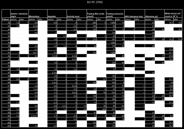
30 30
31 EH PC CPSS Crystalluria Bladder ultrasonography Complications Check-up Patient before after before after i.o. p.o. number months p.o. Ultrasonography postoperative Mortality Notes closed 33 yes closed 4 (5 years p.o.) 34 yes 1,2,5 1 1 open 35 yes 3 (2 days p.o.) Shock closed closed unknown 39 yes 1,2 1,2 1 1,5 open 4 (5 years p.o.) 40 yes 1 yes 3 (2 days p.o.) Hemorrhage 41 4 yes yes 3 (1 day p.o.) open 4 (4 years p.o.) 43 yes yes 3 1 1,5 open open and acquired shunting unknown closed 47 yes 2 yes 3 (0 days p.o.) Cardiorespiratory arrest closed open 50 yes closed closed open open ,5 open open ,5 open ,5 open 4 (unknown) open 59 yes 3 (0 days p.o.) Abdominal hemorrhage 60 1,3 yes 1 1 unknown closed 62 4 yes

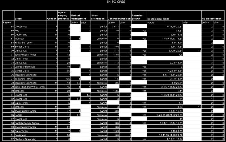
32 32

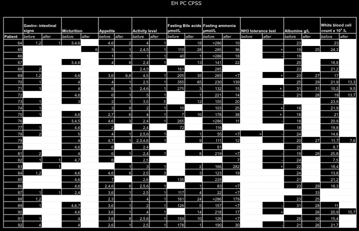
33 33
34 EH PC CPSS Crystalluria Bladder ultrasonography Complications Check-up Patient before after before after i.o. p.o. number months p.o. 64 yes ,5 open open open closed Ultrasonography postoperative Mortality Notes 68 4 yes 3 (0 days p.o.) unknown open closed unknown closed 74 yes unknown 75 yes open 76 yes 1 4 yes 1 1 closed yes yes 1,2 1 yes 2 3 closed open 80 1 yes 3 (3 days p.o.) Cerebral necrosis open unknown ,5 open ,5 unknown 85 yes (2 days p,o.) open ,5 closed 88 yes deviating vessel patern closed closed unknown closed 34


35 1b: Medical records of UKG database, details of patients with PAZ CPSS 35

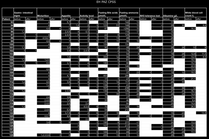
36 36


37 37


38 1c: Medical records of UKG database, details of patients with intrahepatic CPSS. 38

39 39
40 IH CPSS Bladder Crystalluria ultrasonography Complications Check-up Ultrasonography post-operative Mortality Notes Patient before after before after i.o. p.o. number months p.o. shunt 130 yes 1 yes 3 3 open yes 2 4 unknown 132 yes 3(1 day p.o.) Portal hypertensia and hypoglycemia 133 yes 2 2,3 1 1,5 open unknown 135 unknown closed yes 2 3 open yes 2 3 unknown 139 unknown 6 unknown 140 yes 1,2 unknown 6 unknown Urine was negative on crystals before surgery 141 yes 1 unknown open closed open 4(7 months p.o.) Sepsis followed by euthanasia closed unknown closed Complete ligation in two surgeries closed 149 1,2 1,2 3 4 open 150 1,2 yes 3(2 days p.o.) Cerebral necrosis 151 yes unknown yes 1 2 unknown open closed yes yes 3(11 days p.o.) Euthanasia because of lethargy 156 yes 3(25 days p.o) open 4(unknown) 158 2,3 unknown 159 yes 1,2,5 unknown 6 open 4(unknown) unknown yes 1 1,5 open open 4(3 years p.o.) Euthanasia because of heartfailure 163 yes open 164 1,2 2 3 open 4(3 years p.o.) Euthanasia because of chronical hepatitis 165 yes open 4(unknown) 166 1, open 167 1, open 40


41 1d: Caption for 1a-c 41

42 2a: General information 42

43 2b: Quality of Life questionnaire before surgery 43

44 44

45 45

46 46

47 47

48 48

49 2c: Quality of Life questionnaire after surgery 49

50 50

51 51

52 52

53 53

54 54

55 55

56 3a: Results of Quality of Life Questionnaire 56

57 57

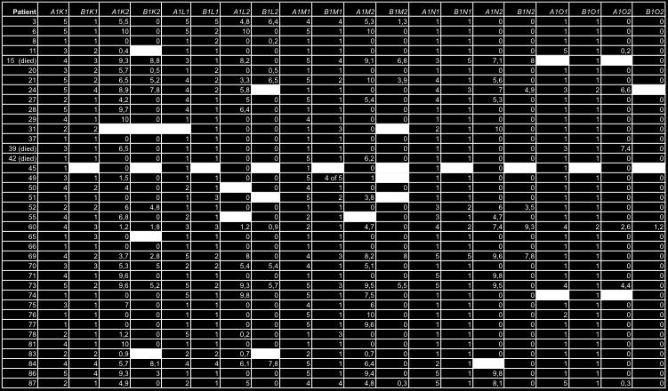
58 58

59 59

60 60

61 61

62 62

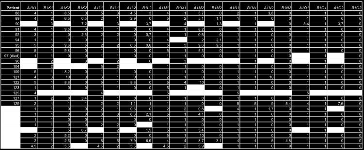
63 63


64 64

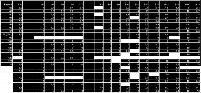
65 65

66 3b: Caption for 3a, Results of Quality of Life Questionnaire (letters referring to the question are also given in appendix 2b-c) 66


67 4: Results of patients at long-term check-up 67
Long-term Outcome After Surgical Ligation for Treatment of Congenital Portosystemic Shunts in Dogs
 Long-term Outcome After Surgical Ligation for Treatment of Congenital Portosystemic Shunts in Dogs H.J.H. GERRITS BSc Master thesis, Department of Small Animal Medicine, Utrecht University, Supervisor:
LIVER SHUNT PORTOSYSTEMIC SHUNT
 LIVER SHUNT PORTOSYSTEMIC SHUNT Karen M. Tobias, DVM, MS, Diplomate American College of Veterinary Surgeons Professor, Small Animal Surgery, University of Tennessee Department of Small Animal Clinical
Advances in congenital portosystemic shunts
 Vet Times The website for the veterinary profession https://www.vettimes.co.uk Advances in congenital portosystemic shunts Author : Marina Dominguez Categories : Companion animal, Vets Date : May 9, 2016
Distribution of extrahepatic congenital portosystemic shunt morphology in predisposed dog breeds
 Van den Bossche et al. BMC Veterinary Research 2012, 8:112 RESEARCH ARTICLE Open Access Distribution of extrahepatic congenital portosystemic shunt morphology in predisposed dog breeds Lindsay Van den
Copper-Storage Liver Disease Basics
 Copper-Storage Liver Disease Basics OVERVIEW Abnormal accumulation of copper in the liver, causing sudden (acute) inflammation of the liver (hepatitis) or long-term (chronic) hepatitis and eventually progressive
SURGICAL ATTENUATION OF CONGENITAL PORTOSYSTEMIC SHUNTS IN DOGS
 SURGICAL ATTENUATION OF CONGENITAL PORTOSYSTEMIC SHUNTS IN DOGS TECHNIQUES, COMPLICATIONS AND PROGNOSIS Anne Kummeling Publication of this thesis was financially supported by Royal Canin, Virbac Animal
Liver regeneration in dogs after CPSS surgery
 Liver regeneration in dogs after CPSS surgery D.J.E. Vrakking Abstract Background: The purpose of this study was to investigate macroscopic liver regeneration after congenital portosystemic shunt (CPSS)
Gastric Dilatation-Volvulus
 Gastric Dilatation-Volvulus The term "ACVS Diplomate" refers to a veterinarian who has been board certified in veterinary surgery. Only veterinarians who have successfully completed the certification requirements
Acute Hemorrhagic Diarrhea Syndrome (AHDS) A Cause of Bloody Feces in Dogs
 A Cause of Bloody Feces in Dogs") Acute Hemorrhagic Diarrhea Syndrome (AHDS) A Cause of Bloody Feces in Dogs No dog parent wants to clean up diarrhea. Cleaning up bloody diarrhea is even more unpleasant. Unfortunately, the development
Acute Hemorrhagic Diarrhea Syndrome (AHDS) A Cause of Bloody Feces in Dogs No dog parent wants to clean up diarrhea. Cleaning up bloody diarrhea is even more unpleasant. Unfortunately, the development
Feline lower urinary tract disease (FLUTD)
") Feline lower urinary tract disease (FLUTD) Feline lower urinary tract disease (FLUTD) is not a specific disease, but rather is the term used to describe conditions that can affect the urinary bladder and/or
Feline lower urinary tract disease (FLUTD) Feline lower urinary tract disease (FLUTD) is not a specific disease, but rather is the term used to describe conditions that can affect the urinary bladder and/or
Understanding your pet s LIVER CONDITION
 Understanding your pet s LIVER CONDITION Why is the liver so important? What causes liver disease in dogs and cats? The liver is one of the largest organs in your pet s body, and it s vital for their good
FELINE LOWER URINARY TRACT DISEASE (Sometimes known as feline urological syndrome)
") FELINE LOWER URINARY TRACT DISEASE (Sometimes known as feline urological syndrome) Introduction Feline Lower Urinary Tract Disease (FLUTD) is sometimes still referred to as feline urological syndrome or
FELINE LOWER URINARY TRACT DISEASE (Sometimes known as feline urological syndrome) Introduction Feline Lower Urinary Tract Disease (FLUTD) is sometimes still referred to as feline urological syndrome or
MONITORING CONSIDERATIONS
 Minnesota Urolith Center UNIVERSITY OF MINNESOTA College of Veterinary Medicine 1352 Boyd Avenue St Paul, MN 55108 Phone 612.625.4221 Fax 612.626.3226 email support@urolithcenter.org Director: Jody P Lulich
My cat has kidney problems and food hypersensitivity what do I do now?
 TROVET Renal (Venison), complete, easily digestible, hypoallergenic dietary food for adult cats with an impaired kidney function My cat has kidney problems and food hypersensitivity what do I do now? reliable
My dog or cat gets diet food and has struvite... what do I do now?
 TROVET Anti Struvite, dietary supplement for the targeted tackling of struvite in dogs and cats My dog or cat gets diet food and has struvite... what do I do now? reliable and affordable dietary pet food
Treatment of septic peritonitis
 Vet Times The website for the veterinary profession https://www.vettimes.co.uk Treatment of septic peritonitis Author : Andrew Linklater Categories : Companion animal, Vets Date : November 2, 2016 Septic
Pathogenesis and treatment of feline lower urinary tract disease
 Vet Times The website for the veterinary profession https://www.vettimes.co.uk Pathogenesis and treatment of feline lower urinary tract disease Author : Sarah Caney Categories : RVNs Date : May 1, 2011
Anesthesia Check-off Form
 Anesthesia Check-off Form 5231 SW 91st Drive Gainesville, FL 32608 (352) 377-6003 The doctors and staff at Haile Plantation Animal Clinic would like to offer the most advanced medical care and services
Australian and New Zealand College of Veterinary Scientists. Membership Examination. Small Animal Surgery Paper 1
 Australian and New Zealand College of Veterinary Scientists Membership Examination June 2018 Small Animal Surgery Paper 1 Perusal time: Fifteen (15) minutes Time allowed: Two (2) hours after perusal Answer
AUSTRALIAN AND NEW ZEALAND COLLEGE OF VETERINARY SCIENTISTS. Sample Exam Questions. Veterinary Practice (Small Animal)
") AUSTRALIAN AND NEW ZEALAND COLLEGE OF VETERINARY SCIENTISTS Sample Exam Questions Veterinary Practice (Small Animal) Written Examination (Component 1) Written Paper 1 (two hours): Principles of Veterinary
AUSTRALIAN AND NEW ZEALAND COLLEGE OF VETERINARY SCIENTISTS Sample Exam Questions Veterinary Practice (Small Animal) Written Examination (Component 1) Written Paper 1 (two hours): Principles of Veterinary
Some important information about the fetus and the newborn puppy
 Some important information about the fetus and the newborn puppy Dr. Harmon Rogers Veterinary Teaching Hospital Washington State University Here are a few interesting medical details about fetuses and
Australian and New Zealand College of Veterinary Scientists. Fellowship Examination. Small Animal Surgery Paper 1
 Australian and New Zealand College of Veterinary Scientists Fellowship Examination June 2017 Small Animal Surgery Paper 1 Perusal time: Twenty (20) minutes Time allowed: Three (3) hours after perusal Answer
Schistosoma mansoni, S. japonicum, S. haematobium
 Schistosoma mansoni, S. japonicum, S. haematobium The Organisms More than 200 million people are infected worldwide with Schistosoma species. The adult worms are long and slender (males are 6 12 mm in
Feline Lower Urinary Tract Disease FLUTD
 Feline Lower Urinary Tract Disease FLUTD What is Feline Lower Urinary Tract Disease? Feline lower urinary tract disease (FLUTD) describes a collection of conditions in cats that can affect the bladder
Therapeutic apheresis in veterinary
 Therapeutic apheresis in veterinary 1 I.P.Pavlov First St.-Petersburg State Medical University, Saint-Petersburg, Russia. Voinov V.A. A. By types of animals on the basis of anatomical and physiological
Subjects Beginning with "U"
 Subjects Beginning with "U" SUBJECT: ARTICLE TITLE: JOURNAL DATE: PAGE: Ucatan miniature pigs SWINE UDDER Teat Laceration Repair in Cattle VCONA/FAP JULY 2008 295 UDDER - MICROBIOLOGY GASTROINTESTINAL
Housesoiling Dogs Basics
 Housesoiling Dogs Basics OVERVIEW Urinating and/or defecating (having a bowel movement), as a means of eliminating or marking territory, in a location that the owner considers inappropriate GENETICS Some
22ND FECAVA. Eurocongress. 31. VÖK Jahrestagung. 31ST VOEK Annual Meeting June 2016 Hofburg, Vienna.
 22ND FECAVA Eurocongress 31. VÖK Jahrestagung 31ST VOEK Annual Meeting 22 25 June 2016 Hofburg, Vienna www.fecava2016.org Proceedings Proceedings 22. FECAVA Eurocongress 31 st Annual Congress of the Association
MANAGEMENT OF FELINE LOWER URINARY TRACT DISEASE Vicky L. Ograin, MBA, RVT, VTS (Nutrition) Academy of Veterinary Nutrition Technicians
 Academy of Veterinary Nutrition Technicians") MANAGEMENT OF FELINE LOWER URINARY TRACT DISEASE Vicky L. Ograin, MBA, RVT, VTS (Nutrition) Academy of Veterinary Nutrition Technicians Introduction Feline lower urinary tract disease (FLUTD) is a term
MANAGEMENT OF FELINE LOWER URINARY TRACT DISEASE Vicky L. Ograin, MBA, RVT, VTS (Nutrition) Academy of Veterinary Nutrition Technicians Introduction Feline lower urinary tract disease (FLUTD) is a term
Developed by Kathy Wonderly RN, MSEd,CPHQ Developed: October 2009 Most recently updated: December 2014
 Developed by Kathy Wonderly RN, MSEd,CPHQ Developed: October 2009 Most recently updated: December 2014 The Center for Medicare and Medicaid (CMS) is moving away from collecting data on the process of care
UNDERSTANDING COLIC: DON T GET IT TWISTED
 UNDERSTANDING COLIC: DON T GET IT TWISTED Today s Topics: What is colic? Anatomy review How to identify colic What to do when you suspect colic What to expect during a colic visit from your veterinarian
Australian College of Veterinary Scientists. Fellowship Examination. Small Animal Surgery Paper 1
 Australian College of Veterinary Scientists Fellowship Examination June 2011 Small Animal Surgery Paper 1 Perusal time: Twenty (20) minutes Time allowed: Three (3) hours after perusal Answer your choice
VCH PHC SURGICAL PROPHYLAXIS RECOMMENDATIONS
 VCH PHC SURGICAL PROPHYLAXIS RECOMMENDATIONS CARDIAC Staphylococcus aureus, S. epidermidis, except for For patients with known MRSA colonization, recommend decolonization with Antimicrobial Photodynamic
DIAGNOSIS AND MANAGEMENT OF CHOLECYSTITIS IN DOGS
 Int. J. Agric.Sc & Vet.Med. 2014 K Satish Kumar and D Srikala, 2014 Research Paper ISSN 2320-3730 www.ijasvm.com Vol. 2, No. 3, August 2014 2014 www.ijasvm.com. All Rights Reserved DIAGNOSIS AND MANAGEMENT
GASTRO-INTESTINAL TRACT INFECTIONS - ANTIMICROBIAL MANAGEMENT
 GASTRO-INTESTINAL TRACT INFECTIONS - ANTIMICROBIAL MANAGEMENT DRAFT AS CURRENTLY OUT FOR CONSULTATION BUT CAN BE UTILISED IN PRESENT FORMAT Name & Title Of Author: Date Revised: Approved by Committee/Group:
Feline Struvite Urolithiasis
 3 CREDITS Article 1 Douglas Palma, DVM Cathy Langston, DVM, DACVIM (Small Animal Internal Medicine) Kelly Gisselman, DVM John McCue, DVM Animal Medical Center New York At a Glance Pathophysiology Page
CANINE HEARTWORM DISEASE
 ! CANINE HEARTWORM DISEASE What causes heartworm disease? Heartworm disease (dirofilariasis) is a serious and potentially fatal disease in dogs. It is caused by a blood-borne parasite called Dirofilaria
Heartworm Disease in Dogs
 Kingsbrook Animal Hospital 5322 New Design Road, Frederick, MD, 21703 Phone: (301) 631-6900 Website: KingsbrookVet.com What causes heartworm disease? Heartworm Disease in Dogs Heartworm disease or dirofilariasis
Medical Conditions Questionnaire
 Medical Conditions Questionnaire (to be completed by the member) Member Full Name.Date of birth. Policy number or scheme name. Please complete the appropriate section(s) only after completing the Member
Struvite uroliths are composed of magnesium ammonium phosphate hexahydrate. Canine Struvite Urolithiasis KEY FACTS
 Vol. 23, No. 5 May 2001 407 Email comments/questions to compendium@medimedia.com CE Article #1 (1.5 contact hours) Refereed Peer Review KEY FACTS Struvite uroliths are usually detected on survey abdominal
Restore life and vitality in your dog. Feel the same results as an owner.
 Restore life and vitality in your dog. Feel the same results as an owner. Your dog, Cushing s syndrome and you This booklet has been designed to help answer questions that you may have about Cushing s
What causes heartworm disease?
 Heartworm Disease: What causes heartworm disease? Heartworm disease (dirofilariasis) is a serious and potentially fatal disease in dogs and cats. It is caused by a blood-borne parasite called Dirofilaria
Heartworm Disease in Dogs
 Customer Name, Street Address, City, State, Zip code Phone number, Alt. phone number, Fax number, e-mail address, web site Heartworm Disease in Dogs Basics OVERVIEW Disease caused by infestation with heartworms
VETERINARY CLINICS. Nutrition and Lower Urinary Tract Disease in Cats SMALL ANIMAL PRACTICE. Joseph W. Bartges, DVM, PhD*, Claudia A.
 Vet Clin Small Anim 36 (2006) 1361 1376 VETERINARY CLINICS SMALL ANIMAL PRACTICE Nutrition and Lower Urinary Tract Disease in Cats Joseph W. Bartges, DVM, PhD*, Claudia A. Kirk, DVM, PhD Department of
Metacam 1.5 mg/ml oral suspension for dogs
 Metacam 1.5 mg/ml oral suspension for dogs Species:Dogs Therapeutic indication:pharmaceuticals: Neurological preparations: Analgesics, Other NSAIDs, Locomotor (including navicular and osteoarthritis) Active
Caution: Federal law restricts this drug to use by or on the order of a licensed veterinarian.
 BOEHRINGER INGELHEIM VETMEDICA, INC. USA Product Label http://www.vetdepot.com 2621 NORTH BELT HIGHWAY, ST. JOSEPH, MO, 64506 2002 Telephone: 800 325 9167 Fax: 816 236 2717 Email: www.bi vetmedica.com
Canine Spay and Neuter Services At Manzini Animal Hospital
 Canine Spay and Neuter Services At Manzini Animal Hospital When your dog is booked in for his/her surgical procedure it can be a very anxious time for you, but here at Manzini we strive to ensure every
Perioperative Care of Swine
 Swine are widely used in protocols that involve anesthesia and invasive surgical procedures. In order to ensure proper recovery of animals, preoperative, intraoperative and postoperative techniques specific
Australian and New Zealand College of Veterinary Scientists. Membership Examination. Small Animal Medicine Paper 1
 Australian and New Zealand College of Veterinary Scientists Membership Examination June 2015 Small Animal Medicine Paper 1 Perusal time: Fifteen (15) minutes Time allowed: Two (2) hours after perusal Answer
Vertebrates. Vertebrate Characteristics. 444 Chapter 14
 4 Vertebrates Key Concept All vertebrates have a backbone, which supports other specialized body structures and functions. What You Will Learn Vertebrates have an endoskeleton that provides support and
A spaghetti sign in feline abdominal radiographs predicts spleno-systemic collateral circulation
 Received: 11 January 2017 Revised: 17 June 2017 Accepted: 18 June 2017 DOI: 10.1111/vru.12555 ORIGINAL INVESTIGATION A spaghetti sign in feline abdominal radiographs predicts spleno-systemic collateral
Nestlé PURINA Scientific Update on Feline Nutrition. Urolithiasis in cats managing the risks
 Nestlé PURINA Scientific Update on Feline Nutrition Urolithiasis in cats managing the risks Urolithiasis in cats managing the risks Dr Andrew H Sparkes BVetMed PhD DipECVIM MRCVS Veterinary consultant
Presentation of Quiz #85
 Presentation of Quiz #85 ***Reminder: Slides are copyrighted and cannot be copied for publication. A 36 year old male from Columbia was admitted to the hospital with seizures. This patient had previously
Jeff Baier MS DVM Birds of Prey Foundation Broomfield, CO
 Jeff Baier MS DVM Birds of Prey Foundation Broomfield, CO drjeffbaier@gmail.com Squamates Chelonians Snakes Lizards Varanids Monitor Lizards Crocodilians Reptilian adaptations Anaerobic glycolysis Low
Australian and New Zealand College of Veterinary Scientists. Membership Examination. Veterinary Radiology (Small Animal) Paper 1
 Paper 1") Australian and New Zealand College of Veterinary Scientists Membership Examination June 2016 Veterinary Radiology (Small Animal) Paper 1 Perusal time: Fifteen (15) minutes Time allowed: Two (2) hours after
Australian and New Zealand College of Veterinary Scientists Membership Examination June 2016 Veterinary Radiology (Small Animal) Paper 1 Perusal time: Fifteen (15) minutes Time allowed: Two (2) hours after
Overview. Clinical signs. Will you treat? Owner willing to treat? Surgical vs. Medical. Medical options
 Part II (cushing s disease is hard to diagnose) Cushing s Disease Is Easy To Treat Why test? When to test? How to test? Will you treat? How to treat? Overview Thomas Schermerhorn, VMD, DACVIM(SAIM) Kansas
SOP #: Page: 1 of 6 Rodent Analgesia
 Comparative Medicine Page: 1 of 6 Rodent Analgesia The intent of this Standard Operating Procedure (SOP) is to describe commonly used analgesics provided to rodents housed at Comparative Medicine (CM).
Understanding your cat s URINARY CONDITION
 Understanding your cat s URINARY CONDITION Understanding your cat's urinary condition. Common urinary crystals in cats. Finding out that your cat is suffering from a urinary condition can be very distressing,
Specialist Referral Service Willows Information Sheets. Rigid endoscopy
 Specialist Referral Service Willows Information Sheets Rigid endoscopy Professor Rob White with a state-of-the-art rigid Karl Storz endoscope. Rigid endoscopy What is endoscopy? Endoscopy involves the
Scottish Surveillance of Healthcare Infection Programme (SSHAIP) Health Protection Scotland SSI Surveillance Protocol 7th Edition 2017 Question &
 Health Protection Scotland SSI Surveillance Protocol 7th Edition 2017 Question &") Contents General... 4 Pre-op... 4 Peri-op... 5 Post-op... 8 Caesarean Section... 12 Orthopaedics... 14 Large Bowel:... 15 Vascular... 17 General Pre-op Q: If a patient is an emergency admission is the
Contents General... 4 Pre-op... 4 Peri-op... 5 Post-op... 8 Caesarean Section... 12 Orthopaedics... 14 Large Bowel:... 15 Vascular... 17 General Pre-op Q: If a patient is an emergency admission is the
Running head: CLOSTRIDIUM DIFFICILE 1
 Running head: CLOSTRIDIUM DIFFICILE 1 Clostridium difficile Infection Christy Lee Fenton Mountainland Applied Technology College CLOSTRIDIUM DIFFICILE 2 Clostridium difficile Infection Approximately 200,000
Restore life and vitality in your dog. Feel the same results as an owner.
 Restore life and vitality in your dog. Feel the same results as an owner. Your dog, Cushing s syndrome and you This booklet has been designed to help answer questions that you may have about Cushing s
Antibiotic prophylaxis guideline for colorectal, hepatobiliary and vascular surgery for adult patients.
 Antibiotic prophylaxis guideline for colorectal, hepatobiliary and vascular surgery for adult patients. Full Title of Guideline: Author (include email and role): Division & Speciality: Scope (Target audience,
Bacterial infections in the urinary tract
 Zurich Open Repository and Archive University of Zurich Main Library Strickhofstrasse 39 CH-8057 Zurich www.zora.uzh.ch Year: 2014 Bacterial infections in the urinary tract Gerber, B Posted at the Zurich
Pectus Excavatum (Funnel Chest) Dr Hasan Nugud Consultant Paediatric Surgeon
 Dr Hasan Nugud Consultant Paediatric Surgeon") Pectus Excavatum (Funnel Chest) Dr Hasan Nugud Consultant Paediatric Surgeon Pectus excavatum Pectus excavatum (PE) is an abnormal development of the rib cage where the breastbone (sternum) caves in,
Pectus Excavatum (Funnel Chest) Dr Hasan Nugud Consultant Paediatric Surgeon Pectus excavatum Pectus excavatum (PE) is an abnormal development of the rib cage where the breastbone (sternum) caves in,
Principles of Anti-Microbial Therapy Assistant Professor Naza M. Ali. Lec 1
 Principles of Anti-Microbial Therapy Assistant Professor Naza M. Ali Lec 1 28 Oct 2018 References Lippincott s IIIustrated Reviews / Pharmacology 6 th Edition Katzung and Trevor s Pharmacology / Examination
Feline Idiopathic Cystitis (icatcare)
") Kingsbrook Animal Hospital 5322 New Design Road, Frederick, MD, 21703 Phone: (301) 631-6900 Website: KingsbrookVet.com Feline idiopathic cystitis (FIC) [1] Feline Idiopathic Cystitis (icatcare) Feline
Kingsbrook Animal Hospital 5322 New Design Road, Frederick, MD, 21703 Phone: (301) 631-6900 Website: KingsbrookVet.com Feline idiopathic cystitis (FIC) [1] Feline Idiopathic Cystitis (icatcare) Feline
Australian and New Zealand College of Veterinary Scientists. Membership Examination. Veterinary Anaesthesia and Critical Care Paper 1
 Australian and New Zealand College of Veterinary Scientists Membership Examination June 2015 Veterinary Anaesthesia and Critical Care Paper 1 Perusal time: Fifteen (15) minutes Time allowed: Two (2) hours
FURTHER NATUROPATHIC SUPPORT
 Product Code AN014 Bladder Health 11 Pages Urinary Syndrome (FUS), or Lower Urinary Tract Disease (FLUTD) e.g. Struvite crystals for all species & ages. Last Updated: 11-06-18 NATURAL MEDICINES - ANIMAL
The Royal College of Veterinary Surgeons DIPLOMA IN EQUINE SOFT TISSUE SURGERY PAPER I. (Basic Sciences) Tuesday 2 May 1995
 Tuesday 2 May 1995") The Royal College of Veterinary Surgeons PAPER I (Basic Sciences) Tuesday 2 May 1995 10.00 a.m. to 1.00 p.m. (3 hours) SECTION A Two long answer questions of which a candidate must choose ONE question
The Royal College of Veterinary Surgeons PAPER I (Basic Sciences) Tuesday 2 May 1995 10.00 a.m. to 1.00 p.m. (3 hours) SECTION A Two long answer questions of which a candidate must choose ONE question
Cell Wall Inhibitors. Assistant Professor Naza M. Ali. Lec 3 7 Nov 2017
 Cell Wall Inhibitors Assistant Professor Naza M. Ali Lec 3 7 Nov 2017 Cell wall The cell wall is a rigid outer layer, it completely surrounds the cytoplasmic membrane, maintaining the shape of the cell
Feline Idiopathic Cystitis: The Veterinary Nurses role in the treatment and management. Nicola Ackerman BSc(Hons) RVN CertSAN CertVN ECC HonMBVNA
 RVN CertSAN CertVN ECC HonMBVNA") Feline Idiopathic Cystitis: The Veterinary Nurses role in the treatment and management. Nicola Ackerman BSc(Hons) RVN CertSAN CertVN ECC HonMBVNA Feline urinary tract issues may result from a number of
Feline Idiopathic Cystitis: The Veterinary Nurses role in the treatment and management. Nicola Ackerman BSc(Hons) RVN CertSAN CertVN ECC HonMBVNA Feline urinary tract issues may result from a number of
Australian and New Zealand College of Veterinary Scientists. Fellowship Examination. Small Animal Medicine Paper 1
 Australian and New Zealand College of Veterinary Scientists Fellowship Examination June 2014 Small Animal Medicine Paper 1 Perusal time: Twenty (20) minutes Time allowed: Four (4) hours after perusal Answer
Suitability of Antibiotic Treatment for CAP (CAPTIME) The duration of antibiotic treatment in community acquired pneumonia (CAP)
 The duration of antibiotic treatment in community acquired pneumonia (CAP)") STUDY PROTOCOL Suitability of Antibiotic Treatment for CAP (CAPTIME) Purpose The duration of antibiotic treatment in community acquired pneumonia (CAP) lasts about 9 10 days, and is determined empirically.
STUDY PROTOCOL Suitability of Antibiotic Treatment for CAP (CAPTIME) Purpose The duration of antibiotic treatment in community acquired pneumonia (CAP) lasts about 9 10 days, and is determined empirically.
Retinal Degeneration Basics
 Retinal Degeneration Basics OVERVIEW Retinal refers to the retina; the retina is the innermost lining layer (located on the back surface) of the eyeball; it contains the light-sensitive rods and cones
Perioperative surgical risks and outcomes of early-age gonadectomy in cats and dogs at People for Animals, Inc.
 Perioperative surgical risks and outcomes of early-age gonadectomy in cats and dogs at People for Animals, Inc. David Croman, VMD; Laurie Heeb, DVM; Jane Guillaume, Alyssa Dillonaire Objective To determine
Irish Medicines Board
 IRISH MEDICINES BOARD ACT 1995 EUROPEAN COMMUNITIES (ANIMAL REMEDIES) (No. 2) REGULATIONS 2007 (S.I. No. 786 of 2007) VPA:10778/003/002 Case No: 7003735 The Irish Medicines Board in exercise of the powers
Naturally occurring hyperadrenocorticism is a wellrecognized
 Evaluation of twice-daily lower-dose trilostane treatment administered orally in dogs with naturally occurring hyperadrenocorticism Edward C. Feldman, dvm, dacvim Objective To evaluate effectiveness and
NUMBER: R&C-ARF-10.0
 1. PURPOSE PAGE 1 OF 6 This policy describes the procedures for keeping and maintaining animal medical records. This procedure is approved by the Creighton University Institutional Animal Care and Use
Biology 323 Human Anatomy for Biology Majors Lecture 13 Dr. Stuart S. Sumida. Gut Tube: Development, Structure, Function
 Biology 323 Human Anatomy for Biology Majors Lecture 13 Dr. Stuart S. Sumida Gut Tube: Development, Structure, Function 1. Implications of Gut Development Foregut Development Midgut Development Hindgut
Senior Pet Care and Early Disease Detection
 Senior Pet Care and Early Disease Detection Thanks to advances in veterinary medicine, pets are living longer than ever before. However, with this increased lifespan comes an increase in the types of ailments
USA Product Label CLINTABS TABLETS. Virbac. brand of clindamycin hydrochloride tablets. ANADA # , Approved by FDA DESCRIPTION
 VIRBAC CORPORATION USA Product Label http://www.vetdepot.com P.O. BOX 162059, FORT WORTH, TX, 76161 Telephone: 817-831-5030 Order Desk: 800-338-3659 Fax: 817-831-8327 Website: www.virbacvet.com CLINTABS
Update in Veterinary Medicine. Dr. Maria M. Crane Zoo Atlanta
 Update in Veterinary Medicine Dr. Maria M. Crane Zoo Atlanta Overview of Discussion Medical management of captive orangutans Preventative Medicine Anesthesia Protocols Vaccinations TB testing Current Health
Hydatid Cyst Dr. Nora L. El-Tantawy
 Hydatid Cyst Dr. Nora L. El-Tantawy Ass. Prof. of Parasitology Faculty of Medicine, Mansoura university, Egypt Echinococcus granulosus Geographical Distribution: cosmopolitan especially in sheep raising
Metacam is an anti-inflammatory medicine used in cattle, pigs, horses, dogs, cats and guinea pigs.
 EMA/CVMP/259397/2006 EMEA/V/C/000033 An overview of Metacam and why it is authorised in the EU What is Metacam and what is it used for? Metacam is an anti-inflammatory medicine used in cattle, pigs, horses,
NSAID Toxicity in Dogs & Cats Beware of Ibuprofen!
 NSAID Toxicity in Dogs & Cats Beware of Ibuprofen! One of the most common drug toxicities in companion animal medicine is accidental ingestion of over-the-counter non-steroid anti-inflammatory (NSAID)
Hope for Healing Liver Disease in Your Dog. Quick Start Guide. by Cyndi Smasal
 Hope for Healing Liver Disease in Your Dog Quick Start Guide by Cyndi Smasal Copyright 2004 by Cyndi Smasal All Rights Reserved. No part of this book may be reproduced, stored in a retrieval system, or
Ylva Sjöström 1) and Anna Lennquist 2)
 and Anna Lennquist 2)") Ylva Sjöström 1) and Anna Lennquist 2) 1) VMD, Swedish specialist in diseases of dogs and cats, Blue Star Animal Hospital, Gjutjärnsgatan 4, SE-417 07 Gothenburg, Sweden 2) PhD in Zoophysiology, Dept.
Ylva Sjöström 1) and Anna Lennquist 2) 1) VMD, Swedish specialist in diseases of dogs and cats, Blue Star Animal Hospital, Gjutjärnsgatan 4, SE-417 07 Gothenburg, Sweden 2) PhD in Zoophysiology, Dept.
The Vet Education Webinar Series Biliary Mucocoeles With Dr Gemma Birnie
 The Vet Education Webinar Series 2016 Biliary Mucocoeles With Dr Gemma Birnie BVSc MACVSc Vet Education is Proudly Supported by Hill s Pet Nutrition Australia Gallbladder Mucocoeles Dr. Gemma Birnie Resident
Treatment of Respiratory Tract Infections Prof. Mohammad Alhumayyd Dr. Aliah Alshanwani
 Treatment of Respiratory Tract Infections Prof. Mohammad Alhumayyd Dr. Aliah Alshanwani 30-1-2018 1 Objectives of the lecture At the end of lecture, the students should be able to understand the following:
Australian and New Zealand College of Veterinary Scientists. Fellowship Examination. Small Animal Surgery Paper 1
 Australian and New Zealand College of Veterinary Scientists Fellowship Examination June 2016 Small Animal Surgery Paper 1 Perusal time: Twenty (20) minutes Time allowed: Three (3) hours after perusal Answer
Non-steroidal anti-inflammatory drugs (NSAIDs) are used widely to relieve pain, with or without
 are used widely to relieve pain, with or without") May 2013 Contents About NSAIDs What about COXselectivity? How effective are NSAIDs? Adverse effects of NSAIDs How frequent are the adverse effects of NSAIDs? General prescribing guidelines for NSAIDs What
May 2013 Contents About NSAIDs What about COXselectivity? How effective are NSAIDs? Adverse effects of NSAIDs How frequent are the adverse effects of NSAIDs? General prescribing guidelines for NSAIDs What
Antimicrobial Stewardship in Continuing Care. Urinary Tract Infections Clinical Checklist
 Antimicrobial Stewardship in Continuing Care Urinary Tract Infections Clinical Checklist December 2014 What is Antimicrobial Stewardship? Using the: right antimicrobial agent for a given diagnosis at the
ANNEX I SUMMARY OF PRODUCT CHARACTERISTICS. Medicinal product no longer authorised
 ANNEX I SUMMARY OF PRODUCT CHARACTERISTICS 1 1. NAME OF THE VETERINARY MEDICINAL PRODUCT Zubrin 50 mg oral lyophilisates for dogs Zubrin 100 mg oral lyophilisates for dogs Zubrin 200 mg oral lyophilisates
NEONATAL Point Prevalence Survey. Ward Form
 Appendix 2 NEONATAL Point Prevalence Survey Ward Form Please fill in one form for each ward included in PPS Date of survey Person completing form (Auditor code) Hospital Name Department/Ward Neonatal departments
Oral and intestinal candidiasis. As adjuvant treatment with other local nystatin preparations to prevent reinfection.
 1. NAME OF THE MEDICINAL PRODUCT Nystatin Orifarm, 100 000 IU/ml oral suspension 2. QUALITATIVE AND QUANTITATIVE COMPOSITION 1 ml contains 100 000 IU nystatin. Excipients with known effect: - Methyl parahydroxybenzoate
Enteric Clostridia 10/27/2011. C. perfringens: general. C. perfringens: Types & toxins. C. perfringens: Types & toxins
 C. perfringens: general Enteric Clostridia Formerly called C. welchii Thick rods, forming spores Non motile Grow fast Habitats: Soil and sewage and in the intestines of animals and humans Double zone hemolysis
The surgical site infection risk in developing countries. Yves BUISSON Société de Pathologie Exotique
 The surgical site infection risk in developing countries Yves BUISSON Société de Pathologie Exotique Surgical site infections Health-care-associated infections occurring within 30 days after surgery, or
Hudson, a 10-year-old MC Cocker spaniel, was referred for evaluation of severe polyuria and polydipsia (PU/PD) of 3 months in duration...
 of 3 months in duration...") VCAWLAspecialty.com David Bruyette, DVM, DACVIM Hudson, a 10-year-old MC Cocker spaniel, was referred for evaluation of severe polyuria and polydipsia (PU/PD) of 3 months in duration... 1. Physical Examination
VCAWLAspecialty.com David Bruyette, DVM, DACVIM Hudson, a 10-year-old MC Cocker spaniel, was referred for evaluation of severe polyuria and polydipsia (PU/PD) of 3 months in duration... 1. Physical Examination
SUMMARY OF PRODUCT CHARACTERISTICS
 SUMMARY OF PRODUCT CHARACTERISTICS 1. NAME OF THE VETERINARY MEDICINAL PRODUCT Rycarfa 100 mg tablets for dogs (BE, DE, ES, FR, IE, IT, NL, PT, UK) Rycarfa vet 100 mg tablets for dogs (DK, FI) Carprox