Life cycle and host-parasite relationships of Schistotaenia tenuicirrus (Cestoda: Amabiliidae)
|
|
|
- Charla Holmes
- 6 years ago
- Views:
Transcription

1 Retrospective Theses and Dissertations 1966 Life cycle and host-parasite relationships of Schistotaenia tenuicirrus (Cestoda: Amabiliidae) Stanley Benjamin Boertje Iowa State University Follow this and additional works at: Part of the Zoology Commons Recommended Citation Boertje, Stanley Benjamin, "Life cycle and host-parasite relationships of Schistotaenia tenuicirrus (Cestoda: Amabiliidae) " (1966). Retrospective Theses and Dissertations This Dissertation is brought to you for free and open access by Iowa State University Digital Repository. It has been accepted for inclusion in Retrospective Theses and Dissertations by an authorized administrator of Iowa State University Digital Repository. For more information, please contact
2 This dissertation has been microfihned exactly as received 66-10,407 BOERTJE, Stanley Benjamin, LIFE CYCLE AND HOST-PARASITE RE LATIONSHIPS OF SCHISTOTAENIA TENUICIRRUS (CESTODA: AMABILIIDAE). Iowa State University of Science and Technology Ph.D., 1966 Zoology University Microfilms, Inc., Ann Arbor, Michigan
3 LIFE CYCLE AND HOST-PARASITE RELATIONSHIPS OF SCHISTOTAENIA TENUICIRHUS (CESTODA; AMABILIIDAE) by Stanley Benjamin Boertje A Dissertation Submitted to the Graduate Faculty in Partial Fulfillment of The Requirements for the Degree of DOCTOR OF PHILOSOPHY Major Subject: Parasitology Approved; Signature was redacted for privacy. J or Work Signature was redacted for privacy. Head of Major Department Signature was redacted for privacy. duate College Iowa State University Of Science and Technology Ames, Iowa 1966
4 11 TABLE OP CONTENTS Page INTRODUCTION 1 HISTORICAL REVIEW 2 MATERIALS AND METHODS 6 Definitive Hosts 6 Intermediate Hosts 9 SUMMARY OF LIFE CYCLE 11 ADULT 13 General Statement 13 Strobila 17 Scolez 18 Male Reproductive System 20 Female Reproductive System 22 Related Species 24 EGG AND ONCOSPHERE 26 INTERMEDIATE HOST 29 General Statement 29 Location of Parasite 31 Natural Infections 33 Pathology 35 DEVELOPMENT AND MORPHOLOGY OP STHOBILOCERCOID 40 DEFINITIVE HOSTS 51 General Statement 51 Location of Parasite 52 Natural Infections 54 Experimental Infections 57 Pathology 66 Host Specificity 69 DISCUSSION 71 SUMMARY AND CONCLUSIONS 74
5 ill Page UTEEATUBE CITED 78 ACKNOWLEDGMENTS 83 PLATES 84
6 1 INTRODUCTION The cestode, Schlstotaenla tenulolrrus Chandler, 1948, is an intestinal parasite of the pied-billed grebe (Podilymbus podiceps L, ) of Iowa, Minnesota, Michigan, Ohio, and Illinois, It has also been reported from the horned grebe, Colymbus auritus L,, in the same areas. Schistotaenla, together with the genera Amabilia Diamare, and Tatria Kowalewski, 1904, comprise the family Amabiliidae Bansom, No published accounts of life cycles of any members of Amabilia have appeared, and until the present study, no life cycles of members of Schistotaenia were known. Several accounts dealing with intermediate stages in the life cycles of members of Tatria, however, have appeared in heiminthological literature. Since pied-billed grebes of northwestern Iowa are known to be infected with Schistotaenia tenuicirrus, a study was undertaken in 1962-I965 to ascertain its life history and host-parasite relationships. Field collections and experimental studies were conducted at the Iowa Lakeside Laboratory in northwest Iowa. The discovery of the unusual intermediate stage of S. tenuicirrus1 for which the term strobilocercoid is proposed, provides an additional example of the varied larvae known to occur among cyclophyllidean tapeworms.
7 2 HISTORICAL REVIEW The prenus Schlstotaenla was created by Gohn (I900) to include small avian cyclophyllidean tapeworms presently assigned to the family Amabiliidae Ransom, Tapeworms of this penus are craspedote, possess conspicuous lateral outgrowths, and are parasites of grebes. Because a thorough historical review and taxonomic revision of the family Amabiliidae was presented by Johri (1959)> only a brief account is presented below. Braun (I9OO) established the subfamily Amablliinae of the family Taeniidae to receive the genus Amabilia Dlamare, Later, Puhrmann (I907) elevated the status of the subfamily Amablliinae Braun, I900, to that of a family, Amabilinidae, which was later correctly designated as Amabiliidae Ransom, Fuhrmann (I907) placed three genera in this family: Amabilia Diamare, 1893; Schistotaenla Cohn, I900; and Tatria Kowalewski, The distinguishing characteristics of the family include the following: small cestodes with a strongly armed, tenacious rostellum; strobila with craspedote proglottids having conspicuous lateral appendages upon which the male genital pores open; genitalia single, alternating regularly or irregularly, or double; true vagina and vaginal pore absent and occasionally replaced in function by median accessory ducts opening to the exterior both dor-
8 3 sally and ventrally. Johri (1959)» noting the distinct differences between the characters of Amabilla and those of Schistotaenia and Tatria, proposed that the family Amabiliidae be divided into two subfamilies, Amabiliinae Braun, 1900, for the reception of the genus Amabilia Diamare, 1893, and Schistotiinae Johri, 1959» for the genera Schistotaenia Cohn, 1900, and Tatria Kowalewskl, The subfamily Schistotiinae was changed to Schistotaeniinae by Owen (I96O), so as to conform to the International Code of Zoological Nomenclature. The unique features of the subfamily Amabiliinae enumerated by Johri (1959) include the following: double male genital organs and double male genital pores present in each segment; cirrus spiny; a single and median set of female genital organs; ovary and vitelline gland dendritic, the latter much larger than the ovary. Adult worms are found in the flamingo. The distinguishing characters of the subfamily Schistotaeniinae include the following: single male genital organs and genital pores in each segment, alternating regularly or irregularly; cirrus spiny or non-spiny; female genital organs single in each segment and median in position; ovary bilobed, and larger than the vitelline gland ; true vagina absent; seminal receptacles consisting of thin-walled sacs in the midline and communicating with one another in consecutive seg
9 4 ments. Adult worms occur in the intestine of grebes. The genus ochistotaenia Cohn, I90O, is distinguished from the closely related genus Tatria Kowalewski, 1904, by the presence, in the former, of numerous testes and of irregularly alternating male genital pores. Dorso-ventral canalsreplace the true vagina. Six species are known for the genus Schistotaenia Cohn, 1900, namely: S. macrorhyncha (Hudolphi, 1810) Cohn, I900; S. scolopendra (Diesing, I856) Baer, 1940; 3. macrocirrus Chandler, 1948; S. tenuicirrus Chandler, 1948; S. colymba Schell, 1955; and S. indica Johri, The significant characters for separating these six species are summarized in Table 1. Hr. John Gallimore, Department of Zoology, University of Alberta, iidmonton. Alberta, Canada, has informed me (personal communication) that he has found a new, as yet undescribed, species of Schistotaenia. The present report Involves the life cycle of one of these species, 8. tenuicirrus, a common parasite of piedbilled grebes of northwest Iowa. The adult worn; was named and described thoroughly by Chandler (1948). No life cycle studies on any members of the genus have appeared prior to an abstract by Boertje and Ulmer (I965). Wardle and McLeod (1952) have suggested that the genera of the family Amablliidae are indiscriminately and unsatisfactorily grouped according to aberrant features, and that
10 5 the discovery of new material or a closer re-examination of existing material may entirely change the present system of classification. Lopez-Keyra (1953) expressed the belief that the family Amabiliidae is an unnatural group based on teratologica], specimens. Matevosyan and Okorokov (1959), however, disagreed with this interpretation by Lopez-Neyra and,considered Tatria a valid genus. The experimental studies on adults of S. tenuicirrus presented in this study, as well as the peculiar larval stage (strobilocercoid) involved in its life cycle, indicate that these cestodes, together with other amabiliids, probably constitute a unique group which should, for the present at least, be retained.
11 6 MATERIALS AND METHODS Definitive Hosts Most of the pied-billed grebes collected for this investigation were shot in ponds, sloughs, and lakes near the Iowa Lakeside Laboratory in northwest Iowa during the months of May through August of I A 12-gauge automatic Remington shotgun was used for obtaining juvenile and adult birds. All grebes were examined for parasites within one hour of the time of death. Forty-three wild grebes were examined for the presence of helminths during this study. For experimental purposesj eggs of pied-billed grebes were collected from nests in the spring of I963 and From eggs incubated at approximately 38 degrees centigrade,. four juveniles were hatched. A diet of boiled crayfish successfully kept two birds alive until after exposure to larval cestodes. Live crayfish were collected from adjacent rivers and ponds with seines, dip nets, and traps. By means of refrigeration, these crustaceans were preserved for several weeks. During the spring of 19^5» a wild juvenile grebe, estimated to be two or three days old, was caught by hand and was also used as an experimental host. The entire diet of this bird consisted of living crayfish. A wire cage (24" x 24" x 24") was designed for rearing the young grebes. A portion of
12 7 the floor of the cage was elevated 5" to the height of two plastic water basins (12" x 14" x 5" high), which were placed within the cage. Laboratory-raised mallard ducks and wild grackles were also used for experimental exposures to strobilocercoids in studies concerning host specificity of Schistotaenia. For collecting the internal parasites of grebes, the body cavity was opened ventrally, the digestive tract removed, and the viscera washed in 0.8^ sodium chloride solution. The proventriculus and ventriculus were examined to determine the bird's food habits, as well as to check for the presence of larval parasites. An incision of the small intestine was made to obtain tapeworms for this study. To facilitate release of scoleces from the gut wall, sections of the host intestine with attached Schistotaenia were placed in Petri dishes with avian Ringer's solution and examined under the dissecting microscope. Tapeworms were relaxed in distilled water before fixation. Following fixation in AFA (alcohol, acetic acid, ard formalin), the worms were stored in 70/^ ethyl alcohol. - Mayer's paracarmine with a fast green counterstain was employed for staining all whole mounts, which were then cleared in xylene or methyl salicylate, and mounted in a synthetic resin (Permount). Tapeworms i^ situ were embedded in paraffin and cut at 10 to 12 microns on a rotary microtome. Stains utilized for these sections were
13 8 Mallory's triple or Harris's hematoxylin followed by an eosin counterstain. Experiments involving the exposure of the epgs of Sohistotaenia to various arthropods required the removal of gravid proglottids from adult worms. Some of these were fed immediately, others were kept from one to five days in distilled water and then fed to suspected intermediate hosts. In other experiments,gravid proglottids were teased apart with dissecting needles to remove intra-uterine eggs. These were stored in filtered, boiled lake water or in distilled water, kept under refrigeration or at room temperature for periods of one to ten days, and the culture water changed daily. Eggs were then transferred to Petri dishes or Syracuse watch glasses for exposure to various invertebrates. An Eberbach shaking machine was employed for agitation of some eggs during incubation. A higher percentage of fully developed eggs was obtained with aeration and agitation at room temperature. Identifications of birds were verified with the use of Birds of America, Pearson (1936), A Field Guide to the Birds, Peterson (1963), and the Handbook of North American Birds, Palmer (1962). ;
14 9 Intermediate Hosts Many aquatic invertebrates near the Iowa Lakeside Laboratory were examined for larval stages of cestodes. Some of the arthropods collected were exposed to the eggs of Schistotaenia in an attempt to determine the intermediate host of this tapeworm. When the peculiar strobilocercoid larvae of Schistotaenia were found to occur in dragonfly naiads, many collections of these insects were made and the body cavities examined for larval stages with the aid of a dissecting microscope. Naturally infected dragonfly naiads were collected from Jemmerson Slough, Marble Lake, Hale's Slough, and Prairie Lake in Dickinson County, Iowa. The most efficient collecting device for obtaining dragonfly naiads from vegetation was a long-handled bottom net with a rectangular bag 18" x 8" x 10" deep and a 6* handle of 1 1/4" diameter. Naiads were placed in separate containers in the laboratory, killed by removing the head, the hemocoel opened with dissecting needles, and the contents examined. Dragonfly naiads for experimental studies were collected from the Big Kettle Hole located about six miles southwest of the Iowa lakeside Laboratory. No grebes were observed on this pond, and no larval cestodes of Schistotaenia were ever recovered from 162 dragonfly naiads examined for naturally
15 10 occurring' larval cestodes. Because of their cannibalism, these experimental hosts were isolated in finger bowls and small bottles. The amphipod, Hyallela azteca, collected from the Big Kettle Hole, was used as the primary food source. Larval stages of Schistotaenia were fixed in APA for whole mounts and in Bouin's for sections. Whole mounts were stained with Mayer's paracarmine and counterstained with fast green. Mayer's hemalum and Delafield's hematoxylin were used for staining sections. These were counterstained with eosin. Crustaceans and insects were identified using keys by Needham and Westfall (1955)» Pennak (1953)» and Ward and Whipple (1959). Verification of the correct determination of the dragonfly naiad, Anax.1 uni us, was by Dr. Jean L. Laffoon, Department of Zoology and Entomology, Iowa State University. Drawings were made with the use of a Leitz microprojector and a camera lucida.
16 11 SUMMARY OP LIFE CYCLE The pied-billed grebe, Podilymbus podiceps, serves as the definitive host for Schlstotaenla tenuicirrus in northwest Iowa. Natural infections in juvenile birds are considerably higher than In adult grebes. Sexually mature birds show a characteristic periodicity for the presence of adult worms ; infected adult grebes are commonly found in May and June, but they lose their Schlstotaenla Infections during or soon after the nesting season. The dragonfly naiad, Anax.1 uni us, serves as the intermediate host for the unusual strobllocercoid larva of S. tenuicirrus. Dragonfly infections are strictly seasonal, never occurring before July. Embryonated eggs of S. tenuicirrus require an incubation period of five days before they are viable. Under experimental conditions, Anax.1 un lus naiads become infected with the larvae of 8. tenuicirrus after exposure to embryonated eggs. The larvae occur free within the hemocoel 48 hours post-exposure. Rapid growth and development follows; in 21- day larvae, the scolex and stroblla have very nearly completed development and strobllizatlon occurs. The strobllocercoid larva is fully developed 28 days post-infection, and measures approximately 20 mm by 3 mm. Two distinct membranes surround the strobllocercoid, the inner one possessing
17 12 spirally arranged papillae, which constitutes one of the most distinctive features of the fully formed larva. Experimental feedings of naturally infected dragonfly naiads containing strobilocercoids of S. tenuicirrus to laboratory-raised grebes conclusively establish the manner in which these birds acquire their infection. Gravid worms appear within 33 days post-exposure.
18 13 ADULT General Statement The pied-billed grebe, Podilymbus podiceps, serves as the principal definitive host for S. tenuicirrus, which has also been reported from the horned grebe (Colymbus auritus) and from the crow, Corvus brachyrhynchos, by Chandler (19^8). Previous investigations by Jones (1929)» Baer (1940), and Chandler (19^8) indicate that four distinct species of Schistotaenia parasitize the pied-billed grebe, namely, S. macrorhyncha, S. scolopendra, S. macroclrrus, and S. tenuicirrus. Characters used in differentiating species of Schistotaenia include number, shape, and size of rostellar hooks; shape and size of scolex; diameter and form of the suckers; presence of spines on rostellum; shape and length of strobila; number of testes per segment; size of cirrus and cirrus pouch; regular or irregular alternation of male genital pores; size and form of seminal receptacles. Comparative measurements, principal hosts, and geographic distribution of the adult worms of the six known species of Schistotaenia are given in Table 1. Data for S. tenuicirrus are derived both from Chandler (19^8) and the present study. During the present investigation, 13^ mature adults were collected and examined from naturally infected hosts. Some
19 14 Table 1. Comparison of characters of known species of Schistotaenia S. tenuicirrus 3. macrorhyncha S. scolopendra Strobila, length mm 40.5 mm 7-11 mm Scolex, length mm 0.9 mm Scolex, diameter mm mm Sucker, diameter mm mm mm mm Ro stellar spines present present present Number of rostellar hooks Hook, length y i [I Base of hook, length p. 158 ^ Alternation of male genital pores irregular irregular irregular Number of testes per segment Cirrus, length 1,5 mm 0.94 mm Cirrus pouch, length mm mm mm Cirrus pouch, diameter mm mm mm
20 15 Table 1. (continued) S. tenuicirrus S. macrorhyncha S. scolopendra Seminal receptacle, diameter mm Principal hosts Podilymbus podlceps Podiceps nlg-rlcollis Podilymbus podiceps Colymbus auritus Podilymbus podiceps Podiceps dominicus Colymbus auritus Colymbus dominicus Locality Minn., Mich., Ohio, 111., Iowa, Georgia iiurope, Texas Antigua, South America Reference Chandler (1948); Boertje (present account) Cohn (1900) Baer (1940) Strobila, length Scolex, length Scolex, diameter S. macrocirrus S. colymba mm mm mm mm mm mm (width) S. indica mm mm mm (width) Sucker, diameter 0, mm mm x 0.35 i&m Rostellar spines absent absent absent
21 16 Table 1. (continued) S. macrocirrus S. colymba S. indica Number of rostellar hooks Total hook, length n i n Base of hook, length n 84-8? [i Alternation of male genital pores irregular regular irregular Number of testes per segment Cirrus, length 3.5 mm 0,4-0,5 mm Cirrus pouch, length 0,34-0,38 mm 0,37-0,38 mm 0,37-0,4 mm Cirrus pouch, diameter ,21 mm 0,11-0,12 mm mm Seminal receptacle, diameter 0,7 mm 0,24 mm Principal hosts Podilymbus podiceps Colymbus. auritus Podiceps ruficollis Locality Ohio Idaho India Reference Chandler (1948) Schell (1955) Johri (1959)
22 17 of the morphological differences of these specimens from Iowa, as contrasted with those described by Chandler (1948) from various Midwestern states, include length of strobila, size of scolex, size of rostellum, diameter of suckers, size of cirrus pouch, number of testes, and width of seminal receptacles. These are discussed below, and emendations relating to this species appear in the discussion section. Strobila The total length of the strobila of gravid S. tenuicirrus (Figures 1, 2) varies from 20 to 96 mm, with an average length of 35 mm. The maximum number of proglottids per worrr is approximately 200. Chandler (1948) indicated his specimens to be from 20 to 30 mm long. It is rather surprising that he used body length as a key character, since in an earlier paper (1939)» he showed that such factors as method of preparation, state of relaxation at time of death, age of worms, and number per host, had marked effects in determining the size of tapeworms. Of 212 adult S, tenuicirrus examined during this investigation, 91 were over 30 mm in length. A distinct neck is present (Figure 6), and segmentation begins a short distance behind the scolex. All proglottids of the strobila are wider than they are long, and increase in length posteriorly. Proglottids are narrowest immediately behind the scolex, and their lateral margins are parallel for
23 18 most of the length of the worm. Each proglottid has a contractile, finger-like, lateral appendage on each side, measuring from 0,6 to 1,0 mm in length. Mature proglottids (exclusive of these contractile extensions) measure 0,35 mm long by 1,7 mm wide; gravid proglottids are 0,4 mm long by 1,85 mm wide, The muscles (Figure 9) of the strobila are strongly developed. Under the cuticle and basement membrane lies a narrow region of subcuticular muscles separated by parenchyma from the underlying parenchymal (medullary) muscle fibers. The longitudinal muscles of the medullary region are not arranged in distinct layers, as described by Baer (19^0) for S. scolopendra. Circular or transverse parenchymal muscles, also well developed, form a single muscle layer. Dorso-ventral muscles are scattered through the medullary region between the reproductive organs. The cortical parenchyma contains a large number of calcareous bodies, which also occur in the lateral appendages. Scolex The scolex (Figure 6) is large, 0,6 to 1.8 mm long, and posteriorly (at its widest portion) 0,9 to 1,6 mm in diameter. The narrowest region, immediately behind the rostellar hooks, has a diameter of 0,3 to 0,4? mm, depending on the state of
24 19 contraction. Great variations in the shape and size of the scolex (Figures 3-5) occur in accordance with the state of contraction of this region. The four spinose muscular suckers are spherical or oval and measure.24 to 0.3 mm by 0.3 to 0.39 mm in diameter. In young worms (Figure 30), suckers are much smaller. The rostellum, large and heavily muscularized, resembles very closely that described by Cohn (1900) for S. macrorhyncha. Two muscular rostellar sacs (Figures 6, 7) are present, one inside the other. These unite anteriorly and are termed inner and outer rostellar sacs. The inner sac, about 0.4 to 0.46 mm long by 0.2 to 0.3 mm wide, consists of two muscle layers (Figure 7)» a thick inner circular layer and an outer longitudinal layer. The outer muscular sac (about 0.8 mm long) consists of an inner circular and an outer longitudinal muscle layer. Some of these longitudinal muscles extend from the scolex into the strobila, are continuous with the longitudinal parenchymal muscles, and serve as retractor muscles. Additional extensions of the longitudinal body muscles (inner retractors, according to Cohn (1900)) penetrate the rostellar sac posterolaterally, extend anteriorly between the outer and inner rostellar sacs, and insert at the anterior rostellar region. They appear as four distinct muscle bundles within the outer rostellar sac, and between them lie glandular masses, the rostellar glands. These stain deeply with hema-
25 20 toxylin and hemalum, as previously reported by Cohn (I900) for S. macrorhynoha and by Baer (1940) for S. scolopendra. The entire surface of the rostellum is covered with small spines, and the apex bears a single crown of 26 very large hooks. Each hook (Figure 8) measures 135 to 14^ microns in total length, from proximal end of base to tip of blade; from proximal end of base to tip of guard, 115 to 125 microns; from tip of blade to tip of guard, 5?- to 59 microns; and from the anterior margin of hook to tip of guard, 82 to 86 microns. The extremely well-developed musculature of the scolex enables the adult to attach tenaciously to its host, so that the rostellar region is often lost if worms are not carefully removed. Male Reproductive System 8. tenuicirrus is protandric, testicular follicles (Figure 10) appearing very early in proglottids posterior to the scolex, prior to the formation of the ovary. In mature proglottids (Figure 11) they are located dorsally in the posterior half of each proglottid, extend across each segment exclusive of lateral, appendages, and are separated by the seminal receptacle and yolk gland into two lateral areas, designated the poral and aporal sides. Testes number from 21 to 26 on the poral side and from 24- to 32 on the aporal side. This is the largest number of testes for any of the six
26 21 presently described species of Schistotaenia. The testes are spherical or oval and vary considerably in size, measuring 31.5 by 31.5 microns to 70 by 100 micrors. They disappear abruptly in subsequent proglottids where seminal vesicles no longer contain sperm cells. Genital pores are lateral and alternate irregularly. A. muscular cirrus pouch (Figure 11) forms early, measuring 0.I5 to 0.24 mm long by 0.09 to 0.11 mm in diameter when the cirrus is withdrawn. After evagination of the cirrus, the pouch contracts to a small pear-shaped body, measuring 0.05 to 0.12 mm long by O.O8 to 0.09 Jiim in diameter. The evaginated cirrus is long and slender, attaining a maximum length of I.5 mm. It has a diameter of about 30 microns at its densely spinose base, diminishing to about 10 microns in diameter for the greater extent of the delicate flagellum. Under high power, the labter is seen to be covered with extremely delicate, short spines not discerned by Chandler (19^8) in his original description of the species. A small seminal vesicle (Figure 10) appears at the proximal end of the cirrus pouch in young proglottids. In mature proglottids, the seminal vesicles (Figure 11) reach a maximum size of 0.26 to 0.39 mm long by 0.2 to 0.28 mm in diameter. Between the seminal vesicle and the cirrus pouch, a smaller intermediate or prostatic vesicle (Figure 11) develops; because of compression between these larger structures, the
27 22 prostatic vesicle varies considerably in shape and size, measuring O.O6 to 0.1 mm lonp" by 0.1 to 0.14 mm in transverse diameter. In several segments posterior to those in which the cirrus is evaginated, the cirrus pouch, seminal vesicle, and intermediate vesicle disappear. All the previously mentioned male penital organs are normally absent from segments containing a well-developed uterus, as in S. colymba and S. macrocirrus. Although Clerc (I907) for S. macrorhyncha and Chandler (1948) for S. macrocirrus suggested that the sudden disappearance of the copulatory apparatus resulted from extrusion or having been torn out by the roots, no evidence to support this view could be seen in S. tenulcirrus. Female Reproductive System The ovary (Figure 11) is densely lobed and located on the ventral side, stretching across the anterior width of the proglottid. Its greatest width is 1.4 mm. Medially, the vitelline gland lies dorsal and posterior to the ovary, and is 0.2 to 0.35 nim in transverse diameter. The most unusual structure of the female genitalia is the seminal receptacle (Figures 10, 11). This structure appears first in very yo^ung proglottids as an ovoid sac located at the anterior border and in the mid-line. In mature segments the seminal receptacles are vase-shaped, their widest portion lying medially in the posterior portion
28 23 of. the proglottid. À narrower portion projects anteriad and communicates with the receptacle of the preceding proglottid by a thin-walled duct. Sagittal sections show the seminal receptacles and longitudinal ducts in a linear series. Consequently, spermatozoa may migrate throughout the entire strobila, as they do in the closely related genus, Tatria. The maximum width of the seminal receptacle is 0.15 mm, its length corresponding to that of the proglottids (0.27 to 0.4 mm in proglottids possessing filled seminal vesicles). A true vagina is absent. In copulation, cirri are inserted directly into the seminal receptacles, by way of small dorsal and ventral canals extending from the body surfaces to the widest portion of the seminal receptacle. These canals, known as accessory dorso-ventral vaginae, have been described by Puhrmann (190?) and Baer (1940). In whole mounts, these canals appear as narrow slit-like openings (Figure 11). The uterus first appears before maturation of the testes as a median transverse tube. Eventually it appears somewhat lobulated and finally fills the entire medullary region of the gravid proglottid (Figure 12). The uterus ultimately becomes completely packed with embryonated eggs, described in another section of this paper. In terminal gravid segments, the lateral appendages begin to degenerate noticeably."
29 24 Related Species According to Chandler (1948), S. tenuicirrus differs from S. macrocirrus in the shape and greater length of the body, larger rostellar hooks, smaller cirrus and cirrus pouch, and in shape and smaller size of the seminal receptacles. Results of the studies herein reported confirm Chandler's accdunt. From the description of S. scolopendra by Baer (1940), S. tenuicirrus may be distinguished by a larger and differently shaped scolex, greater number of hooks, greater length of the strobila, larger number of testes, and a larger cirrus pouch, S. macrorhyncha Cohn (1900) is differentiated from S. tenuicirrus on the basis of hook size and shape, and on its geographic distribution. S. macrorhyncha has hooks with a relatively short blade, the hook length being 2 to 5 microns less than the base. It is considered an Old World species according to the key of Chandler (1948). However, Jones (1929) reported Podilymbus podiceps as a new host, and the state of Texas as a new locality for this species. The two remaining species of Schistotaenia, which are not reported from the pied-billed grebe, are S. colymba and S. indica. Schell (1955) reported and described S. colymba from the horned grebe, Colymbus auritus, in Idaho. He indi-
30 25 Gated major differences between this new species and S. tenuicirrus as the following; S. colymba has a scoleoc that is longer than it is wide, smaller hooks, smaller suckers, longer cirrus pouch, same number of testes in both halves of the proglottid, and genital pores alternating regularly instead of irregularly. In regard to the last characteristic noted, Schell's observations appear to be contrary to those of all other workers with reference to this important generic character of Schistotaenia. Johri (1959) reported S. indica as a new species of Schistotaenia found in the little grebe, Podiceps ruficollis, in India, The major distinguishing features are size, shape, and fewer rostellar hooks, fewer testes, and longer cirrus pouch.
31 26 EGG AND ONCOSPHERE Eggs of Schlstotaenla tenuiclrrus were obtained from the uteri of gravid worms within the small intestine of piedbilled grebes in two ways, by removing intra-uterine eggs from gravid proglottids or by allowing proglottids to disintegrate in distilled water. The first method proved to be the more satisfactory. Eggs were incubated under various conditions. Cultures were maintained in filtered, boiled lake water or distilled water for periods from 1 to 10 days. Incubation, of the eggs was at room temperature (25-27 C.) under daylight conditions or in unlighted, constant-temperature conditions of 4, 10, 15 J 20 C., respectively. Water was changed daily and the eggs examined under a compound microscope to detect any movement or change in development. One of the most difficult problems in this study was to acquire viable eggs which would be infective in the proper intermediate host. When it was found that eggs recently removed from adult worms were not viable, a shaking machine was utilized. Cultures were agitated and aerated at room temperature as suggested for the propagation of Splrometra by Mueller (1959). At the end of three days, active oncospheres were noted. Such activated eggs were then placed under refrigeration for later use or were used immediately for exposing dragonfly naiads.
32 27 Fully developed eggs (Figure 13) are produced within the uteri of worms within four weeks of the time of infection of the avian host. These were obtained from an experimentally infected, laboratory-raised grebe in However, previous daily examination of the feces showed no eggs or proglottids. The thin-shelled non-operculate eggs average 87 (85-89) by 93 (90-96) microns. They are transparent, yolkless, and each contains a typical hexacanth embryo. Although a detailed description of the eggs of the genus Schistotaenia has not appeared previously in helminthological literature, the membranes associated with the eggs of S. tenulcirrus appear to be similar to those associated with the eggs of other cyclophyllidean cestodes. The terminology used by various authors to describe such membranes has not been uniform, but that proposed by Smyth (I963) is followed here. The outermost membrane or capsule of the egg of S. tenuicirrus is thin, and beneath it is a "gelatinous layer" or vitelline layer. Underlying this layer is the relatively thick, non-striated embryophore. Within the embryophore lies the oncosphere (Figure 14), surrounded by a very thin, delicate oncospheral membrane. The oncosphere is spherical and averages 50 (46-' 54) by 53 (50-56) microns. Previous descriptions of Schistotaenia have included no reference to embryonic hooks. Six slender oncospheral hooks are clearly seen in S. tenuicirrus eggs even while ijn utero. The lateral hooks average 12
33 28 microns in length, whereas those of the somewhat longer median pair average 15 microns. Additional details of the internal structure of the oncosphere were not studied. Eggs of S. tenuicirrus are apparently much larger than those reported for other species of Schistotaenia. Chandler (19^8) reported eggs and oncospheres of S. macrocirrus to be 50 to 55 microns and 22 to 24 microns in diameter, respectively. Schell (1955) indicated that the eggs of S. colymba measure 18 microns in diameter, and that none were embryonated when removed from gravid proglottlds. According to Johri (1959)» eggs and oncospheres of S. indica measured 52 to 56 microns and 34 to 36 microns in diameter, respectively, and embryonic hooks were not visible within eggs removed from gravid worms.
34 29 INTERMEDIATE HOST General Statement Prior to the present study, the intermediate hosts for species of the cyclophyllidean genus Schistotaenia were unknoim. During this investigation, strobilocerooids of S. tenuicirrus were found to parasitize naiads of the dragonfly, Anax.lunlus Drury, 1773 (Figures 15, 16). This is one of our commonest and most widely distributed dragonflies, among the earliest to appear in the spring, and one of the last to disappear before winter. The adult Anax.iunius, known as the "big green darner", has a robust, olivegreen body with distinct trimmings of blue and brown. The face is yellow and appendages brown. The female Anax possesses an ovipositor adapted for cutting holes in the stems of aquatic plants where the eggs are usually laid. The yellowish eggs, approximately a millimeter in length, require about three weeks for development, and thé recently emerged naiad is tiny, long-legged, and scarcely a tenth of an inch in length. The smooth, slender body is at first pale green with dark brown longitudinal streaks. The depth of coloring varies with environment and age. The legs are long and fitted with strong tarsal claws. The lower lip or labium, by means of which the naiad secures its food, is a fine grasping mechanism. It may be rapidly extended to a
35 30 length nearly a fourth of the entire body. At its tip, the labium bears two lobes armed with powerful hooks. When a victim is seized, the lobes close and the labium is withdrawn, thus bringing the prey into a position where it may be torn by the powerful mandibles. An Anax naiad will apparently devour any living organism that it is capable of handling. Naiads are cannibalistic and are extremely clever hunters. They cling to the stems of aquatic plants, preferably hanging head downward, and conceal themselves as much as possible. They remain motionless until prey comes within reach, and with a swift stroke of the labium capture the victim. They climb rapidly, and swim well by means of ejections of water from the tracheal gill chamber. The rates of growth and duration of the nymphal life of Anax have been determined by Calvert (193^). Identification and differentiation of the 13 instars of Anax.1 uni us in this study were made by comparisons with his observations and measurements. Other aquatic invertebrates in northwest Iowa were examined for the presence of strobilocercoids of S. tenuicirrus. Many dragonfly naiads of the gei;us.libellula, damselfly naiads, water boatmen, backswimmers, water scuds, and crayfish were exposed to the embryonated eggs of S. tenuicirrus during the summers of 1962 and No infections with cestode larvae of this species were established, however. Two naturally
36 31 infected water boatmen. Si gara sp., harboring- small cystioercoids of an undetermined ty^e, were recovered from Jemmerson Slough on August 13» The only other genus included in the subfamily Schistotaeniinae is Tatria Kowalewskii, Various species of damselfly and dragonfly naiads have been reported as the intermediate hosts for cestodes of this genus. Linstow (I892) found cysticercoids of T. acanthorhyncha Wedl, I855» iri naiads of the damselfly, Agrion puella. A related species, T. decacantha, was reported by Yamagut,! (19^0) in dragonfly naiads of Pseudothemis zonata, Crocothemis servilia, and Anax parthenope. Golikova (196O) found larvae of T. decacantha in damselfly naiads of the genus Agrion. Observations on the life cycle of T. acanthorhyncha were published by Mrazek (1927). All known life cycles of members of the genus Tatria, however, involve small cysticercoids with no indication of any larval stage characterized by a distinctly strobilate body. Location of Parasite The strobilocercoid larva of Schistotaenia tenuicirrus is invariably found within the posterior portion of the hemocoel of the dragonfly naiad, Anax junius. The diaphragm of the Anax larva is a strongly developed sheet of transverse muscle fibers closely surrounding the posterior end of the
37 mesenteron and the ventral tracheal trunk between the ^-th and 5th abdominal segments, and just anterior to the Malpighian tubules. The diaphragm thus divides the abdominal cavity into an anterior compartment continuous with the thoracic and head cavities, and a posterior compartment containing the respiratory chamber of the intestine. The larval cestode always develops in this posterior compartment, lying lateral to the intestine and the rectal gills of the dragonfly naiad (Figure 28). Prom one to three larvae of S. tenuicirrus were recovered from natural infections. The larva (Figure l6) is unusually large and in multiple infections completely fills the hemocoel of the dragonfly naiad. Even in single infections, the strobilocercoid, because of its length, cannot be extended, but is doubled upon itself, taking on a horse-shoe shaped appearance. Usually the exoskeleton of an infected dragonfly naiad is sufficiently translucent to permit observation of the internal, distinctly white strobilocercoid without dissection of the host. Immediately following ecdysis of the dragonfly, the degree of visibility of the larval cestode is greatly increased. To the naked eye, infected dragonflies may sometimes be easily distinguished from non-infected naiads due to the presence of the large chalky-white strobilocercoids. The location of the strobilocercoid of S. tenuicirrus
38 33 within the hemocoel of its host corresponds to many other cyclophyllidean cestodes having an arthropod intermediate host and a cysticercoid cestode larva. Cysticercoids of the closely related genus Tatria, as noted above, have also been reported from the body cavity of dragonfly and damselfly naiads. Natural Infections During 1963 through 19^5» 2,056 Anax junius naiads collected from lakes and sloughs in northwest Iowa were examined for larval helminths. A summary of these insects collected and those found to be parasitized with strobilocercoids of S. tenuicirrus is presented in Table 2. A total of 30 (1.5,^) infected naiads were collected from areas where infected grebes are known to occur. Multiple infections of strobilocercoids were present in several of the parasitized insects. Two harbored triple infections, one contained two strobilocercoids, and all other infected naiads harbored a single larval cestode. The percentage of infection never exceeded 14-^. The 30 infected naiads constituted 2.3^ of the total number (1,281) of dragonflies taken from areas of known larval infections. A striking characteristic of the larva of S. tenuicirrus is its marked seasonal distribution in dragonfly naiads. The percentage of infection of A..1 uni us with strobilocercoids
39 < 34 Table 2. Infection of dragonfly naiads (Anax.junius) with S. tenuicirrus during 19^3 through I Number of Number infected Collection Anax.1 uni us with S. tenuiarea examined cirrus June Jemmerson Slough 49 0 July Jemmerson Slough 30 0 August Jemmerson Slough 28 4 (14^^ September Jemmerson Slough 32 1 (3#) 1964 June Yaeger Slough 16 0 Jemmerson Slough 28 0 July Jemmerson Slough Marble "Lake 32 0 August Jemmerson Slough (3.8% Prairie Lake 42 0 September. Prairie Ls.ke 16 2 (13^^ Jemmerson Slough 64 7 (11#^ October Jemmerson Slough 55 1 (2%) 196$ Kay Jemmerson Slough 5 0 June Hale's Slough Jemmerson Slough July Jemmerson Slough (0.7# August Jemmerson Slough (5#) Totals 2,056 30
40 35 from areas known tc harbor the infection during certain months of I963 through I965 is illustrated in Graph 1. No infected dragonflies were recovered during the months of May or June, indicating a distinct seasonal periodicity. During July of 1963 and 1964, of 220 dragonfly naiads examined, none were infected. However, during the month of July in I965, of 605 naiads harbored stro&ilocercoids. This was a very low percentage of infection: 0.7% compared to the higher average rate of infection of 5.8^ for the months of August, September, and October I963-I965. These field studies show the complete absence of infected dragonfly naiads during the spring of each year. Insects apparently acquire their infections of larval S. tenuicirrus in early summer. Field collections indicate the appearance of strobilocercoids during the month of July, and their continued survival within the intermediate hosts until late October. Strobilocercoids do not appear to overwinter, as they were never found_ln early spring. Pathology Although fully developed strobilocercoids within the hemocoel result in death of the dragonfly naiad, early devel- opmental stages of the parasite do not appear to interfere with normal growth and development of the host. From experimental studies it was observed that the parasite does not '
41 Graph 1. Natural Infections of Anai.lunlus with strobllocercolds of S. tenulclrrus,
42 o 24 UJ a 22 u O 18 < < a» u" I lof (C O U. O 8 6 (U o a: tij a. 4 2 JE. JU. AUG. SEPT JE JU. AUGSEPTOCT 1964 MAY JE. JU. AUG. 1965
43 37 interfere with the molting process during development from the 9th (the earliest natural infections encountered) to the 13th instars. Five naturally infected Anax.iunius naiads were continuously observed in the laboratory until death occurred during the 13th or final instar. Collection details and time of death are given in Table 3. Uninfected naiads of Table 3. Summary of naturally infected A. junius naiads harboring strobilocercoids of S. tenuiclrrus during 19^5 Observation number Date of collection Ju 13 Ju 20 Ju 22 Ju 22 Au 3 Length of naiad when collected 39 mm 36 mm 33 mm 39 mm 36 mm Date of death of naiad Ju 22 Au 26 Au 18 Sept 9 Sept 14 Length of naiad at time of death 41 mm 44 mm 44 mm 45 mm 43 mm A..iunius, according to Calvert (1934), normally metamorphose after having attained a length of ^1.5 to 50 mm. As indicated in Table 3» infected naiads failed to metamorphose even after having attained this size. This is evidence that mortality of naiads due to the presence of strobilocercoids occurs only at the last instar. The parasitized naiads were
44 38 unable to undergo metamorphosis to the adult stare. Three of the five naiads expired after they had crawled from the water and after a split of the exuvia had occurred in this transition to the adult stage. The emerging adult was unable to free itself from the nymphal skin. The other two parasitized naiads died during the final instar before transformation to the adult form had begun. One of these two dragonfly hosts harbored a multiple infection of three strobilocercoids of S. tenuicirrus, and the other harbored a single parasite. The very large size of the larval cestode suggests a need for considerable absorption of nutrients from^the hemolymph of its host. However, this food is apparently adequately provided during the nymphal life since the naiad has a voracious appetite and is an extremely clever hunter. Nevertheless, the pathological effect on the final instar could very well be that of a nutritional deficiency. It is known from previous observations by Needham and Westfall (1955) that the naiad takes no food for several days before the time of transformation to the adult dragonfly. The nutritional requirements of the parasite at this critical period in the metamorphosis of the dragonfly could sufficiently weaken the host so as to produce a lethal result. The great size of the fully-formed strobilocercoid may also produce seriously detrimental effects on the respiratory system of the dragonfly naiad, in that the larva may inter
45 39 fere with the normal functioning of the "branchial basket of its host. The rectal region acts as a diffusion membrane, allowing the passage of oxygen inward from the water to the tracheal capillaries. Interference with normal rectal respiration of the dragonfly at the critical period of metamorphosis from naiad to the adult may have serious consequences. It is doubtful that the presence of the strobilocercoid within the anisopteran naiad has a harmful effect on the functioning of the rectum in the excretion of the fecal pellets or in propulsion. The death of the aquatic naiad, before it leaves the water for an aerial existence as an adult, is unquestionably advantageous to the survival of the parasite; it provides an effective method for the intermediate host to be ingested by the definitive host. The rapid flight of the adult dragonfly probably prevents it from serving as a common prey of the juvenile or adult grebe.
46 40 DEVELOPMENT AND MOliPHOLOGY OP 3TH0BIL0CEHC0ID Experimental infections of Anax.junius naiads with the egsrs of S. tenuicirrus were conducted with epgs obtained from worms occurring in naturally infected pied-billed grebes. Experimental exposures of cestode eggs to dragonfly naiads during the summer of 1964 produced negative results. However, during the summer of I965» developmental stages of the strobilocercoid of S. tenuicirrus were recovered from experimentally infected A..1 uni us naiads. Gravid proglottids of S. tenuicirrus were fed directly to A..lunius naiads on six, occasions during June, July, and August of Gravid segments were fed immediately upon removal from the definitive host or were kept in distilled water from one to five days before feeding. Exposure was accomplished by moving a gravid proglottid (held by a forceps) in front of the dragonfly naiad previously starved for 3 to 4 days. The labium of the naiad was quickly extended to capture the proglottid, and ingestion immediately followed. However, none of these feeding experiments, summarized in Table 4, were successful in producing infections. The use of embryonated eggs of S. tenuicirrus in producing infections proved more satisfactory. Eggs were obtained by teasinr apart-gravid proglottids in distilled water. Intra-uterine eggs, released in this way, were kept
47 41 Table 4. Summary of six feeding experiments conducted with Anax.junlus naiads and gravid proglottids of S. tenulcirrus in I965 Experiment number é Number of dragonfly naiads exposed Proglottid incubation time, in days prior to feeding Number of dragonfly naiads infected 2-3 weeks post-feeding under refrie-eration or at room temperature for 1 to 10 days prior to use in feeding experiments. As indicated in a previous section dealing with the egg and oncosphere, agitation and aeration of some eggs at room temperature increased the percentage of fully developed eggs. Feeding experiments were conducted by pipetting embryonated eggs into a Petri dish containing filtered, boiled lake water. Plastic dividers were used to provide eight individual compartments in each container. These barriers prevented cannibalism among the dragonfly naiads, but permitted circulation of the cestode eggs throughout the medium. Prior to the exposure of dragonfly naiads to such embryonated eggs, naiads were starved for several days.
48 42 Immediately after exposure, amphipods (Hya*lella azteca) were placed in the Petri dish so that naiads feeding on these aquatic organisms might accidentally ingest tapeworm eggs. Dragonfly naiads were exposed to the embryonatéd eggs for 24 hours, and. were then removed from the Petri dish and placed in separate finger "bowls. During 19d5> dragonfly naiads of the genera Libellula and Anax were exposed to embryonated eggs of S. tenuicirrus. Experimental feedings were attempted, with 2? Libellula and 93 Anax naiads. Infections were established, however, only in A..iunlus. In nature, 629 dragonfly naiads younger than the 9th instar were examined; none were found to harbor larvae of S. tenuicirrus. During experimental studies, infections of these earlier instars were also unsuccessful. Results of five feeding experiments of embryonated eggs of S. tenuicirrus conducted with Anax Junius (7th to 12th instar) in I965 are summarized in Table 5. Although the number of infected insects is very low, it must be remembered that ingestion of eggs by naiads is apparently accidental and that some of the eggs may not have been viable. Results indicate that- an incubation period of five days is necessary before eggs become infective. The rectal tracheal gills of dragonfly naiads may very well have been involved in the intake of viable eggs. Onco-
49 43 Table 5- Summary of five feeding experiments conducted with embryonated e^çs of S. tenulcirrus and the intermediate host, Anax junius, during I965 Experiment number Number of dragonfly naiads exposed Egg incubation time, in days, prior to exposure Number of dragonflies infected Number of days post-feeding Number of multiple infections spheres could then penetrate the gut and come to lie in the posterior portion of the hemocoel. If this mode of infection does occur, it would explain why no infections were successful by. feeding proglottids directly. Eggs of S. tenulcirrus were found in the intestine of exposed dragonfly naiads after two hours post-exposure. Young larvae, lying free within the hemocoel of the intermediate host, were first found after 48 hours post-exposure. Penetration of the intestinal wall by the oncosphere was not observed. The shape of the 48-hour larva (Figure 17) recovered from the hemocoel was spherical, approximately 60
50 44 microns in diameter; the three pairs of slender oncospheral hooks, located peripherally at one pole of the sphere, were still present. No cavity or structural differentiation of the internal cells was observed. This first stage corresponds in general to the level of development of cysticercoid formation established in earlier studies by Venard (1938) on development of the double-pored dog tapeworm, Dipylidium caninum, in studies of Wisseman (I945) on the fowl cestode, Raillietina cesticillus, and those of Voge and Heyneman (1957) on two species of hymenolepids, Hymenolepis nana and Plymenolepis diminuta in the intermediate host, Tribolium confusum. A single 72-hour larva was recovered from the hemocoel of a dragonfly naiad on August 7, I965. Mo discernible differentiation from the 48-hour stage was observed. The delicate larva was accidentally broken before measurements could be made. Unfortunately, intermediate stages of development between 72 hours and 2 weeks old. were not available from experimentally infected naiads. However, subsequent development of the strobilocercoid apparently occurs very rapidly. A single naturally infected dragorfly naiad captured on August 2, 1964, contained a strobilocercoid of unknown age, but its degree of development was intermediate to cestode larvae (72-hour and l4-day post-exposure) recovered, from experimental hosts. This developing strobilocercoid measured
51 4 mm long by 0.8 mm in width. A longitudinal section (Figure 18) showed the presence of an outer sac, which at the posterior region, is continuous with the embryo proper. In later stages, however, this membrane is clearly separated from the embryo by a fluid-filled cavity. Two l4-day larvae were recovered from two experimental hosts during 196^: one on June 25 and the other on July 26. These elongate larvae measured approximately 8 mm by 1.5 mm when recovered from their host. Fixed specimens (Figure 19) were somewhat smaller due to contraction at the time of fixation. An outer transparent covering or sac (Figure 21) encloses an opaque, greatly elongated whitish body, one end of which has partially differentiated into the definitive scolex. No evidence of strobilization appears in the l4-day It tf larvae. The outer sac or "parenchymatosen Hulle" of Linstow (1892) is very elastic, almost constantly changing its appearance and shape through its contraction and elongation. It appears to be very similar to the outer covering of the larva of Tatria as described by Linstow (I892) and Mrazek (1927). An extensive fluid-filled cavity has developed between the outer.membrane and the embryo proper. No evidence of oncospheral hooks could be discerned in this or in subsequent stages. Mrazek (192?) reported the larva of Tatria acanthorhyncha to be attached to the intestine of its host by a stalk, but no such attachment appears to be present in
52 46 Schistotaenla tenuiclrrus. The origin of the outer covering was not determined in this study but warrants closer examination. Anteriorly, the developing embryo is one of great cellular density. Development of the scolex and rostellum has occurred, these areas being clearly visible (Figure 19). Rostellar hook formation has already begun, and hooks appear to be soft, movable, and fingerlike. First indications of sucker development are shown by relatively dense lateral areas of the scolex primordium. Posterior to the scolex, the body appears to be filled with dense granular material. Ko segmentation was noted at 14 days. At the posterior end of the body is a minute invagination leading to a small cavity, similar to one described by Hendtorff (1948) in the developing larva of Oochoristica symmetrica, by Milleman (1955) in 0. deserti, and by Hickman (1963) in 0. vacuolata. Voge (i960), however, with reference to an apparently similar structure in the fully developed cysticercoid of liaillietina cesticillus, considers it merely as a "posterior fold" since its relationship to an excretory function has not been established. But longitudinal sections of the strobilocercoid of S. tenuiclrrus show many excretory ducts in- this posterior region and provide additional evidence of the existence of an excretory bladder. On July 2, 1965, and August 25, 19^5, two 21-day larvae
53 were recovered from two experimentally infected dragonfly naiad's. Strobilization had occurred, and each larva consisted of approximately 8 segments. The scolex and strobila now constituted the embryo proper. These larvae had increased in size by approximately one-third and measured up to 12 mm in length. No additional structural differentiation of the outer sac could be observed. However, numerous granules, not observed in preceding stages, but present in all succeeding ones, were distributed throughout the fluid-filled cavity. As a result of incipient withdrawal of the strobila and scolex (Figure 20), the embryo proper begins to become enveloped by a second (inner) membrane of the strobilocercoid. This membrane, continuous with the base of the strobila, exhibits on its outer surface a long spiral line extending throughout the length of the embryo. In fully formed strobilocercoids this spirally arranged configuration is highly modified by numerous Dapillae-like evaginations (Figure 24). In 3-week strobilocercoids, however, this spiral line shows only slight evidence of such evaginations. The scolex and strobila have very nearly completed development in the 21-day larvae. The scolex shows increased ) motility, and also, contractions and expansions of the body are frequent. The rostellum and rostellar hooks are well differentiated and fully formed. Sucker musculature, espe-
54 dally the radial muscles, is distinct. The scolex and strobila evidently become well defined before withdrawal. This pattern of growth and differentiation of the scolex before invagination corresponds to that described for cysticercoids of Hymenolepis (Alicata and Chang (1939)» Voge and. Heyneman (1957))» Raillietina (Wisseman (19^5))» and Oochoristica (Killeman (1955), Hickman (I963)). Fully developed 28-day strobilocercoids were recovered from three experimentally infected hosts. Two infected naiads were examined on July 9» I965, and two strobilocercoids were recovered from a single host on August 9» These, in all respects, were similar to fully formed strobilocercoids from naturally infected naiads '(Figure 23). The mature larva is very large, long and slender. It is approximately 20 mm long and 3 mm broad in the central region and tapers to a point at each end. Its transparent, muscular, external sac, highly variable in shape due to muscular movements, surrounds a fluid-filled cavity containing -a number of calcareous granules. The spirally arranged papillae (Figure 24) associated with the greatly twisted internal membrane (Figure 22) constitutes one of the most distinctive features of the fully formed larva. The withdrawal of the scolex and strobila leads to the formation of a long narrow tube (invagination canal), which connects the embryo proper and the anterior end of the inner membrane (Figures 24, 27). The combined
55 49 length of the scolex and strobila is now 2 to 4 mm and varies in width from 0.6 to 0.8 ram (Figure 25). The scolex is from 0.6 to 0.87 mm long and 0.75 to 0.9 mm broad across the posterior region. The rostellum rr.ay be retracted or evaginated from the rostellar sac. The muscular rostellum, armed with a single row of 26 hooks, is 0.35 to 0.42 mm long; the length of the rostellar sac is 0.52 to 0.63 mm. The suckers (Figure 26) are about 0.28 to 0.3 mm in diameter. Measurements of,the rostellar hooks of the mature larvae are similar to those of adult worms. Distinct strobilizatlon has increased with usually 6 to 20 segments present, depending on the degree of maturity. Proglottids just behind the scolex are narrow and the development of the lateral aupendages is distinctly visible on the posterior segments. This strobilizatlon of the larval tapeworm while still encysted in the intermediate host is similar to that of Hydatigera taeniaeformis strobilocerci in rats and mice. Hutchinson (1958) studied the growth of the larval stage of Hydatigera taeniaeformis in the liver of mice. His studies showed that, on the 30th day after infection, an invaginated cysticercus developed. This larva evaginated on the 42nd day, and by the 48th day a strobilocercus had formed. The activity of the strobilocercoid when removed from surrounding membranes is very unusual. The rostellum and
56 50 suckers show a high degree of muscular contraction and relaxation. The strobilate portion also undergoes constant contraction ard elongation, with the lateral appendages assisting in locomotion. Fully developed strobilocercoid bodies are similar in all aspects to the youngest Schistotaenia tenuicirrus adults from grebes (Figures 29» 30).
57 51 DEFINITIVE HOSTS General Statement The pied-bllled grebe, Podllymbus podlceps L. (Figure 37), is the only grebe which is a simmer resident of the Okoboji region of northwestern Iowa. During the breeding season, this species is found mainly in lakes, marshes, and sloughs having emergent vegetation and areas of open water..although members of the cestode genus Schistotaenia have been reported from grebes since 1900, only four of the presently known species have been associated with the piedbilled grebe. Jones (I929) found S. macrorhyncha in this avian host in Texas, although it had been previously reported from grebes in Europe, According to Baer (1940), S. scolopendra also parasitizes the pied-billed grebe in Antigua, one of the Leeward Islands of the British West Indies. Two additional species of Schistotaenia were described by Chandler (19^8), who reported S. macrocirrus from Podilymbus podice-ps in Ohio and S. tenuicirrus from pied-billed grebes of Minnesota, Michigan, Ohio, and Illinois. Neylans later (1952) described S. tenuicirrus from P. podiceps in Georgia. During I962 through I965, a total of 43 pied-billed grebes in northwest Iowa were collected and examined for the presence of helminths. Two species of trematodes, Petasiger nitldus Linton, I928, and Echinostoma sp., and five species
58 52 of cestodes, Ligula intestlnalls Goeze, I782, Kymenolepls lobulata Mayhew, HaploparaxIs sp., Tatrla duodecacantha Olsen, 1939» and Schlstotaenla tenulclrrus Chandler, 19^8, were recovered. Avian hosts examined and the incidence of S. tenuiclrrus infections are listed in Table 6. Location of Parasite Adult S. tenuicirrus are found only in the small intestine of adult and juvenile pied-billed grebes. According to other workers, all species of Schistotaenia occupy this region of the digestive tract of their avian hosts. Scoleces of all specimens are deeply embedded in the submucosa of the duodenum or the proximal end of the jejunum. The minimum and maximum distances of the cestodes from the ventriculus of the grebe were 4 and 12 inches, respectively. Sexually mature adult grebes appear to lose these cestodes at the time of the nesting season or soon thereafter. Some of the birds examined at this time showed seriously damaged areas of the intestine but no Schistotaenia were present. Although other adult grebes harbored these cestodes, the worms were old, appeared very sluggish, and contained no eggs. In infected juvenile birds, however, worms were vigorous, very active, and possessed gravid proglottids. When cestodes from juvenile birds were placed in avian Ringer's
59 53 Table 6. Summary of pied-billed grebes examined, I Number Number infected examined with 3. tenuicirrus No. worms' Juveniles Adults Juveniles Adults per bird 1?62 July ,5,9 August ,3,7, June ,1,9 July ,2,2,4 5,6,6,7 August ,1,5,12, August , May June ,5 July ,2,2,5,11 Totals (92^) 9 (52 solution, scoleces and strobila expanded and contracted continually. The suckers and rostellum were particularly active.
60 .54 Natural Infections During examination of 43 Podilymbus -podiceps collected from sloughs and lakes in northwest Iowa during the summers of 1962 through 1965» 33 (77/o) were parasitized with 3. tenuicirrus. Of 26 juvenile birds, 24 (92/o) were infected, compared to only nine (52.9;'^) infected adult grebes out of 1? examined. In I962, of 10 pied-billed grebes collected during the months of July and August, seven were infected with S. tenuicirrus and three were negative. Of the seven infected hosts, six were juveniles and only one was sexually mature. The average number of Schistotaenia per infected bird was five (1-10). The only infected sexually mature bird was taken on August 7, I962, and is believed to have been reinfected with a single cestode after losing its former worms. The basis for this assumption was the presence of characteristic intestinal scars presumably caused by previous S. tenuicirrus, and the tapeworm present was gravid and very active, which is not characteristic of old worms. The three negative grebes were all sexually mature and the absence of Schlstotaenla suggests a characteristic periodicity of adult worms during and after the nesting season. Possibly a change in feeding habits by the adults, such as preying on larger organisms, like crayfish, fish, and amphibians, would account for a low percentage of reinfection.
61 55 In June, July, and August I963» of I7 pied-billed grebes examined, all were infected with S. tenuioirrus except one sexually mature adult collected on July 30. The small intestine of this negative bird displayed necrosis and several distinct scars indicating previous attachment of former worms. After the month of June, all infected grebes examined were juveniles except one sexually mature adult collected on July 26. This adult bird contained six S. tenuicirrus which ranged in length from 2 to 14 ram. The small size of these worms indicated that the infective larvae were ingested by the definitive host earlier in the same month. The number of worms present indicated that this bird's diet contained many dragonfly naiads. During 1963, the average number of Schistotaenia per infected grebe was six (1-27). Only two juvenile grebes were examined in 1964 during the month of August. One bird was parasitized with two S. tenuicirrus; the other, with 36. This latter bird had the highest number of Schistotaenia recovered from a single host. This grebe was sexually immature, indicating that all parasites were acquired during the summer months, and that the larval parasite was indigenous in large numbers to this particular locality. Of the 36 worms, four were less than 10 mm in length, nine were from 11 to 20 mm, nine were from 21 to 29 mm, seven were from 30 to 4-0 mm, and seven were from 4l to 51 mm.
62 During the I965 collections of 14 pied-billed grebes examined, three juveniles and five adults were infected with S. tenuicirrus. The average number of these cestodes per infected bird was six (1-18), The three infected adult grebes taken during Kay and June harbored gravid worms; however, during July, only immature Sch.istotaenia were recovered from two sexually mature birds. Of the six negative Podilymbus taken in the month of June, four were sexually mature and two were very young, estimated to be approximately 3 to 5 days old. ^ No relationship appears to exist between the sex of the grebe and the rate of infections of S. tenuicirrus. During this investigation, 14- {70%) of 20 females and I9 (83,^) of 23 male pied-billed grebes were parasitized with this cestode. From these collections, a significant characteristic concerning the age of the worms was observed. In all six of the infected grebes taken during the months of May and June, all 36 of the S. tenuicirrus were erravid. The average length of the 36 worms was 37 nun (26-96 mm). This was not observed during the other summer months of the year when immature, mature, and gravid worms were present. These data, together with evidence obtained from feeding experiments, suggest that the larval stage of S. tenuicirrus is not developed sufficiently during May and June to be infective in northwest Iowa. This conclusion is further supported by the absence of
63 57 natural infections of Anax.1 uni us with the strobilocercoid of S. tenuicirrus during these months. Experimental Infections Following the discovery of the peculiar strobilocercoid larva within the hemocoel of dragonfly naiads, three feeding experiments were designed to establish it as the larval stage of S. tenuicirrus. Seven eggs of Podilymbus podiceps were collected from Hale's Slough and were incubated on June 19, Cf these seven eggs, only two hatched on June 23. Approximately a month later on July 20, one juvenile bird suddenly died. On August 26, the remaining laboratory-raised grebe was fed a single naturally.infected dragonfly naiad harboring a strobilocercoid larva. Four weeks later, a single gravid S. tenuicirrus was recovered from the small intestine. This experimentally reared worm was 33 mm in length, and the strobila consisted of 162 proglottids. Previous examination of the feces of the definitive host for eggs and proglottids did not indicate the presence of this adult tapeworm. After this initial, successful feeding experiment, another collection of grebe eggs was made on June 13, 1964, from Yaep-er's Slough. Of eight eggs incubated, one hatched on June 19 and another on June 23. One juvenile bird died on July 9 before exposure to Infective larvae could be conducted. On August 7» the remaining bird was experimentally
64 53 fed a naturally infected dragonfly naiad which contained a larval S. tenuicirrus. ThZ'ee days later this laboratory grebe died of unknown causes. A postmortem examination revealed the presence of a single immature S. tenuicirrus. This cestode (Figure 29) was 3.2 mm long and consisted of 23 segments. Kc- internal organs were yet present. The final feeding experiments attempted during this study were made during the summer of A very young grebe, estimated to be 2 or 3 days old, was hand caught in Jemmerson Slough on July 9. Three consecutive exposures with larval S. tenuicirrus, from the hemocoel of three naturally infected Anax.junlus naiads, successfully produced infections in this bird. The first Larva of S. tenuicirrus within the intermediate host was fed on July 13, a second on July 20, and a third was fed four hours before the grebe was sacrificed on July 27. From this experimentally infected grebe, two adult S. tenuicirrus were recovered from the small intestine. One mature worn;; measured 18 mm in length and consisted of 105 segments. The other adult worm was immature. It was 5 long and the strobila consisted of 4-2 proglottids. This cestode, believed to be 7 days old, did possess testes in the posterior segments. A single larval S. tenuicirrus was recovered from the ventriculus of the bird. The dragonfly naiad harboring the strobilocercoid and the outer sac of the latter had been com
65 59 pletely difrested during' the four-hour period within the digestive tract of the definitive host. However,-the inner membrane of the strobilocercoid remained intact and undamaged. It appeared white, fluid filled, and the scolex and strobila remained invaginated within the inner membrane. The length of the entire larva was,l6 mm; the scolex and strobilate portion measured a'oprozimately 3 mm long with 18 proglottids present. These feeding experiments, summarized in Table 7, definitely establish the manner in which pied-billed grebes acquire infections of S. tenulcirrus. Wetmore (1924), studying the feeding habits of P. podiceps, examined 180 stomachs and reported, the contents as 46.3^ insects, 27fa crayfish, 24.2# fish, 4.1% other crustaceans, and 2.1;^' miscellaneous animals. During the Tjarmer months of Kay to October, dragonflies and damselflies represented 8;^ of the stomach contents. In July and August, these two insects form a considerably greater part of the food; for in 19 stomachs representing these two months, their remains amounted to 34,%. The greater part were naiads of dragonflies, as damselflies figured in the food of only one bird. During my investigations, 4l stomachs of P. podiceps showed many dragonfly naiads, other insects, fish, amphibians, crayfish, other crustaceans, mollusks, spiders, and aquatic, niants. Feathers also formed a major part of the stomach contents.
66 Table 7, Summary of experimental infections of pled-billed grebes fed strobllocercoids of 3. tenuicirrus within naturally infected Anax Junius Date Number of grebes exposed Number of infected dragonfly naiads fed each grebe Age of worm recovered Results 1963 August days 1 gravid adult 33 mm long; 162 proglottlds 1964 August days 1 immature tapeworm 3.2 mm long; 23 proglottlds 1965 July days 1 mature adult 18 mm long; 105 proglottlds July days 1 Immature tapeworm 5 mm long; kz proglottlds July hours 1 larval cestode, length of entire larva 16 mm; scolex and stroblla portion 3 mm long; 18 proglottlds
67 61 In addition, the final experimental feeding established the fact that successive exposures of the definitive host to the strobilocercbid of S, tenuicirrus do not appear to result in any type of immunity to reinfection. This is supported by natural infections of S. tenuicirrus of varying size and age within wild pied-billed grebes. One of the striking characteristics of S, tenuicirrus illustrated by these experimental studies is the rapid increase in total body length at various ages (Graph 2). As indicated by Wardle and McLeod (1952)» linear length, commonly used as a criterion of growth, while applicable to small tapeworms such as hymenolepids is less applicable to large tapeworms. Wardle and Green (1941) studied the growth of Dibothriocephalus latum in dogs, and their conclusions were based on a comparison of the average weights of worms recovered from a series of 10 dogs killed at 3-day intervals from 3 to 30 days. They concluded that little growth of D. latum occurred during the first 6 days; from the 6th to the 15th day growth was exponential, and following maturation about the I8th day, the weight fluctuated rhythmically because of periodic egg production and apolytic loss of exhausted proglottids. Archer and Hopkins (1958) studied the rate of growth of Diphyllobothrium sp, in the small intestine of a rat. The growth rate was calculated by dividing the average weight of
68 Graph 2. Increase in length of S. tenuicirrus in the definitive host
69 62b AGE (IN DAYS)
70 63 the worms recovered, by the average weight of the pleurocercoids fed to albino rsts. Archer and Hopkins concluded that the growth pattern of Diphyllobothrium sp. consisted of three phases. An initial lag phase, during which there was a loss in weight, was followed by an interphase period of accelerating growth rate. By the 60th hour, a maximum rate of growth was reached and remained constant until the 6th day. On the 7th day, another interphase period was passed through during which growth decelerated. Only a few results were obtained for the third phase, when the growth rate was much slower after the 7th day, and fully formed eggs had appeared in the uterus. Takahashl (1959) described the development of Diphyllobothrium mansoni in the dog. The bothria began to differentiate within 24 hours. After 96 hours, reproductive structures within the segments began to appear, and in 172 hours worms had developed to the stage of oviposition. Studies by Penf-old et aj_. (1937) indirectly demonstrated the rate of growth of Taenia saginata, by counting the number of proglottids shed per day by fully developed worms. They found an increase of 9-12 proglottids per day and a loss of 8-9 proglottids during the same period. Studies on the growth of the adult phase of HydatIgera (Taenia) taeniaeformis were published by Hutchinson (1959) He found that the strobllocercus of H. taenlaeformls sheds a
71 64 portion of its strobila on entering: the intestine of the cat. In his paper, the fraction of the strobilocercus which survives is referred to as the true larval strobila, and that which is shed, the pseudostrobila. During the present investigation, no shedding of the strobilocercoid of S. tenulcirrus was noted. Hutchinson (1959) further reported that H. taeniaeformls commenced frrowth immediately after shedding the pseudostrobila, with no intervening lag period. Growth was based on a comparison of the estimated weight of the infecting worms after 24 hours in the cat, with the actual weight of the worm observed at a later date. His figures indicated that there were two distinct exponential phases of adult growth. During the first phase, the worms doubled their weight in 8 days, and this phase terminated on the l8th day of infection. After a short transition period which coincided with the commencement of egg pinduction, there was a second phase of exponential growth with a doubling time of l6 days. Fully developed hexacanth embryos were not observed until the 33rd day. Chandler (1939) found a very slow linear growth in liymenolepis diminuta during the 5-d.ay to 7-day period in the host, as compared with rapid growth during the 7-day to 18- day period. The rapid linear growth of S. tenuicirrus after the first 7 days in its host is clearly illustrated by
72 65 Graph 2. Schiller (1959) provided data for the growth, development, and reproduction of the strobilate phase of H. nana in different mammalian host species. He observed in worms, from indirect experimental infections in albino mice, that the normal development of the adult involved a rapid growth in the first 10 days, maintenance of size between 10 and l4 days, and progressive senescence with decreasing size after 14 days. Comparisons made of H. nana infections in albino mice, albino hamsters, and gray squirrels indicated that growth r-jte, strobila size, and egg productivity vary according to the host-species. Berntzen (I96I) successfully cultured 5^ vitro the cestode H. diminuta from the cysticercoid stage in beetles to adults. He found little change in length between the 3rd and 4th day; however, between the 4th and 15th day, there appeared a share.daily increase of total length. The growth of the rat tapeworm,.h. diminuta, was also studied during the first 5 days in the final host by Goodchild and Harrison (I96I). They determined, that length increased by 1.3 times the first day, 2.6 times the second day, 3.4 times the third day, 3 times the fourth day, and 2.6 times on the fifth day. The rate of growth, therefore, reached a maximum on the third day during which the length was more than tripled. However, Chandler (1939) indicated
73 66 that the fifth day represents the most productive of the first 5 days In the host, with a total elongation four times that of the length of H. dlmlnuta on the fourth day. The growth of S. tenulclrrus shows a greater similarity to that of the cestode genus Hymenolepls. In the present Investigation, S. tenulclrrus increased only a few proglottids the first three days. During the next four days, there was an Increase of approximately five proglottids per day. This was Immediately followed by a rapid development of 9-10 proglottids per day during the next 7 days. The rate of growth then began to lag, and only about three proglottids per day were produced during the next 19 days. Pathology Attachment of S. tenulclrrus to the intestinal lining of its definitive host produces striking pathological modifications. In most Instances, the scolex is deeply embedded in the submucosa of the small Intestine, and appears very similar to that illustrated by Baer (1940) in his study of S. scolopendra. The anterior end of the scolex, bearing 26 rostellar hooks, is in direct contact with the muscularis externa. Inflammatory reactions and loss of blood by the host occur in the Immediate vicinity of the scolex. Frequently the mucosa, submucosa, lamina propria, and muscularis mucosae are destroyed. Worms embedded in this way can be re
74 6? moved only by tearing the liost tissue to free the hooks. In some instances, a most unusual pathological situation was observed. In only two naturally infected grebes, large amber-colored vesicles, arranged in ^-partite or tetrad formations, appeared on the surface of the sii^all intestine (Fiffure 31). "^ach tetrad, averaging I5 mm x 15 mm, consists of four vesicles, and marks the point of attachment of a sinp-le worm. In one instance a group of 12 vesicles (I5 mm X 24 mm) was observed (Pipure 32). Upon dissection, three gravid 3. tenuicirrus (Figure 33) were seen, so deeply embedded that the rostellum of each could, not be removed without its destruction. Sections of such deeply embedded scoleces demonstrated a unique modification of the rostellar reprion. Each tetrad consists of four greatly swollen areas of the rostellum which have Invaded the host tissue, producing inflammatory reactions of such magnitude that the host tissue layers are difficult to identify. The fluid-filled vesicles, however, as seen in section, are embedded in the muscularis externa, but do not separate this region from the serosa. Figure 34 illustrates the great extent of the penetration of only one of the four vesicles comprising each tetrad. Closer study of worms in si tu indicates that suckers do not penetrate as deeoly as does the rostellum (Figure 35) When the rostellum is withdrawn (Figure 5)» a collar-like area appears
75 68 external to the suckers. This collar-like region appears also in worms 3^ situ (Figures 35» 36) and may be easily mistaken for suckers. The latter, however, are much smaller and in sections (Figure 35) appear anterior to the collar. In Baer's (19^0) illustration (his Figure 31) of S, scolopendra, what apparently have been drawn as suckers, very probably constitute this collar-like region. Muscles of the rostellum (Figure 35) are clearly seen extending into the fluid-filled rostellar vesicles. The center of each edematous vesicle (Figure 34) appears to be filled with a finely granular fluid. Peripherally, each vesicle appears to consist of a hydropic parenchyma (Figure 35). Scattered muscle fibers are clearly seen near the base of each vesicle. The presence of rostellar glands in S. macrorhyncha was noted by Cohn (I900) and in S. scolopendra by Baer (1940). Such glands are clearly seen in sections of S. tenulclrrus (Figure 7), but were reported by Chandler (1948) and Johrl (1959) to be lacking in S. macrocirrus and S. indlca, respectively. Rostellar glands have also been observed in pseudophyllldean, tetraphyllidean, and other cyclophyllldean tapeworms. Smyth (1964a, b) has recently reviewed the occurrence and cytochemistry of these glands in cestodes, and has suggested that in Echlnococcus granulosus, they may serve a three-fold function. Possibly, they are hormonal and may in-
76 69 fluenoe growth; they may produce proteolytic secretions of nutritional significance to the cestode, or may be responsible for immunity to reinfection. In S. tenuicirrus» the latter is unlikely, since evidence reported in an earlier section of this thesis demonstrates that grebes are not immune to reinfection. A further study of these glands of S. tenuicirrus, and their possible relationship to the enormous rostellar vesicles, is certainly warranted. Host Specificity According to Baer (1952), Schistotaenia shows a very high degree of host specificity in that it occurs exclusively in grebes. However, Chandler (1948) reported that Dr. Robert Eausch, at the University of Wisconsin, recovered a single specimen of S. tenuicirrus from a crow, Corvus brachyrhynchosi taken near Marysville, Ohio. The present investigation included experimental exposures of mature larvae of S. tenuicirrus to bronzed grackles, Quiscalus versicolor, and mallard ducks. Anas platyrhynchos, to determine if the host specificity of this tapeworm is ecological rather than phylogenetic. Two laboratory-raised female mallard ducks were each force-fed a single dragonfly naiad containing a mature strobilocercoid of S. tenuicirrus. The first feeding experiment involved a 10-week-old bird exposed on September 20, 1964.
77 70 This mallard was killed and examined for cestodes on October '21, ISSk. No parasites were found. A similar experimental feeding, us in?: a 13-month old female mallard, was conducted on July 26, Two weeks later, this bird was examined, for helminths and results were also negative. Since wild adult crackles were readily available, two experimental feedings with larvae of S. tenuicirrus were undertaken during the month of August in 196^ and I965. Two mature n:ale bronzed grackles were each force-fed naturally infected dragonfly naiads. These birds were sacrificed and examined after 2 and 3 weeks post-feeding, and no S. tenuicirrus were recovered. Although these experiments were not numerous, they do support previous investigations indicating a high degree of host specificity for Schistotaenia.
78 71 DISCUSSION This study on the life cycle of Schistotaenia tenuicirrus provides additional evidence for grouping members of the genus and Tatria (as suggested by Johri (1959)) within the subfamily Schistotaeniinae. In both genera, larval development is strikingly similar. These two cestodes are enclosed within similar larval envelopes, employ similar Intermediate hosts and definitive hosts. Further evidence is provided by certain morphological features of adult worms. In contrast to Amabilia. the other genus of the family, both Schistotaenia and Tatria have single male and female genital organs in each segment. Both genera have bilobed ovaries which are larger than the vitelline glands; true vaginae are absent. The seminal receptacles consist of thin-walled sacs in the midline which communicate with one another in consecutive segments. The study of numerous specimens, furthermore, casts serious doubt upon Lopez-Neyra*s (1953) statement that the family Amabiliidae is an unnatural group based on teratological specimens. Baer (19^0) suggested that the genera Tatria and Schistotaenia should be united. However, the unusual strobllocercold larva of S. tenuicirrus provides additional evidence for considering them as distinct genera. Although
79 72 studies by Mrazek (1927) on larvae presumed to be those of Tatrla (from naturally infected damselflies), have indicated certain developmental features common to both p-enera, the occurrence of a strobilate larva in Schistotaenia differentiates it from any known larvae of Tatria. This study necessitates certain changes in the peneric diagnosis of Schistotaenia, as presented by Yamaguti (1959)» and hence is modified as follows (changes or additions indicated by underscoring): Of the family Amabiliidae. Small forms. Scolex may be sninose; rostellum muscular, with, a single crown of bifurcate hooks whose p-uard is very strong; suckers may be spinose. Proglottids with marginal extensions on each side. Inner longitudinal muscles strongly or rather weakly developed. One set of genitalia per proglottid. Excretory stems of three pairs. Testes usually numerous but sometimes reduced in number, situated dorsally, extending whole width of proglottid near its posterior border. External seminal vesicle present. Cirrus pouch strongly or weakly developed; cirrus usually spinose. Male genital pores alternating regularly or irregularly. Ovary. more or less lobed but not dendritic, elongated transversely and occupying whole width of medulla with a median vitelline gland behind. Uterus a transversely elongated sac; eggs rounded. Vagina passing between two excretory steii.s, but ending blindly near cuticle; accessory dorsoventral duct arising from median seminal receptacle and opening on both surfaces. Parasites of grebes. S. tenuicirrus was initially named and described by Chandler (19^8). The specific diagnosis is modified (changes and additions indicated by underscoring) as follows:
80 73 Total length of frravld worms, 20 to 96 mm with a maximum number of profrlottids being- about 200. Strobila with approximately parallel sides for most of lenp-th; maximum width exclusive of lateral appendages from l.'i to 3 mm, the appendages reaching a length of 0.7 to 1 mm, but seldom extending straight out from worm. Scolex lar^e, 0.6 to 1.3 mm long and 0.9 to 1.6 mm broad across the posterior margin; partly spinose. Muscular rostellum 0.4 to 0.46 mm long. Finely spinose suckers, spherical or oval, measuring 0.24 to 0.3 by 0.3 to 0.39 mm. Hooks number 26. Hooks 135 to 142 microns long, base 115 to 125 microns, '5?. to 59 raierons from blade to guard, ard 84 to 86 microns from anterior margin to tip of guard. Cirrus oouch 0.15 to 0.24 mm lonf, and 0.09 to 0.11 mm broad, filled with coils of cirrus. Testes numbering- 21 to 26 on poral ' side and 24 to 32 on aporal side. Length of cirrus about 1.5 mm ; densely spined at base and bearing minute spines on flagellum. Diameter of cirrus about 30 microns at base. Maximum width of seminal receptacles 0.15 mm. Ovary densely lobulated, extending across the anterior width of the proglottid. Maximum width of ovary 1.4 mm. Vitelline,g-land located medially and posterior to ovary, and measuring 0.2 to 0.35 mm in greatest diameter. Eggs and oncospheres spherical, 87 to 9 3 microns and 50 to 53 microns in diameter, respectively. Six embryonic hooks present. Strobilocercoid larvae developing in dragonfly naiad, Anax.1 un lus.
81 74 SUMMARY AND CONCLUSIONS 1. Life cycle stagps of the cyclophyllidean cestode, Schistotaenia tenuicirrus Chandler, 1948, have been studied in naturally and experimentally infected hosts during 1963-I965. Pied-billed grebes (Podilymbus podiceps L.) from the Okoboji region of northwestern Iowa served as definitive hosts. 2. Comparative measurements, structural differences, principal hosts, and geographic distribution of the adult worms of the six known species of Schistotaenia are presented. 3. Experimental feedings indicate that an incubation period of five days is necessary before eggs of S. tenuicirrus are viable. The first detailed descriptions of the egg and oncosphere of S. tenuicirrus are included. 4. Oncospheres of S. tenuicirrus develop into strobilocercoid larvae, lying free within the hemocoel of the dragonfly naiad, Anax Junius. 5. Natural infections with the strobilocercoid of S. tenuicirrus were found only in the dragonfly naiad, Anax.iunius Drury. Infected dragonflies are never found during the months of May or June, but may appear during July through October. The average rate of infected naiads for the months of, August, September, and October
82 during was ^.8%. Strobilocercoids do not appear to survive within their host durinp the winter months. Experimental studies indicate th.v.t the strobilocercoid does not interfere with normal growth and development of the dragonfly naiad until the final instar, at which time death of the host occurs. Damselfly naiads, water boatmen, backswiramers, water scuds, crayfish, and dragonfly naiads were exposed to the embryonated ergs of S. tenuicirrus; infections occurred only in Anax.junius. During five feeding experiments, 11.of these insects acquired infections. -Ingestion of gravid proglottids by dragonfly naiads does not produce infections. Dragonfly naiads younger than the 9th instar do not appear to harbor strobilocercoids of S. tenuicirrus. Hatching of the oncosphere and its penetration of the dragonfly Intestinal wall were not seen. However, young cestode larvae were found within the hemocoel of experimental hosts 48 and 72. hours post-exposure. It was not determined if the cestode egps entered the dragonfly naiad by means of the mouth or the rectal tracheal gills. The strobilocercoid is a new form of cysticercoid larva. The origin of its various membranes was not determined in this study, but warrants additional
83 76 examination. Fully formed, experimentally developed strobilocercoids (averaging 20 mm x 3 mm) occur 28 days post-exposure. The scolex and strobila of these larvae closely resemble the youngest adult S. tenuicirrus recovered from the intestine of naturally infected grebes. 11. Grebe infections are very seasonal. Sexually mature birds harbor gravid S. tenuicirrus in May, but appear to lose these worms at the time of the nesting season or soon thereafter (July). During July, adult grebes were negative or contained only immature and mature worms. Juvenile birds have a higher percentage of infections than do adults. Of 26 juveniles examined, 24 (92#) were infected with S. tenuicirrus compared to 9 (52.9#) parasitized adult grebes of 1? examined. 12. The average number of 8. tenuicirrus per infected bird was six. The greatest number of worms recovered from a single grebe was Experimental feedings of naturally infected dragonfly naiads to laboratory-raised pied-billed grebes clearly establish the manner in which these birds acquire infections of S. tenuicirrus. Gravid worms develop within 33 days post-exposure. 14. Experimental and natural infections of the definitive host with the strobilocercolds of S. tenuicirrus establish the fact that successive exposures to the cestode
84 77 larvae do not appear to result In any type of immunity to reinfection. The growth rate of adult S, tenuicirrus within its host is slow during the first three days; however, during the next 4 days there is an increase of approximately five proglottids per day. Bapid linear growth of 9-10 proglottids per day follows the first 7 days in the host. The rate of growth then declines to about three proglottids daily during the next 19 days, Scoleces of S. tenuicirrus are deeply embedded in the submucosa of the duodenum or the proximal end of the jejunum of their avian host. Considerable destruction of host tissue is apparent. In some instances, adult worms are tenaciously attached by the extrusion of large vesicles formed as modifications of the rostellum. These penetrate the host's gut, and lie embedded in the muscularis mucosae. Experimental exposures of mature larvae of S. tenuicirrus to bronzed grades and mallard ducks produced no infections, thus supporting previous investigations indicating a high degree of host specificity for this cestode. The genus Schistotaenia is recharacterized, and the specific diagnosis of S. tenuicirrus formulated by Chandler (1948) is modified.
85 78 LITERATUEE CITED Allcata, J. E. and S. Chang The life history of Hymenolepis exigua. Journal of Parasitology 25; Archer, D, K. and C. A. Hopkins, Studies on cestode metabolism. III. Growth pattern of Diphyllobothrium sp. in a definitive host. Experimental Parasitology 7: Baer, J. G. 19^0. Some avian tapeworms from Antigua. Parasitology 32: Ecology of animal parasites. Urbana, Illinois, University of Illinois Press. Berntzen, A, K. I96I. The in vitro cultivation of tapeworms, I. Growth of Hymenolepls dimlnuta (Cestoda: Cyclophyllidea), Journal of Parasitology 47: Boertje, S, B, and M. J, Ulmer, I965. Life cycle stages of the cestode, Schistotaenia tenuicirrus Chandler, 1948 (Cyclophyllidea: Amabiliidae). Journal of Parasitology 51: 60. Braun, M Vermes. IB; Cestodes. In H. G. Bronn. Klassen und Ordnungen des Thierreichs 4: Calvert, P. P. 1934, The rates of growth, larval development and seasonal distribution of dragonflies of the genus Anax (Odonata; Aeshnidae). Proceedings of the American Philosophical Society 73: I-70. Chandler, A. C The effects of number and age of worms on development of primary and secondary infection with Hymenolepls dimlnuta in rats, and an investigation of the true nature of "premunition" in tapeworm infections, American Journal of Hygiene 29: , New species of the genus Schistotaenia, with a key to the known species. Transactions of the American Microscopical Society 67 : I Clerc, W, Notes sur les cestodes d*plseauz de l'oural, III, Centralblatt fur Bakterlologle, ParasItenkunde und Infektionskrankheiten, Originale 43: ,
86 79 Cohn, L Zur Anatomie der Vogelcestoden, Part I, Zeitschrift fur wissenschaftliche Zoologie 67: Puhrmann, 0, Bekannte und neue Arten und Genera von Vogeltaenien. Centralblatt fur Bakteriologle, Parasi tenkunde und Infektionskrankheiten, Originale 45: 5^ Golikova, M, N. I96O. Biology of some species of tapeworms of aquatic "birds, Akademii Nauk SSSR, Doklady 131î Goodchild, C. G. and D. L. Harrison» I96I. The growth of the rat tapeworm, Hymenolepis diminuta, during the first five days in the final host. Journal of Parasitology 47: Hickman, J. L The biology of Oochoristica vacuolata Hickman (Cestoda). Papers and Proceedings of the Eoyal Society of Tasmania 97: Hutchinson, W. M, Studies on Hydatigera taeniaeformis. I, Growth of the larval stage. Journal of Parasitology 44: 57^-582. ; Studies on Hydatigera (Taenia) taeniaeformis. II, Growth of the adult phase. Experimental Parasitology 8: Johri, G, N Descriptions of two Amabilid cestodes from the little grebe, Podiceps ruficollis, with remarks on the family Amabiliidae Braun, I900. Parasitology 49: Jones, M Proceedings of the Helminthological Society of Washington, Journal of Parasitology 15: , Linstow, 0. F, B, von,^ Beobachtungen an Helminthenlarven. Archiv fur Mikroskop Anatomie 39: Lopez-Neyra, C. H, Consideraciones sobre Acoleidae, Amabiliidae y Nematoparataenildae. Hevista Iberica de Parasitologia 13: Matevosyan, E. M, and-v. I, Okorokov, Two new cestode species from Podiceps rufloollls and an opinion on the genus Tatrla, Trudi Vsesoyuznogo Instltuta Gelmlnthologii Im, K, I, Skryablna 6: (In Russian; English summary p, 138),
87 80 Milleman, E. E., Studies on the life history and biology of Oochorlstloa desertl n. sp, (Cestoda: Llnstowl- Idàe) from desert rodents. Journal of Parasitology 4l: Mrazek, A. 192?. Organisasce a ontogenie larvy druhu Tatrla acanthorhyncha Wedl. Vestnik Kralovske Ceske Spolecnostl Nauk 1926: Mueller, J. P. 1959* The laboratory propagation of Splrometra mansonoldes as an experimental tool. I, Collecting, Incubation and hatching of the eggs. Journal of Parasitology 45î Needham, J. G. and M. J. Westfall, Jr Dragonflles of North America, Berkeley, California, The University of California Press. Neylans, 0, 8., Jr Observations on the helminth parasites of the pled-bllled grebe, Podllymbus podlceps (Linn.), from Géorgla. Journal of the Tennessee Academy of Science 2?; 205. Owen, I, L. I96O. HeImlnthological Abstracts 29: 301. Palmer, E. S Handbook of North American birds. Volume I. New Haven, Conn., Yale University Press. Pearson, T. G Birds of America, Garden City, N.Y., Garden City Books. Penfold, W. J., H. B, Penfold, and M. Phillips Taenia saglnata; Its growth and propagation. Journal of Helmlnthology 15: Pennak, fi. W Fresh-water Invertebrates of the United States. New York, N.Y., The Ronald Press Company. Peterson, B. T A field guide to the birds. Cambridge, Massachusetts, The Riverside Press. Bendtorff, R. C Investigations on the life cycle of Oochorlstlca rattl, a cestode from rats and mice. Journal of Parasitology 34: Schell, S. C. 1955* Schlstotaenia colymba n. sp. from the horned grebe (Colymbus aurltus L. ). Transactions of the American Microscopical Society 74:
88 81 Schiller, E. L Experimental studies on morphological variation in the cestode genus Hymenolepis, II, Growth, development and reproduction of the strobilate phase of H. nana in different host species. Experimental Parasitology 8: Smyth, J. D The biology of cestode life-cycles. Technical communication no. 34 of the Commonwealth Bureau of Helminthology, St. Albans, Herts, England a. The biology of the hydatid organisms. In Dawes, B,, ed. Advances in Parasitology. Volume 2. pp New York, N.Y., Academic Press b. Observations on the scolez of Echinococcus granulosus, with special reference to the occurrence and cytochemistry of secretory cells in the rostellum. Parasitology 5^: Takahashi, T Studies on Diphyllobothrium mansoni. I. Life cycle and host specificity. Japanese Journal of Parasitology 8: (English summary, p. 572) Venard, C. E Morphology, bionomics and taxonomy of the cestode, Dipylidium caninum. Annals of the New York Academy of Science 37: Voge, Marietta, i960. Studies in cysticercoid histology. III. Observations on the fully developed cysticercoid of Baillietlna cesticillus (Cestodaî Cyclophyllidea). Proceedings of the HeIminthological Society of Washington 27: Voge, Marietta and D. Heyneman Development of Hymenolepis nana and Hymenolepis diminuta (Cestoda: Hymenolepididae) in the intermediate host Tribolium confusum. University of California Publications in Zoology 59: Ward, H. B. and G. C. Whipple Fresh-water biology. New York, N,Y,, John Wiley and Sons, Inc, Wardle, R, A. and N. K, Green The rate of growth of the tapeworm, Diphyllobothrium latum (L.). Canadian Journal of Research 19: Wardle, R. A. and J. A. McLeod The zoology of tapeworms, Minneapolis, Minnesota, University of Minnesota Press.
89 82 Wetmore, A, 192^. Pood and economic relations of North American grebes. United States Department of Agriculture, Department Bulletin No, II96: 1-24, Wisseman, C, L,, Jr. 19^5«Morphology of the cysticercoid of the fowl tapeworm, Baillietlna cesticillus (Molin), Transactions of the American Microscopical Society 64: Yamaguti, S, 1940, Studies on the helminth fauna of Japan. Part 30. Cestodes of birds, II. Japanese Journal of Medical Science 1: * Systema helminthum. Vol. 2: The cestodes of vertebrates. New York, N.Y., Interscience Publishers, Inc.
90 83 ACKNOWLEDGMENTS The author is particularly grateful to Dr. Martin J, Ulmer of the Department of Zoology and Entomology for his direction and counsel during this investigation and in preparation of this manuscript. Appreciation is expressed to the graduate students of helminthology at Iowa Lakeside Laboratory during the summers of 1964 and 1965 for assistance in field collections and photographing living specimens. The author also wishes to recognize the financial support provided by a fifteen-month National Science Foundation Science Faculty Fellowship from the National Science Foundation, Washington, D.C. Additional support for these studies was provided by K.S.F. grants GB-2384 and GB-23597»
91 84 PLATES Abbreviations, used in the illustrations which follow, are explained below: AV - opening of accessory vagina CM - circular muscle CP - cirrus pouch DBV - dorsal blood vessel ED - excretory duct H I - hemo.coel - intestine ST - strobilocercoid SU - sucker SV - seminal vesicle T - testis TR - trachea U - uterus V - vitellaria IC - Invagination canal IM - inner membrane les - inner rostellar sac LM - longitudinal muscle 0 - ovary ORS - outer rostellar sac OS - outer sac of» strobilocercotd - PV - prostatic (intermediate) vesicle R - rostellum EG - rostellar gland S - scolex SR - seminal receptacle.
92 Plate I Figure 1. Schlstotaenia tenuioirrus from intestine of pied-billed grebe, Podilymbus podiceps Figure 2, Three S, tenuioirrus adults ^ situ, in small intestine of pied-billed grebe
93 86 \ i.>
94 Plate II Figures 3 through 5» Modifications of scoleces of S. tenuicirrus; ail specimens from naturally infected definitive hosts

95 88
96 I Plate III Figures 6 through 7* Scoleces of S. tenuicirrus from naturally infected definitive hosts (drawn to same scale as that shown in Figure 6) Figure 6, Whole mount Figure 7. Section showing details of rostellum (note rostellar gland) Figure 8. Rostellar hook of S. tenuicirrus Figure 9. Cross-section through gravid proglottid of S, tenuicirrus Figures 10 through 12. Proglottids of adult worms (all drawn to same scale as that shown in Figure 9) Figure 10. Whole mount of mature segments showing testes Figure 11. Whole mount of mature segments showing seminal vesicles, ovaries and seminal receptacles Figure 12. Whole mount of gravid segments
97 90 ïrnmâ ssesgw^
98 Plate IV Figure 13. Photomicrograph of eggs of S. tenulclrrus Figure 14. Photomicrograph of egg containing fully developed oncosphere

99 92
100 Plate V Figure 15. Probable 9th instar of Anax.lunius Figure 16. Dragonfly naiad of Anax lunius (12th or I3th instar) with strobilocercoid of S. tenuioirrus removed from hemocoel, natural infection

101 9^ #- O m
102 Plate VI Figure hour larva from hemocoel of experimentally infected Anax.limius Figure 18, Developing strobilocercoid from naturally infected dragonfly naiad, showing attachment of embryo to its enveloping outer sac Figure 19. Experimentally developed l4-day strobilocercoid from hemocoel of dragonfly naiad Figure 20. Anterior region of 21-day strobilocercoid showing incipient withdrawl of scolex and strobila
103 96
104 Plate VII Figure 21. Photomicrograph of anterior end of living l4-day strobllocercold showing transparent outer sac Figure 22. Photomicrograph of living, fully formed strobllocercold of S, tenulclrrus (outer sac has been removed)

105 98
106 Plate VIII Figure 23. Fully developed strobilocercold of S. tenulclrrus from hemoooel of A..jiailus (note the double membranes enclosing the embryo proper) Figure 24. Anterior portion of strobilpoercoid with outer sac removed (note the numerous papillaelike evaginations of the inner membrane) Figure 25 Embryo proper removed from external membranes (note the distinct strobilization)
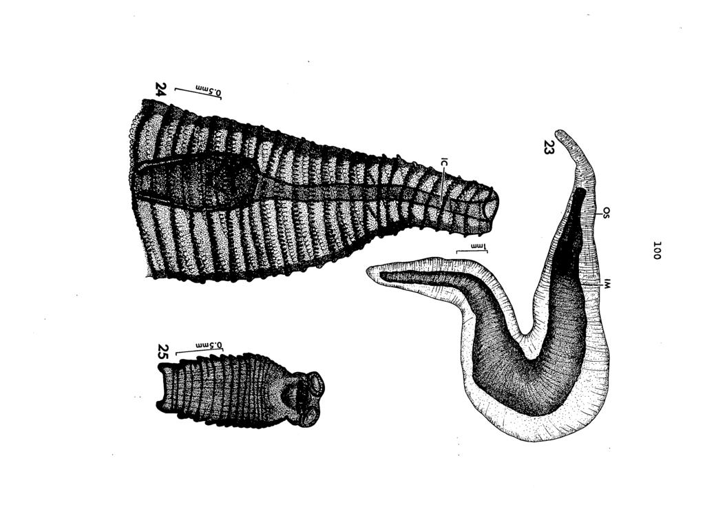
107
108 Plate IX Figure 26. Cross-section through base of rostellar region of strobilooercoid Figure 2?. Cross-section through strobilooercoid anterior to the rostellum (note the invagination canal) Figure 28, Cross-section through A, Junius naiad showing a strobilooercoid within the hemocoel Figure 29. Immature S, tenuicirrus recovered from the intestine of a laboratory-raised piedbilled grebe, after exposure to an infected dragonfly naiad
109 102 *00000 Ww4»4w i#w## 0.3mni ffltilesïâibi TR DBV m
110 Plate X Figure 30, Photomicrograph of irerj young S«tenuicirrus recovered from intestine of & naturally infected grebe, P. podiceps Figure 31. Surface view of Intestine of pied-billed grebe showing tetrad formation resulting from attachment of S, tenuicirrus

111 104
112 Plate XI Figure 32. Three tetrads, surface view, from intestine of pied-billed grebe Figure 33. Dissected intestine showing three adult S. tenuicirrus in situ (same region of intestine as shown in Figure 32)

113 10 &
Title. Author(s)YAMASHITA, Jiro; OHBAYASHI, Masashi; KONNO, Seiji. CitationJapanese Journal of Veterinary Research, 4(3): Issue Date
YAMASHITA, Jiro; OHBAYASHI, Masashi; KONNO, Seiji. CitationJapanese Journal of Veterinary Research, 4(3): Issue Date") Title STUDIES ON ECHINOCOCCOSIS : III. ON EXPERIMENTAL INF DEVELOPMENT OF ECHINOCOCCUS GRANULOSUS (BATSCH, 1786 Author(s)YAMASHITA, Jiro; OHBAYASHI, Masashi; KONNO, Seiji CitationJapanese Journal of Veterinary
Title STUDIES ON ECHINOCOCCOSIS : III. ON EXPERIMENTAL INF DEVELOPMENT OF ECHINOCOCCUS GRANULOSUS (BATSCH, 1786 Author(s)YAMASHITA, Jiro; OHBAYASHI, Masashi; KONNO, Seiji CitationJapanese Journal of Veterinary
Phylum Platyhelminthes Flatworms
 Phylum Platyhelminthes Flatworms The Acoelomates The acoelomates are animals that lack a coelom. Acoelomates lack a body cavity, and instead the space between the body wall and the digestive tract is filled
VARIATION IN MONIEZIA EXPANSA RUDOLPHI
 VARIATION IN MONIEZIA EXPANSA RUDOLPHI STEPHEN R. WILLIAMS, Miami University, Oxford, Ohio In making a number of preparations of proglottids for class study at the stage when sex organs are mature and
PSEUDANDRYA MKUZll sp. nov, ( CESTODA: HYMENOLEPIDl DAE) FROM /CHNEUMIA ALBICAUDA
 FROM /CHNEUMIA ALBICAUDA") Onderstepoort J. Vet. Res. (1963), 30 (2), 127-132 Printed by the Government Printer, Pretoria PSEUDANDRYA MKUZll sp. nov, ( CESTODA: HYMENOLEPIDl DAE) FROM /CHNEUMIA ALBICAUDA R. J. ORTLEPP, Veterinary
Onderstepoort J. Vet. Res. (1963), 30 (2), 127-132 Printed by the Government Printer, Pretoria PSEUDANDRYA MKUZll sp. nov, ( CESTODA: HYMENOLEPIDl DAE) FROM /CHNEUMIA ALBICAUDA R. J. ORTLEPP, Veterinary
MORPHOTAXONOMICAL STUDY OF A NEW CESTODE GANGESIA (GANGESIA) CHOPARAI N.SP. FROM A FRESH WATER FISH, WALLAGO ATTU FROM JALAUN (U.P.
 CHOPARAI N.SP. FROM A FRESH WATER FISH, WALLAGO ATTU FROM JALAUN (U.P.") FLORA AND FAUNA 2016 Vol. 22 No. 1 PP 115-120 ISSN 0971-6920 MORPHOTAXONOMICAL STUDY OF A NEW CESTODE GANGESIA (GANGESIA) CHOPARAI N.SP. FROM A FRESH WATER FISH, WALLAGO ATTU FROM JALAUN (U.P.) INDIA ALOK
FLORA AND FAUNA 2016 Vol. 22 No. 1 PP 115-120 ISSN 0971-6920 MORPHOTAXONOMICAL STUDY OF A NEW CESTODE GANGESIA (GANGESIA) CHOPARAI N.SP. FROM A FRESH WATER FISH, WALLAGO ATTU FROM JALAUN (U.P.) INDIA ALOK
WITH THE TABLE OF THE MORPHOLOGICAL FEATURES OF TAPEWORMS IN VAMPIROLEPIS. (Received: December 22nd, 1965)
") Japan. J. Med. Sci. Biol. 19, 51-57, 1966 *ON A NEW TAPEWORM, VAMPIROLEPIS ISENSIS, FOUND IN BATS WITH THE TABLE OF THE MORPHOLOGICAL FEATURES OF TAPEWORMS IN VAMPIROLEPIS ISAMU SAWADA Biological Laboratory,
Japan. J. Med. Sci. Biol. 19, 51-57, 1966 *ON A NEW TAPEWORM, VAMPIROLEPIS ISENSIS, FOUND IN BATS WITH THE TABLE OF THE MORPHOLOGICAL FEATURES OF TAPEWORMS IN VAMPIROLEPIS ISAMU SAWADA Biological Laboratory,
Rec. zool. Surv. India, 85(4); , 1989
; , 1989") Rec. zool. Surv. India, 85(4); 583-588, 1989 CSTODS OF DOMSTIC FOWL AT VISAKHAPATNAM WITH DSCRIPTION OF A NW SPCIS OF RAILLITINA (RAILLITINA) By SR RAMULU KOLLURI AND C. VIJAYA LAKSHMI Department of Zoology,
Rec. zool. Surv. India, 85(4); 583-588, 1989 CSTODS OF DOMSTIC FOWL AT VISAKHAPATNAM WITH DSCRIPTION OF A NW SPCIS OF RAILLITINA (RAILLITINA) By SR RAMULU KOLLURI AND C. VIJAYA LAKSHMI Department of Zoology,
Contains most of the medically important tapeworms Scolex has 4 suckers and compact vitelline gland are characteristic Range from mm to >10m
 Cyclophyllidae Contains most of the medically important tapeworms Scolex has 4 suckers and compact vitelline gland are characteristic Range from mm to >10m Family Taeniidae Taenia saginata: beef tapeworm
Title. Author(s)OHBAYASHI, Masashi. CitationJapanese Journal of Veterinary Research, 15(1): 1-3. Issue Date DOI. Doc URL.
OHBAYASHI, Masashi. CitationJapanese Journal of Veterinary Research, 15(1): 1-3. Issue Date DOI. Doc URL.") Title GRYPORHYNCHUS NYCTICORACIS YAMAGUTI, 1956 (DILEPIDID APHARYNGOSTRIGEA ARDEOLINA VIDYARTHI, 1937 (STRIGEID CINEREA JOUYI CLARK Author(s)OHBAYASHI, Masashi CitationJapanese Journal of Veterinary Research,
Title GRYPORHYNCHUS NYCTICORACIS YAMAGUTI, 1956 (DILEPIDID APHARYNGOSTRIGEA ARDEOLINA VIDYARTHI, 1937 (STRIGEID CINEREA JOUYI CLARK Author(s)OHBAYASHI, Masashi CitationJapanese Journal of Veterinary Research,
Avian Reproductive System Female
 extension Avian Reproductive System Female articles.extension.org/pages/65372/avian-reproductive-systemfemale Written by: Dr. Jacquie Jacob, University of Kentucky For anyone interested in raising chickens
THE EFFECT OF MUTILATION ON THE TAPEWORM TAENIA TAENIAEFORMIS
 THE EFFECT OF MUTILATION ON THE TAPEWORM TAENIA TAENIAEFORMIS JOE N. MILLER AND WM. P. BUNNER The reader is undoubtedly aware of work which has been done by Child (1910) and others in mutilating certain
Proteocephalus filicollis (Rud. 1810) in the Netherlands
 in the Netherlands") Proteocephalus filicollis (Rud. 1810) in the Netherlands by J.J. Willemse AND A.L.M. Veltman Zoological Laboratory, University of Amsterdam INTRODUCTION in another glass dish containing about 50 specimens
Proteocephalus filicollis (Rud. 1810) in the Netherlands by J.J. Willemse AND A.L.M. Veltman Zoological Laboratory, University of Amsterdam INTRODUCTION in another glass dish containing about 50 specimens
New Species of the Ptychobothridean Tapeworm Circumoncobohrium from Mastacembalus armatus
 New Species of the Ptychobothridean Tapeworm Circumoncobohrium from Mastacembalus armatus M B Sonune and C R Kasar 1 Department of Zoology, Shri. Shivaji Science, and Arts College, Chikhali, Dist- Buldhana,
Flatworms Flatworms Platyhelminthes dorsoventrally free-living planarian parasitic fluke tapeworm label three body layers ectoderm mesoderm
 Flatworms Flatworms are in the phylum Platyhelminthes. Flatworms are flattened dorsoventrally (top to bottom). The group includes the freshwater, free-living planarian and the parasitic fluke and tapeworm.
A Scanning Electron Microscopic Study of Eggshell Surface Topography of Leidynema portentosae and L. appendiculatum (Nematoda: Oxyuroidea)
") The Ohio State University Knowledge Bank kb.osu.edu Ohio Journal of Science (Ohio Academy of Science) Ohio Journal of Science: Volume 88, Issue 5 (December, 1988) 1988-12 A Scanning Electron Microscopic
The Ohio State University Knowledge Bank kb.osu.edu Ohio Journal of Science (Ohio Academy of Science) Ohio Journal of Science: Volume 88, Issue 5 (December, 1988) 1988-12 A Scanning Electron Microscopic
Fischthal and Kuntz (1964) reported the
 reported the") Zoological Studies 41(3): 283-287 (2002) Meristocotyle provitellaria sp. nov. (Digenea: Meristocotylidae) from Varanus salvator in China Wei Liu 1, Qing-Kui Li 2, Hsiu-Hui Shih 3 and Zhao-Zhi Qiu 1, *
Zoological Studies 41(3): 283-287 (2002) Meristocotyle provitellaria sp. nov. (Digenea: Meristocotylidae) from Varanus salvator in China Wei Liu 1, Qing-Kui Li 2, Hsiu-Hui Shih 3 and Zhao-Zhi Qiu 1, *
Frog Dissection Information Manuel
 Frog Dissection Information Manuel Anatomical Terms: Used to explain directions and orientation of a organism Directions or Positions: Anterior (cranial)- toward the head Posterior (caudal)- towards the
Ectoparasites Myobia musculi Radfordia affinis Radfordia ensifera
 Ectoparasites Fleas, ticks, and lice are uncommon in modern laboratory facilities, but may be seen on wild or feral rodents. Most ectoparasite infestations seen in rats and mice used for research are various
SEMESTER ONE 2007 INFECTION and IMMUNITY GRADUATE ENTRY PROGRAMME PARASITOLOGY PRACTICAL 9 Dr TW Jones NEMATODES
 SEMESTER ONE 2007 INFECTION and IMMUNITY GRADUATE ENTRY PROGRAMME PARASITOLOGY PRACTICAL 9 Dr TW Jones NEMATODES Objectives After this class I expect you to be able to: 1. Describe and recognise the range
Chapter 5 Male and female reproductive systems
 Chapter 5 Male and female reproductive systems This chapter begins with a description of the male and female reproductive systems followed by a section on sex determination. A good knowledge of the anatomy
Title. Author(s)YAMASHITA, Jiro; OHBAYASHI, Masashi; KITAMURA, Yukit. CitationJapanese Journal of Veterinary Research, 6(2): 89-92
YAMASHITA, Jiro; OHBAYASHI, Masashi; KITAMURA, Yukit. CitationJapanese Journal of Veterinary Research, 6(2): 89-92") Title STUDIES ON ECHINOCOCCOSIS VII. : ON THE DEVELOPMENT IN THE TAPEWORM STAGE Author(s)YAMASHITA, Jiro; OHBAYASHI, Masashi; KITAMURA, Yukit CitationJapanese Journal of Veterinary Research, 6(2): 89-92
Title STUDIES ON ECHINOCOCCOSIS VII. : ON THE DEVELOPMENT IN THE TAPEWORM STAGE Author(s)YAMASHITA, Jiro; OHBAYASHI, Masashi; KITAMURA, Yukit CitationJapanese Journal of Veterinary Research, 6(2): 89-92
TWO NEW SPECIES OF WATER MITES FROM OHIO 1-2
 TWO NEW SPECIES OF WATER MITES FROM OHIO 1-2 DAVID R. COOK Wayne State University, Detroit, Michigan ABSTRACT Two new species of Hydracarina, Tiphys weaveri (Acarina: Pionidae) and Axonopsis ohioensis
Grasshopper Dissection
 Grasshopper Dissection External Observation Locate the head, thorax, and abdomen. Observe the head. Locate the two compound eyes and the three simple eyes. 1. Why do you think grasshoppers have two types
Cestodes. Tapeworms from man and animals
 Cestodes Tapeworms from man and animals Taenia sp. The common (beef) tapeworm is several meters long. Courtesy Peters W. & Gilles H. Courtesy CDC Courtesy CDC Taenia sp. Unstained egg with four (visible)
Recommended Resources: The following resources may be useful in teaching this
 Unit B: Anatomy and Physiology of Poultry Lesson1: Internal Anatomy of Poultry Student Learning Objectives: Instruction in this lesson should result in students achieving the following objectives: 1. Identify
Vertebrates. Vertebrate Characteristics. 444 Chapter 14
 4 Vertebrates Key Concept All vertebrates have a backbone, which supports other specialized body structures and functions. What You Will Learn Vertebrates have an endoskeleton that provides support and
Biosystematic studies on Raillietina (R.) domestica (Davaineidae, Fuhrmann, 1908) from Gallus gallus domesticus
 domestica (Davaineidae, Fuhrmann, 1908) from Gallus gallus domesticus") RESEARCH PAPER Asian Journal of Bio Science, Volume 8 Issue 1 April, 2013 86-90 Received 26.10.2012; Revised 01.03.2013; Accepted 02.04.2013 Biosystematic studies on Raillietina (R.) domestica (Davaineidae,
RESEARCH PAPER Asian Journal of Bio Science, Volume 8 Issue 1 April, 2013 86-90 Received 26.10.2012; Revised 01.03.2013; Accepted 02.04.2013 Biosystematic studies on Raillietina (R.) domestica (Davaineidae,
Introduction to Helminthology
 Introduction to Helminthology HELMINTHES (WORMS) - Characteristics Eukaryotic, multicellular animals that usually have digestive, circulatory, nervous, excretory, and reproductive systems. Worms with bilateral
V. Subclass Eucestoida (Chapters 20 & 21, BLY 459, 2010)
") V. Subclass Eucestoida (Chapters 20 & 21, BLY 459, 2010) A. Characteristics (Ignore Cestodaria) 1. Differences from trematodes a. No digestive tract (1) No mouth, gut, nor anus (2) All nutrients absorbed
V. Subclass Eucestoida (Chapters 20 & 21, BLY 459, 2010) A. Characteristics (Ignore Cestodaria) 1. Differences from trematodes a. No digestive tract (1) No mouth, gut, nor anus (2) All nutrients absorbed
Title. Author(s)KAMIYA, Masao; SUZUKI, Hiroshi; VILLA-R, Bernand. CitationJapanese Journal of Veterinary Research, 27(3-4): 67. Issue Date
KAMIYA, Masao; SUZUKI, Hiroshi; VILLA-R, Bernand. CitationJapanese Journal of Veterinary Research, 27(3-4): 67. Issue Date") Title A NEW ANOPLOCEPHALINE CESTODE, ANOPLOCEPHALOIDES ROM VOLCANO RABBIT, ROMEROLAGUS DIAZI Author(s)KAMIYA, Masao; SUZUKI, Hiroshi; VILLA-R, Bernand CitationJapanese Journal of Veterinary Research, 27(3-4):
Title A NEW ANOPLOCEPHALINE CESTODE, ANOPLOCEPHALOIDES ROM VOLCANO RABBIT, ROMEROLAGUS DIAZI Author(s)KAMIYA, Masao; SUZUKI, Hiroshi; VILLA-R, Bernand CitationJapanese Journal of Veterinary Research, 27(3-4):
Beaufortia. (Rathke) ZOOLOGICAL MUSEUM - AMSTERDAM. July. Three new commensal Ostracods from Limnoria lignorum
 ZOOLOGICAL MUSEUM - AMSTERDAM. July. Three new commensal Ostracods from Limnoria lignorum") Beaufortia SERIES OF MISCELLANEOUS PUBLICATIONS ZOOLOGICAL MUSEUM - AMSTERDAM No. 34 Volume 4 July 30, 1953 Three new commensal Ostracods from Limnoria lignorum (Rathke) by A.P.C. de Vos (Zoological Museum,
Beaufortia SERIES OF MISCELLANEOUS PUBLICATIONS ZOOLOGICAL MUSEUM - AMSTERDAM No. 34 Volume 4 July 30, 1953 Three new commensal Ostracods from Limnoria lignorum (Rathke) by A.P.C. de Vos (Zoological Museum,
Reproductive physiology and eggs
 Reproductive physiology and eggs Class Business Reading for this lecture Required. Gill: Chapter 14 1. Reproductive physiology In lecture I will only have time to go over reproductive physiology briefly,
1. Examine the specimens of sponges on the lab table. Which of these are true sponges? Explain your answers.
 Station #1 - Porifera 1. Examine the specimens of sponges on the lab table. Which of these are true sponges? Explain your answers. 2. Sponges are said to have an internal special skeleton. Examine the
Presentation of Quiz #85
 Presentation of Quiz #85 ***Reminder: Slides are copyrighted and cannot be copied for publication. A 36 year old male from Columbia was admitted to the hospital with seizures. This patient had previously
30-3 Amphibians Slide 1 of 47
 1 of 47 What Is an Amphibian? What Is an Amphibian? An amphibian is a vertebrate that, with some exceptions: lives in water as a larva and on land as an adult breathes with lungs as an adult has moist
Name Class Date. After you read this section, you should be able to answer these questions:
 CHAPTER 14 4 Vertebrates SECTION Introduction to Animals BEFORE YOU READ After you read this section, you should be able to answer these questions: How are vertebrates different from invertebrates? How
Vol. XIV, No. 1, March, The Larva and Pupa of Brontispa namorikia Maulik (Coleoptera: Chrysomelidae: Hispinae) By S.
 By S.") Vol. XIV, No. 1, March, 1950 167 The Larva and Pupa of Brontispa namorikia Maulik (Coleoptera: Chrysomelidae: Hispinae) By S. MAULIK BRITISH MUSEUM (NATURAL HISTORY) (Presented by Mr. Van Zwaluwenburg
Vol. XIV, No. 1, March, 1950 167 The Larva and Pupa of Brontispa namorikia Maulik (Coleoptera: Chrysomelidae: Hispinae) By S. MAULIK BRITISH MUSEUM (NATURAL HISTORY) (Presented by Mr. Van Zwaluwenburg
Title. Author(s)YAMASHITA, Jiro; OHBAYASHI, Masashi; KITAMURA, Yukit. CitationJapanese Journal of Veterinary Research, 6(4): 226-2
YAMASHITA, Jiro; OHBAYASHI, Masashi; KITAMURA, Yukit. CitationJapanese Journal of Veterinary Research, 6(4): 226-2") Title STUDIES ON ECHINOCOCCOSIS IX. : DIFFERENCES IN DEVEL BETWEEN ECHINOCOCCUS GRANULOSUS (BATSCH, 786) AND E Author(s)YAMASHITA, Jiro; OHBAYASHI, Masashi; KITAMURA, Yukit CitationJapanese Journal of
Title STUDIES ON ECHINOCOCCOSIS IX. : DIFFERENCES IN DEVEL BETWEEN ECHINOCOCCUS GRANULOSUS (BATSCH, 786) AND E Author(s)YAMASHITA, Jiro; OHBAYASHI, Masashi; KITAMURA, Yukit CitationJapanese Journal of
Redescription of Anoplocephaloides indicata (Sawada et Papasarathorn, 1966) comb. nov. (Cestoda, Anoplocephalidae) from Tapirus indicus
 comb. nov. (Cestoda, Anoplocephalidae) from Tapirus indicus") Acta Parasitologica, 2005, 50(2), 118 123; ISSN 1230-2821 Copyright 2005 W. Stefañski Institute of Parasitology, PAS Stefański Redescription of Anoplocephaloides indicata (Sawada et Papasarathorn, 1966)
Acta Parasitologica, 2005, 50(2), 118 123; ISSN 1230-2821 Copyright 2005 W. Stefañski Institute of Parasitology, PAS Stefański Redescription of Anoplocephaloides indicata (Sawada et Papasarathorn, 1966)
VERTEBRATE READING. Fishes
 VERTEBRATE READING Fishes The first vertebrates to become a widespread, predominant life form on earth were fishes. Prior to this, only invertebrates, such as mollusks, worms and squid-like animals, would
Chimaerula bonai sp. n. (Cestoda: Dilepididae) from the bare-faced ibis, Phimosus infuscatus (Lichtenstein) (Aves: Threskiornithidae) in Paraguay
 from the bare-faced ibis, Phimosus infuscatus (Lichtenstein) (Aves: Threskiornithidae) in Paraguay") FOLIA PARASITOLOGICA 47: 303-308, 2000 Chimaerula bonai sp. n. (Cestoda: Dilepididae) from the bare-faced ibis, Phimosus infuscatus (Lichtenstein) (Aves: Threskiornithidae) in Paraguay Boyko B. Georgiev
FOLIA PARASITOLOGICA 47: 303-308, 2000 Chimaerula bonai sp. n. (Cestoda: Dilepididae) from the bare-faced ibis, Phimosus infuscatus (Lichtenstein) (Aves: Threskiornithidae) in Paraguay Boyko B. Georgiev
BREATHING WHICH IS NOT RESPIRATION
 BREATHING WHICH IS NOT RESPIRATION Breathing vs. Respiration All animals respire. A lot of people think respiration means breathing- this is not true! Breathing is the physical process of inhaling oxygen
Animal Diversity 3. jointed appendages ventral nervous system hemocoel. - marine
 Animal Diversity 3 Lab Goals To learn the bauplan (body plan) and identifying characteristics of the phyla Arthrodopa, Echinodermata, and Chordata along with the main subphyla and classes. Include, in
The Worms / Chapter 34 and Partial 35 (pgs )
") Name: The Worms / Chapter 34 and Partial 35 (pgs.712-716) 1-6. Worms are not the simple organisms most people think of at first sight. List three specific features that support the idea that worms are
Name: The Worms / Chapter 34 and Partial 35 (pgs.712-716) 1-6. Worms are not the simple organisms most people think of at first sight. List three specific features that support the idea that worms are
The family Gnaphosidae is a large family
 Pakistan J. Zool., vol. 36(4), pp. 307-312, 2004. New Species of Zelotus Spider (Araneae: Gnaphosidae) from Pakistan ABIDA BUTT AND M.A. BEG Department of Zoology, University of Agriculture, Faisalabad,
Platyhelminthes (flat worms) as parasites of medical interest
 as parasites of medical interest") Platyhelminthes (flat worms) as parasites of medical interest Type of parasites Protozoa Helminths Arthropods Nematodes Trematodes Cestodes Platyhelminthes Platyhelminthes Trematoda Planaria Monogenea
Platyhelminthes (flat worms) as parasites of medical interest Type of parasites Protozoa Helminths Arthropods Nematodes Trematodes Cestodes Platyhelminthes Platyhelminthes Trematoda Planaria Monogenea
Diversity of Animals
 Classifying Animals Diversity of Animals Animals can be classified and grouped based on similarities in their characteristics. Animals make up one of the major biological groups of classification. All
Phylum Arthropoda. Chapter 13 Part 2 of 3
 Phylum Arthropoda Chapter 13 Part 2 of 3 Phylum Arthropoda: Jointed feet General Characteristics: Exoskeleton made of chitin present and must be molted when out grown, segmented body, Jointed appendages
Laboratory 7 The Effect of Juvenile Hormone on Metamorphosis of the Fruit Fly (Drosophila melanogaster)
") Laboratory 7 The Effect of Juvenile Hormone on Metamorphosis of the Fruit Fly (Drosophila melanogaster) (portions of this manual were borrowed from Prof. Douglas Facey, Department of Biology, Saint Michael's
Laboratory 7 The Effect of Juvenile Hormone on Metamorphosis of the Fruit Fly (Drosophila melanogaster) (portions of this manual were borrowed from Prof. Douglas Facey, Department of Biology, Saint Michael's
The Rat Lungworm Lifecycle
 Hawaii Island Rat Lungworm Working Group Daniel K. Inouye College of Pharmacy University of Hawaii, Hilo The Rat Lungworm Lifecycle Rat Lungworm IPM RLWL-3 It is important to understand the lifecycle of
This is the smallest tapeworm that can affect human being but it s not really proper human tapeworm (the human is not the primary host).
.") Echinococcus Granulosus Small Tapeworm (1 cm), Cestode. This is the smallest tapeworm that can affect human being but it s not really proper human tapeworm (the human is not the primary host). The primary
Echinococcus Granulosus Small Tapeworm (1 cm), Cestode. This is the smallest tapeworm that can affect human being but it s not really proper human tapeworm (the human is not the primary host). The primary
SOME ERYTHRONEURA OF THE COMES GROUP (HOMOPTERA: CICADELLIDAE)
") SOME ERYTHRONEURA OF THE COMES GROUP (HOMOPTERA: CICADELLIDAE) DOROTHY M. JOHNSON During a study of the Erythroneura of the Comes Group, chiefly from Ohio, several undescribed species and varieties were
SOME ERYTHRONEURA OF THE COMES GROUP (HOMOPTERA: CICADELLIDAE) DOROTHY M. JOHNSON During a study of the Erythroneura of the Comes Group, chiefly from Ohio, several undescribed species and varieties were
Birds & Mammals. Chapter 15
 Birds & Mammals Chapter 15 What is a Bird? Vertebrate Endothermic Feathered 4 chambered heart Egg laying Fore-limbs adapted for flight Bones nearly hollow (allow for lighter weight) Bird Internal Anatomy
ON THE TRANSPLANTABILITY OF THE LARVA OF TEh'IA CRASSICOLLIS AND THE PROBABLE R~LE OF THE LIVER IN CYSTICERCUS DISEASE OF RATS
 ON THE TRANSPLANTABILITY OF THE LARVA OF TEh'IA CRASSICOLLIS AND THE PROBABLE R~LE OF THE LIVER IN CYSTICERCUS DISEASE OF RATS E'. 1). BULLOCI< AND M. 1%. CURTIS (Prom Coltil?lhin Uiiroersity, Iiistitute
Antihelminthic Trematodes (flukes): Cestodes (tapeworms): Nematodes (roundworms, pinworm, whipworms and hookworms):
: Cestodes (tapeworms): Nematodes (roundworms, pinworm, whipworms and hookworms):") Antihelminthic Drugs used to treat parasitic worm infections: helminthic infections Unlike protozoa, helminthes are large and have complex cellular structures It is very important to identify the causative
Antihelminthic Drugs used to treat parasitic worm infections: helminthic infections Unlike protozoa, helminthes are large and have complex cellular structures It is very important to identify the causative
SILICIFIED TURBELLARIA FROM CALICO MOUNTAINS NODULES
 ^os BULLETIN, SO. CALIF. ACADEMY OF SCIENCES Vol. 59, Part 3, 1960 SILICIFIED TURBELLARIA FROM CALICO MOUNTAINS NODULES W. DWIGHT jplerce Drawings by the author. The following is the fifth report of the
 Title EUDISTOMA LAYSANI (SLUITER) THAILAND FROM TH Author(s) Senawong, Chokechai Citation PUBLICATIONS OF THE SETO MARINE BIO LABORATORY (1972), 19(6): 427-430 Issue Date 1972-03-31 URL http://hdl.handle.net/2433/175735
By H. G. JOHNSTON, Ames, Iowa.
 Dec., 19930 Bulletin of the Brooklyn Entomological Society 295 FOUR NEW SPECIES OF MIRIDAE FROM TEXAS (HEMIPTERA).* By H. G. JOHNSTON, Ames, Iowa. Phytocoris conspicuus n. sp. This species is readily distinguished
Poultry Skillathon 2017
 Age Divisions: Junior (8-11) Intermediate (12-14) Senior (15-18) Exhibitors will participate in age-based Skillathons. This study guide includes all topics an exhibitor might be tested on. Youth will only
Millipedes Made Easy
 MILLI-PEET, Introduction to Millipedes; Page - 1 - Millipedes Made Easy A. Introduction The class Diplopoda, or the millipedes, contains about 10,000 described species. The animals have a long distinguished
Comparing Life Cycles
 Image from Wikimedia Commons Pre-Visit Activity Grade Two Comparing Life Cycles Specific Learning Outcomes 2-1-01: Use appropriate vocabulary related to the investigations of growth and changes in animals.
Phylum Echinodermata. Biology 11
 Phylum Echinodermata Biology 11 General characteristics Spiny Radial symmetry Water vascular system Endoskeleton Endoskeleton Hard, spiny, or bumpy endoskeleton covered with a thin epidermis. Endoskeleton
DLS Sample Preparation Guide
 DLS Sample Preparation Guide The Leica TCS SP8 DLS is an innovative concept to integrate the Light Sheet Microscopy technology into the confocal microscope. Due to its unique optical architecture samples
Brine Shrimp Investigation AP Biology Name: Per:
 Brine Shrimp Investigation AP Biology Name: Per: Background Have you ever gone on a hike and come across an animal that blends in so well with its surroundings that you almost did not notice it? Camouflage
A comparison of placental tissue in the skinks Eulamprus tympanum and E. quoyii. Yates, Lauren A.
 A comparison of placental tissue in the skinks Eulamprus tympanum and E. quoyii Yates, Lauren A. Abstract: The species Eulamprus tympanum and Eulamprus quoyii are viviparous skinks that are said to have
HISTOPATHOLOGY. Introduction:
 Introduction: HISTOPATHOLOGY Goats and sheep are the major domestic animal species in India. Much of the economy of the country has been depend upon the domestication of these animals. Especially economy
NECROPSY FORM STRAND LOCATION: FLOATING IN VAQUITA REFUGE BY MX TIME: 10 AM
 NECROPSY FORM FIELD #: Ps 9 NECROPSY DATE: April 4 2018 SPECIES: PHOCOENA SINUS STRAND DATE: March 28 2018 AGE CLASS: ADULT STRAND LOCATION: FLOATING IN VAQUITA REFUGE BY MX NAVY, BAJA CALIFORNIA, MX SEX:
posterior part of the second segment may show a few white hairs
 April, 1911.] New Species of Diptera of the Genus Erax. 307 NEW SPECIES OF DIPTERA OF THE GENUS ERAX. JAMES S. HINE. The various species of Asilinae known by the generic name Erax have been considered
ECHINOCOCCUS GRANULOSUS
 48 ECHINOCOCCUS GRANULOSUS 48.1 INTRODUCTION E granulosus are small tape worms that parasitize the intestines of carnivores like dogs. About one million people are infected with this tape worm worldwide.
Insects Associated with Alfalfa Seed Production
 Agdex 121/620-1 Insects Associated with Alfalfa Seed Production This field guide was prepared to enable growers of seed alfalfa to quickly identify their pest and beneficial insects. The important distinguishing
Feline and Canine Internal Parasites
 Feline and Canine Internal Parasites Internal parasites are a very common problem among dogs. Almost all puppies are already infected with roundworm when still in the uterus, or get the infection immediately
4. List 3 characteristics of all arthropods. a. b. c. 5. Name 3 main arthropod groups.
 Arthropod Coloring Worksheet Arthropods (jointed appendages) are a group of invertebrate animals in the Kingdom Animalia. All arthropods have a hard exoskeleton made of chitin, a body divided into segments,
Fishes, Amphibians, Reptiles
 Fishes, Amphibians, Reptiles Section 1: What is a Vertebrate? Characteristics of CHORDATES Most are Vertebrates (have a spinal cord) Some point in life cycle all chordates have: Notochord Nerve cord that
cyst&' appeared to be of two kinds-one smaller and Smnith "is inclined to regard these epithelial cell parasites as
 COCCIDIA IN SUBEPITHELIAL INFECTIONS OF THE INTESTINES OF BIRDS PHILIP B. HADLEY From the Agricultural Experiment Station of the Rhode Island State College' Received for publication, July 10, 1916 In an
Diurus, Pascoe. sp. 1). declivity of the elytra, but distinguished. Length (the rostrum and tails 26 included) mm. Deep. exception
. declivity of the elytra, but distinguished. Length (the rostrum and tails 26 included) mm. Deep. exception") 210 DIURUS ERYTIIROPUS. NOTE XXVI. Three new species of the Brenthid genus Diurus, Pascoe DESCRIBED BY C. Ritsema+Cz. 1. Diurus erythropus, n. sp. 1). Allied to D. furcillatus Gylh. ²) by the short head,
210 DIURUS ERYTIIROPUS. NOTE XXVI. Three new species of the Brenthid genus Diurus, Pascoe DESCRIBED BY C. Ritsema+Cz. 1. Diurus erythropus, n. sp. 1). Allied to D. furcillatus Gylh. ²) by the short head,
A NEW AUSTROSQUILLA (STOMATOPODA) FROM THE
 FROM THE") A NEW AUSTROSQUILLA (STOMATOPODA) FROM THE MARQUESAS ISLANDS BY ALAIN MICHEL Centre O.R.S.T.O.M., Noumea, New Caledonia and RAYMOND B. MANNING Smithsonian Institution, Washington, U.S.A. The At s,tstrosqzlilla
A NEW AUSTROSQUILLA (STOMATOPODA) FROM THE MARQUESAS ISLANDS BY ALAIN MICHEL Centre O.R.S.T.O.M., Noumea, New Caledonia and RAYMOND B. MANNING Smithsonian Institution, Washington, U.S.A. The At s,tstrosqzlilla
Nematoda. Round worms Feeding and Parasitism
 Nematoda Round worms Feeding and Parasitism Nematoda Have pseudocoelom Live in many environments Parasitic Important decomposers Covered with cuticle Trichinella spiralis see fig 18.8B Nematode Diets and
Field and Laboratory Study Evaluating the Possibility of Manodistomum syntomentera Causing Malformations In Frogs of the Mississippi River Valley
 11 Field and Laboratory Study Evaluating the Possibility of Manodistomum syntomentera Causing Malformations In Frogs of the Mississippi River Valley Laurie Carter Faculty Sponsor: Dr. Daniel Sutherland,
Honey Bees. Anatomy and Function 9/26/17. Similar but Different. Honey Bee External Anatomy. Thorax (Human Chest): 4 Wings & 6 Legs
: 4 Wings & 6 Legs") Honey Bee Anatomy and Function How Honey Bees are Built and How the Function People Eat: Everything - Meat and Potatoes Omnivores Meat and Vegetables Digest: Stomach & Intestines Excrete: Feces and Urine
Honey Bee Anatomy and Function How Honey Bees are Built and How the Function People Eat: Everything - Meat and Potatoes Omnivores Meat and Vegetables Digest: Stomach & Intestines Excrete: Feces and Urine
TWO NEW PINE-FEEDING SPECIES OF COLEOTECHNITES ( GELECHIIDAE )
") Journal of the Lepidopterists' Society 32(2), 1978, 118-122 TWO NEW PINE-FEEDING SPECIES OF COLEOTECHNITES ( GELECHIIDAE ) RONALD W. HODGES l AND ROBERT E. STEVENS2 ABSTRACT. Two new species of moths,
Journal of the Lepidopterists' Society 32(2), 1978, 118-122 TWO NEW PINE-FEEDING SPECIES OF COLEOTECHNITES ( GELECHIIDAE ) RONALD W. HODGES l AND ROBERT E. STEVENS2 ABSTRACT. Two new species of moths,
Title ON DAUGHTER CYSTS OF COENURUS SERIALIS GERVAIS, Author(s)YAMASHITA, Jiro; OHBAYASHI, Masashi; KONNO, Seiji
YAMASHITA, Jiro; OHBAYASHI, Masashi; KONNO, Seiji") Title ON DAUGHTER CYSTS OF COENURUS SERIALIS GERVAIS, 1847 Author(s)YAMASHITA, Jiro; OHBAYASHI, Masashi; KONNO, Seiji CitationJapanese Journal of Veterinary Research, 5(1): 14-18 Issue Date 1957-03-25
Title ON DAUGHTER CYSTS OF COENURUS SERIALIS GERVAIS, 1847 Author(s)YAMASHITA, Jiro; OHBAYASHI, Masashi; KONNO, Seiji CitationJapanese Journal of Veterinary Research, 5(1): 14-18 Issue Date 1957-03-25
Echinoderms are marine animals with spiny endoskeletons, water-vascular systems, and tube feet; they have radial symmetry as adults.
 Section 1: Echinoderms are marine animals with spiny endoskeletons, water-vascular systems, and tube feet; they have radial symmetry as adults. K What I Know W What I Want to Find Out L What I Learned
NOTE XVII. Dr. A.A.W. Hubrecht. which should he in accordance with. of my predecessors. alive or in excellent. further
 further either EUROPEAN NEMERTEANS. 93 NOTE XVII. New Species of European Nemerteans. First Appendix to Note XLIV, Vol. I BY Dr. A.A.W. Hubrecht In the above-mentioned note, published six months ago, several
INSTITUTE FOR STRATEGIC BIOSPHERIC STUDIES CONFERENCE CENTER HUNTSVILLE, TEXAS
 INSTITUTE FOR STRATEGIC BIOSPHERIC STUDIES CONFERENCE CENTER HUNTSVILLE, TEXAS Mantis/Arboreal Ant Species September 2 nd 2017 TABLE OF CONTENTS 1.0 INTRODUCTION... 3 2.0 COLLECTING... 4 3.0 MANTIS AND
Some Facts about... Amphibians
 Amphibians Amphibians are cold-blooded vertebrates that live part of their lives in water and part on land. Amphibians eggs are laid in water and they are born there. They begin their lives with gills
FROG DISSECTION. a. Why is there a difference in size proportion between the hind and fore limbs?
 FROG DISSECTION External Anatomy 1. The division of a frog s body includes the head, trunk and limbs. Examine the front and hind limbs of the frog. The hind limbs are the long, more muscular limbs of the
Phylogeny of Animalia (overview)
") The Diversity of Animals 2 Chapter 23 Phylogeny of Animalia (overview) Key features of Chordates Phylum Chordata (the Chordates) includes both invertebrates and vertebrates that share (at some point in
The Diversity of Animals 2 Chapter 23 Phylogeny of Animalia (overview) Key features of Chordates Phylum Chordata (the Chordates) includes both invertebrates and vertebrates that share (at some point in
A DESCRIPTION OF THE LABORATORY-REARED FIRST AND SECOND ZOEAE OF PORTUNUS X At IT US it (STIMPSON) (BRACHYURA, DECAPODA)
 (BRACHYURA, DECAPODA)") REPRINT FROM Calif. Fish and Game, 60(2) : 74-78. 1974. A DESCRIPTION OF THE LABORATORY-REARED FIRST AND SECOND ZOEAE OF PORTUNUS X At IT US it (STIMPSON) (BRACHYURA, DECAPODA) J. R. RAYMOND ALLY & r*
REPRINT FROM Calif. Fish and Game, 60(2) : 74-78. 1974. A DESCRIPTION OF THE LABORATORY-REARED FIRST AND SECOND ZOEAE OF PORTUNUS X At IT US it (STIMPSON) (BRACHYURA, DECAPODA) J. R. RAYMOND ALLY & r*
Body Parts and Products (Sessions I and II) BROWARD COUNTY ELEMENTARY SCIENCE BENCHMARK PLAN
 BROWARD COUNTY ELEMENTARY SCIENCE BENCHMARK PLAN") activities 22&23 Body Parts and Products (Sessions I and II) BROWARD COUNTY ELEMENTARY SCIENCE BENCHMARK PLAN Grade K Quarter 3 Activities 22 & 23 SC.F.1.1.1 The student knows the basic needs of all living
activities 22&23 Body Parts and Products (Sessions I and II) BROWARD COUNTY ELEMENTARY SCIENCE BENCHMARK PLAN Grade K Quarter 3 Activities 22 & 23 SC.F.1.1.1 The student knows the basic needs of all living
Emerging Adults BROWARD COUNTY ELEMENTARY SCIENCE BENCHMARK PLAN. SC.F The student describes how organisms change as they grow and mature.
 activity 27 Emerging Adults BROWARD COUNTY ELEMENTARY SCIENCE BENCHMARK PLAN Grade K Quarter 3 Activity 27 SC.F.1.1.3 The student describes how organisms change as they grow and mature. SC.H.1.1.1 The
Phylum Echinodermata -sea stars, sand dollars, sea
 Echinoderms Phylum Echinodermata -sea stars, sand dollars, sea urchins & sea cucumber -marine -deuterostomes -more closely related to chordates, than to other invertebrates -no head or any other sign of
Diplurans. Classification Life History & Ecology Distribution. Major Families Fact File Hot Links
 DIPLURA Diplurans The name Diplura, derived from the Greek words "diplo-" meaning two and "ura" meaning tails, refers to the large cerci at the rear of the abdomen. Classification Life History & Ecology
New species of Cinclotaenia Macy, 1973 (Cyclophyllidea: Dilepididae) from Cinclus leucocephalus Tschudi (Passeriformes: Cinclidae) in Bolivia
 from Cinclus leucocephalus Tschudi (Passeriformes: Cinclidae) in Bolivia") University of Nebraska - Lincoln DigitalCommons@University of Nebraska - Lincoln Faculty Publications from the Harold W. Manter Laboratory of Parasitology Parasitology, Harold W. Manter Laboratory of October
University of Nebraska - Lincoln DigitalCommons@University of Nebraska - Lincoln Faculty Publications from the Harold W. Manter Laboratory of Parasitology Parasitology, Harold W. Manter Laboratory of October
Science Read. 06 Feb. 2.8m-long tapeworm found in Singapore patient who had no symptoms
 Science Read Issue 04 06 Feb Career Guidance Interesting Science Real Life Application Real Time News Upper Secondary 2.8m-long tapeworm found in Singapore patient who had no symptoms Janice Tai, Social
BREEDING ECOLOGY OF THE LITTLE TERN, STERNA ALBIFRONS PALLAS, 1764 IN SINGAPORE
 NATURE IN SINGAPORE 2008 1: 69 73 Date of Publication: 10 September 2008 National University of Singapore BREEDING ECOLOGY OF THE LITTLE TERN, STERNA ALBIFRONS PALLAS, 1764 IN SINGAPORE J. W. K. Cheah*
EGG STAGE. 1. How many eggs does a female Monarch usually lay on one milkweed plant? Given a choice, what age plant, or leaves, does she prefer?
 EGG STAGE 1. How many eggs does a female Monarch usually lay on one milkweed plant? Given a choice, what age plant, or leaves, does she prefer? 2. The egg stage lasts 1-3 days. Look at the egg that you
Parasites of the relict fauna of Ceylon.
 Annales de Parasitologie (Paris), t. 46, 1971, n 5, pp. 575 à 588 Parasites of the relict fauna of Ceylon. II. New species of Cyclophyllidean Cestodes from small hill-vertebrates by H. CRUSZ and V. SANMUGASUNDERAM
NEW SPECIES OF BEAR-ANIMALCULE FROM THE
 NEW SPECIES OF BEAR-ANIMALCULE FROM THE COAST OF NORTH CAROLINA. By W. P. Hay, Of the Washington (Diftfr-ict of Columbia) High Schools. For many years the presence of marine species of tardigrades, or
Characteristics of a Reptile. Vertebrate animals Lungs Scaly skin Amniotic egg
 Reptiles Characteristics of a Reptile Vertebrate animals Lungs Scaly skin Amniotic egg Characteristics of Reptiles Adaptations to life on land More efficient lungs and a better circulator system were develope
Capture and Marking of Birds: Field Methods for European Starlings
 WLF 315 Wildlife Ecology I Lab Fall 2012 Capture and Marking of Birds: Field Methods for European Starlings Objectives: 1. Introduce field methods for capturing and marking birds. 2. Gain experience in