HUMAN PARASITOLOGY LABORATORY
|
|
|
- Dominic Baldwin
- 6 years ago
- Views:
Transcription


")


")
")
1 HUMAN PARASITOLOGY LABORATORY Biology (.pdf edition) Steve J. Upton Clonorchis sinensis (Chinese liver fluke) (Drawing by Jarrod Wood) Division of Biology, Kansas State University
2 2 HUMAN PARASITOLOGY LABORATORY OUTLINE - BIOLOGY 546 Tuesdays 8:30-10:20 (228 Ackert) JAN 16 JAN 23 JAN 30 FEB 06 FEB 13 FEB 20 FEB 27 MAR 06 MAR 13 MAR 20 MAR 27 APR 03 APR 10 APR 17 APR 24 MAY 01 INTRODUCTION AND SLIDE BOX ASSIGNMENTS DIGENES CESTODES DIGENES & CESTODES (Review) LAB EXAM #1 9:30 am (60 points) (Digenes & Cestodes) NEMATODES NEMATODES LAB EXAM #2 9:30 am (60 points) (Nematodes) PROTOZOA (flagellates) SPRING BREAK PROTOZOA (amoebae) PROTOZOA (apicomplexa, ciliates, miscellaneous groups) PROTOZOA (review of all phyla) LAB EXAM #3 9:30 am (60 points) (Protozoa) ARTHROPODA LAB EXAM #4 9:30 am (60 points) (Arthropoda) TOTAL POINTS POSSIBLE IN LAB: 240 (grading will be on a 90%, 80%, 70%, 60%... grading scale) 2
3 3 HUMAN PARASITOLOGY (BIOL. 546) LABORATORY MANUAL (Revised November, 2006) This laboratory is designed to teach students at Kansas State University the basics of identification of common eukaryotic parasites of humans. This course is targeted for sophomore/junior students and at least one course in General Biology is required as a prerequisite. In addition, Biology 545 (human parasitology lecture) is required either as a prerequisite or co-requisite. It is often helpful to also bring the Biology 545 text with you to each laboratory. Students will be required to work in groups of 2-3 and share an assigned slide box containing just under 100 permanently preserved specimens. We do not have enough slide boxes or microscopes for students to work alone. So, early during the first class meeting, find someone with a favorable phenotype and pair up. Then, once slide boxes are assigned, please check to insure that all slides are present. Students will be responsible for slides "disappearing" from the boxes at the end of the semester. Of course, an occasional slide is inadvertently broken during the semester and we have budgeted for that. However, as specimens average about $5.00 each, we ask students to be particularly careful when handling the slides. BE SURE TO OPEN SLIDE BOXES SO THAT THE LID IS FACING UPRIGHT. Each student will use microscopes extensively this semester, and students need to be familiar with the use of these microscopes prior to examination of the slides. Try not to pulverize slides and coverslips by jamming the high objective lens down onto the slide. One common way of misplacing slides is to accidentally leave them on the stage of your microscope at the end of the class period, so please check microscope stages before you leave at the end of each period. The laboratory will only be open Tuesday mornings between 8:30 and 10:20 am. This provides ample time to examine slides and learn the identifications. Please note that the laboratory will NOT be open at any other time, and there will be NO exceptions. If you have another course that conflicts with a portion of the laboratory, then you simply need to make a decision about which course you want to take. A duplicate laboratory will NOT be created for you, and a duplicate laboratory practical will NOT be created for you. Perhaps a no brainer for most of you, but I receive multiple requests for these types of special favors each year. In addition, you may NOT take slides or microscopes out of the laboratory or to another room. If you miss a class, you have 4 options: 1) study intensely during the next laboratory to make up the material; 2) go to the www and view images that I and others have provided; 3) spend time looking at images on the CD ROM accompanying the Biology 545 textbook if you ordered one; or 4) drop the course. During some laboratories, demonstrations may be set up to supplement the slide collections. These demonstrations generally consist of specimens either too large, too valuable, or too rare to be included in the slide boxes. You may be tested over these demonstrations so it is important that you examine them carefully during the laboratory period (NOTE: be aware that most demonstrations will be set up for ONE laboratory period only). The following pages represent the laboratory notebook to accompany the slide collections. This notebook is designed to answer many of the questions students have asked over the years, although new questions arise continually. In order to save laboratory time, I suggest that you read about each group of parasites prior to the beginning of the laboratory period. On an exam, students will not only be responsible for identification of specimens, but also for the content of the laboratory manual, as well as the life-cycles, sites of infection, intermediate hosts that may be utilized, basic morphology, and any associated pathology. Most of this material can be found below in the text and tables accompanying each section. All of this material and much more will be covered in the lecture portion of the course, Biology 545, which you are supposed to be taking anyway. TEXT: The laboratory manual for this course is contained on these pages and can be downloaded directly. In addition, the text for Human Parasitology lecture (Biology 545) often proves highly useful. EXAMS: Four exams are scheduled for this laboratory. Each exam will be at 9:30-10:20 am. These will be practical exams, where students cycle through 15 stations and view a specimen at each station (often through a microscope). There will be two, 2 point questions pertaining to a specimen at each station. The first question will generally ask "what is the species?" The second question will ask about some aspect of parasite biology (i.e. "what is the intermediate host," "where in the host are the adult stages located," etc.). The third exam, over protozoa, will be a kodachrome practical. Separate practicals will NOT be set up for students who miss exams. 3
4 4 TABLE OF CONTENTS Subject page Laboratory Outline Introductory Remarks Table of Contents Digenes Fig. 1 (structure of hypothetical digene) Adapted from Fig. 1, P Medical Protozoology & Helminthology, 1965, US Naval Medical School, National Naval Medical Center, Bethesda. Fig. 2 (morphologic types of digenes redrawn from originals) Fig. 3 (digene larvae) Adapted from Fig. 31, P Medical Protozoology & Helminthology, 1965, US Naval Medical School, National Naval Medical Center, Bethesda. Fig. 4 (line drawings of digenes & eggs) Composite from Figs , P Human Protozoology and Helminthology, 1960, L.R.S. MacFarlane, ed. Williams & Wilkins Co., Baltimore. Table 1 (digene biology) Cestodes Fig. 5 (line drawings of cestodes) Adapted from Fig. 44, P. 146 & Fig. 82, P Medical Protozoology & Helminthology, 1965, US Naval Medical School, National Naval Medical Center, Bethesda. Table 2 (cestode biology) Nematodes Fig. 6 (nematode eggs, larvae, & hookworms) First two rows from Figs. 24, 25c, 26, 27, 29b,c, 30a, 31b, P Human Protozoology & Helminthology, 1960, L.R.S. MacFarlane, ed. Williams & Wilkins Co., Baltimore. Third row from P and P. 3-38, Laboratory Procedures in Parasitology, 1961, TM , Dept. of the Army Technical Manual, Washington, DC. & Fig. 80, P. 198, Medical Protozoology & Helminthology, 1965, US Naval Medical School, National Naval Medical Center, Bethesda. Table 3 (nematode biology) Table 4 (characteristics of human microfilariae) Protozoa Fig. 7 (flagellates & ciliates) From Figs. 10, 11, 13-17, P Human Protozoology & Helminthology, 1960, L.R.S. MacFarlane, ed. Williams & Wilkins Co., Baltimore. Trypomastigote and amastigote from Fig. 24, P Medical Protozoology & Helminthology, 1965, USN Medical School, National Naval Medical Center, Bethesda. Fig. 8 (amoebae) From Figs. 1-9, P Medical Protozoology & Helminthology, 1965, USN Medical School, National Naval Medical Center, Bethesda. Table 5 (protozoan biology) Arthropoda Fig. 9 (arthropods) From figures on P.3,15,31,32,38,41,66,168. CDC Pictoral keys to Arthropods, Reptiles, Birds, & Mammals of Public Health Significance, US Dept of Health, Education, and Welfare, PHS, Atlanta, Georgia. Fig. 10 (common ticks of Kansas) Adapted from plates 07 and 39, Ticks of Veterinary Importance, USDA handbook 485, 1976; plate 3, P. 22, The genera Dermacentor and Otocentor (Ixodidae) in the United States, with studies in variation, 1938, Cooley, R.A., U.S. Treasury Dept. 4
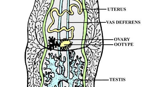
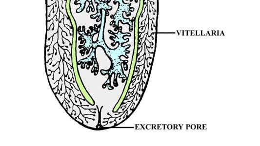
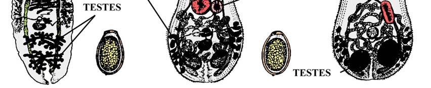
5 5 SECTION 1. DIGENETIC TREMATODES (FIGS. 1-4) The digenetic trematodes ("flukes") are parasitic flatworms in the phylum Platyhelminthes and are found in a variety of animals, mainly vertebrates. Most are dorso-ventrally flattened and possess a muscular oral sucker that surrounds the mouth. In addition, the majority also possess a midventral or posterior acetabulum (ventral sucker) (Fig. 1). The digestive tract of a fluke normally consists of a short, muscular esophagus (often surrounded by a muscular pharynx), which then splits into a pair of blind intestinal cecae. Generally, tissues are drawn into the mouth which are then eroded by the strong pumping action of the pharynx. Species such as the schistosomes, which live in the blood vessels and suck blood, do not have a pharynx. Most trematodes are hermaphroditic (except the schistosomes) and many self-fertilize. The male reproductive system usually consists of two testes (1-several hundred), each of which has a vas efferens that connect to form a common duct, the vas deferens. The vas deferens leads to the genital pore, which usually has associated structures such as an internal seminal receptacle for sperm storage, a prostate gland that may add secretions to the sperm, and a cirrus, the male copulatory organ. The female reproductive system is more complicated than the male system and consists of a single ovary, an oviduct, a seminal receptacle for sperm storage, vitelline glands along the lateral margins of the body that provide material for egg shell formation, a series of glandular structures that aid in egg shell maturation (i.e Mehlis gland, ootype, Lauer's canal, etc.), a uterus which may be filled with eggs, and perhaps a muscular modification of the end of the uterus termed a metraterm. The body of a fluke is covered by a living layer of cells termed a tegument, which functions in nutrient absorption. Thus, flukes can digest and absorb nutrients not only across the gut wall, but also across the outer body. Ornamentation, such as spines, are often present within the tegument and can often be seen with the light microscope. Flukes can be "loosely" catagorized based on the location of suckers (Fig. 2). A monostome is a fluke that has an oral sucker only; an amphistome has an oral sucker plus a ventral sucker at the posterior end of the body; a distome has an oral sucker and a ventral sucker, although the latter is located somewhere other than the posterior end; a holostome is a type of distome that has the body split into distinct anterior and posterior portions; and a echinostome has long tegumental spines surrounding the oral sucker. The life cycle of a digene is indirect (Fig. 3), involving two or more hosts in the life cycle. Eggs of most species are operculate (with a lid; except schistosomes), and laid either embryonated or unembryonated. The eggs of some species hatch in water while those of others require ingestion by the appropriate host. Once the egg hatches, however, a ciliated miracidium emerges which penetrates into the tissue of the host (usually a snail) with anterior penetration glands. Depending upon the species, the miricidium may develop into a sporocyst (an asexual reproductive structure without mouth or intestine that may give rise to daughter sporocysts, rediae, or cercariae) or redia (an asexual reproductive structure with mouth and gut that may give rise to daughter rediae or cercariae). Whichever phases of asexual development a species possesses, the final end product of asexual multiplication are tailed cercariae. Depending upon the species, these sexually immature forms may do one of several things: Some species encyst on vegetation as metacercariae and remain dormant until eaten by an appropriate host; some penetrate the skin of an animal (especially fish) and remain dormant as metacercariae until the fish is eaten by the final host; others may penetrate the skin of the final host directly (i.e. schistosomes); thus, forming no metacercaria. Once inside the final vertebrate host, the cercaria casts off its tail, migrates to its target organ(s), and finally matures into an adult. In the first two laboratories, you are required to be able to identify 9 species of adult flukes and differentiate between six types of trematode eggs (Fig. 4). In addition, you should be able to distinguish leeches from digenes. DO NOT USE OIL IMMERSION to examine any of the specimens. All adult trematodes should be viewed using either the dissecting microscopes or the lowest magnification (4x or 10x) of your brightfield microscope. Eggs should be examined using 4x, 10x, or 40x magnification. The following is a list of slides that you will be responsible for knowing: 5
6 6 SLIDE SERIES (Slides 1-14) Needed for each student group: Laboratory manual; compound dissecting microscope (and light) to view adult specimens; compound (regular brightfield) microscope for eggs; slide boxes with slides Slide 1. Fasciolopsis buski, adult. This is a relatively common parasite of humans and pigs in the orient. Adults live in the intestine and produce eggs similar to Fasciola hepatica. Note the large size of the worm, although small Fasciolopsis buski may appear similar to large Fasciola hepatica. The 2 species can be distinguished as 1) Fasciolopsis buski does not usually possess a "snout" (cephalic cone) whereas Fasciola hepatica usually does, and 2) Fasciolopsis buski has unbranched intestinal ceca whereas Fasciola hepatica has branched cecae. This latter feature is considered the most important. Slide 2. Fasciola hepatica, adult. This parasite is not native to the Western hemisphere, but was introduced two or three hundred years ago. Adults are large worms with dendritic, tandem testes, a dendritic ovary, and oral and ventral suckers that are relatively small in proportion to the body and located anteriorly. Internal details are often difficult to discern because of the dendritic nature of so many key structures. Note the "snout" (cephalic cone) on the worm, where the oral sucker resides, and especially the branched intestinal cecae which can be best viewed laterally near the anterior end of the worm. Adult worms live in the gall bladder and bile ducts of various mammals, only occasionally infecting humans. Maturing worms migrate through the liver for several weeks consuming tissue, often resulting in pronounced liver damage. Slide 3. Fasciola hepatica, eggs. These are very large eggs, easy to see under low power, and measure x micrometers. They do not possess shoulders and are passed unembryonated in the feces. The eggs are almost identical to those of Fasciolopsis buski, the giant intestinal fluke of Asia. Slide 4. Echinostoma trivolvus, adult. These are fairly large, elongate worms. Note that the ventral sucker is larger than the oral sucker. Testes are tandem, located posteriorly, and the oral sucker has a collar of large spines which is an important taxonomic feature. Several genera and species may infect humans, and in North America the most common species found is Echinostoma trivolvus which should have 37 spines comprising its collar. Slide 5. Paragonimus westermani, adult. These are large, fleshy, ovoid worms with a spined tegument, measure about one-half centimeter in length, possess testes that are lobed and opposite to one another, and have a ventral sucker near the middle of the body. They occur as adults in the lungs of mammals. The Paragonimus westermani and several other species infect humans in Asia whereas Paragonimus kellicotti lives in mammals in North America. Other species occur in other countries. Internal details of these worms is very difficult to discern for the novice, even when viewing a well prepared specimen. Slide 6. Paragonimus sp., eggs. Eggs are large, operculate, often possess distinct shoulders (a lip running around the edge of the operculum), and measure x micrometers. They are found in the sputum and feces, and are unembryonated when they are passed in the feces. Slide 7. Clonorchis sinensis, adult. This is a relatively small but elongate worm, with dendritic (branched) testes located posteriorly, a ventral sucker in the anterior one-third of the body, and lateral vitelline follicles that are posterior to the ventral sucker but anterior to ovary. Two closely related species, Opisthorchis felineus (Europe, Asia) and O. viverrini (Asia), have lobate (rather than dendritic) testes. Adults live in the bile ducts of humans in some Asiatic countries. A related worm, Metorchis conjunctus, occurs in mink in the Hudson Bay watershed in Northern Ontario. Occasional human infections are reported from eating undercooked fish containing metacercariae. Clonorchis sinensis is an excellent worm for viewing most of the internal details depicted in Figure 1. Slide 8. Clonorchis sinensis, eggs. These eggs are very small, measuring x micrometers. They may be tan or yellowish in color. Some eggs have shoulders, they are passed embryonated, and one can sometimes see a tiny knob at the end opposite of the operculum. Eggs are so similar to those of Metagonimus, Heterophyes and others that it is nearly impossible for a novice to distinguish the three genera apart based solely on egg structure. Eggs are found in the feces. 6
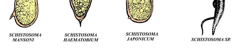
7 Slide 9. Dicrocoelium dendriticum, adult. This is an elongate worm with a ventral sucker in the anterior 1/4 of the body, lateral vitelline follicles, and irregularly-shaped testes that are tandem or oblique and in the anterior 1/2 of body (the latter a feature which helps distinguishes it from Clonorchis). This fluke lives in the bile ducts of mammals, such as cattle and deer, and is distributed in Europe and (following its introduction) portions of the Northeastern United States. Humans are accidental hosts. Slide 10. Nanophyetus salmincola, adult. This is a pin-head size ovoid worm with a ventral sucker in the middle of the body and 2 large, ellipsoidal testes that are opposite from one another (when stained properly and not obscured by a uterus filled with eggs). It lives in the intestine of many mammals and occurs along the NW coast of North America, and Siberia. Slide 11. heterophyid, adult. This group contains 50+ species worldwide capable of infecting humans. All are small and possess both oral & ventral suckers. One common species, Metagonimus yokagawi, has a submedian (off to one side) ventral sucker and oblique testes. Another species, Heterophyes heterophyes, has a median rather than submedian ventral sucker, opposite rather than oblique testes, and a muscular outgrowth of the ventro-genital sac that envelops the genital pore. This outgrowth resembles a second ventral sucker. Since many of these worms look similar, and because many specimens were mis-labeled by the supplier plus they were poorly processed, simply learn these as a "heterophyids." Slide 12. Schistosoma mansoni, adults. These are slender and elongate, blood-vascular worms, with separate sexes and very anterior suckers. Both male and female worms are represented, the latter of which is within the gynecophoral canal of the male. Slide 13. Schistosoma mansoni, eggs. The eggs are found in the feces, are large and ellipsoidal, and measure x micrometers. Eggs are passed embryonated and possess a large, lateral spine which is the most important, distinctive characteristic. Slide 14. Schistosoma japonicum, eggs. Also found in the feces, these eggs are spherical or subspherical, embryonated, and measure x micrometers. If positioned properly, each egg can be seen to possess a tiny, rudimentary spine laterally. Slide 15. Schistosoma haematobium, eggs. Eggs are found in the urine, are ellipsoidal, and measure x micrometers. They are passed embryonated and possess a sharp, terminal spine that is distinctive. 7 Demonstration: Leeches Several specimens of leeches will be made available. These highly muscular and segmented annelids are sometimes confused with amphistome flukes. Note among other things that although many leeches are dorso-ventrally flattened and have a posterior sucker, they have a complete (rather than incomplete) gut, display segmentation, and possess a highly developed musculature. Demonstrations: Bottled speciemens A variety of bottled specimens of digenes will be made available. These are intended to show students how the actual digenes appear prior to staining and mounting onto microscope slides. 7

8 8 FIGURE 1. HYPOTHETICAL DIGENE 8

9 9 FIGURE 2. MORPHOLOGICAL TYPES OF DIGENES FIGURE 3. DIGENE LARVAE 9


10 10 FIGURE 4. DIGENES AND EGGS 10
11 11 Table 1. Basic information on digene biology SPECIES PRINCIPLE DEFINITIVE HOST(S) SITE OF INFECTION METACERCARIAE LOCATION OF EGGS Clonorchis sinensis humans bile ducts FW fish feces Dicrocoelium dendriticum; Dicrocoelium hospes Echinostomes ruminants bile ducts ants feces fish eating mammals; birds small intestine or bile ducts fish or invertebrates Fasciolopsis buski primates; swine small intestine FW plants feces feces Fasciola hepatica; Fasciola gigantica ruminants gall bladder and bile ducts FW plants feces Gymnophalloides seoi oystercatchers; humans small intestine oysters feces Heterophyids fish eating mammals small intestine fish feces Metorchis bilis; Metorchis conjunctus carnivores bile ducts FW fish feces Nanophyetus salmincola fish eating mammals small intestine salmonids feces Opisthorchis felineus carnivores bile ducts FW fish feces Opisthorchis viverrini humans bile ducts FW fish feces Paragonimus kellicotti Paragonimus westermani and others medium size mammals humans; other mammals lungs FW crayfish feces; sputum lungs FW crabs feces; sputum Schistosoma haematobium humans mesenteric veins around urinary bladder none; cercariae penetrate directly urine Schistosoma japonicum humans mesenteric veins around small intestine none; cercariae penetrate directly feces Schistosoma mansoni humans mesenteric veins around large intestine none; cercariae penetrate directly feces 11
12 12 SECTION 2. CESTODES Cestodes (tapeworms) are also flatworms in the phylum Platyhelminthes. Most are segmented and all lack a digestive system. Therefore, they must absorb all nutrients through the tegument. Figure 5 shows many of the structures of typical tapeworms. The strobila (body) of most cestodes consists of individual segments termed proglottids. Each proglottid typically consists of both male and female reproductive organs, similar to those found in the digenes. Formation of new strobila occurs in the neck region, more or less continually during the life of most cestodes, and is termed strobilation. Each proglottid moves toward the posterior end as a new one takes its place and, during the process, maturation occurs. By the time proglottids have reached the posterior end they have matured, copulated (either with themselves, with other proglottids in the strobila, or with those of other worms), and produced eggs. After a proglottid contains fully developed shelled embryos it is said to be gravid. Proglottids detach and pass out of the body of the animal with the feces, often rupturing before exiting the host. Spent or "rotten" proglottids are often said to be senile, a term commonly employed by many students following one of my exams. Most tapeworms have a scolex (head) at the anterior end equipped with various holdfast organs to maintain the position of the worm within the gut of the host. The scolex may have grooves, hooks, suckers, spines, glands, tentacles, or any combination of structures. Some cestodes have a protrusible, dome-shaped area on the apex of the scolex termed the rostellum, which often hosts 1-2 rows of hooks. With rare exceptions, the life cycle of cestodes is usually indirect. Adults live in the intestinal tract and generally have one of two basic strategies (at least, for those species infecting humans): AQUATIC LIFE-CYCLES often involve 3 host life-cycles, with the eggs usually being shed unembryonated (and usually in the water); a six-hooked (=hexacanth) larval stage termed a coracidium eventually develops. The coracidium is composed of an oncosphere (embryo) and outer ciliated envelope. Once mature, the coracidium emerges and is then eaten by an aquatic arthropod (i.e. crustacean). Sometimes, the entire egg containing the coracidium is eaten and the larva hatches within the digestive tract of the arthropod. The coracidium casts off its outer ciliated layer, the oncosphere crosses the gut wall, metamorphosis occurs, and a new larval stage termed a procercoid develops. The hooks of the hexacanth larva migrate posteriorly during maturation of the procercoid and become incorporated into a posterior structure termed a cercomer. When this infected arthropod is eaten by the second intermediate host (usually a fish), the procercoid penetrates the intestine, migrates to the skeletal muscle, and develops into a plerocercoid. The pleurocercoid is elongate, has a scolex, and often some degree of strobilation. Pleurocercoids can generally be passed paratenically from host to host through predation. If the second intermediate host is eaten by the appropriate vertebrate final host, the worm latches on to the intestinal epithelium and develops into an adult. Examples are the Pseudophyllidean tapeworms, some of which are found in humans. TERRESTRIAL LIFE-CYCLES generally involve 2 host life-cycles and the eggs are usually passed out of the host fully embryonated. After being ingested by an appropriate host (arthropod or herbivore), the onchosphere penetrates the gut and develops into a larval stage somewhere within the tissues or body cavity of the animal. This stage may be a cysticercoid (solid body, invaginated, often in arthropods) or cysticercus (scolex on germinative membrane enclosing a fluid-filled bladder, invaginated, introverted). There are several types of cysticerci besides the simple type, such as the coenurus (few to may scolices, termed protoscolices, that arise from the germinative membrane of the cyst, each with a simple stalk invaginated into the common bladder), unilocular hydatid cyst (up to several million protoscolices with endogenous budding of brood cysts), and multilocular hydatid cysts (extensive exogenous budding in abnormal hosts resulting in infiltration of tissues like a cancer). Following ingestion of the larval stage by the appropriate final host, the worm will begin growth and become mature. Examples are the Cyclophyllidean tapeworms, some of which infect humans. In the second two laboratories, you are responsible for being able to distinguish between seven different species of human tapeworms. In addition, you should be able to distinguish the eggs of five species. Here again, DO NOT USE OIL IMMERSION. Use only low power on your microscopes. The following provides a guide. 12
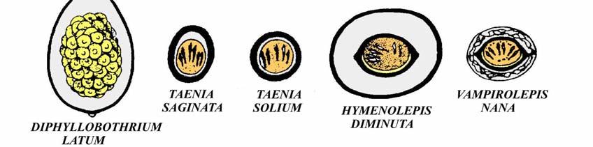
13 13 SLIDE SERIES (Slides 17-32) Needed for each student group: Laboratory manual; compound dissecting microscope (and light) to view adult specimens; compound regular brightfield microscope for eggs; slide boxes with slides Slide 17. Diphyllobothrium scolex. This is a large worm with a scolex with dorsal and ventral longitudinal grooves called bothria. These grooves may be difficult to discern, and may only appear as longitudinal, lightly stained areas on the scolex. It is this genus of the order Pseudophyllidea that most commonly infects humans, although incidental findings of other genera do occur. The first intermediate host is a copepod where the procercoid forms; the second intermediate host is a fish, where pleurocercoids can be found embedded in muscle. Slide 18. Diphyllobothrium proglottids. The proglottids are wider than long and have a characteristic rossette-shaped uterus centrally filled with eggs. Slide 19. Diphyllobothrium eggs. These eggs are similar to those of trematodes, being operculate and easily confused with Paragonimus spp. The ova measure about 60 x 40 micrometers, possess no shoulders, and many possess a tiny knob at the end opposite the operculum. Slide 20. Taenia solium scolex. These scolices have four suckers and a row of hooks. Because the pork tapeworm (Taenia solium) can be a serious pathogen of humans causing cysticercosis, differentiation of this hooked scolex from the non-hooked scolex of Taenia (=Taeniarhynchus) saginata (see demonstration) is of high priority in diagnostic labs. Slide 21. Taenid proglottids. Both mature and gravid proglottids are present. Some slides may also have a scolex. This is Taenia pisciformis from the dog (dog/rabbit life-cycle), but the proglottids are similar to Taenia solium. Compare the mature proglottid with pictures in Figure 5 and see if you can find the vitellarium, genital pore, ovary, vagina, testes, uterus, and sperm duct. The gravid proglottids should have a branched uterus filled with eggs, and you should become familiar with how to discern and count the lateral uterine branches in the proglottid. Note that the genital pore is single and lateral on each proglottid. Slide 22. Taenia saginata eggs. Eggs of the various Taenia spp. and Echinococcus spp. all look somewhat similar. They are spherical or subspherical (generally, eggs of Taenia solium are more spherical than those of Taenia saginata) and contain an onchosphere surrounded by a thick embryophore (inner wall) that is riddled with numerous pores giving the egg a striated appearence. Slide 23. Echinococcus granulosus adult. This worm has four suckers and several circles of hooks. It's a tiny worm with three or four segments only. Although these adults are not found in humans, finding a high prevalence in canines and other carnivores in a local population suggests ample opportunity for human hydatid infection. Slide 24. Dipylidium caninum adult. This dog and cat tapeworm, occasionally found in children, has a retractable rostellum with several circles of tiny rose thorn-like hooks. The proglottids are delicate, elongate, and have two genital pores; one on each side of the proglottid. The presence of these bilateral genital pores is a key diagnostic feature of this worm. Infections are achieved by ingestion of an arthropod (i.e. fleas) containing the cysticercoid. Slide 25. Dipylidium caninum egg packets. These eggs are of the taenid type, but easily diagnosed because they occur in groups (packets) of 8-20 eggs enclosed by a transparent uterine membrane. 13
14 Slide 26. Hymenolepis diminuta adult. This tapeworm, generally found in rats but may infect humans, may reach up to 90 cm and has four suckers, a rostellum, but no hooks. The proglottids have unilateral genital pores and three testes per proglottid. Because the commercially obtained specimens are so poorly fixed and stained, you will not be required to know how to identify adults of this species for the lab exam. Slide 27. Hymenolepis diminuta eggs. These eggs are relatively large, subspherical (sometimes spherical), and contain an onchosphere with significant "distance" between it and the wall. No polar filaments arise from the oncosphere. The eggs measure about 40 x 50 micrometers. Slide 28. Vampirolepis nana (also termed Hymenolepis nana or Rodentolepis nana) adult. The dwarf tapeworm, often termed the most common tapeworm of humans in the world, is small and has four suckers and hooks. It measures up to 40 mm x 1 mm and has a long, slender neck. Proglottids are wider than long, the genital pores unilateral, and there are three testes per segment. It is likely this species actually consists of a "complex" of many morphologically similar species as nearly indistinguishable specimens are commonly found in rodents and other animals worldwide. Because the commercial specimens in the laboratory are poorly fixed and stained, you will not be required to know the adult of this species for the laboratory exam. However, you should know that this species has hooks on the scolex whereas H. diminuta does not. Slide 29. Vampirolepis nana (=Hymenolepis nana) eggs. These eggs have an onchosphere that fills most of the egg volume, they're smaller and more ellipsoidal than those of H. diminuta, and they possess polar filaments which may or may not be discernible. Sometimes the polar filaments simply appear as "fuzz" throughout the egg. Often, two small knobs can be seen on either side of the oncosphere from which the filaments arise. The eggs measure about 30 x micrometers and in your preparations generally occur near or among the debris. Slide 30. Cysticercoid. This is a larval stage of many tapeworms and is found mainly in arthropods. It is solid bodied, with a fully developed scolex invaginated into its body. This type of larval stage is characteristic for Hymenolepis, Vampirolepis, and Dipylidium. The cysticercoids will be small, stained either reddish or purplish, and may have drifted to one edge of the coverslip. Slide 31. Cysticercus. The cysticercus, too, contains a single scolex. However, the scolex is introverted as well as invaginated and the scolex is enclosed within a fluid-filled bladder. You should be able to distinguish between the cysticercus and the solid bodied cysticercoid (slide 30). Taenia solium and Taenia saginatus both form a cysticercus type larva. Slide 32. Coenurus. (portion of coenurus wall) This type of larval stage is a variation of the cysticercus type, however, few-many scolices (protoscolices) bud from the germinative membrane of the cyst. Each slide contains a piece of a coenurus. This type of larva is typical of Taenia multiceps and T. serialis, both of which have a coenurus capable of infecting humans. 14 Demonstration: Know how to differentiate the scolex of Taenia saginatus (no hooks) from that of Taenia solium (with hooks and in your slide box). Demonstration: Know how to differentiate the proglottids of Taenia saginatus (14-23 lateral uterine branches per side) from that of Taenia solium (7-13 lateral uterine branches per side). 14


15 15 FIGURE 5. LINE DRAWINGS OF CESTODES 15
16 16 Table 2. Basic information on cestode biology SPECIES Diphyllobothrium latum PRINCIPLE DEFINITIVE HOST(S) SITE OF INFECTION IN HUMANS INTERMEDIATE HOST(S) LOCATION OF EGGS IN HUMANS fish eating mammals small intestine copepods and fish feces Dipylidium caninum dogs; cats small intestine arthropods feces Echinococcus granulosus canids hydatid cysts in liver, lungs, viscera many mammals none in humans Echinococcus multilocularis canids; felids hydatid cysts throughout body mainly voles none in humans Hymenolepis diminuta rats; humans small intestine arthropods feces Taenia multiceps canids coenurus in viscera sheep; humans none in humans Taenia saginata humans small intestine cattle feces Taenia saginata asiatica humans small intestine predominately pigs feces Taenia serialis canids coenurus in viscera rabbits; humans none in humans Taenia solium humans adults in small intestine; cysticerci throughout body Vampiolepis nana rodents; humans small intestine swine; humans arthropods (optional) feces feces 16
17 17 SECTION 3. NEMATODA The nematodes are probably the most abundant animals on earth. Although more species of insects have been described, this is only because entomologists outnumber nematologists. The number of undescribed, free living species of nematodes are enormous, and virtually all arthropods (and other animals) examined thus far have one or more species of nematode that is specific only for that host. Typical nematodes are elongate, tapered at both ends, and covered by a non-cellular cuticle that is secreted by an underlying hypodermis. The number of cells within an individual worm (and within members of the same species) is the same; a concept termed eutely. Growth occurs primarily by cell enlargement. The digestive system is complete, with a mouth, gut, and anus (although in some species the anus is atrophied). The mouth leads to a buccal cavity, which may be heavily sclerotized to form a buccal capsule and associated teeth or cutting plates (Fig. 6); in other species the lining is thin. Food ingested by the nematode passes through the buccal cavity and into the esophagus, which is usually muscular and contains glandular cells and, perhaps, one or more muscular bulbs. Food passing through the esophagus enters the intestine, where digestion and absorption occur. Sexes are usually separate and most species show sexual dimorphism; a few species are parthenogenic or monoecious. In the male, a single testes is usually present. Nearly all male nematodes have a pair of sclerotized, acellular, copulatory spicules in the cloaca that aid in copulation. In addition, a gubernaculum may be present, which is a sclerotized structure in the cloacal wall that helps guide the exsertion of the spicules. In the female, two ovaries are usually present. As the oocytes move down the oviduct, they increase in size and eventually reach the spermatheca (sperm storage area). After fertilization in this area, the zygotes continue their trek through the female reproductive tract, undergoing meiosis and eggshell formation. The wall of the uterus contains well-developed muscles that not only move the embryos distally by peristaltic action, but also help mold the shape of the eggs and contribute additional eggshell materials. The distal end of the uterus is usually very muscular and is termed the ovijector. The ovijectors of the uteri fuse to form a short vagina, that opens through a ventral, transverse split in the body wall termed the vulva. The vulva may be located anywhere from near the mouth in some species to in front of the anus in others. The vulva never opens posterior to the anus and, only rarely, into the rectum to form a cloaca. In the laboratories where you examine nematodes, you are required to distinguish between six intestinal and several tissue nematodes, and five types of eggs (Fig. 6). Here again, NO OIL IMMERSION. Intestinal species are Enterobius vermicularis (pinworms), Ascaris lumbricoides (giant intestinal roundworms), Necator americanus and Ancylostoma duodenale (hookworms), Trichuris trichiura (whipworm), and Strongyloides stercoralis (a rhabditid worm). Tissue dwelling nematodes include Dioctophyma renale (giant kidney worm), Trichinella spiralis (trichina), Capillaria hepatica (hepatic worm), and microfilariae of filarids. The following provides a summary of slides and demonstrations: 17
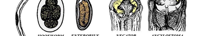
18 18 SLIDE SERIES (Slides 34-47) Needed for each student group: Laboratory manual; compound dissecting microscope (and light) to view adult specimens; compound regular brightfield microscope for eggs and microfilariae; slide boxes with slides Intestinal worms: Slide 35. Enterobius vermicularis, adult female. These worms are one of the most common of all intestinal roundworms. Each female worm has a cuticular inflation of the head, pointed tail, and a relatively large esophageal bulb at the posterior end of the esophagus ("posterior bulb"). They are white or milky in color, and are commonly found around the anus at night laying eggs. A second species of human pinworm, E. gregori, has been described based primarily on the size of the male spicule. However, recent studies now suggest that this species may actually represent young adult E. vermicularis. Slide 36. Enterobius vermicularis, eggs. Eggs occur around the anus or in feces, are partially embryonated, elongate, colorless, and somewhat flattened on one side. They measure x micrometers. Slide 37. Trichuris (=Trichocephalus) trichiura, adults. These common whipworms of the colon and cecum are very long and sometimes coiled, with a thin anterior end and an expanded posterior end where the reproductive organs occur. Heavy infections can result in prolapsed rectum, and growth retardation and finger clubbing may occur in children. Again, note that the head is found at the tiny end, and the expanded body of the worm contains the reproductive organs. Males are often coiled. Slide 38. Trichuris trichiura, eggs. These eggs are easy to distinguish. They are passed unembryonated, measure x micrometers, are often tan, brown, or golden in color, and have very distinct polar plugs at either end. Slide 39. Strongyloides stercoralis, rhabditiform larvae. These are small worms, alternating between a free-living and parasitic life cycle. Adult males are usually only found in the soil and parthenogenic females occur both in the soil and in the small intestine. The eggs produced by the female are so thin walled that they normally rupture, releasing the free 1st stage (rhabditiform) larvae in the feces. Larvae are micrometers long, colorless when unstained, with a short buccal cavity and pointed tail. Slide 40. Ancylostoma sp. adult female or male. Although more common in Europe and Asia, Ancylostoma duodenale (old world hookworm) is also found in North America and other portions of the Western hemisphere. The anterior margin of the buccal capsule has two ventral plates, each with two large cutting teeth fused at the bases. Because virtually all specimens are poorly oriented and the teeth cannot be clearly seen, you do not have to distinguish genera of hookworms from one another on sight. However, you should know the differences on the lab exam if I specifically state whether a particular specimen has teeth or cutting plates. Hint: be careful not to confuse the copulatory bursa of the male with the mouth region. Slide 41. Necator sp., adult female or male. The most common hookworm of the Americas is Necator americanus (new world hookworm). The name literally means "American killer." This worm can be distinguished from Ancylostoma spp. because it has a pair of dorsal and a pair of ventral cutting plates surrounding the anterior margin of the buccal capsule. Because many specimens are poorly oriented so that the plates are not clearly seen, you do not have to distinguish genera of hookworms from one another on sight. However, as stated above, you should know the differences between the two genera if I state whether a particular specimen has teeth or cutting plates. Slide 42. Hookworm eggs. The eggs of hookworms cannot easily be distinguished from one another by a novice. They are fairly thin walled and in the morula stage (generally the 4, 8, 16, or 32 cell stage). The walls of many hookworm eggs appear to have been dissolved during specimen processing (at least with our slides) and, thus, may be difficult to discern initially. The easiest way to find these eggs is to scan your slide using a 10x objective lens (100x total magnification) and identify the morulas. Then, switch the objective to 40x (400x total magnification) to better see the egg wall. 18
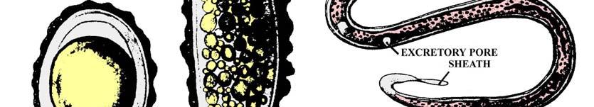
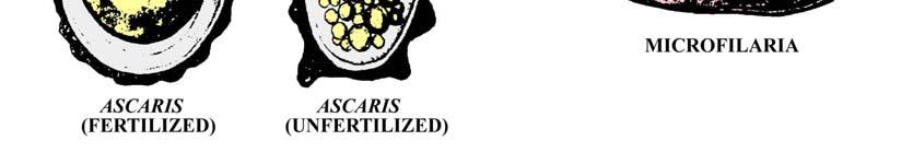
19 Slide 43. Ascaris lumbricoides, eggs. This is the most common pathogenic roundworm of humans, infecting over 1/5 of the world's population. Eggs are easily distinguished because they are subspherical and possess a thick, mammillated wall. The eggs are usually uncleaved when passed in feces. You should be able to distinguish 3 types of eggs: 1) a fertilized egg with the mammillated wall; 2) a fertilized egg where the rough outer wall has been strippped away; and 3) an unfertilized egg which is generally longer and narrower than fertilized ones, the internal details appear as disorganized globules, and and the inner chitinous and lipid wall layers are not formed. Tissue dwelling worms: Slide 44. Trichinella spiralis, larvae in rat skeletal muscle. These larvae are microscopic L1 stages that can be seen coiled within individual skeletal muscle cells. These muscle cells have been modified into supporting "nurse cells." Try viewing the larvae using both low magnification (4x objective lens) on the compound brightfield microscope, and also with the dissecting microscope. Slide 45. Calodium (Capillaria) hepatica, adults and eggs in liver. The technically correct name for this species is Calodium hepaticum, although few textbooks have yet made the conversion. Adults are found in the liver, although it is unlikely that you will see a cross-section through one. The female produces numerous eggs that are retained and encapsulated within the liver parenchyma. Eggs are similar to Trichuris in that they have bipolar plugs in either end, however, they tend to be more squared off at the ends than Trichuris. Once the eggs are liberated into the environment, either by passing through the gut of a carnivore or following host decomposition, eggs embryonate and become infective. Related species known to infect humans and for which eggs may be found in human feces include Aonchotheca philippinensis (Capillaria philippinensis), an intestinal parasite normally using a bird/fish life-cycle, and Eucoleus aerophilus (Capillaria aerophilus), a respiratory tract parasite of carnivores. Slide 46. Onchocerca volvulus, microfilariae in smear. Adults live in subcutaneous nodules and produce microfilariae that wander throughout the skin. These larvae are unsheathed (without an embryonic membrane) and have a tapered, flexed tail. Tail nuclei are absent. Slide 47. Wuchereria bancrofti, Brugia malayi, or Loa loa microfilariae in blood. Adults of the first two species live in lymphatics and produce microfilariae that circulate in the bloodstream. Adults of Loa loa wander throughout the dermis and produce microfilariae that also enter the bloodstream. Larvae of all species have an embryonic sheath, tapered tail, and are difficult to tell apart without a good stain. The distingusihing characteristic is the tail nuclei. The tail nuclei of W. bancrofti are absent. Tail nuclei are present in both of the other species and are terminal in B. malayi. Tail nulcei of Loa loa are present and irregularly spaced. NOTE: Although you are NOT required to distinguish microfilariae of the above 4 species of filarids microscopically (because of our inability to obtain good specimens commercially), you should know some of the key features if asked. Based on the above information and that in your text, you should know the following key characters for differentiating Onchocerca volvulus, Wuchereria bancrofti, Brugia malayi, and Loa loa: 1) Whether the microfiliariae are sheathed or non-sheathed. 2) Whether tail nuclei are absent or present in the microfilariae. 3) If tail nuclei are present, are these nuclei regularly or irregularly spaced. 4) Whether the microfilariae occur in blood or intracellular spaces. 5) Where the adult worms reside. Demonstration: Know how to distinguish female Ascaris lumbricoides (large; straight tail) from males (smaller; hooked tail). Demonstration: Dioctophyma renale are the largest of the worms you will be examining. Adults occur in the kidneys, and eggs with bipolar plugs somewhat like Capillaria hepatica pass out with the urine. Females are much larger than males, and do not have the copulatory bursa of the male. Both are pink or brownish. Be sure that you can distinguish the male from the female


20 20 FIGURE 6. LINE DRAWINGS OF NEMATODES 20
21 Table 3. Basic information on nematode biology 21 SPECIES Aonchotheca philippinensis PRINCIPLE DEFINITIVE HOST(S) SITE OF INFECTION IN HUMANS VECTORS OR INTERMEDIATE HOST(S) birds small intestine fish Ancylostoma duodenale humans small intestine none; L3 larvae penetrate skin LOCATION OF EGGS IN HUMANS feces; many larvae as well feces Ascaris lumbricoides humans small intestine none; eggs ingested feces Brugia malayi humans lymphatics mosquitos none; microfilariae Brugia timori humans lymphatics mosquitos none; microfilariae Calodium hepatica rodents liver none; eggs ingested liver Dioctophyma renale various mammals right kidney various animals urine Enterobius vermicularis humans large intestine none; eggs ingested feces; perianal Loa loa humans dermis deer flies none; microfilariae Mansonella ozzardi humans subcutaneous blackflies; midges none; microfilariae Mansonella perstans humans body cavity midges none; microfilariae Mansonella streptocerca humans dermis midges none; microfilariae Necator americanus humans small intestine none; L3 penetrate feces Onchocerca volvulus humans subcutaneous nodules blackflies Strongyloides stercoralis many mammals intestine none; L3 penetrate Trichinella spiralis Trichinella britovi; Trichinella murrelli; Trichinella nativa; Trichinella nelsoni various domestic mammals various sylvatic mammals intestine; striated muscles intestine; striated muscles Trichuris trichiura humans large intestine none; ingestion of infected meat none; ingestion of infected meat none; ingestion of eggs none; microfilariae few; mainly L1s in feces none; L1 larvae encysted in striated muscles none; L1 larvae encysted in striated muscles feces Wuchereria bancrofti humans lymphatics mosquitos none; microfilariae 21
22 22 Table 4. Major characteristics of human microfilariae SPECIES VECTORS SHEATH LOCATION SIZE (um) TAIL NUCLEI TAIL SHAPE Brugia malayi mosquitos yes blood x 5-6 yes tapered Brugia timori mosquitos yes blood x yes tapered Loa loa deer flies yes blood x 3-5 yes tapered Mansonella ozzardi Culicoides; Simulium no blood x 4-5 no tapered; curved Mansonella perstans Mansonella streptocerca Onchocerca volvulus Wuchereria bancrofti Culicoides no blood x 4-5 yes blunt; straight Culicoides no dermis x 5-6 yes blunt; hooked Simulium no dermis x 5-9 no tapered; bent mosquitos yes blood x no tapered NOTE: You will not be required to learn all of the information contained in Table 4. You will only need to know the five items listed earlier for Brugia malayi, Loa loa, Onchocerca volvulus, and Wuchereria bancrofti. Again, the five items for these four filarid species are as follows: 1) Whether the microfiliariae are sheathed or non-sheathed. 2) Whether tail nuclei are absent or present in the microfilariae. 3) If tail nuclei are present, are these nuclei regularly or irregularly spaced. 4) Whether the microfilariae occur in blood or intracellular spaces. 5) Where the adult worms reside. 22
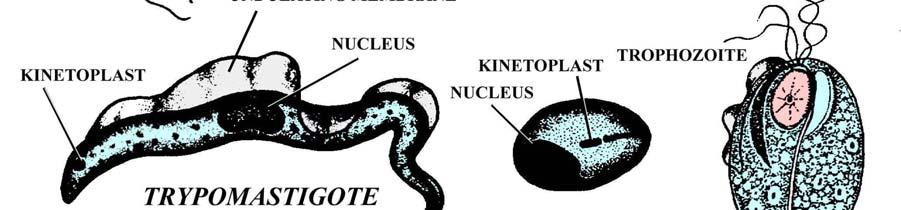
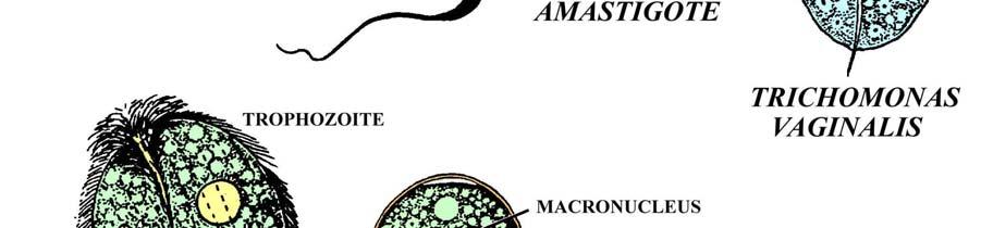
23 23 SECTION 4. PROTOZOA The protozoa, which are mainly single celled organisms capable of independent existence, are now divided into 11 or more phyla. The classification schemes for this group change continually as more organisms are examined at a molecular level. However, we will only examine representatives from three groups within the laboratory: Sarcomastigophora (the amoebae and flagellates), Apicomplexa (malaria, coccidia, and piroplasms), and Ciliophora (the ciliates). You will have the most difficulty with the amoebae, since their small size and amorphous nature makes them difficult to distinguish at first glance. For all protozoa, find them under 10x or 40x, then examine them more closely under oil immersion if you wish. BE CAREFUL NOT TO BREAK SLIDES BY JAMMING INTO THEM WITH THE OBJECTIVE LENS AND BE SURE TO CLEAN IMMERSION OIL OFF LENSES. BE SURE TO CLEAN IMMERSION OIL OFF SLIDES BEFORE PUTTING THEM BACK INTO SLIDE BOX. In the first week, concentrate on the flagellates (Fig. 7). Giardia intestinalis is pathogenic and the trophozoites can be distinguished from all other human flagellates by their teardrop shape, possession of 8 flagella, a ventral adhesive disk, and especially the presence of two distinct nuclei which gives the organism the nickname "monkey-face." Cysts passed in the feces are ellipsoidal and initially may contain two nuclei. They divide quickly so that mature cysts contain four nuclei. The taxonomy of this group has been confused for decades and synonyms include Giardia duodenalis and Giardia lamblia. Trophozoites of Chilomastix mesnili may be confused with Giardia cysts; however, only one nucleus will be present in Chilomastix trophozoites, and you may see a pale, transverse band (the spiral groove) along the middle of the organism. Cysts are very small, 5-8 micrometers in length, contain one nucleus, are ovoidal or lemon-shaped (knobbed), and have prominent cytostomal fibrils that are termed a Shepherd's crook. Trichomonads (Fig. 7) form no cysts and are distinguished by having a single nucleus, prominent axostyle (bundle of microtubules) that often project posteriorly through the organism, an undulating membrane (a recurrent flagellum directed posteriorly that is enveloped by a portion of the plasma membrane), and often a costa (a thin stiffening rod under the undulating membrane). Trichomonas vaginalis is the only human pathogen, infecting the urogenital tract. It has four terminal flagella and an undulating membrane that extends less than one-half of the length of the body. Trichomonas tenax is found only in the mouth, has four anterior flagella, and the undulating membrane extends greater than one-half the body length. It is considered non-pathogenic and you will not need to look at any slides of these. Pentatrichomonas hominis may be found in the colon and is also a tiny, harmless commensal. It has five anterior flagella and the recurrent flagellum within the undulating membrane often trails beyond the body of the trophozoite. You will have no slides of this species, although it is the most common trichomonad of humans. The kinetoplastids are heteroxenous (requiring two hosts) flagellates that possess not only a nucleus, but a highly modified portion of DNA in the mitochondria termed a kinetoplast (Fig. 7). Four principle morphological types of kinetoplastids can be recognized, based on the location of the flagella, kinetoplast, and nucleus. The trypomastigote form is elongate and the kinetoplast is posterior to the nucleus. The flagellum runs along the surface of the organism anteriorly in a fold of the undulating membrane. The epimastigote form is found in some life cycles and the kinetoplast is located between the nucleus and anterior end. A short undulating membrane lies along the proximal portion of the flagellum. The promastigote form is elongate and has the flagellum extending forward as well. However, the kinetoplast is located anterior to the nucleus and no undulating membrane is present. The amastigote form is found mainly within infected cells. The organism is small and ovoid, with a short flagellum projecting only slightly beyond the organism, if at all. The principle types of human African trypanosomes cause African sleeping sickness. In the blood they are generally long, slender trypomastigotes, with a small kinetoplast, prominent nucleus, and undulating membrane. They are transmitted by the bite of any one of a number of Glossina spp (tsetse flies). Trypanosoma rhodesiense and T. gambiense are the species that infect people and are identical morphologically. The former species is found in central and east Africa and causes an acute infection. Native game animals are known reservoirs. The latter species is found in west central and central Africa and causes the chronic form of the disease characteristic in textbooks. Apparently, game animals are not particularly susceptible to T. gambiense although some researchers have found that a few reservoir animals do exist. Both species are thought to be derived from Trypanosoma brucei, a morphologically identical trypanosome found in the bloodstream of native African ruminants. Humans are not susceptible to this parent species, but livestock, horses, swine, and canines are. 23
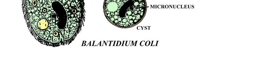
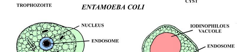
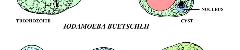
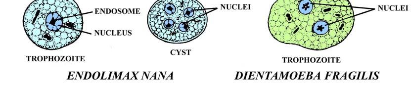
24 24 The American trypanosome, Trypanosoma cruzi, is also a serious pathogen causing Chagas' disease. Trypomastigotes in the blood die in a characteristic C- or?- shape. They have a large kinetoplast at one end and a prominent nucleus. These organisms are engulfed by phagocytic cells and then transform into amastigotes, which undergo binary fission and destroy the host cells. Groups of these amastigotes in the tissues are termed pseudocysts. Infection may lead to either an acute or chronic form of the disease, either of which may be fatal. In humans, T. cruzi is generally transmitted in the feces of reduviid bugs, which defecate when they bite. The parasite enters the wound when the host rubs the contaminated feces into the lesion. The parasite may also infect people via blood transfusions, and dogs have been shown to become infected when they ingest infected reduviids. Leishmania spp. spend their lives in the vertebrate host only as an amastigote. They are transmitted by the bite of sandflies (where they multiply in the gut as promastigotes) and, like T. cruzi, can multiply to large numbers in phagocytic cells. Various species and strains have a predilection for different sites in the body and pathology varies as well. Leishmania donovani is a serious pathogen that can be seen living as clusters of amastigotes within the reticuloendothelial system of the viscera, including spleen, liver, intestine, lymph nodes, and bone marrow. Canids are important reservoirs of the disease. Other Leishmania spp. have predilections for other tissues, such as the dermis or mucus membranes. Leishmaniasis tends to be a chronic, disfiguring illness and most species respond poorly to pharmaceutical intervention. During the second week, review the flagellates but concentrate most heavily on the amoebae (Fig. 8). As stated above, they will prove to be difficult to distinguish, so considerable time must be spent at the microscope (i.e. I advise not cutting this lab early). Amoebae reproduce by binary fission and most species are capable of forming cysts (usually the infective stage). The feeding/reproductive form is termed the trophozoite. The most pathogenic intestinal species is Entamoeba histolytica, whose trophozoites may penetrate the intestine, enter the liver or lungs, and cause serious illness and often death. Several non-pathogenic intestinal species are morphologically indistinguishable from E. histolytica and include E. hartmanni (somewhat smaller), E. dispar, and E. moshkovskii. Entamoeba gingivalis is found in the mouth of many individuals, forms no cysts, but is also similar to trophozoites of E. histolytica. None of these will be included in the laboratory. However, other intestinal amoebae that will be covered include Entamoeba coli, Endolimax nana, and Iodamoeba buetschlii. These all form cysts and are usually only mildly-moderately pathogenic. Dientamoeba fragilis is also intestinal and included here with the amoeba, although electron microscopy has shown that it is actually a flagellate (but forms no flagella). It may cause acute or chronic clinical signs of intestinal distress and forms no cysts. The most serious pathogen, which is almost always fatal, is Naegleria fowleri. It may enter the brain through the nasal passages, erode the olfactory bulbs and rest of the brain, and cause death within six days. Luckily, infection with this organism is relatively rare. Amoebae are distinguished in several ways. First, the structure of the nucleus and endosome (nucleolus); second, the number of nuclei within the trophozoite (Dientamoeba fragilis is the only amoeba with two); third, the presence or absence of glycogen or red blood cells within the trophozoites; and fourth, whether an amoeba forms cysts or not and, if so, the number of nuclei in the cyst, the morphology of the nucleus, and the presence or absence of various inclusions. Naegleria fowleri and other types of CNS invading amoebae are generally diagnosed by histological sections of the brain following death. Cysts are not formed within the brain, which helps differentiate this species from Acanthamoeba spp. During the third week, examine the remaining protozoa. The only important ciliate pathogen in humans is Balantidium coli, which is also commonly found in swine and other primates (Fig. 7). It is the largest protozoan parasite of humans and trophozoites are oblong or spherical, possess cilia, and multiply by binary fission. It can easily be seen at 10x magnification, so there is no reason to use oil immersion. In some individuals, these trophozoites become invasive and are capable of causing colonic ulceration. Large, spherical cysts are formed (ca 50 um in diameter) that pass out with the feces and are capable of infecting new hosts. A large, sausage-shaped macronucleus can be seen easily within both trophozoites and cysts, which is a diagnostic feature of most ciliates. About six species of coccidia or coccidian-like organisms are pathogens of humans. Isospora belli forms resistant cyst stages (oocysts) that are transmitted from host-to-host. Oocysts are elongate-ellipsoidal, passed unsporulated, and the cytoplasm within these oocysts stains reddish in your slides. This is an intestinal species, preceded by two generations of merogony (multiple fission) and finally gamogony (sexual reproduction). The life-cycle is direct and no intermediate hosts are involved. Similarly, oocysts of Cyclospora cayetanensis are passed unsporulated but instead of being elongate they are spherical and much smaller. The cytoplasm stains reddish. This species also has a direct life-cycle, developing in the jejunum. 24
25 25 Cryptosporidium parvum and C. hominis are morphologically identical apicomplexans distantly related to the coccidia. Severe diarrhea, weight loss, and abdominal cramping are the main symptoms. Oocysts are very tiny and contain four sporozoites (vermiform infective stages). These oocysts show up as small, reddish blotches in your slides and are best seen using either a 40x or 100x objective lens. If an oocyst is ingested, sporozoites penetrate intestinal epithelial cells, undergo two generations of merogony, eventually gamogony, and form new oocysts that are passed in the feces (analogous to I. belli). Although normally self-limiting by your immune system, individuals with immune deficiencies are at severe risk with both species of Cryptosporidium since the organisms are capable of recycling within the host. Toxoplasma gondii is also a serious coccidial pathogen of a variety of mammals, which can lead to swollen lymph glands, fever, headache, muscle pain, anemia, blindness, encephalitis, myocarditis, and death. Oocysts are only found within the feces of felines and, upon ingestion by virtually any mammal, the sporozoites penetrate the gut wall and enter various tissues where they multiply rapidly by endodyogeny (formation of daughter cells while still retained within the mother cell) as tachyzoites (rapidly dividing zoites). Accumulations of tachyzoites are termed groups. As infections become chronic, zoites begin to transform and multiply more slowly. These slowly dividing zoites, now called bradyzoites, are contained within spherical cysts um in diameter. These cysts are contained in histological sections of brain on your slides and 20x and 40x objective lenses should be utilized. If a feline ingests oocysts or cysts, merogony, gamogony, and oocyst formation occur within intestinal cells. Surveys suggest 25-40% of the human population is sero-positive for T. gondii. Plasmodium spp. are the malaria. Four principle species infect humans, the most serious of which is Plasmodium falciparum. 50% of all malarias are due to this pathogen. Plasmodium vivax is also common, accounting for 42-43% of all malarias; P. ovale and P. malariae are rarer. Sporozoites injected with the bite of mosquitos of the genus Anopheles make their way to the liver and multiply in hepatocytes. Once this exoerythrocytic cycle is complete, merozoites (the stages resulting from multiple fission) enter red blood cells and begin schizogony (=merogony). New merozoites formed within these cells (segmenters) rupture out, invade new cells and keep this erythrocytic cycle going. Parasites in red blood cells that are early in development and resemble an amorphous mass are termed trophozoites. Some appear ring-like or bandshaped and are termed rings or bands. Some merozoites grow and form gametocytes (gametes) within red blood cells. Using the plates in your textbook, see if you can identify segmenters, gametocytes, trophozoites, and rings in your blood smears. If gametes are ingested by the appropriate mosquito, some form flagella and undergo exflagellation (male gamete formation). These male gametes (microgametes) fuse with female gametes (macrogametes) to form a motile zygote termed an ookinete. Ookinetes penetrate the gut wall and form oocysts containing sporozoites on the hemocoel side of the gut. Sporozoites eventually rupture out of oocysts and migrate to the salivary glands. Babesia spp are piroplasms; tiny Apicomplexans that infect the red blood cells of some mammals. Although humans are not principle hosts and the majority of development occurs in a ticks, the bite of a tick can sometimes transmit tiny vermicules (merozoites) of some species into humans that cause disease. These vermicules undergo binary fission within red blood cells and rupture the cell open when the new merozoites exit. The disease, caused mainly by B. microti, is not uncommon in the New England states; especially in splenectomized individuals. In your blood smears, see if you can see these merozoites undergoing binary fission in red blood cells. Pneumocystis jiroveci (P. carinii of humans) is an ascomycote fungus that parasitologists still embrace as an opportunistic parasite. Pneumocystis is not a problem unless an individual is immunosuppressed. Then, the amoeboid trophozoites in the lungs can multiply to large numbers. Eventually, cysts are formed containing 8 nuclei when mature. Cysts are disseminated through the air. New trophozoites rupture from the cysts and keep the cycle going. Recent studies have shown the various Pneumocystis spp. to be host specific; thus, a rat Pneumocystis sp. will not infect humans. Blastocystis hominis too may not be a true eukaryotic parasite. It is a pathogen of undetermined taxonomic affinity. Molecular biology has shown it to be unrelated to other eukaryotic parasites of humans and is only distantly related to the Apicomplexa/ciliates/dinoflagellate complex. However, various animals may share similar genetic types of Blastocystis, suggesting the parasite may be zoonotic. Evidence suggests that the parasite may be responsible for many cases of undiagnosed diarrhea, particularly in the southwest, or at least it associates itself with intestinal distress. Although the entire life cycle is not known, an amoeboid (invasive) stage has been seen, a granular (reproductive) stage is found during poor conditions, and the common vacuolated form undergoes binary fission. This form contains a large, central vacuole and peripheral nucleus and cytoplasm. In the next four laboratories you will be responsible for learning the differentiation of a variety of protozoa of human importance. The following is a proposed outline: 25
26 26 SLIDE SERIES (Slides 51-79) Needed for each student group: Laboratory manual; compound regular brightfield microscope for all parasites; slide boxes with slides; (NOTE: no dissecting microscopes will be needed) Week 1: Slide 51. Giardia intestinalis (trophozoites) Look for tear-drop shaped trophozoites with a ventral adhesive disk and two nuclei. Specimens will either be stained blue, gray, or pink depending upon the preparation. Giardia lamblia and Giardia duodenalis are synonyms. Slide 52. Giardia intestinalis (cysts) Look for small, ellipsoidal cysts with dark staining flagella and median bodies, and 4 nuclei. Slide 53. Chilomastix mesnili (trophozoites) Tear-drop (tapered at the end) shaped organisms with single large nucleus anteriorly and small endosome. These may be confused with cysts of Giardia intestinalis, which are often found on your slides along with Chilomastix. Slide 54. Chilomastix mesnili (cysts) Small, ovoidal or lemon-shaped structures with dark-staining Shepherd's crook and a single nucleus. Slide 55. Trichomonas vaginalis (trophozoites) Pink or purple trophozoites with anterior flagella and dark staining axostyles. Slide 56. Trypanosoma gambiense, T. rhodesiense, or T. brucei (blood smear) Numerous purple trypomastigotes among blood cells. Note the irregular manner in which the trophozoites have died, the small kinetoplast at one end, and the undulating membrane associated with the flagellum. Slide 57. Trypanosoma cruzi blood smear Low numbers of purple trypomastigotes among blood cells. Note that they are smaller than the African trypanosomes and die in a C- or?-shape. The kinetoplast is slightly larger in proportion to the body of the trypomastigote when compared to the African trypanosomes. Slide 58. Trypanosoma cruzi, amastigotes in heart muscle Small, dark-staining amastigotes (dots) numerous as clusters within pale vacuoles in some muscle fibers. Slide 59. Leishmania donovani, amastigotes in spleen smear The amastigotes represent numerous small, dark dots with a nucleus and adjacent kinetoplast. You should be able to easily see the numerous parasites using a 40x objective lens, then go to oil immersion to take a closer look. NOTE: some of you may need to trade slides with someone else in the class when you go to oil immersion. Unfortunately, the manufacturer seems to have used coverslips that are quite thick, which has resulted in some slides not allowing the 100x objective lens to get close enough for the parasites to be in focus. Week 2: Slide 60. Entamoeba histolytica (trophozoites) Looks like debris but with a smooth edge, either bluish or gray, with a nucleus containing a small, centrally located endosome. Many of the slides have species other than E. histolytica mixed in, and some of the slides may even have been misidentified by the company. Slide 61. Entamoeba histolytica (cysts) Spherical, gray or purple, with 1, 2, or 4 nuceli. Some young cysts may have large, blunt ended chromotoidal bars. Older cysts should have the 4 nuclei, but the chromotoidal bars may have disappeared. 26
27 Slide 62. Entamoeba coli (trophozoites) Large blobs with smooth margins, either purple or gray, with single nucleus containing an endosome that is usually off-center. Overall, the trophozoites are somewhat larger than E. histolytica, the endosomes should be larger and offcenter rather than central, and the peripheral chromatin should be less evenly dispersed. Beware that many of the slides have a mixed infection of E. coli and E. histolytica. Slide 63. Entamoeba coli (cysts) Large, spherical or ellipsoidal, gray-purple cysts; 8, 16, or 32 nuclei. Some may have chromatoidal bars, most of which should be sharply pointed. Slide 64. Endolimax nana (trophozoites) Tiny blobs with smooth margins, gray or purple, with dark staining, prominent endosome. Slide 65. Endolimax nana (cysts) Spherical, tiny, with 4 dark endosomes/cyst. Tiny chromatoidal bars may be present which resemble additional nuclei. Slide 66. Iodamoeba buetschlii (trophozoites) Gray or purple with smooth margins, with single nucleus, large endosome, and sometimes with large glycogen vacuole. Slide 67. Iodamoeba buetschlii (cysts) Often perfect ellipsoids with nice, large, glycogen vacuoles and a prominent nucleus with a large endosome. Slide 68. Dientamoeba fragilis (trophozoites) Blobs or broad ellipsoids with smooth margins, and with 60% or more being bi-nucleate. No cysts are formed. Slide 69. Naegleria fowleri (trophozoites in brain) Areas of cellular inflammation should surround numerous trophozoites, each in a vacuole. Amoebae possess prominent, dark-staining endosomes. 27 Week 3: Slide 70. Balantidium coli (trophozoites) Large, pink or yellow, with large sausage-shaped macronucleus. Slide 71. Balantidium coli (cysts) Large, spherical, with sausage-shaped nucleus. The cyst wall should be distinct. Slide 72. Isospora belli, unsporulated oocysts in fecal smear Clear ellipsoids with reddish cytoplasmic masses (1-2) within. Slide 73. Cyclospora cayetanensis, unsporulated oocysts in fecal smear Small, individual spherical cytoplasmic masses that stain reddish Slide 74. Cryptosporidium parvum oocysts in fecal smear Tiny ellipsoids, should be reddish, often with 1-3 dark staining dots within. The morphologically identical C. hominis is also found in humans. Slide 75. Toxoplasma gondii cysts in brain Find under 10x, then examine at 40x. Spherical masses filled with numerous dark dots. Slide 76. Plasmodium vivax trophozoites & segmenters in blood smear Purple blobs, rings, bands, and dots in red blood cells. 27
28 28 Slide 77. Plasmodium falciparum gametocytes in blood smear Banana-shaped, purplish staining gametes within blood cells. Often they appear to be lying free among blood cells but if you look closely most should be surrounded by remnants of a thin, red blood cell membrane. This is the only species of human malaria with this type of distinctive gametocyte. Slide 78. Babesia sp. merozoites in blood cells Look for 1 or 2 dark staining merozoites within blood cells. Some cells should contain merozoites undergoing binary fission, and sometimes a distinctive "V" shape can be noted as one end of the merozoites is still joined. Slide 79. Pneumocystis carinii trophozoites & cysts in rat lung smear Small, non-staining ellipsoids and circles, sometimes with 1-8 dark nuclei inside. Look for 8 nuceli within a small, clear, circular area, which represent mature cysts. The species in humans is termed Pneumocystis jiroveci. Slide 80. Blastocystis hominis trophozoites in fecal smear Small, variable in size but usually spherical, with pale gray or greenish "vacuolar" area (depending upon the stain) surrounded by thin, peripheral ring of darker cytoplasm. Red or purple dots should be seen in the peripheral cytoplasm. Week 4: Review of the last 3 weeks worth of material. 28


29 29 FIGURE 7. LINE DRAWINGS OF FLAGELLATES AND CILIATES 29


30 30 FIGURE 8. LINE DRAWINGS OF AMOEBAE 30
31 31 SPECIES Table 5. Basic biology of Protozoa PRINCIPLE DEFINITIVE HOST(S) SITE OF INFECTION IN HUMANS VECTORS/ INTERMEDIATE HOST(S) LOCATION OF CYSTS/OOCYSTS/ SPORES IN HUMANS Acanthamoeba spp. none; free-living brain none brain Babesia spp. ticks erythrocytes ticks are vectors; mammals are intermediate hosts none Balamuthia mandrillaris none; free-living brain none brain Balantidium coli primates large intestine none feces Blastocystis hominis various mammals large intestine none feces Chilomastix mesnili primates large intestine none feces Cryptosporidium hominis humans small intestine none feces Cryptosporidium parvum many mammals small intestine none feces Cyclospora cayetanensis humans small intestine none feces Dientamoeba fragilis humans large intestine none; perhaps carried in pinworm eggs unknown Endolimax nana humans large intestine none feces Entamoeba chattoni; (=Entamoeba polecki); (=Entamoeba suis) primates; swine large intestine none feces Entamoeba coli humans large intestine none feces Entamoeba dispar humans large intestine none feces Entamoeba gingivalis humans large intestine none no cysts formed Entamoeba hartmanni humans large intestine none feces Entamoeba histolytica various mammals large intestine none feces Entamoeba moshkovskii humans large intestine none feces Encephalitozoon cuniculi many mammals disseminates none spores disseminated throughout tissues Encephalitozoon hellem humans and budgerigars corneal; may also disseminate none cornea Encephalitozoon intestinalis humans and ruminants small intestine; may also disseminate none feces; urine Enterocytozoon bieneusi humans; other mammals small intestine none feces Giardia intestinalis humans small intestine none feces 31
32 32 SPECIES (CONT.) PRINCIPLE DEFINITIVE HOST(S) SITE OF INFECTION IN HUMANS VECTORS/ INTERMEDIATE HOST(S) LOCATION OF CYSTS/OOCYSTS/ SPORES IN HUMANS Iodamoeba buetschlii primates; swine large intestine none feces Isospora belli humans small intestine none feces Leishmania donovani humans; canids disseminates viscerally sandflies none Leishmania spp. (non-visceral) humans; other mammals cutaneous or mucocutaneous sandflies none Naegleria fowleri none; free-living brain none trophozoites only in brain tissue Pentatrichomonas hominis humans large intestine none none; but rounds up and survives passage Plasmodium spp. mosquitos liver; erythrocytes humans none in humans Pneumocystis jiroveci humans lung none lungs; sputum Sarcocystis hominis humans small intestine Sarcocystis suihominis humans small intestine Toxoplasma gondii felids disseminates cattle (cysts in muscle) swine (cysts in muscle) many mammals and some birds feces feces none in humans Trachipleistophora hominis humans disseminates none disseminated Trichomonas tenax humans mouth none none Trichomonas vaginalis humans urogenital tract none none Trypanosoma cruzi various mammals disseminates; blood reduviids none Trypanosoma gambiense humans blood; CNS tsetse flies none Trypanosoma rhodesiense humans; game animals blood; CNS tsetse flies none Vittaforma corneae humans cornea none cornea 32
33 33 SECTION 5. ARTHROPODA The arthropods are a huge assemblage of animals. They are bilaterally symmetrical, possess a complete gut, and are segmented. They have an exoskeleton composed of chitin, a polysaccharide somewhat similar to cellulose. The more rigid sections of the exoskleton are articulated with each other by thin, flexible, cuticular joints. In the final laboratory, you will examine some of the arthropods of human importance (Fig. 9). Start off by distinguishing the Anoplura (sucking lice) of humans, a group found only on mammals and of which there are about 500 described species (56 known species from North America). They are wingless insects, with flattened bodies and six legs; the ends of which are modified into large claws for clinging onto hair. Pthirus (=Phthirus) pubis, the "pubic" or "crab" louse, dwells primarily in the pubic area; although it may also be found under the armpits and, rarely, eyebrows, eyelashes, beard, and mustache. It is robust, with a body that tapers from anterior to posterior. Bites may cause puritis, but fortunately do not seem to transmit disease often. Nits (eggs) are cemented onto hair by the female, who only lays about 30 eggs in her short life. Pediculus humanus, the other louse that infests humans, is more elongate than the robust Pthirus. Various appellations are used to describe these organisms, including "graybacks," "mechanized dandruff," and "cooties." One subspecies, Pediculus humanus capitus is found primarily on the head. Pediculus humanus humanus (=P. corporis) is the body louse and prefers clothing. It only travels to the body to take an occasional blood meal. It is difficult to distinguish between the two and they are capable of interbreeding. Both subspecies are capable of transmitting typhus, a rickettsia. Hemipterans (true bugs) contain few members that are parasites. One species, Cimex lectularius, is the bedbug and will attack humans. They are reddish-brown, up to 8 mm long, and are dorso-ventrally flattened. Bedbugs are nocturnal, emerging from hiding at night to feed. Two other similar species occur: C. hemipterus tends to be more tropical and Leptocimex boueti is confined to West Africa. Rarely, other species and genera of bedbugs of wildlife will attack humans. Another group of hemipterans of medical importance are the reduviids (kissing bugs, assassin bugs, or cone-nosed bugs). Examine a specimen of Rhodnius prolixus, one of the many reduviids capable of transmitting American trypanosomiasis. Become familiar with its elongate shape, large lateral eyes, and cone-like build. Other genera capable of transmitting Chaga's disease include Triatoma and Panstrongylus. There are some bottled specimens of Triatoma sanguisuga within the laboratory, which is the only species of the genus Triatoma to occur in the Manhattan, Kansas area. This reduviid has eight instars, lives primarily within the nests of woodrats and cotton rats, but will feed on a variety of mammals including humans. Note the bright red stripes on its dorsal surface. The Siphonapterans (fleas) are nearly all parasitic. They are flattened laterally and most species have strong legs for jumping. The head is broadly jointed to the thorax and often bears a genal ctenidium. Ctendia are a series of stout spines that help a flea retain itself within the fur or feathers of the host and an additional ctendium (pronotal ctenidium) may be found posterior to the first thoracic tergite (main dorsal cuticular piece on the thorax) of some species. Additional spines (setae) on the body of the flea also aid in maintaining the position of the flea upon the host. Of the fleas capable of infesting humans, you should know how to distinguish three species. Ctenocephalides felis has both genal and pronotal combs and has been so successful living on our pets that it has pretty much replaced most of the other flea species even on medium-sized wild mammals. Over 95% of all fleas found on medium-sized mammals today in Kansas will be C. felis. Pulex spp. and Xenopsylla cheopis have no combs but do have ocular bristles; the base of the ocular bristle of the former species is beneath the eye while that of the latter is in front. Another way of distinguishing these species is the presence or absence of a meral rod. The meral rod is a vertical, rod-like structure that divides the mesopleuron (sternite located above the middle pair of legs) of some flea species. The rod is present in Xenopsylla and absent in Pulex. Although Pulex irritans is the species labeled on your slides, it is probably the morphologically similar Pulex simulans. Studies have shown that the Pulex simulans occurs in North America rather than Pulex irritans. Although the oriental rat flea (Xenopsylla cheopsis) is rarely found in Kansas anymore, its historic importance in the transmission of bubonic plague and distribution elsewhere makes learning this species a must. Various Diptera (flies) are capable of attacking humans. The tiny blackfly Simulium is a robust animal with a hump back and broad wings. Males do not feed on blood but females of many species do so for proper ovarian development. Blackflies are vectors for Onchocerca volvulus in humans. Culicoides spp. are representative of the midges or no-see-ums, and only 4 genera within the Ceratopogonidae attack mammals. They carry a variety of diseases and can be recognized by their small size. Compare the structure of the midge with that of the more robust blackfly. Especially note the antennae; those of Simulium spp. have 11 segments without bristles whereas those of Culicoides spp. have more than 11 segments and prominent bristles. 33
34 Glossina spp are tsetse flies; large, brownish-gray animals that feed exclusively on blood and give birth to live larvae. They are only found in Africa (or nearby areas) and are the vectors of African sleeping sickness. They are sometimes confused with Chrysops spp. (deer flies), but there are several morphologic differences. First, Glossina spp. have a more elongate, protruding proboscis. Second, most Chrysops spp. have a distinct splotch on each wing which is absent from the wings of Glossina spp. Finally, and perhaps most importantly, Glossina spp. have a cell between the 4th and 5th wing veins that appears as an inverted "axe" or "hatchet." Glossina spp. are the only dipterans with this "hatchet cell." Mosquitoes, too, are dipterans and one genus, Anopheles, transmits malaria. These animals are long, delicate dipterans with slender wings and mouthparts that form a proboscis. Only the females are capable of sucking blood and eggs are deposited singly or in rafts in or near water. Mosquito larvae are called wigglers and the pupae tumblers. Other important mosquitos in addition to Anopholes spp. include Aedes spp., fierce diurnal biters with representative species transmitting such diseases as La Crosse encephalitis, yellow fever, and dengue; and Culex spp. with some members transmitting St. Louis encephalitis, Western equine encephalitis, Wuchererica bancrofti, Dirofilaria immitis, and avian malaria. Compare the Anopheles sp. female with the next slide, Lutzomyia sp. (sandflies). Sandflies have cutting mandibles and, thus, do not have a proboscis (unlike mosquitoes). They are also quite hairy, possess large black eyes and very long legs, and overall appear far more delicate than mosquitoes. Sandflies are vectors of several important diseases, including Leishmania spp., Carrion's disease, and sand fly fever. The Acari (ticks and mites) have eight legs as adults and have reduced segmentation externally. The resulting body can be divided into two parts; the capitulum bearing the mouthparts and the idiosoma containing most internal organs. Although basically similar, ticks and mites have several fundamental differences. Ticks have a hypostome (portion of the mouthparts) that is large and toothed whereas in mites the hypostome is unarmed and hidden. In addition, ticks have a Haller's organ (olfactory/humidity receptor) on the first tarsi (portion of the leg) whereas mites have none. Probably the easiest way to distinguish the two, however, is that ticks are easily seen whereas mites are small, usually microscopic. Ticks are divided into two groups. The Ixodid, or hard ticks, have a terminal capitulum and a large, dorsal sclerite, the scutum. Argasid, or soft ticks, have no snout (the capitulum is subterminal) and there is no scutum. Male and female ixodid ticks can be distinguished because the females have a scutum that only partially covers the dorsum whereas males have a scutum extending over the entire body (Fig. 10). Because of the importance of Lyme disease and other tick borne diseases, it is important in this laboratory that you be able to distinguish between the genera of hard ticks found in Kansas, as well as tell a hard tick from a soft tick. Study the hard tick demonstration (see below) very carefully since I guarantee several questions will be over this material. Mites are divided into several groups, but only a few are of concern here. Sarcoptes scabiei, the mange mite, has a rounded body and short legs. Females burrow throughout the epidermis laying eggs within tunnels, causing an intense rash termed the "seven-year itch." Demodex folliculorum and the stubbier D. brevis, however, are rarely pathogenic. These are elongate mites that live either in hair follicles (D. folliculorum) or sebaceous glands (D. brevis) around the nose and eyes in about one-half of the population of the United States. Occasionally, these latter species may cause facial rosacea. Chiggers are parasitic larvae of free-living mites. They are usually brightly colored and belong to a variety of genera and species. Eutrombicula alfreddugesi is a common outdoor chigger mite in the United States, causing wheals and rash in susceptible individuals. Large numbers of larvae can sometimes be encountered in small foci termed mite islands. You will be shown some kodachromes of chiggers during the course of your studies, but I have provided no specimens for microscopic examination. You are responsible for knowing 18 species of arthropods in this laboratory. All should be viewed either using the dissecting microscopes or the compound microscopes (at 4x or 10x). The following represents the list: 34 34
35 35 SLIDE SERIES (Slides 81-98) Needed for each group: Laboratory manual; compound dissecting microscope (and light) to view adult specimens; compound regular microscope for fleas and mites; slide boxes with slides Anoplura Slide 82. Pthirus pubis (adult) "pubic crab louse" (often erroneous termed "Phthirus pubis") Slide 83. Pthirus pubis (nits) "pubic crab louse" Slide 84. Pediculus humanus (adult) "body/head louse" Hemiptera Slide 85. Cimex lectularis (adult) "bedbug" Slide 86. Rhodnius prolixus (adult) "reduviid" Siphonaptera Slide 87. Ctenocephalides felis (adult) "cat flea" Slide 88. Pulex irritans (adult) "human flea" Slide 89. Xenopsylla cheopis (adult) "oriental rat flea" Diptera Slide 90. Simulium sp. (adult) "blackfly" Slide 91. Culicoides sp. (adult) "gnat" Slide 92. Anopheles sp. (adult female) "mosquito" Slide 93. Lutzomyia sp. (adult) "sandfly" Slide 94. Glossina sp. (adult) "tsetse fly" Slide 95. Chrysops sp. (adult) "deerfly" Acari Slide 96. Amblyomma americanum (hard tick) "lone star tick" Slide 97. Dermacentor variabilis (hard tick) "American dog tick" Slide 98. Argas persicus (soft tick) "fowl tick" or Otobius megnini (soft tick) "spinose ear tick" Slide 99. Sarcoptes scabiei (adults) "scabies mite" Slide 100. Demodex folliculorum (adults) "follicular mite" DEMONSTRATIONS (Figs. 9, 10) Within specimen jars in the laboratory, a variety of bottled arthropods will be on display. These will include various species of reduviids, bottled diptera such as Stomoxys calcitrans (stable flies), Glossina sp. (tsetses), and Simulium sp. (blackflies), as well as an assortment of lice, fleas, and ticks. The only one of these demonstrations that may be on the laboratory practical exam will be Triatoma sanguisuga, a reduviid known to occur in Kansas. Many of these other representatives are also found mounted on microscope slides in your slide boxes. You will also be provided bottled specimens of ticks commonly found in Kansas. Know how to tell males from the females, and the differences between 4 of the following species: Amblyomma americanum (Lone star tick) There are 2 species of Amblyomma in Kansas. Amblyomma americanum, the lone star tick, is one of the most common ticks on mammals in Kansas and can be found in the eastern 1/3 of the state. Look for the rounded body shape and elongate second segment on the palps. Females have a light spot on the base of the scutum and males have two pale, donut-shaped designs at the posterio-lateral margins. The other species, A. maculatum or Gulf coast tick, is rarely seen and occurs in southeast Kansas. You are only responsible for Amblyomma americanum. 35
36 36 Dermacentor variabilis (American dog tick) There are 3 species of Dermacentor known from Kansas. Dermacentor variabilis, the American dog tick, is the most common and widespread tick on mammals in Kansas. Look for the short, stout segments that make up the palps and triangular body shape. The other two species are D. albipictus or Winter tick, which is found commonly on deer, elk, and moose, and D. parumapertus or rabbit dermacentor, which is found on rabbits in southwest Kansas. You are only responsible for Dermacentor variabilis. Ixodes scapularis (=Ixodes dammini) (Black-legged tick) At least 8 species of the genus Ixodes occur in Kansas, and there may be more. Look for the dark body, lack of festoons, anal groove anterior to the anus, and more equal lengths of the palp segments. Ixodes scapularis, the blacklegged tick, is found commonly on deer as adults (sometimes dogs and people) and is capable of transmitting Lyme disease. Note that the males are much smaller than the females. This is the only member of the genus that you will be responsible for knowing. Rhipicephalus sanguineus (Brown dog tick) The Brown dog tick is found mainly on canines, but may also bite humans occasionally. It is worldwide in distribution, and was introduced into the Western hemisphere along with dogs. It is considered the most widespread (geographically) of all species of ticks. The brown dog tick is the only member of the genus to have truly gained a significant foothold in North America. Look for the hexagonal structure of the basis capituli. You will be responsible for knowing this species. Haemaphysalis spp. (you will NOT be tested for this genus) There are 2 species in Kansas and you will NOT need to know either of them. Haemaphysalis leporispalustris, or rabbit tick, is generally found only on rabbits. The other species, H. cordelius, is found only on birds. There is a flare at the base of each secondary palp segment, characteristic of the genus. Acknowledgement: Dr. David G. Young, University of Florida in Gainesville, kindly supplied the sandflies currently used in class. 36


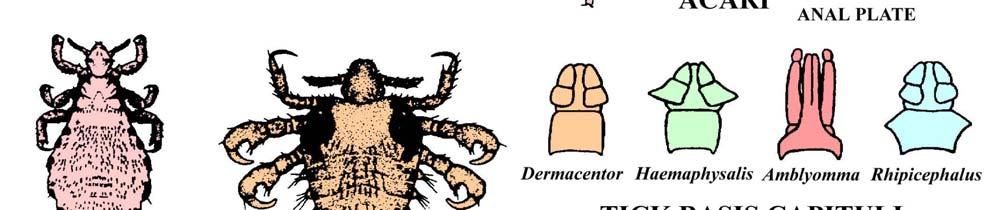
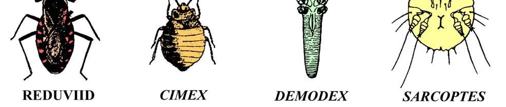
37 37 FIGURE 9. LINE DRAWINGS OF ARTHROPODS 37

38 38 FIGURE 10. COMMON TICKS OF KANSAS 38
Introduction to Helminthology
 Introduction to Helminthology HELMINTHES (WORMS) - Characteristics Eukaryotic, multicellular animals that usually have digestive, circulatory, nervous, excretory, and reproductive systems. Worms with bilateral
This is the smallest tapeworm that can affect human being but it s not really proper human tapeworm (the human is not the primary host).
.") Echinococcus Granulosus Small Tapeworm (1 cm), Cestode. This is the smallest tapeworm that can affect human being but it s not really proper human tapeworm (the human is not the primary host). The primary
Echinococcus Granulosus Small Tapeworm (1 cm), Cestode. This is the smallest tapeworm that can affect human being but it s not really proper human tapeworm (the human is not the primary host). The primary
LABORATORY. Introduction: Objectives: Laboratory 6 Pg.1
 LABORATORY Laboratory 6 Pg.1 6 Platyhelminthes and Acanthocephala Introduction: The Phylum Platyhelminthes contains both the trematodes and cestodes. The trematodes (flukes) have an incomplete digestive
Antihelminthic Trematodes (flukes): Cestodes (tapeworms): Nematodes (roundworms, pinworm, whipworms and hookworms):
: Cestodes (tapeworms): Nematodes (roundworms, pinworm, whipworms and hookworms):") Antihelminthic Drugs used to treat parasitic worm infections: helminthic infections Unlike protozoa, helminthes are large and have complex cellular structures It is very important to identify the causative
Antihelminthic Drugs used to treat parasitic worm infections: helminthic infections Unlike protozoa, helminthes are large and have complex cellular structures It is very important to identify the causative
Cestodes. Tapeworms from man and animals
 Cestodes Tapeworms from man and animals Taenia sp. The common (beef) tapeworm is several meters long. Courtesy Peters W. & Gilles H. Courtesy CDC Courtesy CDC Taenia sp. Unstained egg with four (visible)
أسئلة وأجوبة مادة أوليات والفقاريات )123 ح( )نصف ورقة إمتحانية(
123 ح( )نصف ورقة إمتحانية(") أسئلة وأجوبة مادة أوليات والفقاريات )123 ح( )نصف ورقة إمتحانية( جامعة: بنها كلية: العلوم قسم: علم الحيوان شعبة: حيوان وكيمياء تاريخ اإلمتحان: 2132 32/13/ الممتحنون: أ.د/ جزاء حسن مرسى د/ جيهان حسين الشين
أسئلة وأجوبة مادة أوليات والفقاريات )123 ح( )نصف ورقة إمتحانية( جامعة: بنها كلية: العلوم قسم: علم الحيوان شعبة: حيوان وكيمياء تاريخ اإلمتحان: 2132 32/13/ الممتحنون: أ.د/ جزاء حسن مرسى د/ جيهان حسين الشين
Cestodes (tapeworms) Pro. Dr. Mohammed Sabri
 Pro. Dr. Mohammed Sabri") Cestodes (tapeworms) Pro. Dr. Mohammed Sabri Characters (general):- Tape worms consist of a round head, called a scolex, and a flat body of multiple segments called proglottides. The scolex has specialized
Cestodes (tapeworms) Pro. Dr. Mohammed Sabri Characters (general):- Tape worms consist of a round head, called a scolex, and a flat body of multiple segments called proglottides. The scolex has specialized
Ecology & Evolutionary Biology 4274 Platyhelminthes Lecture Exam #2 October 22, 2014
 Name 1 Ecology & Evolutionary Biology 4274 Platyhelminthes Lecture Exam #2 October 22, 2014 Read through the exam once before you begin. Read the questions CAREFULLY; be certain to provide all of the information
Phylum Platyhelminthes Flatworms
 Phylum Platyhelminthes Flatworms The Acoelomates The acoelomates are animals that lack a coelom. Acoelomates lack a body cavity, and instead the space between the body wall and the digestive tract is filled
Contains most of the medically important tapeworms Scolex has 4 suckers and compact vitelline gland are characteristic Range from mm to >10m
 Cyclophyllidae Contains most of the medically important tapeworms Scolex has 4 suckers and compact vitelline gland are characteristic Range from mm to >10m Family Taeniidae Taenia saginata: beef tapeworm
Parasitology. Echinococcus Spp.
 Parasitology المحاضرة الثالثت ا. صباح النجار Echinococcus Spp. Genus Echinococcus include three different species in which man acts as intermediate host and infecting by the larval stage of these species.
Medical Parasitology (EEB 3895) Lecture Exam #2
 Lecture Exam #2") 1 Name November 2016 Medical Parasitology (EEB 3895) Lecture Exam #2 Read through the exam once before you begin. Read the questions CAREFULLY; be certain to provide all of the information requested. In
1 Name November 2016 Medical Parasitology (EEB 3895) Lecture Exam #2 Read through the exam once before you begin. Read the questions CAREFULLY; be certain to provide all of the information requested. In
V. Subclass Eucestoida (Chapters 20 & 21, BLY 459, 2010)
") V. Subclass Eucestoida (Chapters 20 & 21, BLY 459, 2010) A. Characteristics (Ignore Cestodaria) 1. Differences from trematodes a. No digestive tract (1) No mouth, gut, nor anus (2) All nutrients absorbed
V. Subclass Eucestoida (Chapters 20 & 21, BLY 459, 2010) A. Characteristics (Ignore Cestodaria) 1. Differences from trematodes a. No digestive tract (1) No mouth, gut, nor anus (2) All nutrients absorbed
Flatworms Flatworms Platyhelminthes dorsoventrally free-living planarian parasitic fluke tapeworm label three body layers ectoderm mesoderm
 Flatworms Flatworms are in the phylum Platyhelminthes. Flatworms are flattened dorsoventrally (top to bottom). The group includes the freshwater, free-living planarian and the parasitic fluke and tapeworm.
ECHINOCOCCUS GRANULOSUS
 48 ECHINOCOCCUS GRANULOSUS 48.1 INTRODUCTION E granulosus are small tape worms that parasitize the intestines of carnivores like dogs. About one million people are infected with this tape worm worldwide.
Platyhelminthes (flat worms) as parasites of medical interest
 as parasites of medical interest") Platyhelminthes (flat worms) as parasites of medical interest Type of parasites Protozoa Helminths Arthropods Nematodes Trematodes Cestodes Platyhelminthes Platyhelminthes Trematoda Planaria Monogenea
Platyhelminthes (flat worms) as parasites of medical interest Type of parasites Protozoa Helminths Arthropods Nematodes Trematodes Cestodes Platyhelminthes Platyhelminthes Trematoda Planaria Monogenea
Schistosoma mansoni, S. japonicum, S. haematobium
 Schistosoma mansoni, S. japonicum, S. haematobium The Organisms More than 200 million people are infected worldwide with Schistosoma species. The adult worms are long and slender (males are 6 12 mm in
SEMESTER ONE 2007 INFECTION and IMMUNITY GRADUATE ENTRY PROGRAMME PARASITOLOGY PRACTICAL 9 Dr TW Jones NEMATODES
 SEMESTER ONE 2007 INFECTION and IMMUNITY GRADUATE ENTRY PROGRAMME PARASITOLOGY PRACTICAL 9 Dr TW Jones NEMATODES Objectives After this class I expect you to be able to: 1. Describe and recognise the range
Eukaryotic Parasites. An Illustrated Guide to Parsitic Life Cycles to Accompany Lecture. By Noel Ways
 Eukaryotic Parasites An Illustrated Guide to Parsitic Life Cycles to Accompany Lecture By Noel Ways Giardia lamblia Life Cycle Reservoir: Beavers strongly implicated. Also, many other wild animals as well
Ectoparasites Myobia musculi Radfordia affinis Radfordia ensifera
 Ectoparasites Fleas, ticks, and lice are uncommon in modern laboratory facilities, but may be seen on wild or feral rodents. Most ectoparasite infestations seen in rats and mice used for research are various
Ecology & Evolutionary Biology 4274 Platyhelminthes Lecture Exam #2 October 30, 2009
 Name _ 1 Ecology & Evolutionary Biology 4274 Platyhelminthes Lecture Exam #2 October 30, 2009 This exam consists of TWO (2) SECTIONS. You must complete both Sections. SECTION I: You must answer all 12
The Worms / Chapter 34 and Partial 35 (pgs )
") Name: The Worms / Chapter 34 and Partial 35 (pgs.712-716) 1-6. Worms are not the simple organisms most people think of at first sight. List three specific features that support the idea that worms are
Name: The Worms / Chapter 34 and Partial 35 (pgs.712-716) 1-6. Worms are not the simple organisms most people think of at first sight. List three specific features that support the idea that worms are
Eukaryotic Organisms
 Eukaryotic Organisms A Pictoral Guide of Supportive Illustrations to accompany Select Topics on Eukaryotic Oranisms Bacteria (Not Shown) Agent of Disease Reservoir Vector By Noel Ways Favorable Environmental
Ecology & Evolutionary Biology 4274 Platyhelminthes Lecture Exam #2 October 26, 2011
 1 Name Ecology & Evolutionary Biology 4274 Platyhelminthes Lecture Exam #2 October 26, 2011 Read through the exam once before you begin. Read the questions CAREFULLY; be certain to provide all of the information
Breast Mass on Mammography
 Breast Mass on Mammography Bobbi Pritt, MD 2014 MFMER slide-1 Clinical Presentation 68 year old woman was noted to have 2 adjacent masses in the right breast on screening mammography Well-circumscribed,
Ascarids, Pinworms, and Trichocephalids
 LABORATORY Laboratory 3 Pg. 1 3 Introduction: Ascarids, Pinworms, and Trichocephalids The ascarids are large parasitic nematodes that usually live in the lumen of the small intestine of their host. All
Ascarids, Oxyuris, Trichocephalids
 LABORATORY Laboratory 4 Pg. 1 4 Introduction: Ascarids, Oxyuris, Trichocephalids The ascarids are large parasitic nematodes that usually live in the small intestine of their host. All ascarids have 3 lips
Presentation of Quiz #85
 Presentation of Quiz #85 ***Reminder: Slides are copyrighted and cannot be copied for publication. A 36 year old male from Columbia was admitted to the hospital with seizures. This patient had previously
BIO Parasitology Spring Trichostrongylines. Lecture 20. Hairworms in Horses. Stephen M. Shuster Northern Arizona University
 BIO 475 - Parasitology Spring 2009 Stephen M. Shuster Northern Arizona University http://www4.nau.edu/isopod Lecture 20 Trichostrongylines Hairworms in Horses Eggs hatch when eaten by the horse. Larvae
BIO 221 Invertebrate Zoology I Spring Ancylostoma caninum. Ancylostoma caninum cuticular larval migrans. Lecture 23
 BIO 221 Invertebrate Zoology I Spring 2010 Stephen M. Shuster Northern Arizona University http://www4.nau.edu/isopod Lecture 23 Ancylostoma caninum Ancylostoma caninum cuticular larval migrans Order Ascarida
Title. Author(s)YAMASHITA, Jiro; OHBAYASHI, Masashi; KONNO, Seiji. CitationJapanese Journal of Veterinary Research, 4(3): Issue Date
YAMASHITA, Jiro; OHBAYASHI, Masashi; KONNO, Seiji. CitationJapanese Journal of Veterinary Research, 4(3): Issue Date") Title STUDIES ON ECHINOCOCCOSIS : III. ON EXPERIMENTAL INF DEVELOPMENT OF ECHINOCOCCUS GRANULOSUS (BATSCH, 1786 Author(s)YAMASHITA, Jiro; OHBAYASHI, Masashi; KONNO, Seiji CitationJapanese Journal of Veterinary
Title STUDIES ON ECHINOCOCCOSIS : III. ON EXPERIMENTAL INF DEVELOPMENT OF ECHINOCOCCUS GRANULOSUS (BATSCH, 1786 Author(s)YAMASHITA, Jiro; OHBAYASHI, Masashi; KONNO, Seiji CitationJapanese Journal of Veterinary
General introduction
 Spirometra mansoni General introduction Distributed worldwide, mainly in southeast Asia. Larval infection of S. mansoni may cause serious clinical disease ---Sparganosis Morphology Adult worm measures
Frog Dissection Information Manuel
 Frog Dissection Information Manuel Anatomical Terms: Used to explain directions and orientation of a organism Directions or Positions: Anterior (cranial)- toward the head Posterior (caudal)- towards the
Hazem.K.Al-Khafaji FICMS College of medicine- Al-Qadissyia university
 Hazem.K.Al-Khafaji FICMS College of medicine- Al-Qadissyia university Cestodes(Tapeworms) - Morphology - Tapeworm parts: Flat, segmented body with various length (several mm,25mm as hymenolepis nana ~
Diphyllobotrium latum Sparganosis Taenia spp.
 Introduction of Cestodes Diphyllobotrium latum Sparganosis Taenia spp. (317221 inter) Asst. Prof. Dr. Atiporn Saeung Additional Readings น ม ตร มรกต, เกต ร ตน ส ขว จน, บรรณาธ การ.ปรส ตว ทยาทางการแพทย โปรโตซ
Parasitology PAR 311
 Parasitology PAR 311 Basic Information Program Title Bachelor of Medicine and Surgery; MB,BCh Department Offering the Course Parasitology Academic Year / Level Third year Date of Specification Approval
Recommended Resources: The following resources may be useful in teaching this
 Unit B: Anatomy and Physiology of Poultry Lesson1: Internal Anatomy of Poultry Student Learning Objectives: Instruction in this lesson should result in students achieving the following objectives: 1. Identify
1. Examine the specimens of sponges on the lab table. Which of these are true sponges? Explain your answers.
 Station #1 - Porifera 1. Examine the specimens of sponges on the lab table. Which of these are true sponges? Explain your answers. 2. Sponges are said to have an internal special skeleton. Examine the
Medical Parasitology PAR 311
 Medical Parasitology PAR 311 Basic Information Program Title Bachelor of Medicine and Surgery; MB,BCh Department Offering the Course Academic Year / Level Date of Specification Approval Total Teaching
Review of the Parasites of Large Animals
 LABORATORY Laboratory 10 Pg. 1 10 Introduction: Review of the Parasites of Large Animals In labs 2 through 10 we presented you with the various parasites of veterinary importance in a taxonomic manner.
VARIATION IN MONIEZIA EXPANSA RUDOLPHI
 VARIATION IN MONIEZIA EXPANSA RUDOLPHI STEPHEN R. WILLIAMS, Miami University, Oxford, Ohio In making a number of preparations of proglottids for class study at the stage when sex organs are mature and
Ecology & Evolutionary Biology 4274 Lecture Exam #3. Nematoda, Misc. phyla, Arthropoda, ecology and evolution December 10, 2014
 Name 1 Ecology & Evolutionary Biology 4274 Lecture Exam #3 Nematoda, Misc. phyla, Arthropoda, ecology and evolution December 10, 2014 Read through the exam once before you begin. Read the questions CAREFULLY;
PARASITOLOGY (A)
") Syllabus PARASITOLOGY (A) - 65814 Last update 29-03-2015 HU Credits: 2 Degree/Cycle: 2nd degree (Master) Responsible Department: Veterinary Medicine Academic year: 2 Semester: 1st Semester Teaching Languages:
Syllabus PARASITOLOGY (A) - 65814 Last update 29-03-2015 HU Credits: 2 Degree/Cycle: 2nd degree (Master) Responsible Department: Veterinary Medicine Academic year: 2 Semester: 1st Semester Teaching Languages:
PSEUDANDRYA MKUZll sp. nov, ( CESTODA: HYMENOLEPIDl DAE) FROM /CHNEUMIA ALBICAUDA
 FROM /CHNEUMIA ALBICAUDA") Onderstepoort J. Vet. Res. (1963), 30 (2), 127-132 Printed by the Government Printer, Pretoria PSEUDANDRYA MKUZll sp. nov, ( CESTODA: HYMENOLEPIDl DAE) FROM /CHNEUMIA ALBICAUDA R. J. ORTLEPP, Veterinary
Onderstepoort J. Vet. Res. (1963), 30 (2), 127-132 Printed by the Government Printer, Pretoria PSEUDANDRYA MKUZll sp. nov, ( CESTODA: HYMENOLEPIDl DAE) FROM /CHNEUMIA ALBICAUDA R. J. ORTLEPP, Veterinary
Animal Parasites. endoparasite parasite lives on the inside of host digestive system often very simple or gone altogether eg.
 Animal Parasites Parasitism most common form of symbiosis endoparasite parasite lives on the inside of host digestive system often very simple or gone altogether eg. tapeworms ectoparasites parasite lives
Feline and Canine Internal Parasites
 Feline and Canine Internal Parasites Internal parasites are a very common problem among dogs. Almost all puppies are already infected with roundworm when still in the uterus, or get the infection immediately
Vertebrates and Parasites
 Vertebrates and Parasites Parasites indicators of biodiversity o Lots of parasites with complex life histories = area of high biodiversity with a good ecosystem o Provide deep phylogenetic and ecological
Fischthal and Kuntz (1964) reported the
 reported the") Zoological Studies 41(3): 283-287 (2002) Meristocotyle provitellaria sp. nov. (Digenea: Meristocotylidae) from Varanus salvator in China Wei Liu 1, Qing-Kui Li 2, Hsiu-Hui Shih 3 and Zhao-Zhi Qiu 1, *
Zoological Studies 41(3): 283-287 (2002) Meristocotyle provitellaria sp. nov. (Digenea: Meristocotylidae) from Varanus salvator in China Wei Liu 1, Qing-Kui Li 2, Hsiu-Hui Shih 3 and Zhao-Zhi Qiu 1, *
Nematoda. Round worms Feeding and Parasitism
 Nematoda Round worms Feeding and Parasitism Nematoda Have pseudocoelom Live in many environments Parasitic Important decomposers Covered with cuticle Trichinella spiralis see fig 18.8B Nematode Diets and
Chapter 5 Male and female reproductive systems
 Chapter 5 Male and female reproductive systems This chapter begins with a description of the male and female reproductive systems followed by a section on sex determination. A good knowledge of the anatomy
HYDATID CYST DISEASE
 HYDATID CYST DISEASE Hydatid disease, also called hydatidosis or echinococcosis, is a cystforming disease resulting from an infection with the metacestode, or larval form, of parasitic dog tapeworms from
Diversity of Animals
 Classifying Animals Diversity of Animals Animals can be classified and grouped based on similarities in their characteristics. Animals make up one of the major biological groups of classification. All
TISSUE NEMATODES MODULE 49.1 INTODUCTION OBJECTIVES 49.2 FILARIASIS. Notes
 MODULE Tissue Nematodes 49 TISSUE NEMATODES 49.1 INTODUCTION Some nematodes cause infection in the tissues and may be found in the blood or lymphatics as well as in the muscle and other advetitious tissue.
Hydatid Disease. Overview
 Hydatid Disease Overview Hydatid disease in man is caused principally by infection with the larval stage of the dog tapeworm Echinococcus granulosus. It is an important pathogenic zoonotic parasitic infection
WITH THE TABLE OF THE MORPHOLOGICAL FEATURES OF TAPEWORMS IN VAMPIROLEPIS. (Received: December 22nd, 1965)
") Japan. J. Med. Sci. Biol. 19, 51-57, 1966 *ON A NEW TAPEWORM, VAMPIROLEPIS ISENSIS, FOUND IN BATS WITH THE TABLE OF THE MORPHOLOGICAL FEATURES OF TAPEWORMS IN VAMPIROLEPIS ISAMU SAWADA Biological Laboratory,
Japan. J. Med. Sci. Biol. 19, 51-57, 1966 *ON A NEW TAPEWORM, VAMPIROLEPIS ISENSIS, FOUND IN BATS WITH THE TABLE OF THE MORPHOLOGICAL FEATURES OF TAPEWORMS IN VAMPIROLEPIS ISAMU SAWADA Biological Laboratory,
The Rat Lungworm Lifecycle
 Hawaii Island Rat Lungworm Working Group Daniel K. Inouye College of Pharmacy University of Hawaii, Hilo The Rat Lungworm Lifecycle Rat Lungworm IPM RLWL-3 It is important to understand the lifecycle of
Animal Parasites. Parasitism most common form of symbiosis
 Animal Parasites Parasitism most common form of symbiosis endoparasite parasite lives on the inside of host digestive system often very simple or gone altogether eg. tapeworms ectoparasites parasite lives
Name Class Date. After you read this section, you should be able to answer these questions:
 CHAPTER 14 4 Vertebrates SECTION Introduction to Animals BEFORE YOU READ After you read this section, you should be able to answer these questions: How are vertebrates different from invertebrates? How
Vertebrates. Vertebrate Characteristics. 444 Chapter 14
 4 Vertebrates Key Concept All vertebrates have a backbone, which supports other specialized body structures and functions. What You Will Learn Vertebrates have an endoskeleton that provides support and
1.0 INTRODUCTION. Echinococcosis, a cyclozoonotic helminthosis caused by the dwarf dog
 INTRODUCTION 1.0 INTRODUCTION Echinococcosis, a cyclozoonotic helminthosis caused by the dwarf dog tapeworm Echinococcus granulosus is highly endemic and is considered to be one of the most important parasitic
Cestodes P & S, Joshua Stillman MD, MPH Department of Emergency Medicine Assistant Professor, Columbia University. Helminths
 Cestodes P & S, 2008 Joshua Stillman MD, MPH Department of Emergency Medicine Assistant Professor, Columbia University Helminths Phylum Nematoda (Roundworms) - Nematodes Pinworm, Whipworm, Ascaris + VLM,
Title. Author(s)YAMASHITA, Jiro; OHBAYASHI, Masashi; KITAMURA, Yukit. CitationJapanese Journal of Veterinary Research, 6(2): 89-92
YAMASHITA, Jiro; OHBAYASHI, Masashi; KITAMURA, Yukit. CitationJapanese Journal of Veterinary Research, 6(2): 89-92") Title STUDIES ON ECHINOCOCCOSIS VII. : ON THE DEVELOPMENT IN THE TAPEWORM STAGE Author(s)YAMASHITA, Jiro; OHBAYASHI, Masashi; KITAMURA, Yukit CitationJapanese Journal of Veterinary Research, 6(2): 89-92
Title STUDIES ON ECHINOCOCCOSIS VII. : ON THE DEVELOPMENT IN THE TAPEWORM STAGE Author(s)YAMASHITA, Jiro; OHBAYASHI, Masashi; KITAMURA, Yukit CitationJapanese Journal of Veterinary Research, 6(2): 89-92
30-3 Amphibians Slide 1 of 47
 1 of 47 What Is an Amphibian? What Is an Amphibian? An amphibian is a vertebrate that, with some exceptions: lives in water as a larva and on land as an adult breathes with lungs as an adult has moist
Nematodes 2. Lecture topics. Ascarid life cycle. Main features of the Ascarids. Adults L 5 L 1 L 4 L 2 L 3. Groups that you need to know about
 Lecture topics Nematodes 2 BVM&S Parasitology T.W.Jones The Ascarids Migratory & non-migratory species Hypobiosis Paratenic hosts The Strongyles Tissue feeders Migratory & non-migratory species The Hookworms
ECHINOCOCCOSIS. By Dr. Ameer kadhim Hussein. M.B.Ch.B. FICMS (Community Medicine).
.") ECHINOCOCCOSIS By Dr. Ameer kadhim Hussein. M.B.Ch.B. FICMS (Community Medicine). INTRODUCTION Species under genus Echinococcus are small tapeworms of carnivores with larval stages known as hydatids proliferating
ECHINOCOCCOSIS By Dr. Ameer kadhim Hussein. M.B.Ch.B. FICMS (Community Medicine). INTRODUCTION Species under genus Echinococcus are small tapeworms of carnivores with larval stages known as hydatids proliferating
Title ON DAUGHTER CYSTS OF COENURUS SERIALIS GERVAIS, Author(s)YAMASHITA, Jiro; OHBAYASHI, Masashi; KONNO, Seiji
YAMASHITA, Jiro; OHBAYASHI, Masashi; KONNO, Seiji") Title ON DAUGHTER CYSTS OF COENURUS SERIALIS GERVAIS, 1847 Author(s)YAMASHITA, Jiro; OHBAYASHI, Masashi; KONNO, Seiji CitationJapanese Journal of Veterinary Research, 5(1): 14-18 Issue Date 1957-03-25
Title ON DAUGHTER CYSTS OF COENURUS SERIALIS GERVAIS, 1847 Author(s)YAMASHITA, Jiro; OHBAYASHI, Masashi; KONNO, Seiji CitationJapanese Journal of Veterinary Research, 5(1): 14-18 Issue Date 1957-03-25
Nematodes 2. BVM&S Parasitology T.W.Jones
 Nematodes 2 BVM&S Parasitology T.W.Jones Lecture topics The Ascarids Migratory & non-migratory species Hypobiosis Paratenic hosts The Strongyles Tissue feeders Migratory & non-migratory species The Hookworms
COMMON INTESTINAL WORMS IN DOGS AND CATS W. JEAN DODDS, DVM :: HEMOPET/NUTRISCAN :: SALINAZ AVENUE :: GARDEN GROVE, CALIF.
 The following is a quick reference guide for pet caregivers about intestinal worms found in dogs and cats. At the end, I have provided my thoughts on treatment options. Roundworms Intestinal Parasite Most
Proteocephalus filicollis (Rud. 1810) in the Netherlands
 in the Netherlands") Proteocephalus filicollis (Rud. 1810) in the Netherlands by J.J. Willemse AND A.L.M. Veltman Zoological Laboratory, University of Amsterdam INTRODUCTION in another glass dish containing about 50 specimens
Proteocephalus filicollis (Rud. 1810) in the Netherlands by J.J. Willemse AND A.L.M. Veltman Zoological Laboratory, University of Amsterdam INTRODUCTION in another glass dish containing about 50 specimens
Field and Laboratory Study Evaluating the Possibility of Manodistomum syntomentera Causing Malformations In Frogs of the Mississippi River Valley
 11 Field and Laboratory Study Evaluating the Possibility of Manodistomum syntomentera Causing Malformations In Frogs of the Mississippi River Valley Laurie Carter Faculty Sponsor: Dr. Daniel Sutherland,
Grasshopper Dissection
 Grasshopper Dissection External Observation Locate the head, thorax, and abdomen. Observe the head. Locate the two compound eyes and the three simple eyes. 1. Why do you think grasshoppers have two types
Cestodes P & S, Joshua Stillman MD, MPH Department of Emergency Medicine Assistant Professor, Columbia University. Helminths
 Cestodes P & S, 2009 Joshua Stillman MD, MPH Department of Emergency Medicine Assistant Professor, Columbia University Helminths Phylum Nematoda (Roundworms) - Nematodes Pinworm, Whipworm, Ascaris + VLM,
Name Class Date. After you read this section, you should be able to answer these questions:
 CHAPTER 14 2 The Animal Kingdom SECTION Introduction to Animals BEFORE YOU READ After you read this section, you should be able to answer these questions: What is diversity? What are vertebrates? What
Balantidium coli Morphology of 2 stages. Balantidium coli
 Balantidium coli It causes balantidiasis or balantidial dysentery, is the largest intestinal protozoan of humans. The trophozoite is a ciliated, oval organism 60 X 45 μm or larger. It has a steady progression
Phylum:Apicomplexa Class:Sporozoa
 Phylum:Apicomplexa Class:Sporozoa The most characteristic features of sporozoa are 1-unique appearance of most protozoa makes it possible for knowledge able person to identifiy them to level of genus and
COMPARATIVE VERTEBRATE HISTOLOGY ZOO 4756c Syllabus for Fall 2018
 COMPARATIVE VERTEBRATE HISTOLOGY ZOO 4756c Syllabus for Fall 2018 Instructor: Frank T. Logiudice Office: Biology Building, Room 202c Office Phone Number: (407) - 823-2495 Email Address: Frank.Logiudice@ucf.edu
The Scoop on Poop 2019 Country Living Expo Classes #123 & #223
 The Scoop on Poop 2019 Country Living Expo Classes #123 & #223 TIM CUCHNA, DVM NORTHWEST VETERINARY CLINIC STANWOOD (360) 629-4571 NWVETSTANWOOD@GMAIL.COM WWW.NWVETSTANWOOD.COM Fecal Exam Today s plan
THE EFFECT OF MUTILATION ON THE TAPEWORM TAENIA TAENIAEFORMIS
 THE EFFECT OF MUTILATION ON THE TAPEWORM TAENIA TAENIAEFORMIS JOE N. MILLER AND WM. P. BUNNER The reader is undoubtedly aware of work which has been done by Child (1910) and others in mutilating certain
Title. Author(s)OHBAYASHI, Masashi. CitationJapanese Journal of Veterinary Research, 15(1): 1-3. Issue Date DOI. Doc URL.
OHBAYASHI, Masashi. CitationJapanese Journal of Veterinary Research, 15(1): 1-3. Issue Date DOI. Doc URL.") Title GRYPORHYNCHUS NYCTICORACIS YAMAGUTI, 1956 (DILEPIDID APHARYNGOSTRIGEA ARDEOLINA VIDYARTHI, 1937 (STRIGEID CINEREA JOUYI CLARK Author(s)OHBAYASHI, Masashi CitationJapanese Journal of Veterinary Research,
Title GRYPORHYNCHUS NYCTICORACIS YAMAGUTI, 1956 (DILEPIDID APHARYNGOSTRIGEA ARDEOLINA VIDYARTHI, 1937 (STRIGEID CINEREA JOUYI CLARK Author(s)OHBAYASHI, Masashi CitationJapanese Journal of Veterinary Research,
PARASITES OF PIGS By Dr. Fred O. Obonyo
 TRENDS IN VETERINARY PARASITOLOGY A TWO-DAYS COURSE DEPARTMENT OF VETERINARY PATHOLOGY, MICROBIOLOGY & PARASITOLOGY FACULTY OF VETERINARY MEDICINE UNIVERSITY OF NAIROBI 10 TH & 11 TH AUGUST 2011 PARASITES
Avian Reproductive System Female
 extension Avian Reproductive System Female articles.extension.org/pages/65372/avian-reproductive-systemfemale Written by: Dr. Jacquie Jacob, University of Kentucky For anyone interested in raising chickens
Hydatid Cyst Dr. Nora L. El-Tantawy
 Hydatid Cyst Dr. Nora L. El-Tantawy Ass. Prof. of Parasitology Faculty of Medicine, Mansoura university, Egypt Echinococcus granulosus Geographical Distribution: cosmopolitan especially in sheep raising
Module 1. Introduction to Targeted Neglected Tropical Diseases (NTDs)
") TARGETED FOR CONTROL OR Module 1. Introduction to Targeted Neglected Tropical Diseases (NTDs) Overview Road map to NTDs targeted for Preventive Chemotherapy (PC) Disease specific epidemiology and control
TARGETED FOR CONTROL OR Module 1. Introduction to Targeted Neglected Tropical Diseases (NTDs) Overview Road map to NTDs targeted for Preventive Chemotherapy (PC) Disease specific epidemiology and control
HOOKWORM FAQ SHEET (rev ) Adapted from the CDC Fact Sheet
 Adapted from the CDC Fact Sheet") HOOKWORM FAQ SHEET (rev 3-1-10) Adapted from the CDC Fact Sheet Hookworm Infection FAQ Sheet Contents What is hookworm? Where are hookworms commonly found? How do I get a hookworm infection? Who is at
HOOKWORM FAQ SHEET (rev 3-1-10) Adapted from the CDC Fact Sheet Hookworm Infection FAQ Sheet Contents What is hookworm? Where are hookworms commonly found? How do I get a hookworm infection? Who is at
Sec KEY CONCEPT Amphibians evolved from lobe-finned fish.
 Wed 4/26 Activities Learning Target Class Activities *attached below (scroll down)* Website: my.hrw.com Username: bio678 Password:a4s5s Students will describe the adaptations of amphibians that help them
2018 Copyright Jolie Canoli and Friends. For personal and educational use only. Find more resources at joliecanoli.com
 PHYLUM: BODY PLANS After the classification of Kingdom comes the category Phylum. The Phylum category of Animals puts animals with similar body types together. There are many phylum, but we will study
A Scanning Electron Microscopic Study of Eggshell Surface Topography of Leidynema portentosae and L. appendiculatum (Nematoda: Oxyuroidea)
") The Ohio State University Knowledge Bank kb.osu.edu Ohio Journal of Science (Ohio Academy of Science) Ohio Journal of Science: Volume 88, Issue 5 (December, 1988) 1988-12 A Scanning Electron Microscopic
The Ohio State University Knowledge Bank kb.osu.edu Ohio Journal of Science (Ohio Academy of Science) Ohio Journal of Science: Volume 88, Issue 5 (December, 1988) 1988-12 A Scanning Electron Microscopic
Digestive System Dissection
 Digestive System Dissection THE TERMS YOU NEED FOR THE PRACTICAL ARE IN THIS DISSECTION GUIDE. Instructions: Do one of the 2 respiratory dissections, and then the digestive dissection. Wordlist for cat
Sustainable Resources 11. Poultry Unit: Chicken Anatomy
 Sustainable Resources 11 Poultry Unit: Chicken Anatomy The Chicken Birds: Class AVES are winged, bipedal, endothermic (warm-blooded), egg-laying, vertebrates. Chicken: Gallus gallus are a domesticated
UNIT: INVERTEBRATE ANIMALS 1º ESO BIOLOGY AND GEOLOGY
 UNIT: INVERTEBRATE ANIMALS 1º ESO BIOLOGY AND GEOLOGY 2015/2016 What do they have in common? What are their differences? What is the theme for the next unit? Vertebrates and Invertebrates 1 Label the animals
Order Strongylida. Superfamilies: Trichostrongyloidea Strongyloidea Metastrongyloidea Ancylostomatoidea (hookworms)
") Order Strongylida Superfamilies: Trichostrongyloidea Strongyloidea Metastrongyloidea Ancylostomatoidea (hookworms) ORDER STRONGYLIDA - Bursate worms Superfamily - Ancylostomatoidea HOOKWORMS *dorsally
Order Strongylida Superfamilies: Trichostrongyloidea Strongyloidea Metastrongyloidea Ancylostomatoidea (hookworms) ORDER STRONGYLIDA - Bursate worms Superfamily - Ancylostomatoidea HOOKWORMS *dorsally
Giardia and Apicomplexa. G. A. Lozano UNBC
 Giardia and Apicomplexa G. A. Lozano UNBC NINE Protozoan diseases/parasites Ciliphora, Ichthyophthirius, Ick Sarcomastigophora, Giardia, giardiasis Apicomplexa: Eimeria, Toxoplasma, Sarcocystis, Cryptosporidium.
Aquaculture and human health
 Aquaculture and human health Jimmy Turnbull Institute of Aquaculture University of Stirling Scotland UK 1 Introduction zoonosis The transmission of a disease from an animal or nonhuman species to humans.
Science Read. 06 Feb. 2.8m-long tapeworm found in Singapore patient who had no symptoms
 Science Read Issue 04 06 Feb Career Guidance Interesting Science Real Life Application Real Time News Upper Secondary 2.8m-long tapeworm found in Singapore patient who had no symptoms Janice Tai, Social
Most amphibians begin life as aquatic organisms and then live on land as adults.
 Section 3: Most amphibians begin life as aquatic organisms and then live on land as adults. K What I Know W What I Want to Find Out L What I Learned Essential Questions What were the kinds of adaptations
THE MICROSCOPE PATHOGEN IDENTIFICATION
 CONTENTS 5 ABOUT THE AUTHOR 5 ACKNOWLEDGEMENTS 6 OVERVIEW 6 What is the Purpose of this Book? 6 What are the Limitations of Light Microscopy as a Diagnostic Tool? 7 When Should I Contact a Veterinarian?
THE STRUCTURE OF ECHINOCOCCAL CYSTS AND HISTOPATHOLOGICAL CHANGES IN LIVER
 THE STRUCTURE OF ECHINOCOCCAL CYSTS AND HISTOPATHOLOGICAL CHANGES IN LIVER Michal Juszynski Helena Palenga, Danuta Cielecka PhD Department of General Biology and Parasitology Medical University of Warsaw
VERTEBRATE READING. Fishes
 VERTEBRATE READING Fishes The first vertebrates to become a widespread, predominant life form on earth were fishes. Prior to this, only invertebrates, such as mollusks, worms and squid-like animals, would
Guard against intestinal worms with Palatable All-wormer
 Guard against intestinal worms with Palatable All-wormer WHIPWORMS HOOKWORMS TAPEWORMS ROUNDWORMS Palatable All-wormer, for superior, flexible protection of dogs and cats. GENTLE ON PETS, TOUGH ON WORMS.