MEDICAL VECTORS. Advanced Vector Surveillance Workshop 30 October 1 November Prepared by Mosquito Consulting Services (NZ)
|
|
|
- Richard Shaw
- 5 years ago
- Views:
Transcription


1 MEDICAL VECTORS Advanced Vector Surveillance Workshop 30 October 1 November 2013 Prepared by Mosquito Consulting Services (NZ)
2 ii
3 Disclaimer and Acknowledgement This document has been constructed as revision notes for the Ministry of Health s Border Health courses and workshops only and not permitted for distribution. Several of the images in this document were sourced from the ICPMR website ( it/mosquitoes.htm). Thank you to Richard Russell and Stephen Doggett who kindly gave permission for these images to be used in these notes. iii
4 iv
5 Table of Contents 1. INTRODUCTION TO VECTORS BASIC VECTOR INFORMATION PUBLIC HEALTH DISEASE THREAT VECTOR AND DISEASE CONTROL Background Personal Protection Surveillance Control MOSQUITOES LIFE CYCLE The Egg The Larva The Pupa The Adult HABITATS Larvae Adults HOSTS BEHAVIOUR DISEASES Malaria Dengue Yellow fever Japanese encephalitis Ross River & Barmah Forest West Nile Virus Chikungunya Other Diseases EXOTIC MOSQUITOES WHY ARE WE CONCERNED ABOUT EXOTIC MOSQUITOES? PATHWAYS UNWANTED ORGANISMS MOSQUITO PROFILES Aedes (Stegomyia) aegypti (Linnaeus) Aedes (Stegomyia) albopictus (Skuse) Aedes (Finlaya) japonicus (Theobald) MOSQUITOES AND MOSQUITO-BORNE DISEASE CONTROL Mosquito Personal Protection Mosquito Surveillance Mosquito Control TICKS THE TICK LIFE CYCLE Hard ticks Soft ticks HABITATS Hard ticks Soft ticks HOSTS Hard Ticks Soft Ticks BEHAVIOUR Hard Ticks Soft Ticks DISEASES v
6 3.5.1 Lyme disease Rocky Mountain spotted fever (RMSF) Tick-borne relapsing fever (TBRF) Ehrlichiosis Babesiosis Tick-Borne Encephalitis (TBE) Tularemia Some other tick borne diseases TICKS AND TICK-BORNE DISEASE CONTROL Tick Personal Protection Tick Surveillance Tick Control SAND FLIES LIFE CYCLE Egg Larva Pupa Adult HABITATS HOSTS BEHAVIOUR DISEASES Leishmaniasis Sandfly (Phlebotomus) Fever Other Diseases SAND FLIES AND SAND FLY-BORNE DISEASE CONTROL Sand Fly Personal Protection Sand Fly Surveillance Sand Fly Control MITES LIFE CYCLE Eggs Larvae Nymphs Adults HABITATS HOSTS BEHAVIOUR DISEASES Scrub typhus Rickettsial Pox Scabies MITES AND MITE-BORNE DISEASE CONTROL Mite Personal Protection Mite Surveillance Mite Control FLEAS LIFE CYCLE Eggs Larvae Pupae Adults HABITATS HOSTS BEHAVIOUR DISEASES Plague Dipylidiasis Other Diseases vi
7 6.6 FLEAS AND FLEA-BORNE DISEASE CONTROL Flea Personal Protection Flea Surveillance Flea Control LICE LIFE CYCLE The Egg The Nymph The Adult HABITATS HOSTS BEHAVIOUR DISEASES Louse-borne relapsing Fever Louse-borne Typhus Trench Fever Pediculosis LICE AND LOUSE-BORNE DISEASE CONTROL Louse Personal Protection Lice Surveillance Lice Control BED BUGS LIFE CYCLE The Egg The Larvae (Nymph) The Adult HABITATS HOSTS BEHAVIOUR DISEASES BED BUG CONTROL Bed bug Personal Protection Bed Bug Surveillance Bed Bug Control COCKROACHES LIFE CYCLE The Egg The Nymph The Adult HABITATS HOSTS BEHAVIOUR DISEASES Typhoid Amoebiasis Biology of disease: Shigellosis Polio COCKROACH CONTROL Cockroach Personal Protection Cockroach Surveillance Cockroach Control RATS LIFE CYCLE BEHAVIOUR Rattus rattus Rattus norvegicus DISEASES RAT CONTROL vii
8 Indications of Rodent Infestation Integrated Pest Management SAMPLING METHODOLOGIES SAMPLING MOSQUITOES Trapping Adults Larval sampling Biting/Landing Collections Aspirating Adults SAMPLING TICKS Removing ticks from hosts Free-living ticks Trapping Ticks SAMPLING SANDFLIES CDC light trap Sticky traps SAMPLING MITES Attached mites Unattached mites SAMPLING FLEAS SAMPLING LICE SAMPLING BEDBUGS Advice to be given before inspection Surveillance Tools and Equipment Indications of a Bed Bug Infestation Other methods used For Detection Important Information SAMPLING COCKROACHES SAMPLING RATS PREPARATION FOR TRANSPORT Mosquito Larvae Mosquito Adults Ticks and Mites Sandflies Fleas Bed Bugs Cockroaches Rats PACKAGING FOR TRANSPORT BORDER BIOSECURITY REQUIREMENTS FOR RETURN OF SAMPLES TO NZ viii
9
10 1. Introduction to Vectors 1.1 Basic vector information In epidemiological terms, vectors are transmitters of disease-causing organisms, i.e. they carry the pathogens from one host to another. By common usage, vectors are generally considered to be invertebrate animals, specifically arthropods. However, technically vertebrates can also act as vectors, e.g. foxes, which can transmit the rabies virus to humans via a bite. An arthropod that transmits a disease-causing organism from one vertebrate host to another is referred to as a disease vector. These are the types of vector we are concerned with in this manual. Arthropods account for over 85 percent of all known animal species and are the most important disease vectors globally. They affect human health either directly by bites, stings and infestation of tissues, or indirectly through disease transmission. There are two types of vector that convey infectious organisms to a host, mechanical and biological. Mechanical vectors only physically transport microorganisms from host to host, microorganisms do not multiply within them. In contrast, microorganisms must propagate within a biological vector before they can be transmitted into a host. The most significant mode of vector-borne disease transmission is biological transmission by bloodfeeding arthropods. The pathogen multiplies within the arthropod vector and is transmitted to the host when the arthropod takes a blood meal. Several groups of arthropod play a role in human and animal disease transmission, with mosquitoes and ticks being the most notable disease vectors. A classic example is the Anopheles mosquito which acts as a vector for the disease malaria by transmitting the malarial parasite Plasmodium to humans. The Plasmodium is harmless to the mosquito vector (its intermediate host), but causes illness in humans (its definitive host). The majority of vector-borne diseases survive in nature, utilizing animals as their hosts. Zoonosis is the term given to infectious disease that is able to be transmitted by a vector from other animals, both wild and domestic, to humans (or from humans to animals often called reverse zoonoses). For a small number of zoonoses, such as malaria and dengue, humans are the major host, with no significant animal reservoirs. Intermediary animal hosts often serve as a reservoir for the pathogens until susceptible human populations are exposed. The vector receives the pathogen from an infected host and transmits it either to an intermediary host or directly to the human host. The different stages of the pathogen's life cycle occur during this process and are intimately dependent upon the availability of suitable vectors and hosts. Key components that determine the occurrence of vector-borne diseases include: (1) the abundance of vectors and intermediate and reservoir hosts; (2) the prevalence of disease-causing pathogens suitably adapted to the vectors and the human or animal hosts; (3) the local environmental conditions, especially temperature and humidity; and (4) the resilience behaviour and immune status of the host population. 1
11 1.2 Public health disease threat Vector-borne diseases are prevalent in the tropics and subtropics and are relatively rare in temperate zones, although climate change could create conditions suitable for outbreaks of diseases such as Lyme disease, Rocky Mountain spotted fever, malaria, dengue fever and viral encephalitis in temperate regions. There are different patterns of vector-borne disease occurrence. Parasitic and bacterial diseases, such as malaria and Lyme disease, tend to produce a high disease incidence but do not cause major epidemics. An exception to this rule is plague, a bacterial disease that does cause outbreaks. In contrast, many vector-borne viral diseases, such as Yellow fever, dengue, and Japanese encephalitis, commonly cause major epidemics. There has been a worldwide resurgence of vector-borne diseases since the 1970s including malaria, dengue, Yellow fever, louse-borne typhus, plague, leishmaniasis, sleeping sickness, West Nile encephalitis, Lyme disease, Japanese encephalitis, Rift Valley fever and Crimean-Congo hemorrhagic fever. Reasons for the emergence or resurgence of vector-borne diseases include: the development of insecticide and drug resistance; decreased resources for surveillance, prevention and control of vector-borne diseases; deterioration of the public health infrastructure required to deal with these diseases; unprecedented population growth; uncontrolled urbanization; changes in agricultural practices; deforestation; and increased travel. Changes have been documented in the distribution of important arthropod disease vectors. For example, the Yellow fever mosquito, Aedes aegypti has re-established in parts of the Americas where it had been presumed to have been eradicated; the Asian tiger mosquito, Aedes albopictus was introduced into the Americas in the 1980s and has spread to Central and South America; and the blacklegged tick, Ixodes scapularis, an important transmitter of Lyme disease and other pathogens, has gradually expanded its range in parts of eastern and central North America. It is clear that people will always have to live with vector-borne diseases, but maintenance of a strong public health infrastructure and undertaking research activities directed at improved means of control (possibly utilizing biological and genetic-based strategies), combined with the development of new or improved vaccines for diseases such as malaria, dengue and Lyme disease should lessen the threat to human health. Control measures for vector-borne diseases will always be important as most zoonoses that are maintained in nature in cycles involving wild animals, are not amenable to eradication. Therefore, control methods targeting the arthropod vector help reduce the prevalence of these diseases and the risk to public health. Such methods should include: undertaking personal protective measures by establishing physical barriers such as house screens and bed nets wearing appropriate clothing (boots, apparel that overlap the upper garments, head nets, etc.) using insect repellents Environmental modification to eliminate specific breeding areas, or chemical biological control measures to kill arthropod vector larvae or adults may also be undertaken Areas such as ports and airports should be rigidly monitored, with control measures utilized to prevent important arthropod disease vectors from entering the country Some efforts to control vector-borne diseases focus on the pathogen - for example, vaccines are available for diseases such as Yellow fever, tick-borne encephalitis, Japanese encephalitis, tularemia, and plague 2
12 The vertebrate host may be the target - for example, vaccination of foxes against rabies in Europe and Canada is an effective means to reduce the threat of rabies Reduction of host reservoirs, such as rodents and birds, from areas of human habitation may lessen the risk for contracting certain vector-borne diseases such as plague and St. Louis encephalitis. 1.3 Vector and disease control Background The first discovery of an arthropod-borne pathogen was in 1893, and it was quickly followed by notable successes in the prevention of yellow fever and malaria through mosquito control. Despite continual progress in the technology of vector control during the last century, military forces remain vulnerable to many serious diseases caused by pathogens transmitted by mosquitoes, ticks, and other arthropods that cause considerable morbidity and mortality. The hundreds of recentlyreturning U.S. veterans from Iraq and Afghanistan who had contracted cutaneous leishmaniasis transmitted by sandflies is a testimony to this fact. Other recent U.S. military operations have also been negatively impacted by arthropod-borne infections. In September 2003, when 290 Marines went ashore in Liberia as military advisors to oversee a civil transition, 80 contracted malaria (28% attack rate). Malaria remains a significant threat on the Korean peninsula and elsewhere throughout Asia. Japanese encephalitis is one of approximately 100 viruses spread by insects and ticks and is a significant threat to US forces in the Pacific region. In addition to vector-borne and zoonotic diseases, biological threats during deployments include biting and stinging arthropods (fire ants, mites/chiggers, scorpions, etc.); vertebrate animals (rodents, bats, snakes, etc.); and poisonous plants (e.g., poison oak and poison sumac). Biting and stinging arthropods can degrade mission readiness and combat effectiveness even when they do not transmit disease. These arthropods can cause casualties ranging in severity from secondary infections to death from allergic reaction. Annoyance from persistent pests, itching bites, and loss of sleep can also erode morale Personal Protection Vector-borne diseases and associated discomfort caused by biting arthropods can be largely prevented with proper use of personal protective measures (PPMs) by individuals. Personal protective measures include arthropod repellents, clothing impregnants, and equipment such as bed nets and techniques which, when appropriately applied, will preserve the fighting strength of the troops. U.S. standardized practice for personal protection against arthropods: Apply the standard military clothing repellent (Permethrin clothing treatment, either the impregnation kit [IDA kit, a.k.a. shake and bake ] or the aerosol can) to your uniform, and the standard military skin repellent (DEET insect repellent lotion, made by the 3M Corporation, and also sold commercially as Ultrathon ) to all areas of exposed skin. Also, it s crucial to create a physical barrier to insects by wearing your uniform properly: cover as much skin as possible by wearing your sleeves down; close all openings in your clothing by tucking your pant cuffs into your boots, tucking your undershirt into your pants, and buttoning your shirt at the neck and wrists. Lace up boots completely. Wear your uniform loosely, as mosquitoes can bit through untreated fabric that is taut against the skin. This procedure could be translated into a useful equivalent in NZ. 3
13 Immunizations and Chemoprophylaxis Both immunizations and chemoprophylactic measures are considered personal protective measures although they are not controlled by the individual Soldier. Chemoprophylaxis is available for some of the protozoan (malaria) and bacterial (scrub typhus) pathogens transmitted by arthropods. Vaccines are routinely available for only a few of the viral pathogens (yellow fever virus, Japanese encephalitis virus) and are available on an experimental use protocol for a few others (Venezuelan equine encephalitis virus, Rift Valley fever virus). Even when appropriate chemoprophylaxis or vaccination is available for the disease of greatest concern, their use entails considerable medical management. When risk is unknown or considered to be low, personal protection may be the appropriate strategy for prevention. Therefore, the proper use of other personal protective measures described earlier offer the most practical means of interrupting and preventing arthropod-borne disease transmission Surveillance Disease vector and pest surveillance is designed to: 1. measure the relative population levels of known pests to determine when and where to begin specific management techniques; 2. detect invasions of new and potentially important vectors and pests; 3. detect breeding sites that can be eliminated; and 4. measure the effectiveness of previous management efforts Control Integrated pest management describes many approaches to pest control including non-chemical activities such as sanitation, habitat modification, and development of surveillance programs to specifically target pest locations and activity times. Use of IPM must not compromise the effectiveness of control and must be tailored to best address the specific needs of each pest or disease vector problem. Pesticides are indispensable management tools. As part of any IPM program, when choosing to use chemical control tools, pest managers are directed to select the least hazardous pesticides that will still provide acceptable results. For example, pesticides in the organophosphate and carbamate chemical classes are still used if specific conditions warrant, but effective substitutes such as newer generation pyrethrins or insect growth regulators are preferred choices. 4
14 5
 of the class Insecta and further separated from other flies in the Family Culicidae.")

15 2. Mosquitoes The word mosquito is derived from Spanish and means little fly. Mosquitoes are in fact a type of fly, belonging to the order Diptera ( true flies or two-winged flies ) of the class Insecta and further separated from other flies in the Family Culicidae. There are about 35 genera (species groups) of mosquitoes worldwide, including over 2700 different species. Many mosquito species are vectors of disease. They can carry and transmit disease-causing organisms such as viruses, bacteria and worms, from host to host. WNV Dengue Filarial worms Mosquitoes are the arthropod group responsible for the most human deaths worldwide. 2.1 Life cycle The mosquito life cycle begins with an adult female laying eggs. Aquatic immature stages called larvae emerge and develop through four moults (instars), increasing in size until the final moult when it reaches the non-feeding pupal stage (See Figure b & c). Inside the pupa the adult mosquito develops (either a male or female) and the terrestrial/aerial adult stage emerges from a split in the back of the pupal skin (See Figure a & d). The adult mosquitoes feed, mate, and the female develops eggs to complete the cycle and begin the next generation. Some species of mosquito have only one generation per year. Others have several generations during a single season of favourable climatic conditions. Some continue to breed throughout the year, but may be more abundant in warmer seasons - this depends on the local environment, particularly temperature and rainfall. 6
16 Life cycle of a Culex pipiens mosquito. a) Emerging adult. b) female adult ovipositing egg raft on water surface. c) representative of each larval instar using siphon to breathe at water surface. d) comma-shaped pupa breathing using trumpet at water surface. Diagram ex Gullan, P.J. & Cranston, P.S The Insects. 3 rd Edition. Blackwell Publishing. 505pp The Egg Mosquito females oviposit their eggs on or near water bodies. They are almost transparent when first laid, but gradually darken to brown or black as they mature. Eggs of Culicine mosquitoes (e.g. Culex and Aedes) are usually elongate-oval in shape with the anterior end rounded and the posterior bluntly pointed. Anopheline eggs (e.g. Anopheles) are more cigar-shaped with flotation structures on each side. The eggs are laid singly or in clusters and this can vary depending on the genus. Aedes species lay their eggs as single units and deposit them on moist substrate such as rock surfaces, moist earth and the inside wall of tree holes or containers above the receding water level. They also lay eggs under debris and in crevices in soil and dry mud, where they will be subsequently flooded. These eggs are able to withstand desiccation, and can survive long periods until they are submerged by water, at which time they begin to hatch. Egg rafts of Culex and Coquillettidia spp. float on top of the water surface, while Mansonia spp. egg rafts can be found attached to the underside of leaves or twigs, just below the water surface. Mansonia species eggs differ from other species in that they have one end extended into a spike. Egg rafts cannot withstand desiccation and are usually associated with permanent or semipermanent water bodies. They will hatch after about two days on the water and without constant water, they desiccate and die. 7


, may influence the female in her search for an appropriate oviposition site.")
17 Aedes sp. eggs Coquillettidia sp. egg raft Anopheline sp. egg note the floats on either side of the egg. Mansonia sp. egg raft attached under debris Culex sp. Egg raft The Larva The larval stage must have an aquatic habitat in which to complete its development to the pupal stage. The adult female selects an appropriate larval habitat when she deposits her eggs. They are able to discern physical and chemical properties of different collections of water and choose between sites available. Factors including shade, temperature, salinity, water quality and the texture of the substrate (Aedes species), may influence the female in her search for an appropriate oviposition site. Various combinations of these factors can be identified as being characteristic of a typical breeding site of a particular species. Therefore there are a range of diverse habitats where mosquito larvae can be found, which is dependent on species type as well as environmental factors. 8

 or exuviae (plural).")

 alternans and Toxorhynchites sp.")
 australis larva with mouth brushes for filtering water Head of")
18 Culex quinquefasciatus larvae hatching from eggs The larvae hatch from the eggs and grow through four instars before developing into a pupa. Between each stage they moult their rigid outer skin so they can increase in size. The discarded skin is termed an exuvium/exuvia (singular) or exuviae (plural). The larval instar level is determined by the size of the head capsule, not the body length. Most larvae feed on microscopic organisms in the water and bottom detritus, either by filtering water through their mouth brushes or by grazing with specially adapted mouth appendages. Some larvae are predatory (Aedes (Ochlerotatus) alternans and Toxorhynchites sp.) and their mouth brushes are modified so they are strong enough to grasp prey. Head of 4 th instar Aedes (Ochlerotatus) australis larva with mouth brushes for filtering water Head of Toxorhynchites sp. larva, arrow indicating prey-gripping mouth brushes. Some species feed habitually at the surface (Anopheles), some in the middle range below the surface (Culex) and others typically feed on the bottom of the habitat (Aedes). 9


19 Larvae breathe air from openings (spiracles) at the tail end of the body, generally through a structure termed a siphon (See Figure). They hang below the water surface with only the tip of the siphon exposed to the air. They can remain motionless on the bottom for some time, but need to return to the surface for air to prevent suffocation. Water surface Siphon One of the two main exceptions to breathing behaviour occurs within larvae of the genera Mansonia and Coquillettidia. They attach to plants below the surface of the water after hatching, using a specially adapted piercing siphon and obtain their oxygen directly from the plant tissues. Larvae of these two genera do not visit the water surface during their development and usually feed by filtering food particles from the surrounding water with their mouth brushes. Coquillettidia linealis larvae breathing through a plant stem. The second main exception in breathing behaviour occurs within the genus Anopheles, whose larvae do not have a siphon or breathing tube. Species of this genus lie alongside the surface of the water to breathe. The time taken for development through the larval stages is dependent on a number of environmental factors, the most important 10


20 of which is temperature. Availability of food and the extent of larval crowding within the habitat are also important. During favourable summer conditions, Anopheles species may complete larval development in 7-10 days, Aedes species may complete larval development in as little as 4-5 days and Culex species may require at least 7-10 days. Low temperatures usually delay development and may cause cessation of growth and induce a over-wintering of larvae in some species. Identification of larvae is most easily accomplished with mature larvae, i.e. the fourth instar and microscopic examination is usually required. However, there are some genus characteristics that enable partial identification in the field. For example, Anopheles species lack of a siphon and larvae lying flat at the surface of the habitat when breathing or resting, distinguishes them from Culex and Aedes species which have siphons and hang suspended from the surface. Culex species typically have longer siphons than Aedes species, which also can help assist in recognising the different genera in the field, however this can really only be achieved with experience and should always be checked under the microscope. The larvae of Mansonia and Coquillettidia, although not commonly collected because of their attachment to submerged aquatic vegetation, can be identified as being from one of these two genera either by their attachment to a plant or if separated from the vegetation, by their modified siphon. Anopheles spp. Aedes or Culex spp. Coquillettidia or Mansonia spp The Pupa After the 4 th larval instar completes its development, it moults into a non-feeding but highly mobile stage called the pupa. Within the body casing of the pupa, the immature tissues are breaking down and adult tissues are forming. The pupa breathes through a pair of tube-like organs (trumpet) situated at the head end of the comma-shaped body. 11
, obtain oxygen from plant tissues below the water surface, using")

.")
21 Trumpets Identification of pupae is only possible using microscopy. However, as with the larvae, some groups can be distinguished by their behaviour. Mansonia and Coquillettidia species for example, are different from other mosquitoes in that their pupae (like their larvae), obtain oxygen from plant tissues below the water surface, using modified trumpets (See Figure 9b). Culex annulirostris pupa Coquillettidia linealis pupa breathing through plant stem. breathing at the water surface. The duration of the pupal stage again is dependent on temperature but is generally of the order of 2-3 days for Anopheles, Aedes and Culex species. Once the adult tissues have developed and it is time for emergence, the pupa swims to the water surface and stretches itself out to full length and the pupal skins splits along the back and the teneral adult mosquito emerges above the water surface (see photo below). After emerging from the pupal casing, the adult mosquito rests on the water surface for a short time, to allow its wings and body to dry, before flying off in search of nourishment and a mate. Male mosquitoes develop faster than females, and are usually the first to emerge. 12

22 Emergence The Adult After emerging from the pupal casing, the adult mosquito rests on the water surface for a short time allowing its wings and body to dry, before flying off in search of a mate. In a single generation, the males of a species usually develop marginally more quickly than the females, and males are usually first to emerge from the larval habitat. This is not always noticeable in the field where generations may overlap. Male mosquitoes do not normally travel far from the breeding site and feed on plant juices, sugars from flowers and fruit nectars. The adult female also seeks out a sugar meal of nectar or similar plant juices to replenish expended energy reserves and then mates with a male, usually near a breeding site at dusk. Female mosquitoes mate only once, as the sperm packet introduced by a male during the mating act is sufficient for the female to fertilise all batches of eggs she subsequently produces. For egg production, female mosquitoes require protein via a blood meal. A few species can develop the first batch of eggs using nutritional reserves carried over from the larval stage, this is called autogeny. They usually require a blood source to produce the second and subsequent batches. The preferred source of a blood meal can vary widely between mosquito species. In general terms, mosquitoes are attracted to a warmblooded host by a combination of factors; carbon dioxide, a product of respiration is an important attractant as are various body odours and chemicals such as lactic acid. 13

23 These seem to be the longer range attractants. At closer distances, temperature can be a factor, as can visual perception at very close proximities. Some species may take several blood meals to acquire sufficient protein for egg production. The female searches for secluded refuge where she can rest undisturbed, digest the blood meal and develop a batch of eggs. She will then fly off in search of additional blood meals to repeat this process. Subsequent blood meals may be taken the night of oviposition if a host is nearby, otherwise a day or more may elapse before the next feed. important to the mosquito may not always be apparent. As mentioned above, the oviposition site (and thus, the larval habitat) may be characteristic for a particular species. However, although we know that mosquitoes have sense organs which allow them to choose between physical and chemical features of an aquatic site, the determining features Identification of adult mosquitoes is very complex, even to genus level and microscopes are required. However, the sex of mosquitoes caught in the field can often be determined by eye if they stop flying around for a second! Adult males differ from females in that they have long palps protruding from their head next to their proboscis, and very bushy antennae compared to those of the females. An exception to this occurs in the Anopheles genus, which has both sexes with long palps, but the males still have the more bushy antennae. Resting positions also vary between these genera (see diagram). Summary diagram of the main mosquito genera. Ex: Carpenter & LaCasse (1955). Mosquitoes of North America (North of Mexico). University of California Press, Berkeley. 360pp. 14
24 The life span of adult mosquitoes is not well known. Some species apparently live one or two months during the summer, although under unfavourable conditions this period may be greatly reduced. Adults that hibernate during winter may live for six months or more. In laboratory conditions, Aedes aegypti adults have lasted as much as 240 days (about eight months). All stages in the life cycle of a mosquito are dependent upon a number of environmental factors for their survival and development. Some common and measurable environmental factors, such as wind, light, temperature, rainfall, and humidity, have a known relationship to the survival of mosquitoes and can be used as the basis of an index for use in surveillance and control. 2.2 Habitats The range of habitats utilised by mosquitoes is extremely diverse. With over 3000 species worldwide, mosquitoes have evolved to utilise almost any aquatic system in most parts of the world Larvae An internationally accepted mosquito breeding habitat classification lists the following 11 larval habitats: 1. Flowing stream 2. Ponded stream 3. Lake edge 4. Swamp/Marsh 5. Permanent Pond 6. Temporary Pond 7. Intermittent ephemeral puddle 8. Natural container 9. Artificial container 10. Subterranean habitats-natural 11. Subterranean habitats-artificial The majority of mosquito species can be classified as either Container Breeders or Groundwater Breeders. Container breeders generally utilise smaller habitats such as tree holes, leaf axils and coconut shells. However they have adapted well to artificial habitats, often found in discarded rubbish, tyres, tin cans, plastic sheeting as well as items that are in use, oil drums, buckets and guttering. Some containers may provide more permanent habitat, such as drain sumps and rock pools, but classification of those habitats may be debatable. Container breeders are often more commonly associated with populated areas as these generally provide a much greater opportunity for breeding. Groundwater breeders utilise more expansive habitats; swamps, marshes, lake edges field drains and mangroves etc. Groundwater breeders may be found in and around urban areas, although often their habitat will not occur within cities. An example of this involves saltmarsh habitats which often occur adjacent to urban areas. One of the key characteristics of saltmarsh species is often a long flight range, so the habitat existing outside of urban environments does not necessarily provide protection for the hosts within the city. There are also some species whose behaviour allows for breeding in both container and groundwater e.g. Culex gelidus an important vector of Japanese Encephalitis. 15
25 Temperature plays a vital role in larval mosquito population dynamics. In tropical regions where there is no significant cold season, the seasonal pattern of mosquito population changes is related to the supply of water and rainfall. A slight rise in the level of water may cause an increase in mosquito production by re-establishing the less frequently inundated oviposition sites and increasing the number of temporary bodies of water. Excessively heavy rainfall and runoff during flood conditions may have a flushing effect and reduce the numbers of mosquitoes in the area. Such a reduction in the larval mosquito population is normally of a relatively short duration Adults Adult mosquitoes will utilise different habitat for different purposes. In general however, males will remain near the breeding habitat and only travel short distances to some source of a sugar feed (nectar, fruit etc). Females will generally seek shelter from the environment, somewhere with little air movement, often dark, with sugar feeds nearby but also within distance of blood sources and breeding habitat. This will be significantly affected by the flight range of the mosquito, species with a short flight range will be found near to the breeding habitat, while mosquitoes with a longer flight range may be found sheltering several kilometres from the nearest breeding habitat. Many mosquitoes prefer vegetation to rest in but domestic mosquitoes such as Aedes aegypti will be found in dwellings, resting in closets and under tables etc. 2.3 Hosts Mosquitoes will utilise almost any land-based animal large enough to provide it with a blood feed. Some species are adaptable while others are quite host specific. Hosts include: Birds Mammals Reptiles Amphibians The choice of host species for blood feeding is an important factor in disease transmission. Aedes aegypti is an urban mosquito with a preference for biting man. It s ability to transmit dengue combined with a close association with human populations make it the most significant vector in dengue outbreaks. However with diseases like Japanese encephalitis where two host species are required for the disease cycle, a less specific mosquito species, such as Culex annulirostris is a better vector. 2.4 Behaviour Male and female adult mosquitoes are usually present in about equal numbers following emergence. Typically the male mosquitoes reside near the breeding sites and have a shorter lifespan than females. Females may travel some distance to find a blood source. Only the female mosquitoes blood feed in order to obtain protein to produce fertile eggs. Flight habits vary considerably; Aedes aegypti, arguably the most highly domesticated mosquito, typically flies very short distances (usually less than 500 metres). In studies some individuals have flown less than 35m from the water body they emerged from in their entire lifetime, while Aedes vigilax will comfortably travel 5-10km for sugar and blood feeds and may travel upwards of 300km 16
26 in jetstream wind-assisted migrations. Although a coastal species, Ae. vigilax has been found as far inland in Australia as Alice Springs following a migration dispersal. The possible flight range of Anopheline mosquitoes varies considerably, depending on the species and circumstances in search of food and shelter. Generally they will fly less than 3km, but they have been known to fly 30 kilometres in temperate climates with wind assistance. Times of activity vary from species to species. Some species are active during the day (diurnal or day-biting)and others only at night (nocturnal or night-biting) with many more active at dawn and dusk (crepuscular). 2.5 Diseases Mosquitoes are the most important group of blood sucking insects that cause nuisance and transmit diseases to humans and other warm blooded animals. The nuisance and annoyance caused by mosquitoes is not easy to translate into economic value, however it is as vectors of disease that mosquitoes are most often of concern. Vector mosquitoes and the parasites and pathogens that they transmit, are recognised to have played an important role in the development and dispersal of the human race, being responsible for some events that have shaped the course of history. Although vaccines, chemoprophylaxis, chemotherapy, genetics and vector control measures are becoming more sophisticated, even now, none of the major mosquito-borne diseases of the world can be said to be under complete control. Mosquitoes are responsible for transmitting three types of human pathogenic organisms: Arboviruses viruses causing diseases such as dengue, Yellow fever and various encephalitides. (The term arbovirus is derived from arthropod-borne-virus) Plasmodia protozoans which are the cause of malaria Filarial worms nematodes that cause lymphatic filariasis Mosquitoes can act as transmitters or vectors of pathogens or parasites by both mechanical and biological means. Mechanical transmission occurs where the pathogen has no biological association with the vector, i.e. the pathogen is picked up from one source and deposited in another location. This occurs when the pathogen is carried passively on the biting mouthparts of a mosquito which has fed on an infected host and the pathogen passes passively into a second host at a subsequent feeding. This is most likely to occur where mosquitoes are interrupted during feeding and where any pathogens on the mouthparts remain viable for a short time and are introduced to another host during a subsequent attempt to feed to repletion. Although mechanical transmission of pathogenic organisms occurs via mosquitoes with some animal diseases, biological transmission is predominant for human parasites. Biological transmission refers to the situation where the pathogen or parasite undergoes a period of development and/or multiplication within the vector (which acts as a true intermediate host and is essential for the completion of the cycle) before being passed on to another host following this incubation period (sometimes called the intrinsic incubation period to differentiate it from the incubation period in the vertebrate host which is the extrinsic incubation period). There are three systems that apply: 1. The pathogen develops and multiplies, e.g. malarial parasites there is sexual union of the blood stages in the mosquito gut, encystation in the gut wall, multiple sporozoite formation in the cyst and movement of the sporozoites to the mosquito s salivary glands as infective stages for introduction into a new host during subsequent feeding. 17
27 2. The pathogen develops only, e.g. filarial parasites the microfilarial blood stages taken into the mosquito gut escape from the gut and develop through three stages in the mosquito s tissues before entering the head as infective stages for introduction into a new host during subsequent feeding. 3. The pathogen multiplies only, e.g. arboviruses virus particles taken in with blood into the mosquito gut invade the gut cells, disseminate and multiply in body tissues and penetrate the salivary glands to be introduced into a new host during subsequent feeding. Irrespective of the particular cycle, once a mosquito vector has picked up a pathogen, the vector needs to survive for at least the period of time required by the pathogen to complete its development cycle or multiply to the point that the mosquito becomes infective, before that mosquito can be involved in the transmission of the pathogen to the new host. This intrinsic incubation period varies with pathogen and temperature, but in general is in the order of 1-2 weeks. Thus the mosquito must survive for at least 1-2 weeks for it to become infective and therefore mosquito longevity is a critical factor in the dynamics of transmission of disease pathogens. Some of the diseases spread by mosquitoes are associated with animal reservoirs and are called zoonoses (e.g. Yellow fever, viral encephalitides, Brugian filariasis), while others involve only human reservoirs (e.g. dengue, malaria, Bancroftian filariasis). In all cases, the crucial factor in transmission to man (the epidemiology of the disease) is the amount and type of contact between the mosquito vector and the human host. The incidence and prevalence of disease in an area will depend upon the presence of the disease, susceptible vectors and the amount of human-vector contact. The latter is a product of interaction between habitat and behaviour of the mosquito vector and the habitat, and behaviour of the human host. The more often that a potentially infective mosquito intrudes into the human environment or that the humans intrude into the natural environment where mosquitoes harbour pathogenic organisms, the greater the risk of initiating an urban outbreak or epidemic. Mosquito borne diseases are complicated communicable diseases as they involve the vector as an additional component of the disease system. Social, behavioural, environmental and immunological factors may affect the human component, yet with vector involvement further influences impinge on the system and still more may arise if the disease is a zoonosis, involving other vertebrates as well as humans. Such a complex system may seem formidable, however the more complex the system, the greater the number of opportunities exist for disrupting the disease cycle. Not all mosquitoes can or do act as vectors for all or any pathogens Malaria Excerpts from Malaria parasites are micro-organisms that belong to the genus Plasmodium. There are more than 100 species of Plasmodium, which can infect many animal species such as reptiles, birds, and various mammals. Only four species of Plasmodium infect humans in nature (there are some other species which can, exceptionally or under experimental conditions, infect humans). The four species infecting humans are: Plasmodium falciparum, which is found worldwide in tropical and subtropical areas. It is the only species that can cause severe, potentially fatal malaria. It is estimated that every year 700,000 to 2.7 million people are killed by P. falciparum, especially in Africa where this species predominates. P. falciparum can cause severe malaria because it multiples rapidly in the blood, and can thus cause severe blood loss (anaemia). In addition, the infected parasites can clog small blood vessels. When this occurs in the brain, cerebral malaria results, a complication that can be fatal. 18
, it can cause symptoms that are incapacitating. Thus, P.")
28 P. vivax, which is found mostly in Asia, Latin America, and in some parts of Africa. Because of the population densities especially in Asia it is probably the most prevalent human malaria parasite. While P. vivax only exceptionally causes death (most often due to rupture of an enlarged spleen), it can cause symptoms that are incapacitating. Thus, P. vivax contributes substantially to the disease burden (morbidity) of malaria, with a resulting social and economic impact. P. vivax (as well as P. ovale) has dormant liver stages ("hypnozoites") that can activate and invade the blood ("relapse") several months or years after the infecting mosquito bite. Plasmodium ovale is found mostly in Africa (especially West Africa) and the islands of the western Pacific. It is biologically and morphologically very similar to P. vivax. However, differently from P. vivax, it can infect individuals who are negative for the Duffy blood group, which is the case for many residents of sub Saharan Africa. This explains the greater prevalence of P. ovale (rather than P. vivax ) in most of Africa. P. malariae, found worldwide, is the only human malaria parasite species that has a quartan cycle (three-day cycle). (The three other species have a tertian, two-day cycle). Plasmodium malariae causes a long-lasting, chronic infection that in some cases can last a lifetime. In some patients, P. malariae can cause serious complications such as the nephrotic syndrome. Malaria is transmitted among humans by female mosquitoes of the genus Anopheles. Female mosquitoes take blood meals to carry out egg production, and such blood meals are the link between the human and the mosquito hosts in the parasite life cycle. Of the approximately 430 known species of Anopheles, only transmit malaria in nature. Photo ex Life Cycle of Malaria In nature, malaria parasites spread by infecting successively two types of hosts; humans and female Anopheles mosquitoes. In the human hosts, the parasites grow and multiply, first in the liver cells and then in the red cells of the blood. In the blood, successive broods of parasites grow inside the red cells and destroy them, releasing daughter parasites ("merozoites") that continue the cycle by invading other red cells. The blood stage parasites are those that cause the symptoms of malaria. When certain forms of blood stage parasites ("gametocytes") are picked up by a female Anopheles mosquito during a blood meal, they start another, different cycle of growth and multiplication in the mosquito. After days, the parasites are found (as "sporozoites") in the mosquito's salivary glands. When the Anopheles mosquito takes a blood meal from another human, the sporozoites are injected along with the mosquito's saliva and start another human infection once they parasitize the liver cells. Thus the mosquito carries the disease from one human to another (acting as a "vector"). In contrast to the human host, the mosquito vector does not suffer from the presence of the parasites. 19

29 The malaria parasite life cycle involves two hosts. During a blood meal, a malaria-infected female Anopheles mosquito inoculates sporozoites into the human host. Sporozoites infect liver cells and mature into schizonts, which rupture and release merozoites. (Of note, in P. vivax and P. ovale a dormant stage [hypnozoites] can persist in the liver and cause relapses by invading the bloodstream weeks, or even years later.) After this initial replication in the liver (exo-erythrocytic schizogony ), the parasites undergo asexual multiplication in the erythrocytes (erythrocytic schizogony ). Merozoites infect red blood cells. The ring stage trophozoites mature into schizonts, which rupture releasing merozoites. Some parasites differentiate into sexual erythrocytic stages (gametocytes). Blood stage parasites are responsible for the clinical manifestations of the disease. The gametocytes, male (microgametocytes) and female (macrogametocytes), are ingested by an Anopheles mosquito during a blood meal. The parasites multiplication in the mosquito is known as the sporogonic cycle. While in the mosquito's stomach, the microgametes penetrate the macrogametes generating zygotes. The zygotes in turn become motile and elongated (ookinetes) which invade the midgut wall of the mosquito where they develop into oocysts. The oocysts grow, rupture, and release sporozoites, which make their way to the mosquito's salivary glands. Inoculation of the sporozoites into a new human host perpetuates the malaria life cycle. Humans infected with malaria parasites can develop a wide range of symptoms. These vary from asymptomatic infections (no apparent illness), to the classic symptoms of malaria (fever, chills, sweating, headaches, muscle pains), to severe complications (cerebral malaria, anaemia, kidney failure) that can result in death. The severity of the symptoms depends on several factors, such as the species (type) of infecting parasite and the human's acquired immunity and genetic background. 20
30 The successful development of the malaria parasite in the mosquito (from the "gametocyte" stage to the "sporozoite" stage) depends on several factors. The most important are ambient temperature (higher temperatures accelerate the parasite growth in the mosquito), humidity and whether the Anopheles survives long enough to allow the parasite to complete its cycle in the mosquito host Dengue Excerpts from Dengue (DF) and dengue hemorrhagic fever (DHF) are caused by one of four closely related, but antigenically distinct, virus serotypes (DEN-1, DEN-2, DEN-3, and DEN-4), of the genus Flavivirus. Infection with one of these serotypes provides immunity to only that serotype for life, so persons living in a dengue-endemic area can have more than one dengue infection during their lifetime. DF and DHF are primarily diseases of tropical and sub-tropical areas and the four different dengue serotypes are maintained in a cycle that involves humans and the Aedes mosquito. Aedes aegypti, a domestic, day-biting mosquito that prefers to feed on humans, is the most common vector. Infections produce a spectrum of clinical illness ranging from a non-specific viral syndrome to severe and fatal haemorrhagic disease. Important risk factors for DHF include the strain of the infecting virus, as well as the age, and especially the prior dengue infection history of the patient. History of Dengue The first reported epidemics of DF occurred in in Asia, Africa and North America. The near simultaneous occurrence of outbreaks on three continents indicated that these viruses and their mosquito vector have had a worldwide distribution in the tropics for more than 200 years. During most of this time, DF was considered a mild, non-fatal disease of visitors to the tropics. Generally, there were long intervals (10-40 years) between major epidemics, mainly because the introduction of a new serotype in a susceptible population occurred only if viruses and their mosquito vector could survive the slow transport between population centres via sailing vessels. A pandemic of dengue began in Southeast Asia after World War II and has spread around the globe since then. Epidemics caused by multiple serotypes (hyperendemicity) are more frequent, the geographic distribution of dengue viruses and their mosquito vectors has expanded and DHF has emerged in the Pacific region and the Americas. In Southeast Asia, epidemic DHF first appeared in the 1950s, but by 1975 it had become a frequent cause of hospitalization and death among children in many countries in that region. Current Trends In the 1980s, DHF began a second expansion into Asia when Sri Lanka, India, and the Maldive Islands had their first major DHF epidemics; Pakistan first reported an epidemic of dengue fever in The epidemics in Sri Lanka and India were associated with multiple dengue virus serotypes, but DEN-3 was predominant and was genetically distinct from DEN-3 viruses previously isolated from infected persons in those countries. After an absence of 35 years, epidemic dengue fever re-emerged in both Taiwan and the People's Republic of China in the 1980s. The People's Republic of China had a series of epidemics caused by all four serotypes and the first major epidemic of DHF, caused by DEN-2, was reported on Hainan Island in Singapore also had a resurgence of dengue/dhf from 1990 to 1994 after a successful control program had prevented significant transmission for over 20 years. In other countries of Asia where DHF is endemic, the epidemics have become progressively larger in the last 15 years. In the Pacific, dengue viruses were reintroduced in the early 1970s after an absence of more than 25 years. Epidemic activity caused by all four serotypes has intensified in recent years with major epidemics of DHF on several islands. 21
, Kenya (1982, DEN-2), Mozambique (1985, DEN-3), Djibouti (1991-92, DEN-2),")
31 Despite poor surveillance for dengue in Africa, epidemic dengue fever caused by all four serotypes has increased dramatically since Most activity has occurred in East Africa and major epidemics were reported for the first time in the Seychelles (1977), Kenya (1982, DEN-2), Mozambique (1985, DEN-3), Djibouti ( , DEN-2), Somalia (1982, 1993, DEN-2), and Saudi Arabia (1994, DEN-2). Epidemic DHF has not been reported in Africa or the Middle East, but sporadic cases clinically compatible with DHF have been reported from Mozambique, Djibouti, and Saudi Arabia. The emergence of dengue/dhf as a major public health problem has been most dramatic in the American region. In an effort to prevent urban Yellow fever, which is also transmitted by Ae. aegypti, the Pan American Health Organization started a campaign that eradicated Ae. aegypti from most Central and South American countries in the 1950s and 1960s. As a result, epidemic dengue occurred only sporadically in some Caribbean islands during this period. The Ae. aegypti eradication program, which was officially discontinued in the United States in 1970, gradually weakened elsewhere and the mosquito began to re-infest countries from which it had been eradicated. As a result, the geographic distribution of Ae. aegypti in 2002 was much wider than that before the eradication program. In 2005, dengue is the most important mosquito-borne viral disease affecting humans; its global distribution is comparable to that of malaria and an estimated 2.5 billion people live in areas at risk for epidemic transmission. Each year, tens of millions of cases of DF occur and depending on the year, up to hundreds of thousands of cases of DHF. The case-fatality rate of DHF in most countries is about 5%, but this can be reduced to less than 1% with proper treatment. Most fatal cases are among children and young adults. The reasons for the dramatic global emergence of DF/DHF as a major public health problem are complex and not well understood. However, several important factors can be identified: 1. First, major global demographic changes have occurred, the most important of which have been uncontrolled urbanization and concurrent population growth. These demographic changes have resulted in substandard housing and inadequate water, sewer and waste management systems, 22
32 all of which increase Ae. aegypti population densities and facilitate transmission of Ae. aegyptiborne disease. 2. In most countries the public health infrastructure has deteriorated. Limited financial and human resources and competing priorities have resulted in a "crisis mentality" with emphasis on implementing so-called emergency control methods in response to epidemics rather than on developing programs to prevent epidemic transmission. This approach has been particularly detrimental to dengue control because, in most countries, surveillance is (just as in the U.S.) passive; the system to detect increased transmission normally relies on reports by local physicians who often do not consider dengue in their differential diagnoses. As a result, an epidemic has often reached or passed its peak before it is recognized. 3. Increased travel by airplane provides the ideal mechanism for infected human transport of dengue viruses between population centres of the tropics, resulting in a frequent exchange of dengue viruses and other pathogens. 4. Lastly, effective mosquito control is virtually nonexistent in most dengue-endemic countries. Considerable emphasis in the past has been placed on ultra-low-volume insecticide space sprays for adult mosquito control, a relatively ineffective approach for controlling Ae. aegypti. Future Outlook No dengue vaccine is available. Recently, however, attenuated candidate vaccine viruses have been developed. Efficacy trials in human volunteers have yet to be initiated. Research is also being conducted to develop second-generation recombinant vaccine viruses. Therefore, an effective dengue vaccine for public use will not be available for 5 to 10 years. Prospects for reversing the recent trend of increased epidemic activity and geographic expansion of dengue are not promising. New dengue virus strains and serotypes will likely continue to be introduced into many areas where the population densities of Ae. aegypti are at high levels. With no new mosquito control technology available, in recent years public health authorities have emphasized disease prevention and mosquito control through community efforts to reduce larval breeding sources. Although this approach will probably be effective in the long run, it is unlikely to impact disease transmission in the near future. Therefore we must develop improved, proactive, laboratory-based surveillance systems that can provide early warning of an impending dengue epidemic. At the very least, surveillance results can alert the public to take action and physicians to diagnose and properly treat DF/DHF cases Yellow fever Excerpts from Yellow fever is a viral disease that has caused large epidemics in Africa and the Americas. It can be recognized from historic texts stretching back 400 years. The "yellow" in the name is explained by the jaundice that affects some patients. The disease is caused by the Yellow fever virus, which belongs to the Flavivirus group. In Africa there are two distinct genetic types (called topotypes) associated with East and West Africa. South America has two different types, but since 1974 only one has been identified as the cause of disease outbreaks. Infection causes a wide spectrum of disease, from mild symptoms to severe illness and death. The virus remains silent in the body during an incubation period of three to six days. There are then two disease phases. While some infections have no symptoms whatsoever, the first, "acute", phase is normally characterized by fever, muscle pain (with prominent backache), headache, shivers, loss of appetite, nausea and/or vomiting. Often, the high fever is paradoxically associated with a slow pulse. After three to four days most patients improve and their symptoms disappear. 23
33 However, 15% enter a "toxic phase" within 24 hours. Fever reappears and several body systems are affected. The patient rapidly develops jaundice and complains of abdominal pain with vomiting. Bleeding can occur from the mouth, nose, eyes and/or stomach. Once this happens, blood appears in the vomit and faeces. Kidney function deteriorates; this can range from abnormal protein levels in the urine (albuminuria) to complete kidney failure with no urine production (anuria). Half of the patients in the "toxic phase" die within days, the remainder recover without significant organ damage. Yellow fever is difficult to recognize, especially during the early stages. It can easily be confused with malaria, typhoid, rickettsial diseases, haemorrhagic viral fevers (e.g. Lassa), arboviral infections (e.g. dengue), leptospirosis, viral hepatitis and poisoning (e.g. carbon tetrachloride). A laboratory analysis is required to confirm a suspect case. Blood tests (serology assays) can detect Yellow fever antibodies that are produced in response to the infection. Several other techniques are used to identify the virus itself in blood specimens or liver tissue collected after death. These tests require highly trained laboratory staff using specialized equipment and materials. Although an effective vaccine has been available for 60 years, the number of people infected over the last two decades has increased and Yellow fever is now a serious public health issue again. Regions Affected The virus is constantly present with low levels of infection (i.e. endemic) in some tropical areas of Africa and the Americas. This viral presence can amplify into regular epidemics. Until the start of this century, Yellow fever outbreaks also occurred in Europe, the Caribbean islands and Central and North America. Even though the virus is not thought to be present in these areas now, they must still be considered at risk for Yellow fever epidemics. Thirty-three countries, with a combined population of 508 million, are at risk in Africa. These lie within a band from 15 N to 10 S of the equator. In the Americas, Yellow fever is endemic in nine South American countries and in several Caribbean islands. Bolivia, Brazil, Colombia, Ecuador and Peru are considered at greatest risk. There are 200,000 estimated cases of Yellow fever (with 30,000 deaths) per year. However, due to underreporting, only a small percentage of these cases are identified. Small numbers of imported cases also occur in countries free of Yellow fever. Although Yellow fever has never been reported from Asia, this region is at risk because the appropriate primates and mosquitoes are present. Transmission Humans and monkeys are the principal animals to be infected. The virus is carried from one animal to another (horizontal transmission) by a biting mosquito (the vector). The mosquito can also pass the virus via infected eggs to its offspring (vertical transmission). The eggs produced are resistant to drying and lie dormant through dry conditions, hatching when the rainy season begins. Therefore, the mosquito is the true reservoir of the virus, ensuring transmission from one year to the next. Several different species of the Aedes and Haemogogus (S. America only) mosquitoes transmit the Yellow fever virus. These mosquitoes are either domestic (i.e. they breed around houses), wild (they breed in the jungle) or semi-domestic types (they display a mixture of habits). Any region populated with these mosquitoes can potentially harbour the disease. Control programmes successfully eradicated mosquito habitats in the past, especially in South America. However, these programmes have lapsed over the last 30 years and mosquito populations have increased. This favours epidemics of Yellow fever. Infection of humans There are three types of transmission cycle for Yellow fever: sylvatic, intermediate and urban. All three cycles exist in Africa, but in South America, only sylvatic and urban Yellow fever occur. 24
34 Sylvatic (or jungle) Yellow fever: In tropical rainforests, Yellow fever occurs in monkeys that are infected by wild mosquitoes. The infected monkeys can then pass the virus onto other mosquitoes that feed on them. These infected wild mosquitoes bite humans entering the forest resulting in sporadic cases of Yellow fever. The majority of cases are young men working in the forest (logging, etc). On occasion, the virus spreads beyond the affected individual. Intermediate Yellow fever: In humid or semi-humid savannahs of Africa, small-scale epidemics occur. These behave differently from urban epidemics; many separate villages in an area suffer cases simultaneously, but fewer people die from infection. Semi-domestic mosquitoes infect both monkey and human hosts. This area is often called the "zone of emergence", where increased contact between man and infected mosquito leads to disease. This is the most common type of outbreak seen in recent decades in Africa. It can shift to a more severe urban-type epidemic if the infection is carried into a suitable environment (with the presence of domestic mosquitoes and unvaccinated humans). Urban Yellow fever: Large epidemics can occur when migrants introduce the virus into areas with high human population density. Domestic mosquitoes (of one species, Aedes aegypti) carry the virus from person to person; no monkeys are involved in transmission. These outbreaks tend to spread outwards from one source to cover a wide area. Treatment There is no specific treatment for Yellow fever. Dehydration and fever can be corrected with oral rehydration salts and paracetamol. Any superimposed bacterial infection should be treated with an appropriate antibiotic. Intensive supportive care may improve the outcome for seriously ill patients, but is rarely available in poorer, developing countries. Vaccination Vaccination is the single most important measure for preventing Yellow fever. In populations where vaccination coverage is low, vigilant surveillance is critical for prompt recognition and rapid control of outbreaks. Mosquito control measures can be used to prevent virus transmission until vaccination has taken effect. Yellow fever vaccine is safe and highly effective. The protective effect (immunity) occurs within one week in 95% of people vaccinated. A single dose of vaccine provides protection for 10 years and probably for life. Over 300 million doses have been given and serious side effects are extremely rare. However, recently a few serious adverse outcomes, including deaths, have been reported in Brazil, Australia and the United States. Scientists are investigating the cause of these adverse events and monitoring to ensure detection of any similar incidents. The risk to life from Yellow fever is far greater than the risk from the vaccine, so those who may be exposed to Yellow fever should be protected by immunization. If there is no risk of exposure, for example, if a person will not be visiting an endemic area, there is no necessity to receive the vaccine. Since most of the other known side effects have occurred in children less than six months old, vaccine is not administered to this age group. The vaccine should only be given to pregnant women during vaccination campaigns in the midst of an epidemic. Vaccination can be part of a routine preventive immunization programme or can be done in mass "catch-up" campaigns to increase vaccination coverage in areas where it is low. The World Health Organization (WHO) strongly recommends routine childhood vaccination. The vaccine can be administered at age nine months, at the same time as the measles vaccine. Eighteen African nations have agreed to incorporate Yellow fever vaccine into their routine national vaccination programmes. This is more cost effective and prevents more cases (and deaths) than when emergency vaccination campaigns are performed to control an epidemic. Past experience shows the success of this strategy. Between 1939 and 1952 Yellow fever cases almost vanished from French West Africa after intensive vaccination campaigns. Similarly, Gambia 25
35 instituted mass routine vaccination after its 1979/1980 epidemic and later incorporated Yellow fever vaccine into its childhood immunization programme. Gambia reported 85% vaccine coverage in No cases have been reported since 1980, yet the virus remains present in the environment. To prevent an epidemic in a country, at least 80% of the population must have immunity to Yellow fever. This can only be achieved through the effective incorporation of Yellow fever into childhood immunization programmes and the implementation of mass catch-up campaigns. The latter is the only way to ensure that coverage of all susceptible age groups is achieved and will prevent outbreaks from spreading. Very few countries in Africa have achieved this level to date. Vaccination is highly recommended for travellers to high-risk areas. A vaccination certificate is required for entry to many countries, particularly for travellers arriving in Asia from Africa or South America. Fatal cases in unvaccinated tourists have been reported. Surveillance Because vaccination coverage in many areas is not optimal, prompt detection of Yellow fever cases and rapid response (emergency vaccination campaigns) are essential for controlling disease outbreaks. Improvement in Yellow fever surveillance is needed as evidenced by the gross underreporting of cases (estimates as to the true number of cases vary widely and have put the underreporting factor between three- and 250-fold). A surveillance system must be sensitive enough to detect and appropriately investigate suspect cases. This is facilitated by a standardized definition of possible Yellow fever cases, that is "acute fever followed by jaundice within two weeks of onset of symptoms, or with bleeding symptoms or with death within three weeks of onset". Suspect cases are reported to health authorities on a standardized case investigation form. Ready access to laboratory testing is essential for confirming cases of Yellow fever, as many other diseases have similar symptoms. WHO has recently recommended that every at-risk country have at least one national laboratory where basic Yellow fever blood tests can be performed. Training programmes are being conducted and test materials are provided by WHO. Given the likelihood that other cases have occurred (but have not been detected), one confirmed case of Yellow fever is considered to be an outbreak. An investigation team should subsequently explore and define the outbreak. This produces data for analysis, which guides the epidemic control committee in preparing the appropriate outbreak response (e.g. emergency vaccination programmes, mosquito control activities). This committee should also plan for the long term by implementing or strengthening routine childhood Yellow fever vaccination. Future Outlook Over the last 20 years the number of Yellow fever epidemics has risen and more countries are reporting cases. Mosquito numbers and habitats are increasing. In both Africa and the Americas, there is a large susceptible, unvaccinated population. Changes in the world's environment, such as deforestation and urbanization, have increased contact with the mosquito/virus. Widespread international travel could play a role in spreading the disease. The priorities are vaccination of exposed populations, improved surveillance and epidemic preparedness. In March 1998, WHO held a technical consensus meeting in Geneva to identify obstacles to Yellow fever prevention and control. Priorities identified included: prevention through routine immunization and preventive mass immunization campaigns; detection, reporting and investigation of suspect cases; laboratory support; outbreak response; vaccine supply; and furthering research. Guidelines for investigation and control of Yellow fever outbreaks, and a background document reviewing topics of importance discussed at this meeting have been published Japanese encephalitis Excerpts from 26
36 Japanese encephalitis is a viral disease that infects animals and humans. It is transmitted by mosquitoes and in humans causes inflammation of the membranes around the brain. Intensification and expansion of irrigated rice production systems in South and South-East Asia over the past 20 years have had an important impact on the disease burden caused by Japanese encephalitis. Where irrigation expands into semi-arid areas, the flooding of the fields at the start of each cropping cycle leads to an explosive build-up of the mosquito population. This may cause the circulation of the virus to spill over from their usual hosts (birds and pigs) into the human population. Japanese encephalitis (JE) is caused by a flavivirus that affects the membranes around the brain. Most JE virus infections are mild (fever and headache) or without apparent symptoms, but approximately 1 in 200 infections results in severe disease characterized by rapid onset of high fever, headache, neck stiffness, disorientation, coma, seizures, spastic paralysis and death. The case fatality rate can be as high as 60% among those with disease symptoms; 30% of those who survive suffer from lasting damage to the central nervous system. In areas where the JE virus is common, encephalitis occurs mainly in young children because older children and adults have already been infected and are immune. The virus causing JE is transmitted by mosquitoes belonging to the Culex tritaeniorhynchus and Culex vishnui groups, which breed particularly in flooded rice fields. The virus circulates in ardeid birds (herons and egrets). Pigs are amplifying hosts, in that the virus reproduces in pigs and infects mosquitoes that take blood meals, but does not cause disease. The virus tends to spill over into human populations when infected mosquito populations build up explosively and the human biting rate increases (these culicines are normally zoophilic, i.e. they prefer to take blood meals from animals). JE is a leading cause of viral encephalitis in Asia with 30,000-50,000 clinical cases reported annually. It occurs from the islands of the Western Pacific in the east to the Pakistani border in the west, and from Korea in the north to Papua New Guinea in the south. Because of the critical role of pigs, its presence in Muslim countries is negligible. JE distribution is very significantly linked to irrigated rice production combined with pig rearing. Japanese encephalitis is a patchy disease and important outbreaks have occurred in a number of places in the past 15 years, including South India (Arkot district in Tamil Nadu) and in Sri Lanka (Mahaweli System H). Vaccination An effective killed vaccine is available for Japanese encephalitis, but it is expensive and requires one primary vaccination followed by two boosters. This is an adequate intervention for travellers, but has limited public health value in areas where health services have limited resources. An inexpensive live-attenuated vaccine is used in China, but is not available elsewhere Ross River & Barmah Forest From Traditionally known as Epidemic Polyarthritis, both Ross River (RR) and Barmah Forest (BF) disease are caused by viruses which are transmitted to humans through the bite of mosquitoes. A wide variety of symptoms may occur from rashes with fevers, to arthritis that can last from months to years with RR virus infection. There are no specific treatments, actions which reduce mosquito bites are the best form of prevention against these debilitating diseases. 27

37 RR disease is the most commonly transmitted mosquito-borne viral disease to humans in Australia. The number of cases has averaged >5,000 per annum during The virus appears to be endemic in most rural areas, and there has been an increasing incidence near major cities. BF disease is less common, but the number of cases appears to be increasing annually, with several outbreaks occurring during the 1990's. For most of Australia, peak incidence of the two diseases is through the summer and autumn months, particularly from January through to March, when the mosquito vectors are most abundant. However, in southwestern Australia and eastern Victoria, RR activity often begins in the spring months and peaks in early summer. Areas under intensive irrigation and localities close to saltmarshes, are most productive for mosquito populations and hence tend to result in the highest number of human cases of disease. Outbreaks occur when local conditions of rainfall, tides and temperature promote vector abundance. Serological studies and laboratory investigations have indicated that native mammals, most likely kangaroos and wallabies, are natural hosts for RR virus but little is known about the hosts of Barmah Forest virus. RR virus transmission from human to mosquito to human (thus occurring without the involvement of an animal) has been proposed, and there is now little doubt that such a cycle involving only humans and mosquitoes occurs during periods of intense virus activity. RR and BF viruses have been isolated from many mosquito species, indicating wide susceptibility among mosquitoes. In inland regions, the major vector is Culex annulirostris which breeds in freshwater habitats, especially in irrigated areas. Along coastal regions, saltmarsh mosquitoes represent the major threat, including Aedes (Ochlerotatus) vigilax and Ae. camptorhynchus in northern and southern coastal regions respectively. There is some evidence that 'floodwater' Aedes species such as Ae. normanensis play an important role in transmission in inland regions following heavy rains or floods, and Coquillettidia linealis is a secondary vector in areas with established wetlands. In the domestic urban situation, there is evidence to suggest that Ae. notoscriptus may be a vector, while Cx. quinquefasciatus is not. Symptoms Human infection with RR virus or BF virus, may result in the clinical condition known as polyarthritis. The effects range from a symptom-less condition, through a transient rash and mild illness with fever, to polyarthritis affecting chiefly the ankles, fingers, knees, and wrists, but other joints may be affected. The disease is not fatal. For RR virus, symptoms become evident from 3-21 days (average 9 days) after infection, and mild cases may recover in less than one month but many persist for months to years. Recent studies have indicated that the rash may be more florid with BF virus infections but that the arthritic symptoms are greater with RR virus infection. People of working age are most likely to be afflicted with the diseases, whilst symptoms are rare in children. A variety of blood tests are used to demonstrate the presence of specific antibodies to RR and BF virus. Blood samples should be taken during the acute and convalescent phases of the illness and a four-fold rise in antibody levels will confirm the clinical diagnosis. Specific therapies do not exist to treat the disease, rather it is the symptoms that are alleviated. This includes various analgesics to reduce the pain and fevers and anti-inflammatory agents for the arthritic symptoms. 28
38 2.5.6 West Nile Virus Excerpts from West Nile virus (WNV) is in the genus Flavivirus, part of the Flavivirus Japanese Encephalitis Antigenic Complex. This complex includes the Alfuy, Cacipacore, Japanese encephalitis, Koutango, Kunjin, Murray Valley encephalitis, St. Louis encephalitis, Rocio, Stratford, Usutu and Yaounde viruses. West Nile Virus Photo ex Purdue Department of Biological Sciences at WNV has emerged in recent years in temperate regions of Europe and North America, presenting a threat to public and animal health. The most serious manifestation of WNV infection is fatal encephalitis (inflammation of the brain) in humans and horses, as well as mortality in certain domestic and wild birds. WNV has also been a significant cause of human illness in the United States in 2002 and History West Nile virus was first isolated from a febrile adult woman in the West Nile District of Uganda in The ecology was characterized in Egypt in the 1950s. The virus became recognized as a cause of severe human meningitis or encephalitis (inflammation of the spinal cord and brain) in elderly patients during an outbreak in Israel in Equine disease was first noted in Egypt and France in the early 1960s. WNV first appeared in North America in 1999, with encephalitis reported in humans and horses. The subsequent spread in the United States is an important milestone in the evolving history of this virus. West Nile virus has been described in Africa, Europe, the Middle East, west and central Asia, Oceania (subtype Kunjin), and most recently, North America. Outbreaks of WNV encephalitis in humans have occurred in Algeria in 1994, Romania in , the Czech Republic in 1997, the Democratic Republic of the Congo in 1998, Russia in 1999, the United States in , and Israel in Epizootics of disease in horses occurred in Morocco in 1996, Italy in 1998, the United States in and France in 2000, and in birds in Israel in and in the United States in In the U.S. since 1999, WNV human, bird, veterinary or mosquito activity have been reported from all states except Hawaii, Alaska, and Oregon. Transmission Cycle West Nile (WN) virus is amplified during periods of adult mosquito blood-feeding by continuous transmission between mosquito vectors and bird reservoir hosts. Infectious mosquitoes carry virus particles in their salivary glands and infect susceptible bird species during blood-meal feeding. Competent bird reservoirs will sustain an infectious viremia (virus circulating in the bloodstream) for 1 to 4 days after exposure, after which these hosts develop life-long immunity. A sufficient number of vectors must feed on an infectious host to ensure that some survive long enough to feed again on a susceptible reservoir host. 29

39 People, horses, and most other mammals are not known to develop infectious-level viremia very often and are probably "dead-end" or incidental-hosts Chikungunya Chikungunya is an alphavirual disease that is spread by mosquitoes. Two important vectors are Aedes aegypti and Aedes albopictus, which also transmit dengue virus. The name chikungunya derives from a Kimakonde word meaning to become contorted and describes the stooped appearance of sufferers with joint pain. Chikungunya is an acute febrile illness with sudden onset of fever and joint pains, particularly affecting the hands, wrists, ankles and feet. Most patients recover after a few days but in some cases the joint pains may persist for weeks, months or even longer. Other common signs and symptoms include muscle pain, headache, rash and leukopenia. Occasional cases of gastrointestinal complaints, eye, neurological and heart complications have been reported. Symptoms in infected individuals are often mild and the infection may go unrecognized or be misdiagnosed in areas where dengue occurs. Chikungunya occurs in sub-saharan Africa, south-east Asia and tropical areas of the Indian subcontinent, as well as islands in the south-western Indian Ocean (Map). There are no specific antiviral drugs and no commercial vaccine. Treatment is directed primarily at relieving the symptoms, particularly the joint pain Other Diseases There are many other diseases vectored by mosquitoes. Brief summaries of some additional diseases have been included here: 30
40 Filariasis - Lymphatic Filariasis (Philariasis) is a parasitic and infectious tropical disease, caused by three thread-like parasitic filarial worms called nematode worms, Wuchereria bancrofti, Brugia malayi, and Brugia timori, all transmitted by mosquitoes. It is extremely rare in Western countries. Arboviral encephalitides - Mosquito-transmitted viral diseases causing brain inflammation/encephalitis including Japanese Encephalitis and West Nile virus that have also been discussed but also: Eastern Equine encephalitis Western Equine Encephalitis La Crosse Encephalitis St Louis Encephalitis Powassan Encephalitis Venezuelan Equine Encephalitis Murray Valley Encephalitis Kunjin Rift Valley fever (RVF) is an acute, fever-causing viral disease that affects domestic animals (such as cattle, buffalo, sheep, goats, and camels) and humans. RVF is most commonly associated with mosquito-borne epidemics during years of unusually heavy rainfall. The disease is caused by the RVF virus, a member of the genus Phlebovirus in the family Bunyaviridae Exotic Mosquitoes Why are we concerned about exotic mosquitoes? New Zealand has a small number of species, meaning there are a number of underutilised mosquito habitats. There is the potential for exotic species to exploit these habitats, with very little local competition from our own mosquitoes. New Zealand has no endemic diseases of public health significance and the population is largely unaware of mosquitoes and their roles as vectors of disease. Therefore the population is highly susceptible to exotic mosquito-borne diseases, both in terms of the lack of inherent resistance and ignorance, regarding controlling mosquito numbers to prevent epidemics. First there would be a potential risk of transmission of disease; not only to humans but to animals and birds. There would of course be nuisance biting from any human biting species that arrived, to a degree most New Zealanders would not be familiar with. There is also the cost of control and mitigation of all types from monitoring, to sprays to repellents. There are about 2700 species of mosquito described in the world at present, as well as over 1000 arboviruses which are known to cause disease in humans, animals or both. Even Australia, our neighbour has over 300 species of mosquito as well as several mosquito-borne diseases including Dengue, Ross River virus, Barmah Forest virus, Murray Valley Encephalitis and Kunjin virus. Risk Exotic species intercepted (16 responses) Exotic species intercepted (16 responses) Exotic species intercepted (11 responses) Exotic species intercepted (24 responses) Exotic species intercepted (22 responses) Exotic species intercepted (24 responses) Exotic species intercepted (15 Responses) Exotic species intercepted (31 responses) Exotic species intercepted (34 responses) Exotic species intercepted (37 responses) 31
41 exotic species intercepted (12 responses) 2013 to end September- 4 exotic species intercepted (23 responses) some species endemic to New Zealand were collected that were definitely of overseas origin Unwanted Organisms Aedes (Stegomyia) aegypti Yellow fever mosquito Aedes (Stegomyia) albopictus- Asian tiger mosquito # Aedes (Stegomyia) polynesiensis Aedes (Stegomyia) scutellaris* Culex gelidus frosty mosquito Culex annulirostris common banded mosquito Culex pipiens pallens - common house mosquito Culex sitiens # Aedes (Finlaya) japonicus Asian rockpool mosquito Aedes (Ochlerotatus) vigilax - Saltmarsh mosquito # Aedes (Ochlerotatus) camptorhynchus - Southern Saltmarsh mosquito Aedes (Finlaya) sierrensis - western tree hole mosquito Aedes (Finlaya) atropalpus All species from the genus Anopheles Other than the above the following exotics have also been intercepted in the last 11 years Aedes (Mucidus) alternans - scotch grey mosquito Culex fuscocephala Aedes vexans Culex australicus Aedes cooki Uranotaenia novobscura Aedes vittiger Tripteroides bambusa Toxorhynchites speciosus Verrallina funereal Anopheles albimanus Culex ocossa Mansonia titillans Culex australicus Aedes solicitans # Uranotaenia Sp. # # These species have been intercepted already in the last 12 months (September 2011 to September 2012) *These species have not been intercepted in NZ since July
42 2.6.2 Pathways Increased imports and international travel has meant there are several pathways of entry for mosquitoes into NZ. Shipping New Zealand imports a large number of goods, from cars to food stuffs. The large number of vehicles and containers that arrive at our borders contain endless sites for mosquitoes to stowaway in. There have been several discoveries of exotic mosquito adults in containers in the last few years but the bulk of interceptions have been related in some way to used vehicles/machinery. Aircraft This mode of transportation is the second major pathway which, as well as transporting a wide variety of import goods, is also responsible for bringing countless travellers and their luggage into NZ. Interception specimens have been found in air freighted goods and in cabins of aircraft entering NZ for engineering work. Postage The posting of goods is another pathway, associated with both shipping and aircraft. There is a continuous supply of materials entering the country. Generally mosquito interceptions take the form of larval specimens, probably due to the greater visibility of the habitat compared to individual mosquito adults. 33

43 2.6.3 Unwanted organisms Mosquito profiles Aedes (Stegomyia) aegypti (Linnaeus) Yellow-fever Mosquito NZ Status: Not present Unwanted Organism Vector and Pest Status Aedes aegypti is the primary vector of dengue fever and yellow fever (Black et al., 2002). In Asia, Chikungunya virus is thought to be transmitted by Ae. aegypti (Sam and Abu Bakar, 2006). Laboratory studies have shown this species can transmit both Murray Valley encephalitis and Ross River virus efficiently and is considered a potential vector of these arboviruses (Lee et al., 1987). Studies have shown this species is a poor laboratory vector of dog heartworm (Dirofilaria immitis) (Serrao et al., 2001) and it can also transmit Chandipura virus (Rhabdoviridae) (Mavale et al., 2005). Aedes aegypti has been recorded with filarial infections of Wuchereria bancrofti and Dirofilaria immitis (Russell et al., 2005). It is also susceptible to infection and can transmit the avian parasite Plasmodium gallinaceum (Alavi et al., 2003). This species is also capable of mechanical transmission of lumpy skin disease virus (LSDV) from infected to susceptible cattle (Chihota et al., 2001). Geographic Distribution Aedes aegypti is predominantly a coastal species on large continents, sometimes confined to ports, however, in Australia, United States and Brazil this species has spread inland (Lee et al., 1987). Aedes aegypti is widespread throughout the world, including Africa, Argentina, Australia, Brazil, Caribbean Islands, China, Cook Islands, Fiji, India, Hawaii, Japan, Malaysia, Morocco, New Caledonia, Papua New Guinea, Peru, Philippines, Portugal, Samoa, Seychelles, Surinam, Taiwan, Thailand, Vanuatu and the U.S.A ( 34


44 2006 M. Disbury SMS-NZB This map denotes only the country or general areas where this species has been recorded, not actual distribution. The distribution of Ae. aegypti changed over the years as a result of an eradication programme. In the Americas, because of the threat of outbreaks of urban yellow fever, a hemisphere widespread eradication campaign was started in Almost all of the countries of the hemisphere were able to eradicate Ae. aegypti except Venezuela and the USA, and these countries remained a source of reinfestation. Because of funding, technical and administrative problems, most countries were unable to sustain a high level of surveillance once the species had been eradicated from their territory. Reinfestations often escaped attention for some time and when discovered were frequently already widespread; as time went on, funding and national will were less and less available to attempt it again. By 1993 virtually every country in Latin America had become reinfested (Gratz, 1993) Distribution of Aedes aegypti in the Americas. NB was at the end of the mosquito eradication program ( Incursions and Interceptions Aedes aegypti larvae and adults have been intercepted in New Zealand on a number of occasions. More recent interceptions have included larvae and pupae collected at the Ports of Auckland in December 2012, in used tyres from Samoa. And again larvae, pupae and adults intercepted at the Ports of Auckland in October 2010, in tyres on the deck of a ship from Papua New Guinea. Adults were also found at an Auckland devanning site in May On the 30 th July 2005, an adult Ae. aegypti was collected after it flew into a vehicle at the Ports of Auckland, and subsequently, Ae. aegypti and Ae. albopictus was discovered breeding in a hatch cover on the wharf. Taxonomy Aedes aegypti belongs to the Scutellaris group of subgenus Stegomyia. At least three morphologically distinguishable biotypes of this species are known (Christophers, 1960; Lee et al., 1987). Aedes aegypti is a small, dark mosquito with conspicuous white markings and banded legs, a black proboscis and white scaling on the tips of the palps. Adults and larvae may be confused with Ae. notoscriptus and Ae. mallochi (Russell, 1993). Habits and Habitats 35
45 Aedes aegypti is a domestic container breeding species. It commonly breeds in artificial containers including water drums (Chadee and Rahaman, 2000), roof guttering (Montgomery and Ritchie, 2002), rain water tanks, pot plant saucers, tanks, tins, vases, tyres, subterranean waters and refuse filled by rain (Lee et al., 1987). This species will also breed in natural containers such tree holes and leaf axils of bromeliads (Lee et al., 1987; Forattini and Marques, 2000). Aedes aegypti prefers to breed in rainwater with some organic matter, but this species can tolerate brackish and even chlorinated water (Lee et al., 1987). Eggs are laid on the inside of containers just above the water line (Lee et al., 1987) and are desiccation resistant (Cooling, 1924) for up to 1 year (Womack, 1993). Development time for each of the juvenile stages has been recorded for Ae. aegypti in Fiji during the months of September and October (mean temperatures of 23.6 C and 24.4 C respectively); eggs - 2 days, larvae - 11 days, pupae 2 days, a total development period of 15 days (Lever, 1943 in Lee et al., 1987). In America, Ae. aegypti is active during the summer in northern states and active all year in the southern states (Womack, 1993). It does not overwinter in the egg stage in colder climates, but more southern populations remain reproductively active during winter and are periodically inactive during cold periods (Womack, 1993). Larvae have been recorded to die below 10 C, while adults do not survive well at temperatures below 5 C and are killed by temperatures below freezing (Womack, 1993). Adults prefer urban and domestic breeding sites and are commonly found indoors (Lee et al., 1987). They tend to bite indoors (Lee et al., 1987), or in sheltered areas near housing. This species commonly bites during the day (Lee et al., 1987) and is especially active in the morning between 6-7am and late afternoon 5-6pm (Gillett et al., 1969). Aedes aegypti primarily bites humans; however it will feed on a wide range of species including birds and mammals (Lee et al., 1987). There are varying reports on the natural dispersal of Aedes aegypti. In field trials, Harrington et al. (2001) found the greatest distance Ae. aegypti flew was 79m, however Muir and Kay (1998) showed the mean distance travelled by recaptured females and males was 56m and 35m respectively. Results of a study of dispersal within and between rural communities demonstrated that Ae. aegypti generally disperses relatively short distances, although there were a few mosquitoes moving a maximum of 512m from one village to the next (Harrington et al., 2005). In a study in Brazil, rubidium (rb) blood fed females of Ae. aegypti were released to track their dispersal (Honorio et al., 2003). Rb-marked eggs were detected up to 800m from the release point, suggesting that females can fly at least 800m within 6 days (Honorio et al., 2003). 36
 albopictus (Skuse) Asian Tiger Mosquito NZ Status: Not present Unwanted Organism Vector and Pest Status Aedes albopictus is a severe pest species and a competent vector of many")
46 Aedes (Stegomyia) albopictus (Skuse) Asian Tiger Mosquito NZ Status: Not present Unwanted Organism Vector and Pest Status Aedes albopictus is a severe pest species and a competent vector of many exotic arboviruses. It is a known vector of dengue (Mitchell et al., 1987; Hawley, 1988), Japanese encephalitis (Weng et al., 1997; 1999) eastern equine encephalitis (Mitchell et al., 1992; Turell, et al., 1994), western equine encephalitis, Venezuelan equine encephalitis (Fernandez et al., 2003), Ross River virus (Kay et al., 1982; Lee et al., 1984), Chikungunya virus (Tesh et al., 1976; Reiter et al., 2006), yellow fever (Mitchell et al., 1987; Johnson et al., 2002), Cache Valley (Mitchell et al., 1998), and West Nile virus (Tiawsirisup et al., 2005), as well as dog heartworm (Dirofilaria immitis) (Chellappah and Chellappah, 1968; Lee et al., 1984; Cancrini et al., 2003a). It is a potential vector of Dirofilaria repens (Cancrini et al., 2003b), avian malaria (La Pointe et al., 2005), St. Louis encephalitis (Savage et al., 1994) and La Crosse encephalitis (Gerhardt et al., 2001). Geographic Distribution Aedes albopictus is native to Southeast Asia, but now occurs through out the Oriental Region from the tropics of Southeast Asia, the Pacific and Indian Ocean Islands, north through China and Japan and west to Madagascar (Novak, 1992). It has also been introduced to, and subsequently established in North and South America, Europe and Africa (Novak, 1992). Locations where Ae. albopictus has most recently been detected are; Belgium (2000 Schaffner et al., 2004), California ( Linthicum et al., 2003), Trinidad (2002 Chadee et al., 2003), Nicaragua (2003 Lugo et al., 2005), Croatia (2004 Klobucar et al., 2006), Torres Strait, Australia (2005 Ritchie et al., 2006) and New Zealand (2007 unpublished data). 37
.")
47 2006 M. Disbury SMS-NZB This map denotes only the country or general areas where this species has been recorded, not actual distribution. This species has become widespread throughout the world as a result of human activities (Laird et al., 1994; Knudsen, 1995; Reiter, 1998). The major means of dispersal is through transportation of used tyres (Reiter and Sprenger, 1987; Novak, 1992; Reiter, 1998). Breeding populations of Ae. albopictus first became established in the United States in the mid-1980 s via imported used tyres (Hawley et al., 1987; Moore and Mitchell, 1997; Reiter, 1998). The early pattern of dispersal in the United States followed the interstate highway system, which suggested further dispersal via human activity (Moore and Mitchell, 1997). The movement of other water holding containers is also believed to play a role in the expanding range of this species (Novak, 1992). It is suspected that Ae. albopictus was introduced into Italy via a shipment of tyres from the United States (Pozza et al., 1994). In 2001, Ae. albopictus was detected in California in container shipments of lucky bamboo (Dracaena spp.) originating from South China (Madon et al., 2003). Overwintering populations were subsequently found at a number of nursery distributors (Linthicum et al., 2003). In 2005, 42 adult Ae. albopictus were found in BG-Sentinel traps on York Island in the Torres Strait adjoining Cape York Peninsula in Australia (Ritchie et al., 2006). Further surveys found the species to be established on 10 Torres Strait Islands (Ritchie et al., 2006). In the United States, the arrival of Ae. albopictus has been correlated with the decline in the abundance and distribution of Ae. aegypti (Lounibos, 2002). On Yorke Island in the Torres Strait, Ae. albopictus has been observed displacing Ae. scutellaris (Ritchie et al., 2006). The ease with which Ae. albopictus has established in various parts of the world suggests that this species could easily establish in New Zealand. This is risk is accentuated by the high frequency that it is intercepted at the border. Cold tolerant strains in particular, are well suited to become established and spread through out New Zealand. Incursions and Interceptions Aedes albopictus has been intercepted many times in New Zealand, 15 times since 2001 (NZ BioSecure, unpubl. data). Many of these interceptions involve more than one life stage, some even the presence of the four larval instars, pupae and adults of this species. In 2007, Aedes albopictus was intercepted on three occasions, all at the Ports of Auckland. Larvae were found on the 1 st January, in a rubber boat on the deck of a ship. The rubber boat was part of the set for the television series Survivor which had been based in the Cook Islands. Larvae were also detected on the 4 th January in a garbage truck ex Japan. An adult male was also collected in a carbon dioxide-baited light trap at the Ports of Auckland on the 2 nd March. Ae. Albopictus was found at the border three times in 2005, twice in 2008, once in 2010 and once last year on the 12 June where all stages were found in a used tyre on a ship from Vanuatu. 38
48 Taxonomy Aedes albopictus belongs to the scutellaris group of the subgenus Stegomyia. Adults are distinctive in that they have a band of silver scales forming a stripe on the scutum and also silver white bands on the palps and legs. This species is similar in size and colour to Ae. aegypti. It is commonly confused with the widespread New Zealand species Aedes notoscriptus to the untrained eye. Habits and Habitat The biology of Aedes albopictus is extremely variable. This mosquito is a semi-domestic container breeder which has adapted to a wide range of environmental conditions. It exploits a variety of different larval habitats (Hawley, 1988; Miller and Ballinger, 1988 cited in Ayres et al., 2002) and has the ability to colonise new areas. This species has been shown to have distinct cold tolerant and tropical strains (Knudsen, 1995). It is usually found within urban, suburban, rural and forested environments, in tropical, subtropical and temperate climatic regions (Hawley, 1988). In densely crowded urban areas which lack vegetation and outdoor breeding sites, or rural areas where the vegetation has been removed, this mosquito may be rare or absent (Rudnick and Hammon, 1960 cited in Hawley, 1988). Aedes albopictus is a container breeder which is known for the wide range of container types it inhabits, which vary in size and type of material. This species predominantly breeds in fresh water and documented habitats include artificial containers such as used tyres, tins, bottles, vases, buckets, pot plant saucers, plastic drink cups, cans, rain gutters, ornamental ponds, bird baths, concrete mixers (Knight and Hull, 1952; Lee et al., 1984; Novak, 1992; Alto and Juliano, 2001; Lounibos et al., 2001; Snow and Ramsdale, 2002). It also breeds in natural containers including tree-holes, coconut shells, bamboo and fern stumps, leaf axils, rock pools and rock holes (Bohart and Ingram, 1946; Lee et al., 1984; Novak, 1992; O Meara et al., 1997; Lounibos et al., 2001; Snow and Ramsdale, 2002). It has been recorded from subterranean habitats such as underground storm water drains (Blackmore, 1995, cited in Derraik, 2006), and in pools of water on cement floors 20 stories above the ground (Nathan and Knudsen, 1994). The most typical habitats are man-made containers and tree holes. Aedes albopictus is a multivoltine species, with several generations produced in one year (Hawley, 1988). In some tropical areas with sufficient rainfall, a generation time of three weeks results in up to 17 generations being produced per year. In cooler areas the development time can be as long as eight weeks, resulting in 5-7 generations per year (Hawley, 1988). The eggs of Ae. albopictus are laid singly above the water line on the edge of a receptacle (Hawley, 1988). They are desiccation resistant, which allows them to remain viable until they are inundated with water, which stimulates hatching (Hawley, 1988). Eggs may require several inundations before they hatch (Hawley, 1988). Very little research has been carried out on hatching and installment hatching of Ae. albopictus (Vitek and Livdahl, 2006). Maximum egg longevity for Ae. albopictus has been recorded as long as 243 days (Gubler, 1970, cited in Hawley, 1988). An individual female may lay up to 950 eggs in her lifetime, on average eggs (Gubler, 1970, cited in Hawley, 1988). Usually eggs are produced per blood meal for the first gonotrophic cycle (Hawley, 1988). All populations of this species are likely to show some autogenous egg production (Hawley, 1988). In temperate climates, this species overwinters in the egg stage through egg diapause (Hawley, 1988). Diapause seems to be induced mainly by a combination of photoperiod and temperature, and is adaptive in nature (Hawley, 1988). When adult females experience long days (>13-14h daylight), they produce nondiapausing eggs, however during short days they produce eggs that will diapause (Hawley, 1988; Novak, 1992). This photoperiodic response appears to vary with latitude within temperate regions (Pumpuni et al., 1992). Lower temperatures also encourage the production of overwintering eggs (Hong et al., 1971, cited in Hanson, 1995). It has been observed that 78-99% of Ae. albopictus eggs from temperate Asia and the United States are able to survive exposure to -10 C for 24 hours (Hawley et al., 1987). Depending on temperature and the availability of food, this species can complete larval development between 5-10 days and the pupal stage within two days (Hawley, 1988; Novak, 1992). The mean development from egg hatch until pupation may be as long as three weeks at temperatures from C (Udaka, 1959, cited in 39
49 Hawley, 1988). Larval development has been recorded to cease at temperatures of 11 C and below (Udaka, 1959, in Hawley, 1988). A typical Aedes mosquito, the larvae of Ae. albopictus feed on detritus at the bottom of the containers they inhabit, only coming to the surface to breathe (Russell, 1993). The pupae do not feed and also visit the water surface to breathe. Third and fourth instar larvae and pupa have been shown to survive for a day on dry filter paper in the laboratory, at room temperature with 87% humidity (del Rosario, 1963 cited in Hawley, 1988). In tropical and subtropical habitats, Ae. albopictus populations are active throughout the year with no overwintering stage (Hawley, 1988). This species has been observed exhibiting cold acclimatisation within eight years after establishing in Italy (Romi et al., 2006). Females extended their trophic activity to the coldest months of the year and are now active for 10 months each year (Romi et al., 2006). Females show a preference for ovipositing in urban and residential habitats (Barker et al., 2003). The preferred oviposition site is a rough, dark substrate which is vertically oriented (Hawley, 1988). Females actively seek outdoor locations which are well shaded and protected from wind (Gomes et al., 2005). Aedes albopictus is an aggressive daytime biting mosquito, which is also known to bite during the early morning, late afternoon (Knight and Hull, 1952) and at night (Murray and Marks, 1984). The time of peak biting activity varies with habitat, although both early morning and late afternoon peaks were noted by Wang (1962, cited in Hawley, 1988) in China. This species is usually an outdoor-biting mosquito, but it also bites indoors (Hawley, 1988). It usually bites at ground level, but has been collected in traps within the forest canopy (MacDonald and Traub, 1960, cited in Hawley, 1988). Females will bite any area of exposed skin, but prefer the ankles and knees (McClelland et al., 1973 cited in Hawley, 1988; Robertson and Hu, 1935 cited in Hawley, 1988). Aedes albopictus feeds on a wide variety of hosts including humans, domestic and wild animals and birds (Huang, 1972; Hawley, 1988). The preferred hosts of this species are mammals (Hawley, 1988), 83% of the blood meals analysed in a field study by Richards et al., (2006) in the United States were shown to be of mammalian origin while only 7% were from avian hosts, predominantly chickens (Richards et al., 2006). Humans, cats and dogs comprised the bulk of the mammalian component, with 24%, 21% and 14% respectively (Richards et al., 2006). Specific hosts documented include humans, cattle, cats, dogs, rodents, chickens, snakes, lizards and frogs (Hawley, 1988; Ponlawat and Harrington, 2005; Richards et al., 2006). Some female Ae. albopictus have been shown to feed on almost anything when given no alternative in the laboratory (Hawley, 1988). Individual mosquitoes have also been shown to take blood meals from a mix of non-avian and avian hosts (Richards et al., 2006). Adults are found in shady areas, rests in shrubs near the ground (Hawley, 1988; Koehler and Castner, 1997). In a study of urban, suburban and rural areas in Brazil, it was found that adult males and females Ae. albopictus were much more common in rural areas (93%) and outdoors (90%), demonstrating their preference to rest in areas with more vegetation (Lima-Camara et al., 2006). In forested areas, this species is more common at the forest edges than within the interior (Lu et al., 1980 cited in Hawley, 1988). Males are believed to seek mates everyday, while females seek hosts only every 3-5 days (Hawley, 1988). Males are attracted to hosts where they attempt to mate with females coming to feed (Hawley, 1988). Flight sounds of females initiates sexual behaviour of males (Hawley, 1988). Results from field based mark-release-recapture experiments have indicated that adults live up to three weeks in the wild. Bonnet and Worcester (1946) and Rosen et al. (1976) both recaptured individuals after 21 days post release (Hawley, 1988). An average of 80% of the adult population is believed to survive with each successive day (R.C. Russell, ICPMR, pers. com., 2007). The flight range of adults is limited usually less than 1km (Reiter and Sprenger, 1987) and they have not been observed to fly in strong winds (Novak, 1992). Most adults disperse less than 180m during their lifetime (Bonnet and Worcester, 1946), however some have been recorded dispersing greater than 800m within a 6-40

50 day period in Brazil (Honorio et al., 2003). Dispersal will vary depending on availability of shelter, food and breeding sites Aedes (Finlaya) japonicus (Theobald) Japanese Rock Pool or Asian Bush Mosquito NZ Status: Not Present Unwanted organism Vector and Pest Status Aedes japonicus is a known vector of Japanese encephalitis (JE) (Sucharit et al., 1989) and can also transmit St. Louis encephalitis (Sardelis et al., 2003), Eastern Equine encephalitis (Sardelis et al., 2002b) and La Crosse virus (Sardelis et al., 2002a) in the laboratory. It is a highly efficient laboratory vector of West Nile virus and wild specimens have been found containing this virus (Turell et al., 2001a; 2001b; Sardelis and Turell, 2001). Ae. japonicus is also susceptible to infection with Getah virus (Takashima and Hashimoto, 1985). Geographic Distribution Aedes japonicus is widespread throughout Asia and is found in Japan, Korea, the Ryukyu Archipelago (Okinawa and associated islands), Taiwan, South China, and Hong Kong. In 2000, larvae of Ae. japonicus were discovered in a village in north western France in recycled tyres from the USA and Japan (Schaffner et al., 2003). Larval stages found in the area indicate that this species is reproducing locally (Schaffner et al., 2003). The subspecies Ae. japonicus japonicus was found in New York and New Jersey, the United States in 1998, and spread to 19 states and Quebec, Canada by the end of 2003 (Peyton et al., 1999; Savignac et al., 2002; Thielmann and Hunter, 2006). It has been suggested that the method for this species may be via the international transport of used tyres (Peyton et al., 1999; Thielmann and Hunter, 2006). In 2003, Ae. japonicus japonicus was also found on the island of Hawaii, it is the 8 th exotic species to become established in that state (Larish and Savage, 2005). 41

51 2006 M. Disbury SMS-NZB This map denotes only the country or general areas where this species has been recorded, not actual distribution. Incursions and Interceptions Aedes japonicus has been intercepted in New Zealand on ten occasions since The specimens were collected from a water tanker, used tyres (Laird et al., 2004), and used machinery, all offloaded from ships originating from Japan (Derraik, 2004; NZ BioSecure, unpublished data, 2007). Taxonomy Aedes japonicus belongs to the subgenus Finlaya and contains several morphologically similar subspecies. Tanaka et al., (1979) describes the four subspecies that occur throughout most of Japan, Taiwan, Korea, and eastern China. They are: Aedes japonicus japonicus Palaearctic Japan and Korea Aedes japonicus yaeyamensis Ryukyu Archipelago Aedes japonicus amamienis - Ryukyu Archipelago Aedes japonicus shintiensis - Taiwan Habits and Habitat Aedes japonicus is a container breeding species which breeds in natural containers such as tree holes, leaf axils, bamboo stems and rock holes, as well as artificial containers such as tins, tyres, drums, water tanks, vases, bird baths and roof gutters (Kano et al., 1954; Tanaka et al., 1979; Andreadis et al., 2001; Scott et al., 2001). It has also been collected from subterranean catch basins; surface water rain pools and spring fed depressions (Andreadis et al., 2001) Rock holes appear to be the most favoured immature habitat (Tanaka et al., 1979). This species usually prefers shaded breeding areas and water rich in organic matter (Tanaka et al., 1979), and is commonly encountered breeding with other species such as Aedes atropalpus in the USA (Andreadis et al., 2001). Females of this species lay their eggs just above the water line. The eggs are desiccation resistant and may survive for several months in dry conditions. A study of oviposition activity of the subspecies Ae. j. japonicus in the field found that egg laying occurred at sunrise and sunset (Scott, 2003). Tanaka et al. (1979) indicated a preference by larvae for shaded locations, however they have also been observed in containers in sunlit areas in the USA, Japan and Korea (Andreadis et al., 2001 and references there in). Tanaka et al. (1979) suggested Ae. japonicus has adapted to colder conditions and is capable of surviving snowy winters. This species overwinters as eggs in north eastern Japan and larvae in south western Japan (Kamimura, 1976 in Tanaka et al., 1979). In the USA (Connecticut) this species is multivoltine (Andreadis et al., 2001), as it is in Japan (Iriarte et al., 1991). Its presence in Connecticut from late May to early November further indicates that this species is cold tolerant under these climactic conditions (Andreadis et al., 2001) Dispersal of adults depends on the availability of habitat available, but is usually within m from the emergence site. They will disperse further if there is no suitable habitat nearby. Adults live in forested areas and are day biters (Tanaka et. al. 1979). Females feed on an array of species including humans, pigs, dogs, chickens, deer and rodents (Scott, 2003). They have been recorded as being 42
52 reluctant to bite humans (Tanaka et al., 1979) and in the laboratory they have been observed to feed on chickens and mice, but not on reptiles or amphibians (Miyagi, 1972). Host feeding preferences in the field are unknown (Andreadis et al., 2001). 2.7 Mosquitoes and Mosquito-Borne Disease Control A detailed description of all the ways used to control mosquitoes is beyond the scope of this document Mosquito Personal Protection Mosquito repellents applied to exposed skin surfaces, bed nets, or window curtains are the most widely used and successful of these measures. Nearly all repellents applied to skin contain DEET; bed nets and curtains are usually treated with a synthetic pyrethroid, such as permethrin or deltamethrin. Loose fitting, long sleeved and long legged clothing as well as bed nets can be effective, even without the use of repellents, if kept in good repair and used properly. Vaccines and prophylactic drugs also fall under this category. Unfortunately, there are few effective vaccines available for human use, exceptions being the highly effective 1 7-D yellow fever and Japanese encephalitis vaccines. Prophylactic drugs are available that both prevent and cure malaria infections, but widespread resistance by parasites to many of the drugs in various parts of the world continues to hamper their use Mosquito Surveillance See section Mosquito Control The most widely used approach for mosquito abatement and prevention of mosquito-borne diseases worldwide continues to be the application of chemical pesticides to aquatic larval sources (larvicides) and into the air for adult mosquito control (adulticides). Such applications are done with aircraftmounted, truck-mounted or manual equipment. Two popular methods for pesticide application are spreading of granular formulations and fogging with small amounts of concentrated insecticides broken into very small particles, Thermal Fogging and Ultra-Low Volume, or ULV. In the years following World War II, highly effective and persistent insecticides such as DDT, an organochlorine, were used to control both mosquito larvae and adults. The worldwide malaria eradication effort of the 1950s and 1960s was based on treatment of interior walls of houses to kill indoor resting female anopheline mosquitoes. This program was responsible for the complete disappearance of malaria in some countries and the reduction of human cases in others. Unfortunately, a combination of factors, including resistance to DDT, increasing costs, and political instability, eventually doomed the program, and malaria has returned to nearly all the formerly malaria-free areas at incidences as high as or higher than before. Organochlorines were phased out in most areas of the world and replaced by newer classes of conventional insecticides, such as organophosphates (e.g., malathion), carbamates (e.g., carbaryl), and synthetic pyrethroids (e.g., resmethrin). Some of the same problems that arose with DDT (resistance, environmental safety) have occurred with nearly all the classes of synthetic organic insecticides, and few chemical companies are developing new products for mosquito control. This has led to the use of pesticides known collectively as third-generation insecticides. These include 43
53 synthetic materials that affect mosquito development (insect growth regulators), microbial insecticides such as Bacillus thuringiensis israelensis (Bti) and chitin inhibitors, such as diflubenzuron. The use of oils to kill mosquito larvae predates the use of synthetic organic chemicals, and such use continues. Third-generation pesticides are more expensive than conventional pesticides but generally are less toxic to humans and other vertebrates. Because many are highly specific for mosquitoes, they are less disruptive to the environment. The future of insecticides for mosquito abatement is uncertain. Physiological resistance to pesticides in general has been a problem since the introduction of DDT, and resistance is now beginning to show up even among third- generation products, including Bacillus sphaericus (Bs), a microbial insecticide, and altosid, an insect growth regulator. The greatest threat to the continued use of pesticides for mosquito control is economics. The costs involved in conducting vertebrate and environmental safety tests on new pesticides are rarely justified on the basis of a relatively small market for public health pesticides. Consequently, few products based on new active ingredients have become available over the past years. Source reduction, e.g., the management of standing water to avoid mosquito development, is an important tool in mosquito control. In the early days of mosquito control, source reduction usually meant draining of swamps and marshes, and vast areas of wetlands were permanently lost. As appreciation of the value of wetlands increased in the latter part of the 20th century, a more balanced approach to source reduction was adopted by mosquito abatement agencies. Research has shown that mosquito breeding can be minimized by the timing of flooding in artificial freshwater wetlands and by restructuring of water channels in salt marshes in a way that restores natural tidal action. Such approaches are desirable because they actually improve aquatic habitats while minimizing mosquito problems. Biological control of mosquitoes is a persistent long-term goal in mosquito control. Biological control is the depression of population levels of mosquitoes using biological organisms or their products. In practice, this involves the introduction of biological agents into mosquito habitats or the management of habitats to optimize the effect of such agents that occur there naturally. Most biological control agents are parasites or predators of mosquitoes; many different organisms have been tested for their effectiveness. However, only a few organisms can be considered to be effective as biological control agents. Two successful examples are mosquitofish (Gambusia affinis) and microbial toxins (Bti and Bs). Some investigators do not consider toxins to be BC agents, but rather a type of insecticide. Several factors have prevented BC from being a viable alternative to insecticides for control of larval mosquitoes. Cost, difficulty in mass production of agents, and density-dependent effects all present serious challenges to effective mosquito control using this approach. Considerable research emphasis has been placed recently on the use of molecular approaches to produce genetically altered strains of mosquitoes as a means of introducing lethal genes into natural populations or genes that may confer inability to transmit human pathogens. These schemes have yet to be proven practical, but their theoretical promise suggests that this kind of research should continue. Because of the continuing reduction in available mosquito pesticides and the fundamental discoveries that have been made over the last decade or so in the area of molecular genetics of mosquitoes, their application for vector mosquito control seems inevitable. For the near future, the most promising approach for area-wide control seems to be the selective use of combinations of modern, environmentally safe pesticides, source reduction, and biological control. For personal protection, locally implemented programs for repellent- treated bed nets and window curtains will continue to be important. 44

54 3. Ticks Ticks are external parasites (ectoparasites) that feed off the blood of mammals, birds, reptiles and amphibians. There are at least 850 species of tick globally, with approximately 825 species currently described. Ticks belong to the class Arachnida because they usually have eight legs (the exception being the larval stage which has six) and are therefore not actually insects. A number of tick species are vectors of human and animal diseases, as they can carry and transmit a range of viruses as well as haemoparasitic protozoans and bacteria. There are two main groups of ticks; the Ixodids or hard ticks (Family Ixodidae) and the Argasids or soft ticks (Family Argasidae). The hard ticks are referred to as such because of their hard dorsal shield (scutum). They are most commonly seen as they remain attached to their host for long periods of time. The soft ticks lack a hard scutum and feed only for a short time and are therefore seldom seen. There is a third group (Family Nuttalliellidae). It consists of a single obscure species found only in Africa and which is of little medical importance, and so won t be covered further. 3.1 The Tick Life cycle Both hard ticks and soft ticks have four stages in their life cycle; egg, larva, nymph and adults. Individuals transition to each new life stage by moulting, following a blood feed. The four tick life stages Photo ex Hard ticks In general, the life cycle of a hard tick is as follows: A newly hatched larva feeds on a host, drops off to the ground, and moults to a nymph. A nymph seeks out and feeds on a second host, drops off to the ground, and moults to an adult. Hard ticks have only one nymphal stage, unlike soft ticks. Male and female adults seek out a third host, feed, mate, and drop off to the ground. Males die soon thereafter, while females eventually lay eggs on the soil. The duration of egg laying may last several days to a few weeks. Depending on the species, a single female 45
55 may lay 3,000-8,000 eggs and then dies. The female tick does not reattach to a host after laying her eggs, she lives only a short time after her egg batch has been laid. Hard tick life cycle Diagram ex The life cycle of a hard tick varies in the number of host species utilised - one, two or three. One-host ticks remain on a single host during the larval and nymphal stages and only the adult females drop off to lay their eggs. See diagram below of the cattle tick (Rhipicephalus microplus) life cycle. 46

 before moulting and then reattach themselves to a new host.")
56 One-host life cycle of Rhipicephalus (formerly Boophilus) microplus Two-host ticks remain feeding on the first host during their larval and nymphal stages but then drop off for the final moult to an adult. The newly moulted adults will find a second host for feeding and the female will then drop off a second time to lay her eggs. See diagram below of the red legged tick (Rhipicephalus evertsi). Diagram ex Three-host ticks drop off their host at the end of each life stage (larva, nymph, adult) before moulting and then reattach themselves to a new host. The vast majority of hard tick species are three-host ticks. 47

57 Three-host life cycle of the American Dog tick Dermacentor variabilis Diagram ex The life cycles of hard ticks differ depending on several factors, such as the type of hosts on which they feed, the length of each developmental stage, which developmental stage survives winter, and how long it takes to complete a life cycle. For example, depending on the species, hard ticks may spend winter either as larvae, nymphs, or female adults. A few species may have two or three developmental stages that over-winter. Some species complete a life cycle in as few as 90 days, others take a year, and a few require two years to complete a life cycle Soft ticks In soft ticks, the life stages are not so easily distinguished. There is some variation in the number of nymphal moults (up to 7), where hard ticks consistently have only one nymphal stage. In general, the life cycle of a soft tick is as follows; eggs are laid in several batches of hundreds. Once hatched, the larvae require a blood meal from a host and moult to the first of their nymphal stages. For most soft tick species, there are multiple nymphal stages. They gradually increase in size with each moult until they reach the adult stage. Soft ticks will feed several times during each life stage and adult females will lay several small batches of eggs between each blood meal. They usually have a longer life cycle than hard ticks, lasting through several years. 48
, and employs a single host life cycle.")
58 Some soft tick species are also able to survive for long periods of time without feeding. Some species such as Otobius megnini (spinose ear tick) have adapted to a different life cycle. This species lives and feeds inside the ears of the host (naturally deer, mountain sheep and pronghorn antelope but now also cattle, domestic animals and humans), and employs a single host life cycle. The adult ticks do not feed and mating occurs on the ground. Otobius megnini nymph Photo ex Habitats Hard ticks Hard ticks are found in habitats that support large numbers of vertebrate hosts, such as mammals, ground-dwelling birds, and lizards. Some of the most productive habitats are moist woodlands and areas of vegetation around the edge of forests, along forest trails, and in grassy fields. Additional habitats include areas surrounding power line routes made through forests, in and around campgrounds, and in abandoned grassy yards in urban areas. They use vegetation for host seeking and are often found in tall grass and shrubby areas. They crawl up the vegetation and sit on the ends of leaves or blades of grass to seek out their host. Hard ticks are more sensitive to desiccation than soft ticks and are usually found in environments which are protected from high temperatures, low humidity and constant winds. 49
59 3.2.2 Soft ticks The majority of soft tick species are nest parasites, which emerge only briefly to feed at night and hide during the day in or near the nest of the host. They are often found in burrows, caves, nesting material and nearby rock crevices. They have also been known to sometimes inhabit dilapidated dwellings and animal rearing shelters. Many soft ticks thrive in hot and dry conditions. 3.3 Hosts Hard Ticks Hard ticks are less host specific than soft ticks and will feed on a variety of hosts, including mammals, reptiles and birds. They usually come into contact with people through pasture animals like cattle, deer or horses, or on smaller animals like dogs, cats and rodents who have collected questing ticks. There are also some species commonly found on reptiles including the New Zealand species Aponomma sphenodonti, a parasite of the tuatara. The type of hosts utilised varies between life stages of hard ticks. The most common hosts of larvae are small mammals, especially mice, and ground-dwelling birds, but larvae of some species feed on humans. Nymphs tend to feed on small to medium-sized mammals, but nymphs of a few species are attracted to larger hosts, including pets and humans. Adult ticks tend to feed on larger mammalian hosts such as deer, livestock, dogs, and humans Soft Ticks Soft ticks have high host and microhabitat specificity. They are most commonly associated with birds, particularly sea birds such as penguins. Some species are also parasitic on mammals, humans and occasionally bats. 3.4 Behaviour Hard Ticks Host Seeking Being flightless, hard ticks "wait" for passing vertebrate hosts. Larvae, nymphs, and adults detect carbon dioxide, host odours, vibrations and warm, moist air currents. Tick larvae usually remain on the ground, where they encounter potential hosts. Nymphs remain on the ground or climb grassy vegetation from which they are able to grasp a passing host. 50


60 Questing ticks Photos ex and Adult ticks may remain on the ground, but more commonly climb up vegetation, from which they grasp a passing host. They often can be seen at the tops of grass blades or on lower leaves of bushes engaging in "questing". Questing is a behaviour in which the tick reaches upward with waving front legs ahead of an approaching host. Feeding Hard ticks typically take one blood meal in each of the three developmental stages - larval, nymphal and adult. Both sexes are blood feeders, but only the female becomes greatly distended during engorgement. Most species feed on a different host during each stage, but there are some one-host and two-host species as described in the life cycle section above. The act of blood feeding by a hard tick results in a "feeding wound." As it begins to feed, a tick secretes saliva containing compounds that increase blood flow, prevent clotting, and suppress the host's immune response. Ticks imbibe the blood that pools in the wound. At the same time, they regurgitate excess water that has been extracted from the blood meal into the wound. This process increases the possibility for the transmission of pathogens from a tick to its animal host. Transmission of a pathogen typically does not occur until an infected tick has attached and fed for at least 24 hours, and transmission of some pathogens does not begin until an infected tick has fed for 48 hours or more. Feeding tick Photo ex 51

61 Hard ticks commonly feed on their host for long periods of time, sometimes as long as several weeks, with feeding time depending on life stage, species of tick and the type of host. Larvae of hard ticks usually complete a blood meal within a day or two and engorge very little. Nymphs attach to a host and complete a blood meal within a few days. They engorge enough so that red blood can be seen through their body wall. Male adults feed much like nymphs, but do so repeatedly on one host animal. They are only intermittent feeders and do not engorge as the large scutum greatly limits the amount that can be consumed at one time. Females will not undergo feeding and engorgement until they have mated with a male tick. They attach to a host feed to completion in a week or so. Engorgement is dramatic over the course of the last day or two of feeding, resulting in a huge increase in body size. Engorgement of female adults is facilitated by the lack of a large scutum and the possession of an expandable body wall. Fully engorged females of certain species may be over half an inch long and a quarter inch wide. The hard tick s cuticle will grow as the tick feeds, to allow for the increasing amount of blood consumed Soft Ticks Host Seeking Some soft ticks also quest for their host by climbing low lying vegetation. The majority of soft ticks however, are nest parasites which inhabit sheltered environments such as nests, caves or burrows. They remain hidden amongst the nesting material of their host while they are away and will rapidly move to feed on the host once it returns to the nest. Therefore not needing to actively seek hosts. Feeding Soft ticks usually feed rapidly and for only a short amount of time on their host, ranging from just several minutes to a few days. The cuticle of soft ticks will expand as they feed but it doesn t grow as in hard ticks, so they can t consume nearly as much blood at one time. Adults generally feed to repletion in minutes to hours, while larvae and nymphs feed for more extended periods. Soft ticks may expand to anywhere from 5-10 times their unfed body weight. Photo ex 52


62 3.5 Diseases Ticks are excellent vectors for disease transmission. They are second only to mosquitoes as vectors of human disease, both infectious and toxic. Ticks can transmit a wide variety of pathogens, including bacteria, protozoans and viruses to humans and animals. Some of the tick-borne diseases of greatest concern to human health include; Lyme disease, ehrlichiosis, babesiosis, rocky mountain spotted fever, tularemia, tickborne encephalitis and tick-borne relapsing fever. Major animal diseases include babesiosis and anaplasmosis. Most tick-borne diseases are carried by hard ticks. Ticks can harbour more than one disease-causing agent, and therefore their hosts can be infected with more than one pathogen at the same time, compounding the difficulty in diagnosis and treatment Lyme disease Lyme disease is caused by Borrelia burgdorferi, a bacterium carried by the blacklegged tick or deer tick (Ixodes scapularis), the western black legged tick I. pacificus and the sheep tick (I. ricinus) in Europe and by I. persulcatus in Asia. These ticks are usually found feeding on cattle, sheep, horses, dogs and cats. 53
.")
63 Ixodes scapularis, I. pacificus and I. ricinus females Photos ex and Lyme disease is endemic to North America and Eurasia. Symptoms include fever, headaches, fatigue and a distinctive bullseye rash on the skin (Erythema migrans). Infection can spread into the heart, joints and nervous system if it remains untreated. Lyme disease can usually be confirmed through a blood test to detect the presence of antibodies designed to fight the disease. However, it takes six to eight weeks for the antibodies to show up, and so a blood test done soon after contracting the disease may be negative (falsely indicating absence of Lyme disease). Even after the disease has progressed and antibodies are present, the tests may sometimes still be negative when the disease is present (a result called a "false negative"). If there are signs of early symptoms, especially the telltale rash, immediate treatment is usually advised. The blood test will continue to be positive for life. Erythema migrans rash Photo ex _erythematous_rash_in_lyme_disease_-_phil_9875.jpg There is no certain cure for Lyme disease, however it can be effectively treated with antibiotics. The earlier the disease is treated the better the prognosis for complete recovery. However, successful treatment of the disease will not prevent getting Lyme disease again. A Lyme disease vaccine is now available. 54

 in the United States, and Amblyomma cajennense in South America.")

64 3.5.2 Rocky Mountain spotted fever (RMSF) Rocky Mountain spotted fever (RMSF), like all rickettsial infections, is classified as a zoonosis. Zoonoses are diseases of animals that can be transmitted to humans. RMSF is caused by Rickettsia rickettsii, a bacteria that is transmitted to humans by the American dog tick (Dermacentor variabilis) and the Rocky Mountain wood tick (D. andersoni) in the United States, and Amblyomma cajennense in South America. Despite its name, RMSF is found in many areas outside of the Rockies, occurring throughout North, Central and South America. Dermacentor variabilis and D. andersoni females Photos ex and crawford.tardigrade.net/journal/album7212.html Amblyomma cajennense male (left) and female Diagrams ex These ticks are vectors and primary reservoirs for this bacterial pathogen. Mice, deer, grounding-feeding birds, wild rodents, and dogs are also reservoirs. Risk factors for 55

65 contracting the disease are those who are frequently exposed to dogs and who live near wooded areas or regions with tall grass. Symptoms of RMSF include sudden onset of fever, headache, and muscle pain, followed by the development of a rash. It can be difficult to diagnose in the early stages of the disease and can prove fatal if it is not treated. As the name implies, the illness presents with a very distinctive rash that, indeed, looks like spots. The rickettsia are introduced into humans after an infected tick feeds for more than 6 hours. The tick bite is painless and frequently goes unnoticed Tick-borne relapsing fever (TBRF) Tick-borne relapsing fever (TBRF) is caused by several species of spiral-shaped bacteria (Borrelia spp.) that are transmitted to humans through the bite of infected soft ticks of the Ornithodoros genus. Most cases occur in the summer months in mountainous areas of the Western United States. TBRF is a disease characterized by relapsing or recurring episodes of fever, often accompanied by headache, muscle and joint aches and nausea Ehrlichiosis Ehrlichiosis is a tick borne disease caused by several species of bacteria in the genus Ehrlichia, which are pathogens that cause disease in humans, dogs, cattle, sheep, goats, and horses. Currently, three species of Ehrlichia in the United States and one in Japan are known to cause disease in humans. There are three distinct ehrlichioses in the United States. The first is caused by the bacteria Ehrlichia chaffeensis, is transmitted by the lone star tick (Amblyomma americanum) which occurs in South eastern and southern central parts. 56
66 Amblyomma americanum female Photo ex Human granulocytic ehrlichiosis (HGE) represents the second recognized ehrlichial infection of humans in the United States. The name for the species that causes HGE has not been formally proposed, but is carried by the black legged tick (Ixodes scapularis) and the western blacklegged tick (Ixodes pacificus) in the United States. The third and most recently discovered ehrlichiosis is caused by Ehrlichia ewingii has so far, been limited to a few patients in Missouri, Oklahoma, and Tennessee. The full extent of the geographic range of this species, its vectors, and its role in human disease is currently under investigation. Symptoms of Ehrlichiosis include headaches, myalgia, rigors and vomiting. Sennetsu fever, caused by Ehrlichia sennetsu, in Japan is characterized by fever and swollen lymph nodes. This disease is very rare outside the Far East and Southeast Asia, and most cases have been reported from western Japan. Canine ehrlichiosis is caused by Ehrlichia canis which is transmitted by the brown dog tick (Rhipicephalus sanguineus). Symptoms of canine ehrlichiosis include lameness and fever Babesiosis Babesiosis is an uncommon malaria-like parasitic disease caused by piroplasms, protozoan parasites of the genus Babesia. Babesia microti uses the same tick vector (Ixodes scapularis) as Lyme disease and HGE, and frequently occurs in conjunction with them. Babesiosis in humans is a rare, potentially fatal disease, but is a common infection in animals. People can be infected with both babesiosis and Lyme disease at the same time. Babesiosis occurs in the north east of the United States, especially the offshore islands of New York and Massachusetts. Cases have also been reported in Wisconsin, California, Georgia, and in some European countries. Babesiosis causes a disease very similar to Malaria. Infection with Babesia parasites can be asymptomatic or cause a mild non-specific illness, and therefore many cases go unnoticed. In 57

67 mild cases, people may experience mild fevers and anaemia. In more severe cases, fevers go up to 105 F / 40 C with shaking chills, and anaemia (haemolytic anaemia) can become severe. Organ failure may follow including adult respiratory distress syndrome. In animals, there are a number of Babesia species which cause disease. Babesia canis rossi causes canine babesiosis and is vectored by the brown dog tick (Rhipicephalus sanguineus). This tick will feed on a wide variety of mammals, but dogs are the preferred host in the United States and appear to be required to develop large infestations. Canine babesiosis symptoms include fever, anorexia and anaemia. Rhipicephalus sanguineus Photo ex Out of at least six Babesia species that have a considerable impact on livestock health and productivity, two species, Babesia bovis and Babesia bigemina have the greatest affect. Both of these piroplasms cause bovine babesiosis (tick-fever or cattle-fever) in cattle, economically the most important tick-borne disease of cattle worldwide. Babesia bigemina is transmitted by Boophilus ticks while Babesia bovis is vectored by ticks of the genera Boophilus, Rhipicephalus, and Ixodes. Particularly severe forms of this disease can include a severe haemolytic anaemia. B. bigemina is distributed wherever Boophilus ticks are encountered, which includes North and South America, Southern Europe, Africa, Asia and Australia. No vaccine against babesiosis is available Tick-Borne Encephalitis (TBE) Tick-borne encephalitis (TBE) or tick-borne meningoencephalitis (FSME) is a tick-borne viral infection of the central nervous system affecting humans as well as most other mammals. Caused by a member of the genus Flavivirus, the tick-borne encephalitis virus (TBEV) which has two subtypes; the European subtype, vectored by the sheep tick (Ixodes ricinus) and the Far Eastern subtype (Russian spring-summer encephalitis virus (RSSEV)), vectored by the taiga tick (I. persulcatus). 58

68 The ticks act as both the vector and reservoir for the TBEV. The main hosts are small rodents, with humans being accidental hosts. Large animals are feeding hosts for the ticks, but do not play a role in maintenance of the virus. The virus can chronically infect ticks and is transmitted both transtadially (from larva to nymph to adult ticks) and transovarially (from adult female tick through eggs). Ixodes ricinus and I. persulcatus females Photo ex The virus can infect the brain (encephalitis), the membrane that surrounds the brain and spinal cord (meningitis) or both (meningoencephalitis). The disease is incurable once manifest, but infection can be prevented by vaccination, and the virus can be inactivated, halting disease progression. In humans the disease can be fatal. Person-to-person transmission has not been reported. Vertical transmission from an infected mother to foetus has occurred. TBE is an important infectious disease in many parts of Europe, the former Soviet Union, and Asia, corresponding to the distribution of the ixodid tick reservoir Tularemia Tularemia (also known as "rabbit fever" and "deer fly fever") is a disease that was first recognized as a plague-like disease of rodents in 1911 in Tulare, California. It is caused by a highly infectious bacterium (Francisella tularensis) that is widespread "in nature, occurring in a variety of wild animals, in water, and even in soil. The bacterium is not dependent on arthropod transmission, but can be transmitted by the lone star tick (Amblyomma americanum) and also from deer flies. 59

69 Amblyomma americanum female Photo ex This disease has a worldwide distribution, but exists primarily in the northern hemisphere, including Asia and North America. Most cases occur in the south central United States. General symptoms include fever, headache, chills, nausea, and dry cough. An ulcerated lesion at the site of the bacteria inoculation (e.g. a tick bite) occurs in about 80% of patients. Photo ex There is no vaccine for the general public, but one is available for people in high-risk occupations. 60
70 3.5.8 Some other tick borne diseases Epidemiologic features and symptoms of rickettsial diseases ANTIGENIC GROUP DISEASE AGENT VECTOR OR ANIMAL ACQUISITION RESERVOIR MECHANISM GEOGRAPHIC DISTRIBUTION OUTSIDE THE US Spotted fevers African tick bite fever Rickettsia africae Tick Rodents Sub-Saharan Africa Aneruptive fever R. helvetica Tick Rodents Old World Australian fever spotted R. marmionii Tick Rodents, reptiles Australia Far Eastern spotted R. heilongjiangensis Tick Rodents Far East of Russia, fever Northern China Flinders Island R. honei Tick Not defined Australia, Thailand spotted fever, Thai tick typhus Lymphangitis associated rickettsiosis R. sibirica subsp. mongolotimonae Tick Rodents Southern France, Portugal, Asia, Africa Maculatum infection R. parkeri Tick Rodents Brazil, Uruguay Mediterranean spotted fevers R. conorii Tick Dogs, rodents Africa, India, Europe, Middle East, Mediterranean North Asian tick R. sibirica Tick Rodents Russia, China, typhus Mongolia Oriental spotted fever R. japonica Tick Rodents Japan Queensland typhus Tick-borne lymphadenopathy (TIBOLA), Dermacentor- necrosis lymphadenopathy (DEBONEL) tick R. australis Tick Not defined Australia, Tasmania borne and R. slovaca Tick Lagomorphs, rodents Unnamed rickettsiosis R. aeschlimannii Tick Domestic and wild animals Coxiella Q fever Coxiella burnetii Most human Goats, sheep, infections are cattle, domestic acquired by cats, other inhalation of infectious aerosols; tick Anaplasma Anaplasmosis Anaplasma phagocytophilum Tick Small mammals, and rodents Europe, Asia Africa Worldwide Europe, Asia, Africa *This represents only a partial list of symptoms. Patients may have different symptoms or only a few of those listed. Table adapted from Ticks and Tick-Borne Disease Control Tick Personal Protection Wear light-coloured clothing with long pants tucked into socks to make ticks easier to detect and keep them on the outside of the clothes. Unfortunately, surveys show the majority of individuals never tuck their pants into their socks when entering tick-infested 61
71 areas. It is unclear just how effective this prevention measure is without the addition of a repellent. Larval and nymphal ticks may penetrate a coarse weave sock. Do not wear open-toed shoes or sandals. Use of a DEET or permethrin-based mosquito and tick repellent can substantially increase the level of protection. This approach may be particularly useful when working in areas with a high risk of tick exposure. When walking keep to the centre of trails to minimize contact with adjacent vegetation. Unattached ticks brought in on clothing can potentially result in a later tick bite. Blacklegged ticks can survive for many days in the home depending upon the humidity. In the laboratory, nvmphal I. scapularis can survive for over 6 months at % relative humidity (RH). but over half will die in less than 4 days at 65% RH (RH in modern homes is generally <65%). On returning home. remove, wash and dry the clothing. Many blacklegged ticks and lone star ticks can survive a warm or hot water wash, but they cannot withstand one hour in a hot dryer. Carefully inspect the entire body and remove any attached ticks (see below). Ticks may feed anywhere on the body. Tick bites are usually painless and. consequently. most people will be unaware that they have an attached tick without a careful check. Also carefully inspect children and pets. A hypersensitivity reaction to tick bite may aid detection in a few individuals. but most people will be unaware a tick is attached and feeding Tick Surveillance See section Tick Control The removal or regular maintenance of ground cover that provides shelter for ticks, cutting grass and removing covering shrubbery will assist in the control of tick populations. Residual treatment of the ground cover in areas walked over regularly, should remove some of the risk of individuals picking up ticks. However, ticks are able to detect and avoid pesticides. A barrier treatment of the populated area with a residual insecticide such as bifenthrin or permethrin, from the centre out, would provide for good control. 62

72 4. Sand Flies Sand flies are true flies belonging to the order Diptera, family Psychodidae, subfamily Phlebotominae. Sand fly is the common name used to describe the small, hairy, biting flies of the subfamily Phlebotominae. The name sand fly is often mistakenly used to describe other small biting flies such as biting midges of family Ceratopogonidae and blackflies of the family Simuliidae. Members of the family Psychodidae are characterised by their densely hairy wings which may give them a moth-like appearance. The phlebotomines may be distinguished from other members of the family by the vertical v position at which they hold their wings. There are six genera and around 700 species of sand fly worldwide, with about 70 of these considered to be vectors of disease. Only the females are ectoparasites, feeding on the blood of a wide range of vertebrate hosts including lizards, birds, humans and other mammals. The six sand fly genera are grouped into Old World and New World species. There are two genera of sand flies implicated in transmission of disease to humans, the genus Lutzomyia is the only genus of phlebotomine flies that feed on human blood in the New World and the genus Phlebotomus is the main genus to feed on human blood in the Old World although Sergentomyia has been known to rarely bite humans, but this genus does not transmit disease to humans. 4.1 Life cycle The sand fly life cycle begins with an adult female laying eggs. Eggs hatch into tiny terrestrial larvae which develop through four instars (moults) until they reach their final moult and develop into pupae. Inside a pupa an adult male or female sand fly develops before hatching and flying away. The adults then feed and mate, with the female taking a blood meal to complete development of the eggs before laying them 3 8 days later and so starting off the next cycle. Unfortunately, there have been no in depth studies of sand fly development in nature and most studies have been focused on the easier to find adults Egg Unlike others in the family Psychodidae, female sand flies oviposit their eggs onto organically rich soil, rather than water. The eggs must be laid in soil in relatively cool and humid areas such as piles of rubble, cracks in rocks and buildings or animal burrows. 63

73 Eggs are an elongated oval shape and are very pale when first laid but darken with exposure to air and develop a single black eye spot. Females may produce up to a hundred eggs from one blood meal and will deposit these eggs scattered around the breeding site. The eggs and their subsequent larvae are exceedingly hard to find as they are not laid in well defined habitats though different species may have very specific requirements for the habitats in which they lay their eggs Larva Under favourable temperature conditions, the eggs will hatch after a few days to two weeks, with the first instar larvae emerging from the egg through a j-shaped fracture. The larvae are elongated and worm-like but possess a sclerotised head capsule with chewing mouthparts and have thick bristles on the body, with two pairs of long caudal hairs on the posterior end. The first instar larvae differ in that they only have one pair of the caudal hairs. The larvae develop in the soil and may burrow up to 30cm into the ground to feed on decomposing organic material such as dead insects, faeces of small vertebrates, rotting plant material and other detritus. Larval development is temperature dependent so varies greatly from just a few weeks to a few months. In temperate regions the fourth instar larvae may diapause over the cooler winter before moving closer to the surface of the substrate and forming a pupa Pupa The final immature stage of the sand fly is a non feeding, inactive stage, the pupa. Development usually takes between five to ten days during which time the golden brown pupae must remain in humid conditions as they are very sensitive to desiccation. The fourth instar larval skin and caudal bristles remain attached to the pupae at the posterior end, helping to fix it to the substrate on which it is developing Adult Adults emerge from the pupal skin in darkness, usually just before dawn. Shortly before the adult sand fly is ready to emerge, the wings and eyes turn black. Males emerge around 24 hours before the females which gives their genitalia time to rotate 180 to the correct position for mating. Sand fly adults are small with a body size of about 3mm in length. The adult flies possess long slender legs and many long slender scales on the body and wings, giving a hairy appearance. The head is elongated, possessing well-developed eyes and antennae. The antennae are long, may appear beaded, and protrude near a large set of black compound eyes. The mouth parts are at least as long as the head and consist of labrum and mandibles (all building the food channel), 64
74 laciniae and hypopharynx, the latter containing the salivary channel. The five-segmented maxillary palps are well developed. The pointed wings contain numerous parallel veins. Males and females cannot be separated according to their antennae like some other flies, but by the weaker developed mouth parts of males, in which mandibles are absent, and more easily by the long external genitalia of males. The wings are upwardly pointing at rest, and are a distinctive feature of Phlebotomine sand flies as they are never closed or laid flat across the body. This feature explains another vernacular name in Brazil, cangalinha or little yoke. All sand fly adults are brownish in daylight but their bodies are densely covered in oily hairs which give the insects a whitish appearance when illuminated. This explains some of their common names, e.g. manta blanca (white mantle) in Ecuador, palomilla (little dove) in Colombia and asa branca (white wing) in Brazil. 4.2 Habitats Breed in humid, terrestrial habitats, within a great variety of sites where organic matter is present. Specific habitats of sand flies are poorly known or understood. Most research comes from studying sand fly adults at their resting sites. The eggs and larvae are exceedingly hard to find as they are not laid in well defined habitats though different species may have very specific requirements for the habitats in which they lay their eggs. Eggs and larvae are usually found in places such as cracks in walls, piles of rubble and animal burrows. Sand flies are found mainly in the tropics and subtropics, with fewer species found in temperate regions. There are no phlebotomine sand flies in New Zealand. 4.3 Hosts It is only the adult female sand flies which require a host for blood feeding before developing her eggs. Most species have broad host ranges. Known hosts include humans, dogs, equines, sheep and birds. 4.4 Behaviour Adults are mainly active in calm conditions in the early morning, evening and at night (although they can bite during the day if disturbed). When inactive, adult sand flies have habitat-specific resting sites that are characteristic of particular species. Resting sites are often similar or near to the larval breeding sites and are usually places that are cool, humid and dark. Sand flies are able to survive in dry environments by withdrawing to cool, humid resting sites during the day and then becoming active at night when temperatures are cooler 65
75 and humidity increases. Seasonal activity of adults is mainly affected by temperature and rainfall. Mating takes place at or near hosts. The males congregate in leks (a gathering of males for the purposes of competitive mating display) on or near the host and produce sex pheromones. Females are able to sense the hosts using both host odour and the odour produced by the males. Vibration of the wings by males can be important in encouraging females to mate. Biting activity is crepuscular or nocturnal and adults will rest in dark humid areas during the day such as cracks and tree trunks. Their bite is sometimes not felt and leaves a small round, reddish bump that starts itching hours or days later. However, more usually their bite is relatively painful because they are a pool feeder (telmophage) and they inject anticoagulants and antihistamines into a small wound they make in the skin of the host. It has been likened to a tiny drop of hot oil and has earned them the names quemadores (burners) or pringadores (stingers) in Colombia. In most species, the females are gonotrophically concordant meaning they require a blood meal to complete the development of each batch of eggs. A few species are autogenous and so do not require a blood meal. Males do not blood feed but feed on various sugary secretions of plants and some homopteran insects such as nectar and honeydew. Females will also feed from these sources but still require a blood meal for development of mature eggs. Only female sand flies bite. They their mouthparts to create a pool of blood, which is then sucked up. They are annoying biting pests, which may probe several times before feeding producing a pricking sensation. They can cause trouble sleeping in unprotected areas (300+ bites per night). Eventual desensitization may occur over time. In a sand fly carrying Leishmania parasites, the diameter of the foregut may be reduced or blocked by the masses of parasites. Presumably due to the masses of parasites in the foregut, blood ingestion is affected and infected phlebotomines probe much more often than uninfected specimens. This increases the likelihood of Leishmania being transmitted. Phlebotomine sand flies have a weak, direct flight and once on the host progress by a series of small hops. They do not hover around a host and as such are often not recognised as a biting nuisance. They also do not create any buzzing or whining noise audible to the human ear and so may not be noticed until after it has bitten. See Appendix 19.2 Impact of Phlebotomine Sand Flies on U.S. Military Operations at Tallil Air Base, Iraq: 1, for further detail. 4.5 Diseases Sand flies transmit viral and bacterial diseases, as well as protozoans. Sand fly-borne viral fevers include sand fly fever, changuinola fever, and vesicular stomatitis. However, phlebotomines are most widely known as vectors of Leishmania, causing cutaneous, mucocutaneous or visceral leishmaniasis. Detailed data are only available for Leishmania, not for viral or bacterial infections. 66

76 4.5.1 Leishmaniasis Excerpts from website and This disease is also known as Leichmaniosis, Leishmaniose, leishmaniose, and formerly, Orient Boils, Baghdad Boil, kala azar, black fever, sand fly disease, Dum-Dum fever or espundia. Leishmaniasis is a vector-borne disease caused by obligate intracellular protozoa of the genus Leishmania and is transmitted by sand flies. Human infection is caused by about 21 of the 30 species that infect mammals. These include the L. donovani complex with three species (L. donovani, L. infantum, and L. chagasi); the L. mexicana complex with three main species (L. mexicana, L. amazonensis, and L. venezuelensis); L. tropica; L. major; L. aethiopica; and the subgenus Viannia with four main species (L. (V.) braziliensis, L. (V.) guyanensis, L. (V.) panamensis, and L. (V.) peruviana). The different species are morphologically indistinguishable, but they can be differentiated by isoenzyme analysis, molecular methods, or monoclonal antibodies. Leishmaniasis is transmitted by the bite of infected female phlebotomine sand flies. The sand flies inject the infective stage (i.e., promastigotes) from their proboscis during blood meals. Promastigotes that reach the puncture wound are phagocytized by macrophages and other types of mononuclear phagocytic cells. Progmastigotes transform in these cells into the tissue stage of the parasite (i.e., amastigotes), which multiply by simple division and proceed to infect other mononuclear phagocytic cells. Parasite, host, and other factors affect whether the infection becomes symptomatic and whether cutaneous or visceral leishmaniasis results. Sand flies become infected by ingesting infected cells during blood meals (, ). In sand flies, amastigotes transform into promastigotes, develop in the gut (in the hindgut for leishmanial organisms in the Viannia subgenus; in the midgut for organisms in the Leishmania subgenus) and migrate to the proboscis. 67

77 Leishmaniasis is found in parts of about 88 countries. Approximately 350 million people live in these areas. Most of the affected countries are in the tropics and subtropics and habitats range from rain forests in Central and South America to deserts in West Asia. This disease is found in Mexico, Central America, and South America from northern Argentina to Texas (not in Uruguay, Chile, or Canada), southern Europe (leishmaniasis is not common in travellers to southern Europe), Asia (not Southeast Asia), the Middle East, and Africa (particularly East and North Africa, with some cases elsewhere). More than 90% of the world's cases of visceral leishmaniasis are in India, Bangladesh, Nepal, Sudan, and Brazil. Human leishmanial infections can result in two main forms of disease; cutaneous leishmaniasis and visceral leishmaniasis (kala-azar). The factors determining the form of disease include leishmanial species, geographic location and immune response of the host. Cutaneous leishmaniasis is characterized by one or more cutaneous lesions on areas where sand flies have fed. People who have cutaneous leishmaniasis have one or more sores on their skin. The sores can change in size and appearance over time. They often end up looking somewhat like a volcano, with a raised edge and central crater. A scab covers some sores. The sores can be painless or painful. Some people have swollen glands near the sores (for example, in the armpit if the sores are on the arm or hand). Cutaneous leishmaniasis Visceral leishmaniasis is a severe form in which the parasites have migrated to the vital organs. People who have visceral leishmaniasis usually have fever, weight loss and an enlarged spleen and liver (usually the spleen is bigger than the liver). Some patients have swollen glands. Certain blood tests are abnormal, for example, patients usually have low blood counts, including a low red blood cell count (anaemia), low white blood cell count and low platelet count. Some patients develop post kala-azar dermal leishmaniasis. Visceral leishmaniasis is becoming an important opportunistic infection in areas where it coexists with HIV. 68
 Fever SANDFLY fever is a disease of considerable military importance because, although it is never fatal it may nevertheless suddenly incapacitate large numbers of troops who")
78 Visceral leishmaniasis Sandfly (Phlebotomus) Fever SANDFLY fever is a disease of considerable military importance because, although it is never fatal it may nevertheless suddenly incapacitate large numbers of troops who may be urgently required for important operations. It has long been endemic in the Mediterranean area and persists chiefly in the lowlands of those tropical and subtropical countries which have long periods of hot dry weather. The native population seems to be immune, probably because of infection in childhood. Newcomers, however, usually succumb to the disease during the first epidemic. Sandfly fever is due to a small virus which is present in the blood of a patient from twentyfour hours before till twenty-four hours after the onset of the disease. The virus can be readily transferred to volunteers by intravenous or intracutaneous injection. It has not so far been transmitted to lower animals. In nature the disease is transmitted by the bite of the female sand fly and there is no evidence that any other insect is implicated in transmission. Following an incubation period of three to six days the disease has a rapid onset with fever for between two and four days. Occasional cases may be febrile for longer periods. Other symptoms include severe headaches, malaise and general aching of the limbs and back. Many patients complain of stiffness in the neck and occipital pain. After the fever has abated weakness depression and loss of appetite may persist with resulting weight loss Other Diseases Additional disease organisms transmitted by sand flies include: Chandipura virus causes fever, symptoms similar to those of flu and acute encephalitis (inflammation of the brain). Chandipura virus was first isolated in 1965 in a village in Maharashtra State, India. Since then, the virus has been reported in adjoining states in central India. The likely vector of the virus is the female phlebotomine sand fly. The virus has been detected in sand flies in Senegal and Nigeria as well as in India. In 2003, Chandipura virus 69
79 was responsible for an outbreak in southern India in which 329 children developed acute encephalitis and 183 died. The disease progressed rapidly from an influenza-like illness to coma and death. Chandipura virus is a member of the Vesiculovirus genus of the family Rhabdoviridae. This virus should be considered as an important emerging pathogen. Carrion's disease (Oroya fever or Peruvian Wart) is a rare infectious disease found only in Peru, Ecuador, and Colombia. It is endemic in some areas of Peru and is caused by infection with the bacterium Bartonella bacilliformis and transmitted by sand flies of genus Lutzomyia. Toscana virus occurs in the northern and western Mediterranean while Chagres and Punta Toro viruses are found in the New World. 4.6 Sand Flies and Sand Fly-Borne Disease Control Sand Fly Personal Protection If possible stay in well-screened or air-conditioned areas as much as possible. Avoid outdoor activities, especially from dusk to dawn, when sand flies are the most active. When outside, wear long-sleeved shirts, long pants, and socks. Tuck your shirt into your pants. Apply insect repellent (DEET based) on uncovered skin and under the ends of sleeves and pant legs. Follow the instructions on the label of the repellent to ensure effective application. Spray clothing with permethrin-containing insecticides. The insecticide should be reapplied after every five washings. Spray living and sleeping areas with an insecticide to kill insects. If you are not sleeping in an area that is well screened or air-conditioned, use a bed net and tuck it under your mattress. If possible, use a bed net that has been residually treated. Fine-mesh netting (<1.5mm) is needed for an effective barrier against sand flies. This is particularly important if the bed net has not been treated with permethrin Sand Fly Surveillance See section Sand Fly Control Unfortunately due to the lack of understanding of the habitat utilised by immature stages of the sand fly and difficulties associated with surveillance of these stages, chemical control of sand flies before their adult stage is impractical. For controlling the adult stages barrier treatment of vegetation in areas frequented using a residual insecticide such as bifenthrin or permethrin may provide the most effective control. Fogging / ULV type treatments will also provide some control but, as with mosquito control, timing of the treatment is vital to its effectiveness. 70
80 5. Mites Mites are ectoparasites which feed off plants and animals, including insects. There are over 45,000 described species of mites. It is believed that we have only found 5% of the total diversity of mites. Mites are believed to have existed for around 400 million years. Mites belong to the class Arachnida (eight legs), order Acarina (also known as Acari). Members of this order differ from other arachnids in that the body is not segmented and the cephalothorax and abdomen are combined into one body region. Mites are among the most diverse and successful of all the invertebrate groups. They have exploited an incredible array of habitats, and because of their small size (most are microscopic, some can t be seen without a microscope), most go totally unnoticed. A tropical species, Archegozetes longisetosus is one of the strongest animals in the world, relative to its mass (100 μg): It lifts up to 1182 times its own weight, over five times more than would be expected of such a minute animal. Some mite species are vectors of a number of human and animal diseases while others are pests of plants. Some of the plant pests include the so called spider mites (family Tetranychidae), thread-footed mites (family Tarsonemidae), and the gall mites (family Eriophyidae), these will not be discussed further. Some species that attack animals are obligate parasites, ie. they have to eat the tissues of living animals to survive. Mites can be ectoparasites, feeding on the outer skin, or endoparasites, feeding on the underlying tissues. The ectoparasites live on the host's body surface, while the endoparasites dig tunnels under the host's skin in which they live and reproduce. While some parasitic mites transmit disease organisms, many cause diseases themselves. These include scabies and mange contagious skin diseases characterized by inflammation, irritation, and intense itching. Members of the Sarcoptic Mange mites (family Sarcoptidae) which burrow under the skin and the Demodex mites (family Demodicidae), parasites that live in or near the hair follicles of mammals including humans. Many more mites are free living, that is, not parasitic. Some, such as the chigger, are parasitic as larvae but free-living in the nymph and adult stages. Perhaps the best-known, is the house dust mite (family Pyroglyphidae). This mite is well known for causing allergic reactions in humans. The allergy is a hypersensitive reaction to proteins in the excretion of dust mites. The protein attacks the respiratory passages causing hay fever and asthma. It will also aggravate atopic dermatitis in people who have a tendency to this problem. Obviously these reactions are not as a result of a disease and will not be covered further. Insects also have parasitic mites, examples include Varroa destructor which attaches to the body of the honeybee and Acarapis woodi (family Tarsonemidae), which lives in the tracheae of honey bees. There are hundreds of species of mites associated with other bee species and most are poorly described and understood. Some are thought to be parasites, while others beneficial symbionts. These types of mites will not be discussed further. 71
, which hatches into stage two, the six-legged larvae (sometimes called a chigger).")
81 5.1 Life cycle Reproduction in mites is highly variable. They have variable numbers of different developmental stages, one or more stages may be absent in some groups. For the purposes of this manual, a four stage life cycle will be described. The first is the egg (or nit), which hatches into stage two, the six-legged larvae (sometimes called a chigger). In stage three, they moult into the eight-legged nymph, and then again into the final stage, the adult. Each stage varies considerably throughout the mites, even between closely related groups. Due to their diverse appearance a general description is difficult to provide. Information of each stage is very sparse for some groups and therefore only a small amount of detail has been included for each stage. Mites may take only a week to complete their life cycle, while some like the harvest mites, only have one generation per year. Life cycle of a harvest mite Eggs Mite eggs are generally spherical-oval. Some are laid singly while others, in large batches. Most mites are oviparous, i.e. they lay eggs in which the embryos are at an early stage of 72


82 development. Some mites develop their eggs within the female and live larvae are born (termed ovoviviparity). Sarcoptes mite eggs Demodex egg and nymph Photo ex Mite eggs occur in a wide range of habitats, for example, the adult females of harvest mites lay their eggs on the ground, while scabies and demodex mites burrow into the skin of humans to lay their eggs Larvae Larval mites have three pairs of legs, whereas nymph and adult mites have four pairs. They are usually too small for a person to see, although you may be able to see groups of them. A commonly used name for some mite larva e.g. harvest mites is chigger. Chigger larvae do not burrow into the skin, but inject a salivary fluid which produces a hardened, raised area around them. Body fluids from the host are withdrawn through a feeding tube. Larvae feed for about 4 days and then drop off and moult to non-parasitic nymphs. Chigger feeding Diagram ex Sarcoptes scabiei larvae spend their entire life on the host, in a burrow beneath the skin. They consume dead skin cells, skin secretions, fungal spores and bacteria (see section photo of S. scabiei larvae). 73


83 Demodex larva Photo ex Nymphs Mite nymphs can vary significantly in shape, two examples include the two-spotted spider mite in the photo below and the demodex nymph in section above. Two-spotted spider mite nymph Photo modified ex Sometimes the nymphal stage is free-living in parasitic species, while in others like Sarcoptes scabiei, nymphs moult into the adult while still in the burrow Adults Some mites including harvest mites, overwinter as adults. Females lay eggs on the ground in groups of several hundred, and the resulting clumps of larval mites that hatch from these eggs can result in severe infestations of their hosts. Harvest mite Photo ex Sometimes, like the nymph, the adult stage is free-living even in some parasitic species. 74


84 Demodex adult Straw itch mite adult Red spider mite Photo ex and Habitats Mites exist in almost all habitat types including terrestrial, freshwater and marine environments and ranging from deserts to rain forests, mountain tops to tundra and saltwater ocean floors to freshwater lakes. Terrestrial mites are commonly found in soil or leaf litter, under the bark of trees or feeding on the leaves and stems of plants. Many live freely in the soil or water, but there are also a large number of species that live as parasites on plants, animals and invertebrates, and even some that feed on mould. 5.3 Hosts Most species of mites are predatory and will feed on a variety of small invertebrates, while others are more herbivorous, often feeding on plant sap and sometimes causing damage to agricultural crops and garden plants. Mites are generally host specific, meaning they will usually attack only a certain species of host, but will sometimes cross over from one species to another, particularly if their preferred choice of host is not available. 75

85 5.4 Behaviour Parasitic mites normally live on the host or in their nests, burrows, building etc. When the animal dies or abandons the nest/burrow/building, mites migrate to other areas of the structure in search of new hosts. For example, rodent mites often become a nuisance after an infestation of mice or rats has been eliminated and people become aware of the problem as they are being attacked by the mites searching for an alternate food source. Many mites have needle-like piercing-sucking mouthparts, for example spider mites feed by penetrating the plant tissue with their mouthparts and are found primarily on the underside of the leaf. They spin fine strands of webbing on the host plant - hence their name. Harvest mite larvae or chiggers have several instinctive behaviours that help them find food: Light sensitivity: Chiggers move toward shady areas. This protects them from the sun, which can dry out their bodies. Also, when a potential host casts a shadow in a chiggerinfested area, chiggers can flock toward it. Temperature sensitivity: Once a chigger comes into contact with its host's skin, it detects the host's body heat. Touch sensitivity: Tiny, hair-like sensory organs cover a chigger's body. These organs help chiggers find hosts and find a good place to feed. They also help chiggers find each other -- chiggers may form clusters because they can feel one another with their sensory hairs. Upward mobility: Chiggers like to climb. They will climb into vegetation and wait for hosts to brush past. Questing response: Like ticks, chiggers use a posture called questing to find food. In the right conditions, chiggers will stand with their front legs outstretched so they can grasp potential hosts. Shadows and vibrations tend to provoke the questing response. Chigger bites Photo ex These behaviours all help chiggers find hosts, but finding a host is only half the problem. Chiggers also have to find places to feed where they won't be brushed or groomed away. On top of that, the skin in the feeding area has to be thin, because a chigger's microscopic mouthparts can't get through tough, leathery skin. Chiggers often attach themselves to pores or hair follicles or any other natural depression in the skin. 76

86 5.5 Diseases [Excerpts from Thomson, M.C. 1995, Disease Prevention Through Vector Control: Guidelines for Relief, Oxfam. 96pp.] Mites are associated with disease either as vectors or, as in the case of scabies, as the cause through burrowing into human flesh. Many mites also cause several forms of allergic diseases, including hay fever, asthma and eczema and also aggravate atopic dermatitis. It is thought that inhalation of mites during sleep exposes the human body to some antigens which eventually induce hypersensitivity reaction. Examples of mites causing allergic reactions such as itching and dermatitis include; chicken mite (Dermanyssus gallinae), Tropical rat mite (Ornithonyssus bacoti) and the Northern fowl mite (Ornithonyssus sylviarum) Scrub typhus Scrub typhus is an infectious disease that is transmitted to humans from field mice and rats through the bite of mites that live on the animals. It is a rickettsial disease caused by Rickettsia tsutsugamushi (Orientia tsutsugamushi), transmitted by the larvae of trombiculid mites that parasitize rodents. The larva is the only stage that can transmit the disease to humans and other vertebrates. The tiny chiggers (mite larvae) attach themselves to the skin. During the process of obtaining a meal, they may either acquire the infection from the host or transmit the rickettsiae to other mammals or humans. In regions where scrub typhus is a constant threat, a natural cycle of R. tsutsugamushi transmission occurs between mite larvae and small mammals (e.g., field mice and rats). Humans enter a cycle of rickettsial infection only accidentally. The two main vectors are Leptotrombidium akamushi and L. deliense, a relation of the mite in the photo below. Trombiculid mite under SEM (Leptotrombidium pallidum) Photo ex 77

87 In Malaysia, Sumatra, New Guinea, and tropical Queensland, mite islands are associated with the coarse, fire-resistant kunai grass (Imperata cylindrical). Limited studies have shown that rat control may exacerbate scrub typhus transmission, because the mites, with fewer hosts upon which to feed, are more likely to feed on humans. Scrub typhus is also known as tsutsugamushi disease. The name tsutsugamushi is derived from two Japanese words: tsutsuga, meaning something small and dangerous, and mushi, meaning creature. The infection is called scrub typhus because it generally occurs after exposure to areas with secondary (scrub) vegetation. It has recently been found, however, that the disease can also be prevalent in such areas as sandy beaches, mountain deserts, and equatorial rain forests. Therefore, it has been suggested that the names mite-borne typhus, or chigger-borne typhus, are more appropriate. Since the disease is limited to eastern and southeastern Asia, India, northern Australia and the adjacent islands, it is also commonly referred to as tropical typhus. The seasonal occurrence of scrub typhus varies with the climate in different countries. It occurs more frequently during the rainy season. Certain areas such as forest clearings, riverbanks, and grassy regions provide optimal conditions for the infected mites to thrive. These small geographic regions are high-risk areas for humans and have been called scrubtyphus islands. Some of the islands are essentially man-made and are those environments where field rodents have built up large populations, for example, in neglected areas of cultivation. The incubation period of scrub typhus is about 10 to 12 days after the initial bite. The main symptoms are fever, a wound at the site of the bite, a spotted rash on the trunk, and swelling of the lymph glands. Infections in humans can result in a high rate of mortality. Scrub typhus Photo modified ex This disease is found in South, Central, Eastern, and Southeast Asia and Australia Rickettsial Pox The house mouse mite (Liponissoides sanguineus) is primarily a parasite of mice. It leaves its rodent host to wander throughout buildings and bite people. Its major importance is that it has been identified as the vector of rickettsial pox (Rickettsia akari), a mild and nonfatal human disease. 78

88 Symptoms include fever, adenopathy and chicken-pox like rash. The disease begins at the site of the mite bite as a painless, firm, red nodule that develops into a fluid-filled blister that bursts and crusts over. This lesion may be large, almost up to an inch wide. Several days later, the patient develops a fever and chills with sweating (diaphoresis), and muscle pain (myalgia). Over the next 2 to 3 days, a rash that looks like chickenpox develops. This rash clears up within a week. This disease is present in the United States, Russia, South Africa, Korea, Turkey and Balkan countries Scabies Scabies is not caused by a microorganism, but the minute parasitic itch mite (Sarcoptes scabiei), burrowing into the surface layer of a person s skin. This mite also causes mange in a wide range of domestic and wild animals. The inflammation and itching typical of a scabies infestation is caused by the body s response to the activities and faecal debris of mites. Scabies infections can be extremely unpleasant because of the intense itchiness they cause. Secondary infections can occur as a result of scratching. Transmission is mostly through prolonged close contact with other people that are infected with the mite; quick contact (as in a hug or a handshake) is unlikely to spread the mite. The scabies mite is only active above 20 C and is transmitted during host contact under warm conditions, for example in bed. The mites can survive up to 24 hours outside the skin. Scabies mite Photo ex In scabies mites, the adult fertilized female mite is usually the infective life stage. She adheres to the skin using suckers on her legs and burrows into the skin, chewing her way through the skin surface, feeding on skin cells and excavating a tunnel beneath where she lays her oval eggs. In 3 to 5 days these eggs hatch into larvae and move freely over the skin. Soon they 79


89 transform into nymphs and reach maturity 10 to 14 days after hatching. The adults can then either stay in that host or be scratched off and transmitted to a new host. Eventually they mate; then the females quickly borrow back into the skin to tunnel and lay eggs. Females live about two months and never return to the skin surface. Adult females are usually short-lived after they have lain their eggs. Scabies mite adult and larvae Photo ex Domesticated animals can serve as reservoir hosts, but usually different strains have distinct host preferences so infections that are contracted from animals may cause irritation and itching, but are usually short-lived. Signs include a vesicular rash, visible burrows in the skin, intense itching of infected areas, caused by allergic reaction to activities and secretions of the mites. Intense itching may result in disturbed sleep; bleeding and scab formation from scratching can allow for secondary bacterial infection. Itching may be especially bad at night. Scabies 80

90 Photo ex Immuno-compromised individuals may experience Norwegian scabies, which involves extensive scaling and crusting. Norwegian Scabies Scabies is a major public health problem worldwide and can reach epidemic proportions in refugee camps, where crowding and poor environmental conditions enable the mite which causes the infection to spread rapidly. It is related to shortage of water for washing. There are an estimated 300 million cases a year, with immuno-compromised people more likely to develop Norwegian (crusted) scabies. 5.6 Mites and Mite-Borne Disease Control Mite Personal Protection To avoid picking up mites, use insect and mite repellents which contain e.g. DEET on exposed skin. Permethrin treatment of clothing provides protection against mites. Wear protective boots, long sleeved clothes and trousers Mite Surveillance See section
91 5.6.3 Mite Control Indoor treatments with residual sprays or dusts may provide some degree of control against mites. Proper treatment and control of a scabies problem requires: Positive diagnosis of the problem by a physician. Scabies mites are extremely small; females measure about 1/60th inch. In the case of both scabies and straw itch mites, the rash or bites associated with these mites is the primary diagnostic characteristic. Application of an insecticide-containing prescription lotion to the body. Because there is time lag between the initial mite infestation and the appearance of symptoms, family members or people coming in close contact with infested persons may require treatment. Sanitation is extremely critical to successful control. An infested person's undergarments and bed linen should be washed regularly in hot, soapy water. NOTE: Human scabies mites cannot survive off a host for more than about 24 hours. Therefore, insecticide foggers ("bug bombs") and sprays do not help eliminate the problem and are unnecessary. 82

92 6. Fleas Fleas are the common name given to any of the small, wingless and laterally flattened insects of the order Siphonaptera. The name Siphonaptera is derived from the Greek word for a hollow tube, siphon and a meaning without and ptera meaning wings. Fleas are wingless and have tube-like mouthparts for blood feeding. There are about 2100 species of flea worldwide all living on a variety of warm-blooded hosts such as dogs, cats, rodents, birds and humans. Some well known flea species include: Cat flea (Ctenocephalides felis) Dog flea (Ctenocephalides canis) Human flea (Pulex irritans) Northern rat flea (Nosopsyllus fasciatus) Oriental rat flea (Xenopsylla cheopis) Adult fleas and their feeding may cause irritation to the host and in some cases cause the host to develop an allergic reaction. Fleas are also vectors of a number of diseases, most notably the Plague. Adult fleas Photo ex Fleas are small (1 to 10mm, though usually not exceeding 5mm), agile, usually dark coloured (for example, the reddish-brown of the cat flea), wingless insects with tube-like mouthparts adapted to feeding on the blood of their hosts. Their bodies are laterally compressed (flattened side to side), allowing easy movement through the hairs or feathers on the host's body. Their legs are long, the hind pair well adapted for jumping - around 200 times their own body length. The flea body is hard, shiny, and covered with many hairs and short spines directed backward, also allowing the flea a smooth passage through the hairs of its host while preventing it from falling off or being dislodged. Their tough body is able to withstand great pressure, possibly an adaptation to survive scratching etc. Fleas also act as vectors of disease. These include but are not limited to bubonic plague (Yersinia pestis bacteria), murine typhus (endemic typhus) and Hymenolepiasis tapeworm. They have a formidable reputation of claiming more victims than all the wars ever fought, as a result of the "bubonic" (Black Death) plague they spread throughout the world in the 14th 83

93 century causing the deaths of over 200 million people. Now, these insects are better known for their irritation and pest status worldwide. Some people and animals suffer allergic reactions to flea saliva resulting in rashes. The bites often appear in clusters or lines, and can remain itchy and inflamed for up to several weeks afterwards. Fleas can also lead to hair loss as a result of frequent scratching and biting by the animal, and can cause anaemia in extreme cases. 6.1 Life cycle The life stages of a flea population are unevenly distributed with around 34% as eggs, 57% larvae, 8% pupae and 1% as adults. It may take as little as two weeks for a flea to complete its life cycle and a female flea can lay well over 500 eggs during its lifetime, allowing for massive population explosions. Fleas are holometabolous insects, going through four life stages of egg, larva, pupa and imago (adult). The flea life cycle begins when the female lays after feeding on a host. Adult fleas must feed on blood before they are able to reproduce. Diagram ex animals.howstuffworks.com/insects/flea2.htm 84


94 6.1.1 Eggs Fleas lay tiny, white oval shaped eggs in batches of up to 20. They are smooth, oval, pearly white and approximately 0.5 mm in size. Depending on the temperature and humidity, the eggs will begin hatching one and a half days to a week after being laid. Because the eggs are smooth and not laid attached to hair or skin, they easily fall off the host. This means that most eggs end up in areas where the host spends a lot of time such as in bedding, carpets and rest areas. Flea eggs Photo ex Larvae Flea larvae emerge from the eggs to feed on any available organic material such as dead insects, faeces and vegetable matter. They are unable to see and are negatively phototropic, moving away from light and keeping to dark places like cracks, crevices and bedding. Cat flea larva Photo ex Larvae undergo three moults before pupating. With an adequate supply of food this may happen within 1 or 2 weeks. 85

95 6.1.3 Pupae After going through three larval stages, a sticky substance is secreted to spin a silken cocoon and incorporate debris from the surroundings. The cocoon provides a protection barrier resistant to chemicals and pesticides. After one or two weeks, under optimal conditions, the adult flea is fully developed inside the cocoon and is ready to emerge. The pupal stage can vary greatly in length between individuals as emergence may be delayed with the pupa lying dormant until cues alerting a potential host are sensed. Vibrations, including sound, heat and carbon dioxide are all stimuli which may indicate the presence of a host and trigger emergence. Cat flea pupae Photo ex edis.ifas.ufl.edu/in137 Pupae may remain dormant for years if they are not stimulated to hatch. This explains why some people report coming home to a flea plague after being away for some time. Their vibrations when they re-enter the house can trigger a wave of flea emergence in dormant pupae Adults Both the male and the female adult fleas are ectoparasites and require a host to survive. Both feed on the blood of the host and the females require this not only as a source of food but it is essential to the development of her eggs. Adult fleas must take their first blood meal within about a week of emergence, but after this first meal, they may survive for a number of months with no food. Adult fleas may survive in the environment without a host for days, although they usually live on the host they are feeding on. The life span of an adult flea ranges between as little as 12 days to over 100 days. If the host should die before the flea then the flea will vacate to find a new host. 86


96 Adult cat fleas Photo ex Habitats Fleas parasitize hosts in nearly all habitats where their hosts live and are found not only on their bodies but also in their burrows and nests. Bird fleas only parasitize species that reuse their nests year after year, including swallows, seabirds, some ground-dwelling species, and those living in tree holes and cavities. A few flea species that live in coastal, warm and moist, and tropical regions are free-living. Cat, dog, and human fleas all regularly spend time away from their hosts and are commonly found on the floors of homes, foot paths, animal pens, and pet beds. Most larvae are free-living and do not make their home on the body of a bird or mammal. They are usually found in pet beds and nests. 6.3 Hosts About 5% of all flea species occur on birds, while the remaining 95% parasitize, or live off of, mammals. They usually do not parasitize amphibians and reptiles. Some fleas can attack a range of hosts and their ability to move from one host to another allows for the possible transfer of pathogens including viral, bacterial and parasitic diseases. For example, cat fleas are the intermediate host for the dog and cat tapeworm (Dipylidium caninum) which is also easily transmitted to humans. The main flea species that attack humans include the cat flea Ctenocephalides felis, the dog flea C. canis, and the human flea Pulex irritans. The latter two species are relatively rare. The common cat flea is found on both cats and dogs. It is this species which is often identified in attacks on humans and usually responsible for flea plagues. 6.4 Behaviour Newly emerged fleas which have not taken a blood meal are almost black in colour and very flat. As soon as the adult flea has hatched out of the pupa it will keep jumping until it finds a 87
97 suitable host. After they have found a host and taken a blood meal the engorged flea is less flat and turn a red/brown colour. Adult fleas locate their hosts with visual, chemical and physical cues. Carbon dioxide will cause a random jumping response though visual and heat stimuli are their primary means of finding a host. Once fleas have found a suitable host both the male and female will blood feed, then reproduce and the females will lay eggs. Adults feed every four to six hours for around five minutes at a time. Egg production begins two days after the first blood meal is taken by the female. The largest number of eggs is produced six or seven days after the first blood meal. The average female flea will lay between eggs per day and up to 800 eggs over a lifetime. Eggs are laid loosely on the host and quickly fall off into the surrounding environment, usually in a place frequented by the host such as a den or bedding. Once hatched, the larvae remain in this area but seek out the darkest areas, moving away from sources of light. 6.5 Diseases Fleas are capable of transmitting pathogens that cause disease in humans and other animals Plague Plague is an infectious disease of animals and humans caused by the bacterium Yersinia pestis. People usually get plague after being bitten by a flea that is carrying the plague bacterium or more rarely by handling an infected animal. Since fleas bite both people and animals, especially cats and rodents, an infected flea can pass plague to animals or people. Millions of people in Europe died from plague in the Middle Ages, when human homes and places of work were inhabited by flea-infested rats. Today, modern antibiotics are effective against plague, but if an infected person is not treated promptly, the disease is likely to cause illness or death. There are several forms of plague; Pneumonic, Bubonic and Septicemic. Depending on circumstances, these forms may occur separately or in combination. People usually show symptoms 2-6 days after being infected. Symptoms include fever, chills, weakness, and swollen and painful lymph nodes. A few people get pneumonia as a first symptom of plague. The infection then spreads to other parts of the body. If this disease is not treated early, it is often fatal. Pneumonic plague occurs when Y. pestis infects the lungs. This type of plague can spread from person to person through the air. Transmission can take place if someone breathes in aerosolized bacteria, which could happen in a bioterrorist attack. Pneumonic plague is also spread by breathing in Y. pestis suspended in respiratory droplets from a person (or animal) with pneumonic plague. Becoming infected in this way usually requires direct and close contact with the ill person or animal. Pneumonic plague may also occur if a person with bubonic or septicemic plague is untreated and the bacteria spread to the lungs. 88
98 Bubonic plague is the most common form of plague. This occurs when an infected flea bites a person or when materials contaminated with Y. pestis enter through a break in a person's skin. Patients develop swollen, tender lymph glands (called buboes) and fever, headache, chills, and weakness. Bubonic plague does not spread from person to person. Septicemic plague occurs when plague bacteria multiply in the blood. It can be a complication of pneumonic or bubonic plague or it can occur by itself. When it occurs alone, it is caused in the same ways as bubonic plague; however, buboes do not develop. Patients have fever, chills, prostration, abdominal pain, shock, and bleeding into skin and other organs. Septicemic plague does not spread from person to person. Wild rodents in certain areas around the world are currently infected with plague. Outbreaks in people still occur in rural communities or in cities. They are usually associated with infected rats and rat fleas that live in the home. In the United States, the last urban plague epidemic occurred in Los Angeles in Since then, human plague in the United States has occurred as mostly scattered cases in rural areas (an average of 10 to 15 persons each year). Globally, the World Health Organization reports 1,000 to 3,000 cases of plague every year. In North America, plague is found in certain animals and their fleas from the Pacific Coast to the Great Plains, and from southwestern Canada to Mexico. Most human cases in the United States occur in two regions: 1) northern New Mexico, northern Arizona, and southern Colorado; and 2) California, southern Oregon, and far western Nevada. Plague also exists in Africa, Asia, and South America (see map) Dipylidiasis As mentioned earlier, Ctenocephalides flea species are an intermediate host for the tapeworm Dipylidium caninum that infects dogs, cats and occasionally humans and causes Dipylidiasis. 89

99 D. caninum is the most common tapeworms in dogs. Like all tapeworms, it requires an intermediate host and in this case it is the flea. The adult tapeworm is flat, segmented and can be over several metres long. It is attached to the small intestine by suckers and has is own reproductive organs. The eggs are produced in the lower segments of the worm which break off and pass out with the faeces, containing the eggs. The intermediate stage once ingested by the dog can become an egg producing adult in 2-3 weeks. Adult tapeworm Photo ex NSETOS%20E%20%E7CAROS%20DE%20IMPO~de.htm Because human infection is the result of ingestion of infected dog or cat fleas, it occurs more often in children who kiss or are licked by their infected pets. Human infections exist worldwide, having been reported in Europe, the Philippines, China, Japan, Argentina, and the United States. Most infections with Dipylidium caninum are asymptomatic. Pets may exhibit behaviour to relieve anal pruritis (such as scraping anal region across grass or carpeting). Mild gastrointestinal disturbances may occur. The most striking feature in animals and children consists of the passage of proglottids (tape worm segments). The proglottids are motile when freshly passed and may be mistaken for maggots or fly larvae. Only high numbers appear to produce problems such as loss of appetite, weight loss and diarrhoea. Diagnosis is by the presence of rice like segments or eggs in the faeces. 90

100 Gravid proglottids are passed intact in the faeces or emerge from the perianal region of the host. Subsequently they release typical egg packets. On rare occasions, proglottids rupture and egg packets are seen in stool samples. Following ingestion of an egg by the intermediate host (larval stages of the dog or cat flea Ctenocephalides spp.), an oncosphere is released into the flea's intestine. The oncosphere penetrates the intestinal wall, invades the insect's hemocoel (body cavity), and develops into a cysticercoid larva. The larva develops into an adult, and the adult flea harbours the infective cysticercoid. The vertebrate host becomes infected by ingesting the adult flea containing the cysticercoid. The dog is the principal definitive host for Dipylidium caninum. Other potential hosts include cats, foxes, and humans (mostly children),. Humans acquire infection by ingesting the cysticercoid contaminated flea. This can be promulgated by close contact between children and their infected pets. In the small intestine of the vertebrate host the cysticercoid develops into the adult tapeworm which reaches maturity about 1 month after infection. The adult tapeworms (measuring up to 60cm in length and 3mm in width) reside in the small intestine of the host, where they each attach by their scolex. They produce proglottids (or segments) which have two genital pores (hence the name "double-pored" tapeworm). The proglottids mature, become gravid, detach from the tapeworm, and migrate to the anus or are passed in the stool Other Diseases The rabbit flea spreads the myxomatosis virus within rabbit populations, and the Oriental rat flea is the primary vector of Yersinia pestis, the bacterial pathogen for bubonic plague. Despite the large number of flea species there are no known flea-borne arboviruses. 91
101 6.6 Fleas and Flea-Borne Disease Control Flea Personal Protection Repellents such as DEET or permethrin-impregnated clothing may afford some personal protection against fleas Flea Surveillance See section Flea Control Flea control is typically undertaken for one of two reasons, (1) to reduce the risks of disease transmission, or (2) to address a pest problem or economic losses associated with parasitisation of domestic animals by fleas. The strategies used for each situation are often different, and the best results are achieved when the biology and behaviour of the host are taken into account. Flea control is an effective means of reducing the risks of flea-borne plague and murine (fleaborne) typhus. Because both diseases can be transmitted by rat fleas, the same control techniques can be used to control both plague and murine typhus. Typically, this involves using insecticidal dusts to treat rat runways and burrows. In emergencies, liquid spray formulations of insecticides can be applied to runways and burrow entrances, but these should be used only when dust formulations are unavailable. In some situations insecticide can be applied to hosts through the use of bait stations that contain food or some other attractant, along with an appropriate insecticide that is placed on the floor of the station or applied to the host s body by forcing it to brush against an applicator as it enters the station. Limited attempts have been made to use insecticide-treated cotton or other material that rodents take back to their nests. The advantage of this last method is that fleas are controlled not only on the animal but also in the nest. Controlling fleas on pets and domestic animals is of great concern, as suggested by the fact that more than $1 billion U.S. is spent annually in attempts to control cat flea infestations and the problems they cause. Flea control on pets and other domestic animals can take many forms, including the use of insecticidal dust formulations, granules, sprays, flea collars, topically applied liquids, shampoos, and oral systemics. Insect growth regulators (IGR5) that act as chitin synthesis inhibitors or mimic insect juvenile hormones are also popular, especially for controlling fleas on pets. Control measures do not always have to involve the use of insecticides. Vacuuming, when properly done with a suitable vacuum, has been shown to remove about 90% of cat flea eggs and 50% of larvae from carpets. Cleaning or removal of bedding or nests and other environmental modifications also can have favourable effects. 92
 are small (0.5-8mm), wingless ectoparasitic insects of vertebrates.")
102 7. Lice Excerpts in this section from Marquardt, W.C. et al Biology of Disease Vectors, Elsevier Academic Press. 785pp, and Phthiraptera.html. Lice (plural, singular=louse) are small (0.5-8mm), wingless ectoparasitic insects of vertebrates. They are found on all continents, including Antarctica. There are two commonly known groups, chewing (or biting) lice and sucking lice. Sucking lice are more important in terms of human disease through their blood-sucking habits. Fahrenholzia pinnata - sucking louse Photo ex Many species are pale whitish or yellowish, while other species are brown or black. If feeding on blood, a louse s colour may become considerably darker. Some species have colour patterns that help them to blend in with the fur or feathers of the animal on which they live. The extinction of a bird or mammal species leads directly to the extinction of many of their parasites. Nearly 370 species of birds and mammals are listed by the IUCN as Extinct in the Wild or Critically Endangered. At least fifty species of lice share their fate. By 1990 at least eight species of lice had already followed their host birds and mammals to extinction. One species of louse is listed by the World Conservation Union (IUCN), the pygmy hog louse is listed as Endangered or facing a very high risk of extinction in the wild. Its host, the pygmy hog of India, is also listed as Endangered. 93
. Lice have no pupal stage.")
103 Pygmy hog louse biting louse Photo ex Life cycle Lice have a simple life cycle involving the egg, three nymphal stages and the adult (example diagram on the next page). Lice have no pupal stage. Diagram ex 94


104 7.1.1 The Egg Louse eggs or nits are subcylindrical in shape and are glued to the base of a host s hair, feathers or clothing. The exception to this is the body louse, which tends to oviposit in the hosts clothing, particularly along seams. They have an anterior operculum which is pushed off by the emerging first instar nymph. Louse eggs hatch in 4-15 days. A complete head louse egg consists of a tube which encircles the hair shaft with the egg attached to the end furthest from the scalp. Note the operculum forming a lid on the top of the egg. The sides of the egg are rounded when egg is alive, and collapse in when it s dead. A hatched egg has lost the operculum and has a flat top in profile. Living lice eggs tend to be pale white, while dead lice eggs are orangeish. Head lice eggs, unhatched and hatched Photos ex The Nymph There are three nymphal stages/instars which closely resemble the adult louse, but are smaller, lack external genital openings and have progressively more setae, i.e. first instars have less setae than second instars and so on. Each nymphal instar typically lasts 3-8 days before moulting to the next stage The Adult Adult lice live for up to 35 days. Mated females glue eggs per day, one egg at a time onto a hair, feather or clothing, depending on the species. Females are typically larger than males. For most species of lice, it is known that there are both males and females and they reproduce primarily by mating. A few species reproduce by parthenogenesis, a process where the young develop from unfertilized eggs. 7.2 Habitats Chewing and sucking lice are ectoparasites, organisms that live on the outside of their host organism. All species spend their entire lives on the body of the host animal. They require the 95
105 constant temperature and moisture of this habitat to feed and reproduce. Most species of lice are found only on a single kind of host or on small groups of closely related species. Although the host body would seem to be a uniform habitat, it is actually a series of smaller habitats that differ slightly in terms of temperature and moisture. For example, the different parts of a bird's body, such as the head, back, wings, and rump, are completely different habitats from the viewpoint of a louse. These different habitats might allow several species of lice with slightly different temperature and humidity requirements to live on the same host animal without having to compete with one another directly for food and space. Some species occupy more than one part of the body at different times in their lives. For example, a species of lice live inside the throat pouches of pelicans and cormorants where they feed on blood. However, they must return to the head feathers to lay their eggs. 7.3 Hosts Many species are host specific and feed on a single host species. Some are further specialized, in that they predominantly occur only on certain body regions of their hosts. Chewing lice feed mainly on feathers, fur, skin debris, or (rarely) blood of birds or mammals. Sucking lice feed exclusively on the blood of placental mammals. Because of their bloodfeeding habits, sucking lice are much more important as vectors pathogens, especially with respect to human diseases. The geographic distribution of lice is roughly similar to that of the birds and mammals on which they live. However, their distribution within the host population is not uniform. Direct physical contact between hosts is usually the best way for lice to disperse within a host species population. Host animals also pick up new lice by sharing nests and nest materials with other infested animals. 7.4 Behaviour The flattened bodies of lice are perfect for moving in the narrow spaces between feathers and fur. Most louse species remain attached to their host for their entire lives. Their populations vary greatly in size and are strongly influenced by the condition and health of their hosts. For example, birds with damaged bills or feet may have more lice because they are unable to preen or clean themselves efficiently. Some lice escape preening by wedging themselves between feather barbs or by living at the bases of fluffy feathers on the bird's abdomen. They will bite into the feathers with their mouthparts and lock their jaws in place. Some species go to the extreme of actually living inside the quills of wing feathers to escape preening by their shorebird hosts. The dead, dried bodies of lice are found firmly attached to bird and mammal skins in museum collections, sometimes hundreds of years after the collection and death of their host. 96


106 One of the most unusual and rare methods of louse dispersal is by means of phoresy, or hitchhiking. These lice attach themselves to the abdomens of certain flies and hitch a ride to the next host. Wing louse attached to the abdomen of a hippoboscid fly Photo by C.W. Harbison ex Photos ex Lice can vary in the number of times they feed each day, for example, head lice feed regularly every few hours, while body lice feed only once or twice per day when the host is resting. Chewing lice generally feed by chewing the skin, fur or feather on their host. The few hematophagous species typically chew the skin until it bleeds and then imbibe the blood from the wound site. Sucking lice use three sharp stylets to penetrate the host to initiate blood feeding. Before feeding begins these stylets are withdrawn into a stylet sac inside the head. Externally the labrum is modified into a broad partially flattened tube-like structure termed a haustellum with tiny teeth which latch onto the skin. Once in place, the stylet bundle is pushed through the skin until a host blood capillary is penetrated. A cocktail of enzymes, anticoagulants and other compounds is secreted in the saliva. The blood is sucked up through the haustellum. 97

107 7.5 Diseases Lice can be of both direct and indirect importance with regard to humans. For example, human body lice carry and spread bacteria that cause the diseases louse-borne typhus, trench fever, and louse-borne relapsing fever indirect importance. They also cause pediculosis (see section 7.5.4) - direct importance. Although chewing lice have little involvement in human disease, one species, the common dog louse (Trichodectes canis) serves as an intermediate host of the dog tapeworm Dipylidium caninum which can be transmitted to humans. Some chewing lice species are known to transmit pathogens to birds and others are suspected to transmit pathogens to wild mammals. Common dog louse Photo ex Four families of sucking lice contain species that are of direct or indirect importance to humans. Examples of lice of direct importance include the body louse, the head louse and the crab or pubic louse. Of indirect importance to humans are certain rodent-infesting sucking lice which are vectors of zoonotic pathogens. Louse infestations commonly occur among the homeless, or persons in refugee camps and other crowded conditions that result from war and natural disasters. Lice also infest domesticated mammals and poultry. Infested animals and louse control cost farmers and breeders hundreds of millions of dollars every year in lost production and the purchase of expensive chemical controls. For example, infested chickens will lay fewer eggs, resulting in less money earned by poultry farmers Louse-borne relapsing Fever Also known as epidemic relapsing fever. This disease is caused by infection with the spirochaete bacterium Borrelia recurrentis, vectored by body lice. It is most common in Asia, Africa, and Central and South America. The lice become infected after feeding on an infectious person. No animals are affected by this disease. 98

108 After ingestion by the louse, some spirochaetes pass through the gut wall and colonize the haemocoel where they multiply into huge populations. They are effective trapped in the louse and the only way they can be transmitted to another person is by crushing the louse on the skin and causing a small abrasion through which the spirochaetes can enter the body. Body louse Photo ex Symptoms include head and muscle aches, nausea, anorexia, dizziness, coughing, vomiting, decrease in blood platelets and abrupt onset of fever. The most characteristic symptom is the presence of afebrile periods followed by periods of fever. These relapses usually occur 2-5 times before the disease dissipates. In severe infections, the liver and spleen become swollen, breathing becomes painful and the patient typically lies prostrate, shaking and taking shallow breaths. Mortality in untreated cases 5-40%. Antibiotics combat this disease. Louse-borne relapsing fever occurs in epidemics amid poor living conditions, famine and war in the developing world. Historically this disease was responsible for 5 million deaths in eastern Europe and Russia during an epidemic lasting from Louse-borne Typhus Also known as jail fever, epidemic typhus and exanthematic typhus. This disease is caused by the rickettsial bacterium Rickettsia prowazekii and is vectored by body lice which become infected after feeding on an infectious person. Rickettsiae ingested by the louse colonize the cells that line the gut, replicate and burst free into the gut. Some are then voided through the louse s faeces, which are typically deposited on the host while the louse is feeding and are able to penetrate the skin when the bite site is scratched by the host and infection begins. Infectious rickettsiae can remain viable in louse faeces for up to 30 days and it has been suggested that aerial transmission may also be possible. Symptoms usually appear days after the initial exposure, malaise, muscle aches, headaches, coughing, rapid onset of fever and a blotchy rash on the chest or abdomen. In severe cases, the rash will cover much of the host body. Later-stage symptoms in untreated cases include delirium, prostration (total exhaustion or weakness), low blood pressure and coma which may result in death. Fatalities are usually 10-20% but can be up to 50% in untreated outbreaks. Prompt antibiotic administration is usually curative. Several forms of epidemic typhus have been recognised in addition to the classic case outlined above; 99

109 Recrudescent typhus or Brill-Zinsser disease is a recurrence of the disease after individuals were infected months or years previously. Infectious rickettsiae can remain dormant in the human tissues after the host has recovered from the initial bout of the disease, and later cause a second bout of disease in the presence or absence of lice. Intervals as great as 30 years have been recorded. If a patient experiencing a bout of recrudescent typhus also became infested with body lice, the lice could become infected during blood feeding and transfer it to other individuals initiating an outbreak. North American flying squirrels also harbour a zoonotic strain of this bacterium which is infectious to humans. The exact mode of transmission is unknown however, as the principle vector the flying squirrel louse, Neohaematopinus sciuropteri doesn t bite humans. Flying squirrels often colonize attics or eaves of houses and it has been suggested that humans might become infected when frequenting these areas by inhaling aerosolized rickettsiae from infectious louse faeces. Louse-borne typhus is relatively rare today, but persists in some parts of the world including United States, Russia, Asia, Africa, Central and South America. A recently outbreak in refugee camps in Burundi in was the largest epidemic of this disease since World War II and may have involved as many as 500,000 people. This demonstrates that although this disease appears to be less prevalent, it has the potential to emerge rapidly under certain conditions Trench Fever This disease is also known as 5-day fever or wolhynia and is caused by the bacterium Bartonella quintana. Body lice become infected with this bacterium when feeding on the blood of an infectious person who may or may not show clinical symptoms. The bacterium invades the midgut of the louse, replicates and is eventually voided in the faeces. It is transmitted to a new host when the faeces are scratched into the skin. Infection ranges from asymptomatic through mild to severe, but death is a rare outcome. This disease was unknown prior to World War I when in 1916, more than 200,000 (British cases other countries over and above this number) European troops engaged in trench warfare presented with symptoms including headache, muscle ache, fever, and nausea, with disease episodes alternating with afebrile periods. The disease agent was identified, body lice implicated and then the disease virtually disappeared until World War II when it resurfaced in troops under similar conditions. Trench fever is generally considered rare today, although it has been recorded recently as an opportunistic infection of homeless, chronic alcoholic or immuno-compromised individuals. In these cases, it manifests as mainly vascular tissue lesions, chronically swollen lymph nodes and endocarditis (inflammation of the heart valves) and has been called urban trench fever. Trench fever homeless persons leg Photo ex news.bbc.co.uk/1/low/health/ stm 100


110 7.5.4 Pediculosis Infestation by lice is termed pediculosis; body lice - Pediculus corporis head lice - Pediculus capitis crab (pubic) lice Pediculus inguinalis Pediculus corporis Body lice were once common throughout the world. They are now rare in the developed world, but persist in parts of Africa, Asia and Central and South America. Their bites often cause intense irritation for a few days, with each bite site developing into a small red elevation of the skin. Persons subjected to bites over a prolonged period may develop desensitization and little or no reaction occurs at the bite site. Persons with chronic body louse infestations often develop a generalized skin discolouration and thickening known as vagabond disease or hobo disease. They may also develop swollen lymph nodes, edema, elevated body temperature, headaches, joint and muscle pain and a rash. Occasionally people become allergic to the bites and develop generalised dermatitis or a form of asthmatic bronchitis. Body Louse Body lice bites and bite sites infected with Streptococcus pyogenes Photos ex and 101


111 Pediculus capitis Head lice are almost morphologically indistinguishable from body lice, but have a clear predilection for head hair. They are still common throughout the world with an estimated 6-12 million people, primarily children, infested each year in the United States alone. They are passed from one host to the next through infested clothing. Head lice are not directly involved in pathogen transmission, but heavy infestations cause significant irritation and the resultant scratching can lead to secondary infections such as blood poisoning. Swollen cervical lymph nodes may accompany severe head louse infestations, as well as a scabby crust may form on the scalp with large numbers of head lice typically living beneath it. Head Louse and infected bite sites Photo ex medent.usyd.edu.au/fact/headlice.html and Pediculus inguinalis Crab lice are squat lice with robust claws for gripping thick body hairs. Other than pubic regions, these lice can be found in armpits, eyebrows, eyelashes of both sexes, and also in male beards, moustaches and chest hairs. They are common worldwide and are often identified at STD clinics. Purple lesions frequently develop at the intensely itchy bite sites. These lice are not vectors for pathogens but secondary infections may occur at bite sites. Crab louse Photo ex Lice and Louse-Borne Disease Control 102
112 7.6.1 Louse Personal Protection Treat clothing with permethrin. Wear long-sleeved shirts, long pants, and socks. Tuck your shirt into your pants. Apply insect repellent (DEET based) on uncovered skin and under the ends of sleeves and pant legs. Follow the instructions on the label of the repellent to ensure effective application. Spray clothing with permethrin-containing insecticides. The insecticide should be reapplied after every five washings. Avoid prolonged contact with anyone that may have a louse infestation Destroy any clothing or bedding that may have been contaminated with lice Lice Surveillance See section Lice Control The reduced incidence of people infested with body lice has been achieved mainly through insecticidal intervention and increased hygiene standards, predictably accompanied by a global reduction in the prevalence of louse-borne diseases. Lice control will not work if a cleaned person puts on infested clothing, both the infested person and their clothing needs to be treated. Head lice are generally treated using pediculicidal shampoos. These are not completely affective against the nits, so the treatment should be repeated one week later to get any newly hatched nymphs. The use of louse combs are strongly recommended also. Chemical pesticides are commonly used to kill lice on poultry and livestock, however, there are concerns over the safety of using these chemicals on large numbers of animals on a regular basis. There is also evidence that some lice are becoming resistant to pesticides. Louse resistance to pesticides was noted by the fact that fewer and fewer lice are killed with each application of the same amount of chemical. 103

113 8. Bed Bugs 8.1 LIFE CYCLE Bed bugs (Cimex spp.) are insects (True bugs, order hemiptera) that are wingless and dorsoventrally flattened. Adults are a reddish brown, 5-6mm when unfed to almost 10mm when fully blood engorged. Bed bugs develop from egg to adult via simple metamorphosis, with the last larval stage developing into an adult without passing through a non-feeding pupal stage The two common species are Cimex hemipterus (the Tropical bed bug) and Cimex lectularius, (the Common bed bug). Bed Bug Lifecycle 104


114 8.1.1 The Egg Eggs are approximately 1mm, cream in colour with a slight bend. They are laid individually, almost anywhere but tend to be around harbourage sites, and laid in crevices in dark areas, preferably onto textured materials (Fabrics, wood, behind pictures, in furniture, along edges of baseboards, under floor boards etc). They will be cemented firmly onto the surface and not easily removed. Females sometimes randomly lay single eggs while walking, making detection of all eggs virtually impossible. Close up of bed bug egg glued to substrate (Stephen Doggett, Department of Medical Entomology ICPMR) Close up of empty bed bug egg cases on the fabric of a mattress (Stephen Doggett, Department of Medical Entomology ICPMR) 105

115 8.1.2 The Larvae (Nymph) Bed bugs have five larval (nymph) stages. The first instar nymph emerges from the egg approximately 7-10 days after it has been laid. The nymphal stages have a similar body shape to the adults but start out translucent and cream in colour in the first instar, becoming darker in the later instars. The size of the juveniles varies between 1-4mm depending on growth stage The bed bug moults into each consecutive life stage by shedding its exoskeleton & requires a blood meal to do so. They can remain dormant for several months without a blood meal but they do not moult without one. Under optimum conditions all five larval stages can be completed in about a month. Bed bug nymph feeding on the arm of a human host Piotr Naskrecki, CDC The Adult Under favorable conditions the newly emerged female will feed and mate and then start laying eggs 3-6 days later. In perfect conditions 3-6 eggs are laid a day, more commonly 5-7 per week. Females can last 6 months to 2 years, during which time they may lay viable eggs. Both adult male and female bed bugs take repeated blood meals during their lives. Females require blood for the development of eggs. 106


116 An adult male bedbug with pointed abdomen Harold Harlan DPMIAC, armed forces pest management board Adult female bed bug with rounded abdomen Stephen Doggett, Dept medical entomology ICPMR Side view of adult bed bug showing how flat it is even when partially engorged Stephen Doggett Dept of medical entomology, ICPMR 8.2 HABITATS Bed bug adults and larvae are found in the same environments. They are typically active at night and hide during the daytime. Human dwellings provide ideal habitat (Harbourage sites, 107

117 temperature, humidity) as well as a blood source. As they are very flat they can squeeze into almost any cavity, including mattress seams, beneath loose flooring, behind loose wallpaper, inside box springs, behind pictures and headboards, upholstered furniture, within electrical appliances and behind light switches. They can be transported from place to place on clothing or in suitcases but do not typically venture too far once they have established in a new suitable habitat. Bed bugs usually live within 2-3m of where people sleep. However they can travel up to 30 metres for a feed at night up walls, across ceilings, through air conditioning ducts, along wiring, behind walls & even out one window and into another. Bed bugs are often associated with dirty conditions, but can live in very clean new homes as there are still plenty of harborage sites and hosts for feeds. However in very cluttered homes obviously more habitat is provided. Also when buildings are in a state of deterioration more habitat is provided behind peeling wallpaper, cracks around doors, windows, floorboards etc. Bed bugs can shelter from insecticide by living in piles of toys, clothes etc. Bed bugs clustering on wood spacer behind hotel headboard Richard Cooper, Cooper Pest Solutions 108


118 Bed bugs and eggs packed into a crack in a hotel head board Richard Cooper, Cooper Pest Solutions Bed bugs in futon seam Richard Cooper, Cooper Pest Solutions 109


119 Bed bugs around electrical outlets Stop bedbugs.com 8.3 HOSTS Humans are the preferred host for a blood meal. Bed bugs don t tend to live on people like lice. Generally the only real contact is during feeding, which may take five to ten minutes. In the absence of humans, bed bugs will feed on other warm blooded animals including dogs, cats, birds & rodents. Bed bugs lined up along the edge of a blanket while feeding on a human host, showing one way people can end up with a row of bites. Note the presence of adults, nymphs, eggs and shed skins. Michael Potter, University of Kentucky 110

120 Bed bug bites on a woman s arm Richard Cooper, Cooper Pest Solutions 8.4 BEHAVIOUR Bed bugs are very resilient. Nymphs and adults can persist months without feeding. The ability to survive without a blood meal is longer at cooler temperatures - potentially a year or longer at 12 C or less. In temperature-controlled buildings, a more typical duration is about 2 to 6 months. When infested dwellings such as apartments are vacated, bed bugs often disperse to nearby units, or reduce their activity until the unit is reoccupied. Bed bugs are active mainly at night. During the daytime, they prefer to hide close to where people sleep. Their flattened bodies enable them to fit into tiny crevices--especially those associated with mattresses, box springs, bed frames and headboards. Bed bugs do not have nests like ants or bees, but do tend to congregate in habitual hiding places. Characteristically, these areas are marked by dark spotting and staining, which is the dried excrement of the bugs. Also present will be hatched and un-hatched eggs, the tannish shed skins of maturing nymphs, and the bugs themselves. Another possible sign are rusty or reddish smears on bed sheets or mattresses from crushed engorged bed bugs. Although it s often stated that bed bugs have a telltale buggy odor, the smell is seldom evident except in extreme infestations and should not be relied upon for detection. Bed bugs prefer to hide close to where they feed, but if necessary will crawl several feet to obtain a meal. Initially the bugs tend to be situated around sleeping areas, i.e., beds, couches 111

121 and recliners. If infestations are allowed to persist, they also may disperse to other locations within the dwelling making elimination more difficult. 8.5 DISEASES Although bed bugs can harbor various pathogens, transmission to humans has not been proven and is considered unlikely. At least 27 agents of human disease have been found in bed bugs, including viruses, bacteria, protozoa, and parasitic worms. None of these agents reproduce or multiply within bed bugs, and very few survive for any length of time inside a bed bug. There is no evidence that bed bugs are involved in the transmission (Via bite or infected faeces) of any disease agent, including hepatitis B virus ad HIV. Though not known to carry diseases, bed bugs can substantially reduce quality of life by causing discomfort, sleeplessness, anxiety, and embarrassment. Heavy rates of feeding in children can result in significant blood loss and eventually lead to anaemia, especially in malnourished children Their medical significance is most commonly attributed to itching and inflammation from their bites. Many people have mild to severe allergic reaction to the bites, in rare cases, anaphylaxis (severe, whole-body reaction). These bites can also lead to secondary infections of the skin such as impetigo, ecthyma, and lymphanigitis Delayed reaction from bed bug feeding on arm of researcher Harold Harlan, DPMIAC, Armed Forces Pest Management Board 112


 can also help.")
122 8.6 Bed Bug Control Bed bug Personal Protection Conventional insect repellents, like those used to deter ticks and mosquitoes, do not appear to be as effective against bed bugs. Therefore, attempting to avoid being bitten by applying insect repellent at bedtime is not recommended. Sleeping with the lights on is also not likely to deter hungry bed bugs, as they will adjust their feeding cycle to the host s sleeping patterns The best option for preventing bites is by reducing the likelihood of exposure to bed bugs. This can only be achieved by reduction of potential habitat, and regular inspections. Decluttering is one of the best ways of reducing potential harbourage sites and also makes detection easier. General maintenance (i.e. keeping paint, walls, flooring etc. in good condition to reduce number of potential harbourage sites) can also help. To protect yourself when travelling and to reduce the likelihood of collecting hitch-hikers, it is wise to put your luggage on a stand (or other hard surface) while you inspect the room for signs of bed bugs. One of the biggest obstacles to success in bed bug control is excessive clutter, which provides unlimited areas for bed bugs to hide and to lay eggs (And protect them from pesticides!) To help guard against bed bugs while traveling, take a moment to inspect beds. A small flashlight is useful for dimly-lit areas. It is advisable to keep luggage on a stand or other hards surface to prevent bugs crawling into your baggage M. Potter, University of Kentucky 113
123 8.6.2 Bed Bug Surveillance Bed bugs are very elusive so a thorough search must be undertaken to determine the extent of the infestation & target control measures. See section Bed Bug Control One of the major factors for the degree of the bed bug resurgence has been poor pest control and the failure of industry associations around the world to provide guidance to their members on best practice in the management of modern insecticide resistant strains of bed bugs (Doggett et al. 2011) Incorrect preparation for insecticidal treatment as well as poor application of insecticide has lead to the development of resistance in some areas. In other cases poor application has simply lead to dispersal of bugs and compounding of problems. Because bed bugs can shelter under a number of surfaces from pesticides, it is important a thorough inspection is conducted to determine the spread of the infestation, and to prevent dispersal following pesticide application due to the repellant effect of some pesticides. Control is best undertaken by a pest control firm experienced in bed bug control. Even with experienced operators, 100% eradication can never be honestly declared due to the biology & evasive nature of bed bugs. Usually several visits for inspection and control are needed for effective control. A range of Integrated Pest Management techniques may be used to control the infestation, including: Reduction of habitat & Disposal of items Heat treatment Steam Extreme cold Encasement Physical removal Vacating a Room Traps/Barriers Insecticides Reduction of habitat & Disposal of items Reducing the overall biomass of a bed bug infestation can be achieved through discarding infested furnishing, although complete control will not be achieved. This option can be very expensive to the property owner and not always necessary. The exceptions are mattresses that are torn; these are difficult to treat by insecticides and steam, and should be discarded. However they can be covered with an appropriate mattress encasement, heat treated or fumigated. Any item to be removed must be sealed in plastic before removal, ensuring that all openings are securely taped shut. Such furnishings should be treated before discarding. To avoid others acquiring bed bugs from discarded infested items, the furniture should be destroyed or rendered unusable, for example mattresses and bases should be slashed. They should also be clearly labeled with obvious signs indicating that the items are infested with bed bugs and must be destroyed. Disposal of items should be coordinated with waste disposal collection. Around the world, heat is being employed to effectively treat infested mattresses and furniture, and such processes are now becoming available in New Zealand. 114
124 Decluttering (Being careful not to spread the infestation) is a necessary first step to achieving control. Items which can t be encased for treatment off-site should be opened in such a way to allow for surveillance and subsequent penetration of insecticide or heat treatments. Heat Treatment Bed bugs are very sensitive to heat and are rapidly killed when exposed to temperatures over 45 o C. If heat is used for bed bug control it is important that the high temperatures are applied suddenly; a gradual rise in temperature may cause the bed bugs to disperse, thereby potentially spreading an infestation. Laundering Studies from the United Kingdom (Naylor & Boase, 2010) have shown if the water is at 60 o C, then every bed bug stage will be killed in the wash. However, a temperature of 40 o C will not be lethal to all the eggs. Many tap outlets will be below 60 o C for safety reasons so clearly if hot water is to be relied on for bed bug disinsection, the temperature must be confirmed at or above 60 o C. Bed linen, curtains and clothing can be bagged (and sealed) before removal from the room and washed in the hottest water possible (>55 o C) and/or dried in a hot air clothes drier for at least 30 minutes. Alginate bags are preferable for infested linen, as the bags with the linen enclosed can be placed directly into the washing machine and the bags will dissolve. If alginate bags are not available then plastic bags should be used. For tumble drying, the Naylor and Boase investigations found that the dryer had to be operated on the hot setting for 30 minutes for dry clothes to achieve a complete kill of all stages. If clothes are wet, then they should be left in the machine until completely dry. All wardrobes, drawers and cupboards should be emptied and the contents treated as above. After clothing and materials have received the heat treatment, these should not be returned to wardrobes but kept sealed in plastic bags away from the infestation until eliminated. Thermal Heating: large electric or gas driven heating units are increasingly being employed for bed bug control around the world. The most efficient are bubble treatments, where infested items are treated in a small contained area. Heat treating whole rooms is rarely successful without the use of insecticides as there are many harbourages that can protect the bed bugs, and control is especially difficult in heavily cluttered rooms Thermal control for bed bugs in large spaces requires a high level of skill; there have been a series of fires resulting in the complete destruction of dwellings caused by the inappropriate use of heating units; this method should only be undertaken by trained individuals. Solar Heating: It is often claimed that bed bugs can be killed via heat by placing infested materials into black plastic bags and then into the sun. However, a scientific investigation has shown that this can be ineffective with large items such as mattresses, which have a high thermal inertia (Doggett et al., 2006). Since this method can not be relied upon to disinfest items it is not recommended. Steam One practical method of exploiting heat is through the use of steam. The great advantage is that it will kill all bed bug stages including the eggs (most insecticides are non-ovicidal). However, control cannot reliably be achieved with steam alone. 115
125 It is important to note that there are many different brands and types of steam machines on the market, and not all are appropriate; the unit must be able to produce steam of a low vapour flow and high temperature. As with all equipment, the steam machine must be properly maintained and the operating temperatures should be regularly checked with the aid of an infrared thermometer. Immediately after steam treatment the surface should be recording at least a temperature of o C. An experienced operator should be used to ensure bed bugs are not blown about by the steam, that all areas are treated thoroughly with the correct flow of steam- inspections must be diligent and the treatment process must be meticulous. As with any technology, steam has its limitations. Being water based, electrocution is a potential issue and thus power points and other electrical fittings should not be steam treated. Steam may damage heat and water sensitive materials, thus the Pest Manager should always test the item to be treated in a non-conspicuous area. Steam will raise the humidity in a room, which can lead to mould growth leading to other potential health issues. Steam treatments are very time consuming. The greatest disadvantage is that steam is non-residual. Thus bugs that are not directly killed (and it is prudent to assume that a certain percentage will not be contacted) will not be exposed to any further control influence unless an insecticide is present. Thus it is always necessary to complete the control process by following up any steam treatment with a residual insecticide. Cold The alternative to extreme heat is extreme cold, i.e. freezing the bugs. This has the advantage that heat sensitive materials will not be damaged. While this method can often not be directly used by the Pest Manager, it can be recommended to the home owner and Hotelier for small items. Any item for freezing should be placed loosely into a bag, and as always, this must be done in the infested room prior to removal. The amount of time in the freezer would be dependent on the size of the item; the larger the item, the longer in the freezer. If the freezer is operating at or around -20 o C, then two hours at this temperature will kill all stages. However, for the average household freezer, studies have indicated that 10 hours will be required (Naylor & Boase, 2010). Dense items may take several days for the centre to cool sufficiently to kill the bugs and the longer an item is kept frozen, the more likely the bugs will be destroyed. Naylor and Boase suggest around 8 hours of freezing is required per 2.5kg of dry weight of laundry. Many modern freezers are of the frost-free type and go through cycles of varying temperatures. As a result, bed bugs will require a much longer time in the freezer to be killed, even up to several days. Mattress Encasements Seamless mattress covers provide fewer potential harbourage areas than mattresses, thus making them less susceptible to an infestation. The covers can also be readily removed for laundering thereby making control easier and being white makes bed bugs and their spotting easier to notice. The benefits provided by mattress covers have been further extended with the recent development of specialised anti bed bug mattress encasements, which are now available in New Zealand. These encasements have incorporated an in built membrane that is impervious to bed bugs; not only can bed bugs not penetrate these encasements, they are even unable to bite through the material. 116
126 Encasements may be used in two modes; to completely contain and hence inactivate an existing infestation in a mattress and ensemble base, or to prevent the mattress and base becoming infested in the first place. As bed bugs can live for up to six months without feeding at 22 o C, or even longer in cooler climates, if used in containment mode the encasements must be left in place for much longer than this, as removal represents a reinfestation risk. Thus users need to be made aware that encasements should not be removed if being used for bed bug containment. In these circumstances, bed sanitation can be improved by covering the mattress encasement with a seamless mattress cover which can be regularly removed and hot washed and hot dried. It is important to note that mattress encasements cannot by themselves stop bed bugs and should be used as part of an overall bed bug management program. They will not stop a bed bug from climbing up onto a matress but will make them easier to spot & treat. The desirable features of mattress encasements include: small zipper teeth that stop juvenile bed bugs passing through, few seams and tightly stitched joins, an inbuilt bite-proof membrane, end zipper stops that prevent bed bug escape or entry, and anti-removal devices Physical Removal Bed bugs can be physically removed using vacuuming (Or by sticky tape if numbers are small on a mattress). However it is important that the vacuum cleaner does not become the source for further infestations so it must be properly disinsected following use and only be used for pest control. Vacuum units that have the base and all hoses composed of solid plastic can be readily sterilised in hot water. This should be done as soon as possible after use. A vacuum machine that has a disposable dust bag should be used. A crevice nozzle can be used along carpet edges, bed frames, mattress seams and in ensemble bases, furniture, and other potential harbourages. Vacuuming cracks and crevices prior to insecticide treatment will not only remove the bugs but dirt as well, which will allow the chemicals to penetrate better and improve their residual effect. After vacuuming is complete, the contents must be sealed within a plastic bag. This should then be destroyed by incineration if possible, rather than just being placed into the general rubbish. If incineration is not possible, then apply insecticide dust to the contents and seal in a plastic bag prior to disposal. Under no circumstances should an insecticide aerosol or spray be applied to an operating vacuum machine as this may cause an explosion and/or fire. The allergens from bed bugs are known to trigger asthmatic reactions and dispersal of the allergens can occur through vacuuming. Repeated exposure to the allergens can lead to a sensitisation thereby increase the risk of adverse respiratory effects, thus it is important that a vacuum machine fitted with a HEPA filter is used to protect the health of the client and the Pest Manager. When not in use the vacuum unit itself should be stored in a sealed bag. Vacuuming is useful but has limitations so must be combined with a number of other measures to achieve control. Stiff brushes are sometimes suggested for removing bed bug eggs, however they are not recommended as they can disperse the eggs and make control more difficult. 117
127 Vacating a Room Leaving an infested room vacant for extended periods is not an option to control the bugs as they can live for many months without a blood meal. Infested rooms must be inspected and treated. Bed Bug Traps/Barriers There have been a number of devices coming onto the marketplace claimed to capture or detect bed bugs (traps and monitors) or that aim to prevent the insects crawling onto beds (barriers). Neither will eliminate an infestation by themselves and must be used as part of an IPM program. Most traps are active devices that attempt to catch host seeking bed bugs via the use of various attractants such as heat, humidity, carbon dioxide, and/or various other attractants. There are many practicality and health and safety issues with traps currently available and as of August 2011, none of the traps have been tested and found efficacious in an independent scientific study. Thus presently, they are not recommended. The use of sticky tapes for the monitoring of bed bugs have been found ineffective (Doggett et al. 2011). Bed bugs tend to react negatively to gels and other sticky surfaces, and avoid capture. Barriers, also referred to as intercepting devices, are simple passive units that aim to protect the sleeper by preventing bed bugs climbing beyond the bed legs. They are not dissimilar to the various techniques historically used to thwart bed bugs accessing the bed. Barriers are placed either underneath the bed legs/casters or on top of the casters of ensemble bases. Added to the barrier may be additional security devices to reduce the risk of the bed bugs gaining access to the sleeper including the inclusion of various dusts (insecticidal or talc) and/or sticky substances that entrap the insects. For barriers to be effective the bed must be kept away from the wall and valances and sheets must not touch the ground, otherwise bed bugs can then access the bed. The Climbup is a smooth plastic barrier that has been demonstrated efficacious in a scientific study as part of an IPM program (Wang et al. 2009). Like all barriers, this device aims to prevent bed bugs accessing the bed and thence the sleeping host. The success of this device does rely on the assumption that the bed has been cleared of any active infestation. It is supplemented with the addition of a light dusting of talc, which does need refreshing from time to time, but this makes it even more difficult for the bed bugs to breach the device. The BB Secure Ring is a very simple barrier that fits between the bed leg and the bed, and is constructed from an ultra smooth plastic which bed bugs can not climb over. In laboratory trials, the device was able to prevent access by bed bugs of all strains and stages (Doggett et al. 2011). 118


128 The 'Climbup Interceptor' underneath a bed leg The BB Secure Ring on a bed leg Chemical Control Chemical control is necessary as part of an IPM strategy, and effective control is unlikely to be achieved without the use of pesticides. However these should only be undertaken by operators experienced in bed bug control as there are various repellency effects and resistance to certain pesticides. Various surfaces need to be treated with different products and application rates and it is important all harbourage sites are reached by the chemical. Thus the experienced operator needs to survey & prepare the room correctly before any chemicals are applied. Bed bug control can only be maintained through a treatment strategy that includes a variety of techniques plus careful attention to monitoring. Proper use of pesticides may be part of the strategy, but will not by itself eliminate bed bugs. In addition, bed bug populations in different areas (Both between and within countries) have developed resistance to the ways many pesticides work to kill pests. When dealing with a resistant population, some products and application methods may only make the problem worse. It is necessary to consult a qualified pest management professional experienced in bed bugs to achieve control. 119

129 9. Cockroaches 8.1 LIFE CYCLE Cockroaches are hemimetabolous so undergo incomplete metamorphosis whereby the egg hatches out into a nymph which is already similar in appearance to the final adult stage. This nymph undergoes a number of moults before finally developing into the fully reproductive adult stage. Adult females lay clusters of eggs in a case called an ootheca which may be dropped or attached to a surface. It can take anything between just a few weeks, to over a year for a cockroach complete its growth cycle, depending on species and environmental conditions The Egg Eggs of cockroaches are laid in a bean or purse shaped casing called an ootheca. The cases are formed in a special chamber of the abdomen behind the egg pore which can be closed off by flaps. Glands lining this chamber secrete a white fluid that coats the egg. This gradually hardens and as the eggs are laid the flaps are relaxed and the egg can protrude from the abdomen. This process is repeated several times and eventually a ridged casing containing the eggs can be seen attached to the abdomen of the female cockroach. The casing makes the eggs water and pesticide resistant. Once the case of eggs is completed, the female may either carry the eggs around until they hatch, shortly before they hatch or deposit them in a suitable location to develop and hatch. 120
130 9.1.2 The Nymph Nymphs hatch out from their eggs resembling small adults though they have undeveloped wings, and may also be a different colour. Nymphs undergo their first moult at the same time as they hatch out from the egg case and are able to move about and feed upon hatching. Nymphs develop quite slowly and in successive stages called instars. Each stage is completed with a moulting of their exoskeleton which enabled them to increase in size as well as revealing newly developed structures. As the nymphs develop they undergo several moults at the end of each development stage and gradually develop wings, increase number of joints in things such as antennae and increase in size. The exact number of moults undergone before adulthood depends on the species of cockroach The Adult Once the cockroach has reached its final adult form it will not moult again. Cockroaches do not have a pupal form as they undergo incomplete metamorphosis, developing from a small wingless nymph to the winged adult. Cockroach adults may survive without food for an extended period, in some cases up to a month, but cannot survive without moisture for more than a few days as they will desiccate. Male and female cockroaches may be determined by comparing the number of appendages at the tip of their abdomen. Male cockroaches have two pairs of sensory appendages at the tip of their abdomen whereas females only have one. Males have pairs of both styli and cerci, while the females have only a pair of cerci. Females may lay many hundreds of eggs in a lifetime. Some females mate once and are able to continually reproduce after this one insemination from a male. Males mate with females by attaching a spermatophore to her abdomen. 9.2 HABITATS The presence of cockroaches indicates inadequate sanitary practices or ineffective cockroach control measures. Moist, damp, dark and narrow spaces are favoured by cockroach nymphs and adults alike. They can and prefer to hide in very small gaps. During the day both the adults and nymphs shelter inside walls, cluster together at backs of refrigerators, ovens, dishwashers, plumbing, inside crevices, in cupboards and behind mouldings and other fittings. Ideal areas include bathrooms and food preparation areas. The greater a site provides for the insect to conceal itself, the more ideal it becomes as a harbourage for cockroaches. 121

131 Cockroaches also need a fairly warm temperature and moisture. If the environment is too dry then they will quickly dehydrate. However cockroaches are in general notoriously hardy and many species can withstand higher or near freezing temperatures for a short period of time. There are over 4000 species of cockroach in the world, inhabiting a vast array of climates, but their basic needs are the same. The German Cockroach is one of the most commonly encountered pests aboard ships. The way that ships are constructed include numerous gaps, partitions, fittings and abundant moisture, food and warmth making an ideal environment for their survival and reproduction. 9.3 HOSTS Cockroaches are a serious sanitary concern for humans but may also play a role in transmission of some worms and diseases to other animals when they are ingested. Although cockroaches can bite, diseases are almost exclusively passed on through mechanical transmission whereby their bodies are contaminated with bacteria which is then passed on to other surfaces they encounter as they move about. Only a small number of the thousands of identified species play a significant role in transmission of disease to humans, because they are well adapted to life inside buildings. 9.4 BEHAVIOUR Pest species of cockroach live in close association with humans and are well adapted for life in buildings and constructed environments. They are active at night and during the day they will hide in cracks, crevices and narrow spaces such as behind fridges or behind and underneath cupboards. Their habits and body structure enable them to potentially transmit pathogens that cause dysentery and diarrhoea. Because cockroaches are omnivorous they will readily eat and move between food sources such as faecal matter and fresh food intended for immediate human consumption and in doing so enable humans to become exposed to potentially dangerous 122
132 pathogens through contaminated surfaces and food products. They also do not feed exclusively on one food source but will scavenge for a variety of foods. Cockroaches impart a foul odour where infestations are well established. Glands on their bodies discharge a malodorous pheromone which signals safe harbourages to other cockroaches. Cockroaches tend to aggregate because of this. Some species of adult cockroach, such as the German cockroach, are known to be able to bite humans but this event is rare. Diseases associated with cockroaches are linked with their feeding preferences and movement, rather than by an infective bite. Cockroaches may disperse to new habitats by crawling or flying, though very often in the case of pest species they are transported around in food sources, in vehicles including ships and in parts, appliances or fittings they have been sheltering in. They can survive months without food, and some species can survive up to 4 weeks without water. This can make infestations and the prevention of new infestations hard to control as they are a very hardy group of insects. 9.5 DISEASES Although cockroaches are not usually associated with widespread disease outbreaks, their presence is a sign of poor sanitation procedures and they are known to carry a number of bacteria which could give rise to serious illness in humans. They may also induce allergies and asthma symptoms in susceptible people. They are known to be able to carry a vast array of bacteria which may lead to wound infections, food poisoning and gastric upset. Amongst the organisms known to be carried by cockroaches are Salmonella spp. including Salmonella typhi causing typhoid, Entamoeba histolytica causing amoebiasis, Shigella dysenteriae causing dysentery, and potentially also the poliomyelitis virus. Also carried are Proteus spp., Staphylococcus aureus. Staphylococcus epidermalis, Streptococcus faecali, and Escherichia coli Typhoid Excerpts from Typhoid fever is a life-threatening illness caused by the bacterium Salmonella typhi. Typhoid fever is still common in the developing world, where it affects about 21.5 million persons each year. Most cases in developed countries can be attributed to infections picked up overseas. Typhoid fever can be prevented and can usually be treated with antibiotics. Typhoid fever is common in most parts of the world except in industrialised regions such as the United States, Canada, western Europe, Australia, and Japan. Therefore, if you are traveling to the developing world, you should consider taking precautions. Over the past 10 years, travelers from the United States to Asia, Africa, and Latin America have been especially at risk. The easiest ways to avoid typhoid are by being vaccinated and by avoiding risk food and drinks. Water should be bought bottled or should be thoroughly boiled before consumption. 123
133 Raw fruit and vegetables that cannot be peeled should be avoided, and anything which can be peeled should be peeled by the intended consumer with clean hands. Hot food should be freshly cooked and still steaming hot. If infected with the disease the three commonly prescribed antibiotics are ampicillin, trimethoprim-sulfamethoxazole, and ciprofloxacin. Persons given antibiotics usually begin to feel better within 2 to 3 days, and deaths rarely occur. However, persons who do not get treatment may continue to have fever for weeks or months, and as many as 20% may die from complications of the infection. Signs and symptoms of typhoid fever Persons with typhoid fever usually have a sustained fever as high as 103 to 104 F (39 to 40 C). They may also feel weak, or have stomach pains, headache, or loss of appetite. In some cases, patients have a rash of flat, rose-colored spots. The only way to know for sure if an illness is typhoid fever is to have samples of stool or blood tested for the presence of Salmonella Typhi. Transmission Salmonella Typhi lives only in humans. Persons with typhoid fever carry the bacteria in their bloodstream and intestinal tract. In addition, a small number of persons, called carriers, recover from typhoid fever but continue to carry the bacteria. Both ill persons and carriers shed SalmonellaTyphi in their feces (stool). You can get typhoid fever if you eat food or drink beverages that have been handled by a person who is shedding Salmonella Typhi or if sewage contaminated with Salmonella Typhi bacteria gets into the water you use for drinking or washing food. Therefore, typhoid fever is more common in areas of the world where handwashing is less frequent and water is likely to be contaminated with sewage. Once Salmonella Typhi bacteria are eaten or drunk, they multiply and spread into the bloodstream. The body reacts with fever and other signs and symptoms. Even if your symptoms seem to go away, you may still be carrying Salmonella Typhi. If so, the illness could return, or you could pass the disease to other people. In fact, if you work at a job where you handle food or care for small children, you may be barred legally from going back to work until a doctor has determined that you no longer carry any typhoid bacteria Amoebiasis Excerpts from Amebiasis (also known as Entamoeba histolytica infection) is a disease caused by the parasite Entamoeba histolytica. It can affect anyone, although it is more common in people who live in tropical areas with poor sanitary conditions. Diagnosis can be difficult because other parasites can look very similar to E. histolytica when seen under a 124
 stained with trichrome.")

134 microscope. Infected people do not always become sick. If your doctor determines that you are infected and need treatment, medication is available. Image: Trophozoites of E. histolytica with ingested erythrocytes (red blood cells) stained with trichrome. Credit: DPDx Biology of disease: Several protozoan species in the genus Entamoeba colonize humans, but not all of them are associated with disease. Entamoeba histolytica is well recognized as a pathogenic ameba, associated with intestinal and extraintestinal infections. The other species are important because they may be confused with E. histolytica in diagnostic investigations. Life Cycle: 125
135 Cysts and trophozoites are passed in feces. Cysts are typically found in formed stool, whereas trophozoites are typically found in diarrheal stool. Infection by Entamoeba histolytica occurs by ingestion of mature cysts in fecally contaminated food, water, or hands. Excystation occurs in the small intestine and trophozoites are released, which migrate to the large intestine. The trophozoites multiply by binary fission and produce cysts, and both stages are passed in the feces. Because of the protection conferred by their walls, the cysts can survive days to weeks in the external environment and are responsible for transmission. Trophozoites passed in the stool are rapidly destroyed once outside the body, and if ingested would not survive exposure to the gastric environment. In many cases, the trophozoites remain confined to the intestinal lumen ( : noninvasive infection) of individuals who are asymptomatic carriers, passing cysts in their stool. In some patients the trophozoites invade the intestinal mucosa ( : intestinal disease), or, through the bloodstream, extraintestinal sites such as the liver, brain, and lungs ( : extraintestinal disease), with resultant pathologic manifestations. It has been established that the invasive and noninvasive forms represent two separate species, respectively E. histolyticaand E. dispar. These two species are morphologically indistinguishable unless E. histolytica is observed with ingested red blood cells (erythrophagocystosis). Transmission can also occur through exposure to fecal matter during sexual contact (in which case not only cysts, but also trophozoites could prove infective). Life cycle image and information courtesy of DPDx E. histolytica infection can occur when a person ingests or or puts anything into their mouth which has touched faeces or been otherwise contaminated with E. histolytica, or swallows cysts of E. histolytica picked up from contaminated surfaces or fingers. Only about 10% to 20% of people who are infected with E. histolytica become sick from the infection. Symptoms usually develop within 2 to 4 weeks. The symptoms are often quite mild and can include loose feces (poop), stomach pain, and stomach cramping. Amebic dysentery is a severe form of amebiasis associated with stomach pain, bloody stools, and fever. Rarely, E. histolytica invades the liver and forms an abscess. In a small number of instances, it has been shown to spread to other parts of the body, such as the lungs or brain, but this is very uncommon. Diagnosis Diagnosis of amebiasis can be very difficult. One problem is that other parasites and cells can look very similar to E. histolytica when seen under a microscope. Therefore, sometimes people are told that they are infected with E. histolytica even though they are not. Entamoeba histolyticaand another ameba, Entamoeba dispar, which is about 10 times more common, look the same when seen under a microscope. Unlike infection with E. histolytica, which sometimes makes people sick, infection with E. dispar does not make people sick and therefore does not need to be treated. If you have been told that you are infected with E. histolytica but you are feeling fine, you might be infected with E. dispar instead. Unfortunately, most laboratories do not yet have the tests that can tell whether a person is infected with E. histolytica or with E. dispar. Until these tests become more widely available, it usually is best to assume that the parasite is E. histolytica. A blood test is also available but is only recommended when your health care provider thinks that your infection may have spread beyond the intestine (gut) to some other organ of your body, such as the liver. However, this blood test may not be helpful in diagnosing your current illness because the test can be positive if you had amebiasis in the past, even if you are no longer infected now. 126
136 Treatment Several antibiotics are available to treat amebiasis. Treatment must be prescribed by a physician. You will be treated with only one antibiotic if your E. histolytica infection has not made you sick. You probably will be treated with two antibiotics (first one and then the other) if your infection has made you sick Shigellosis Excerpts from Shigellosis is an infectious disease caused by a group of bacteria called Shigella. Most who are infected with Shigella develop diarrhea, fever, and stomach cramps starting a day or two after they are exposed to the bacteria. The diarrhea is often bloody. Shigellosis usually resolves in 5 to 7 days. Persons with shigellosis in the United States rarely require hospitalization. A severe infection with high fever may be associated with seizures in children less than 2 years old. Some persons who are infected may have no symptoms at all, but may still pass the Shigella bacteria to others. The Shigella germ is actually a family of bacteria that can cause diarrhea in humans. They are microscopic living creatures that pass from person to person. Shigella were discovered over 100 years ago by a Japanese scientist named Shiga, for whom they are named. There are several different kinds of Shigella bacteria: Shigella sonnei, also known as "Group D" Shigella, accounts for over two-thirds of shigellosis in the United States. Shigella flexneri, or "group B" Shigella, accounts for almost all the rest. Other types of Shigella are rare in this country, though they continue to be important causes of disease in the developing world. One type found in the developing world,shigella dysenteriae type 1, can cause deadly epidemics. Many different kinds of germs can cause diarrhea, so establishing the cause will help guide treatment. Determining that Shigella is the cause of the illness depends on laboratory tests that identify Shigella in the stools of an infected person. The laboratory can also do special tests to determine which antibiotics, if any, would be best to treat the infection. Persons with mild infections usually recover quickly without antibiotic treatment. However, appropriate antibiotic treatment kills Shigella bacteria, and may shorten the illness by a few days. The antibiotics commonly used for treatment are ampicillin, trimethoprim/sulfamethoxazole (also known as Bactrim* or Septra*), ceftriaxone (Rocephin*), or, among adults, ciprofloxacin. SomeShigella bacteria have become resistant to antibiotics. This means some antibiotics might not be effective for treatment. Using antibiotics to treat shigellosis can sometimes make the germs more resistant. Therefore, when many persons in a community are affected by shigellosis, antibiotics are sometimes used to treat only the most severe cases. Antidiarrheal agents such as loperamide (Imodium*) or diphenoxylate with atropine (Lomotil*) can make the illness worse and should be avoided. Persons with diarrhea usually recover completely, although it may be several months before their bowel habits are entirely normal. About 2% of persons who are infected with one type of Shigella,Shigella flexneri, later develop pains in their joints, irritation of the eyes, and painful urination. This is called post-infectious arthritis. It can last for months or years, and can lead to chronic arthritis. Post-infectious arthritis is caused by a reaction to Shigella infection that happens only in people who are genetically predisposed to it. 127
137 Once someone has had shigellosis, they are not likely to get infected with that specific type again for at least several years. However, they can still get infected with other types of Shigella. Every year, about 14,000 cases of shigellosis are reported in the United States. Because many milder cases are not diagnosed or reported, the actual number of infections may be twenty times greater. Shigellosis is particularly common and causes recurrent problems in settings where hygiene is poor and can sometimes sweep through entire communities. It is more common in summer than winter. Children, especially toddlers aged 2 to 4, are the most likely to get shigellosis. Many cases are related to the spread of illness in child-care settings, and many are the result of the spread of the illness in families with small children. In the developing world, shigellosis is far more common and is present in most communities most of the time. Transmission The Shigella bacteria pass from one infected person to the next. Shigella are present in the diarrheal stools of infected persons while they are sick and for up to a week or two afterwards. Most Shigella infections are the result of the bacterium passing from stools or soiled fingers of one person to the mouth of another person. This happens when basic hygiene and handwashing habits are inadequate and can happen during certain types of sexual activity. It is particularly likely to occur among toddlers who are not fully toilet-trained. Family members and playmates of such children are at high risk of becoming infected. Shigella infections may be acquired from eating contaminated food. Contaminated food usually looks and smells normal. Food may become contaminated by infected food handlers who forget to wash their hands with soap after using the bathroom. Vegetables can become contaminated if they are harvested from a field with sewage in it. Flies can breed in infected feces and then contaminate food. Water may become contaminated with Shigella bacteria if sewage runs into it, or if someone with shigellosis swims in or plays with it (especially in splash tables, untreated wading pools, or shallow play fountains used by daycare centers). Shigella infections can then be acquired by drinking, swimming in, or playing with the contaminated water. Outbreaks of shigellosis have also occurred among men who have sex with men. Prevention Currently, there is no vaccine to prevent shigellosis. However, the spread of Shigella from an infected person to other persons can be stopped by frequent and careful handwashing with soap. Frequent and careful handwashing is important among all age groups. Handwashing among children should be frequent and supervised by an adult in daycare centers and homes with children who have not been fully toilet trained. Basic food safety precautions and disinfection of drinking water prevents shigellosis from food and water. However, people with shigellosis should not prepare food or drinks for others until they have been shown to no longer be carrying the Shigella bacterium, or if they have had no diarrhea for at least 2 days. At swimming beaches, having enough bathrooms and handwashing stations with soap near the swimming area helps keep the water from becoming contaminated. Daycare centers should not provide water play areas. Simple precautions taken while traveling to the developing world can prevent shigellosis. Drink only treated or boiled water, and eat only cooked hot foods or fruits you peel yourself. The same precautions prevent other types of traveler's diarrhea. Some prevention measures in place in most communities help to prevent shigellosis. Making municipal water supplies safe and treating sewage are highly effective prevention measures that have been in place for many years. 128
138 9.5.4 Polio Excerpts from Polio is an infectious disease caused by a virus that lives in the throat and intestinal tract. It is most often spread through person-to-person contact with the stool of an infected person and may also be spread through oral/nasal secretions. Polio used to be very common in the United States and caused severe illness in thousands of people each year before polio vaccine was introduced in Most people infected with the polio virus have no symptoms; however, for the less than 1% who develop paralysis it may result in permanent disability and even death. Approximately 95% of persons infected with polio will have no symptoms. About 4-8% of infected persons have minor symptoms, such as fever, fatigue, nausea, headache, flu-like symptoms, stiffness in the neck and back, and pain in the limbs, which often resolve completely. Fewer than 1% of polio cases result in permanent paralysis of the limbs (usually the legs). Of those paralyzed, 5-10% die when the paralysis strikes the respiratory muscles. The death rate increases with increasing age. Poliovirus is a member of the enterovirus subgroup, family Picornaviridae. Enteroviruses are transient inhabitants of the gastrointestinal tract, and are stable at acid ph. Picornaviruses are small, ether-insensitive viruses with an RNA genome. There are three poliovirus serotypes (P1, P2, and P3). There is minimal heterotypic immunity between the three serotypes. That is, immunity to one serotype does not produce significant immunity to the other serotypes. The poliovirus is rapidly inactivated by heat, formaldehyde, chlorine, and ultraviolet light. Pathogenesis The virus enters through the mouth, and primary multiplication of the virus occurs at the site of implantation in the pharynx and gastrointestinal tract. The virus is usually present in the throat and in the stool before the onset of illness. One week after onset there is less virus in the throat, but virus continues to be excreted in the stool for several weeks. The virus invades local lymphoid tissue, enters the bloodstream, and then may infect cells of the central nervous system. Replication of poliovirus in motor neurons of the anterior horn and brain stem results in cell destruction and causes the typical manifestations of poliomyelitis. Clinical features The incubation period for poliomyelitis is commonly 6 to 20 days with a range of 3 to 35 days. The response to poliovirus infection is highly variable and has been categorized on the basis of the severity of clinical presentation. Up to 95% of all polio infections are inapparent or asymptomatic. Estimates of the ratio of inapparent to paralytic illness vary from 50:1 to 1,000:1 (usually 200:1). Infected persons without symptoms shed virus in the stool and are able to transmit the virus to others. Approximately 4% 8% of polio infections consist of a minor, nonspecific illness without clinical or laboratory evidence of central nervous system invasion. This clinical presentation is known as abortive poliomyelitis, and is characterized by complete recovery in less than a week. Three syndromes observed with this form of poliovirus infection are upper respiratory tract infection (sore throat and fever), gastrointestinal disturbances (nausea, vomiting, abdominal pain, constipation or, rarely, diarrhea), and influenza-like illness. These syndromes are indistinguishable from other viral illnesses. 129
139 Nonparalytic aseptic meningitis (symptoms of stiffness of the neck, back, and/or legs), usually following several days after a prodrome similar to that of minor illness, occurs in 1% 2% of polio infections. Increased or abnormal sensations can also occur. Typically these symptoms will last from 2 to 10 days, followed by complete recovery. Fewer than 1% of all polio infections result in flaccid paralysis. Paralytic symptoms generally begin 1 to 10 days after prodromal symptoms and progress for 2 to 3 days. Generally, no further paralysis occurs after the temperature returns to normal. The prodrome may be biphasic, especially in children, with initial minor symptoms separated by a 1- to 7-day period from more major symptoms. Additional prodromal signs and symptoms can include a loss of superficial reflexes, initially increased deep tendon reflexes and severe muscle aches and spasms in the limbs or back. The illness progresses to flaccid paralysis with diminished deep tendon reflexes, reaches a plateau without change for days to weeks, and is usually asymmetrical. Strength then begins to return. Patients do not experience sensory losses or changes in cognition. Many persons with paralytic poliomyelitis recover completely and, in most, muscle function returns to some degree. Weakness or paralysis still present 12 months after onset is usually permanent. Paralytic polio is classified into three types, depending on the level of involvement. Spinal polio is most common, and during , accounted for 79% of paralytic cases. It is characterized by asymmetric paralysis that most often involves the legs. Bulbar polio leads to weakness of muscles innervated by cranial nerves and accounted for 2% of cases during this period. Bulbospinal polio, a combination of bulbar and spinal paralysis, accounted for 19% of cases. The death-to-case ratio for paralytic polio is generally 2% 5% among children and up to 15% 30% for adults (depending on age). It increases to 25% 75% with bulbar involvement. 9.6 Cockroach Control Cockroach Personal Protection To protect humans from infective diseases associated with cockroaches, all areas likely to attract or harbor the insects should have some form of cockroach control applied. This could mean filling cracks in external walls and foundations, fixing or replacing leaking plumbing, keeping areas dry and clean, removing wastes in an efficient manner, ensuring food and human waste is not accessible by insects, setting baited traps or boards to trap or kill any cockroaches that may be present or applying an insecticide. An effective cockroach control program is essential to prevent infestations. 130

140 In areas or situations where dysentery and other gastric diseases are present, good personal hygiene can help mitigate risks of further spread to the individual. Drinking bottled or thoroughly boiled water, washing all raw fruits and vegetables, and thorough cooking will also help to prevent ingestion of food contaminated with faeces or bacteria tracked around by cockroaches. Where cockroach eradication of a site is not possible and diseases such as typhoid and polio are endemic individuals should seek immunization against these Cockroach Surveillance Refer section Cockroach Control It helps to know the species of cockroach involved in any infestation as this allows for a targeted approach, exploiting their susceptibilities to more effectively eradicate them from the site. Where multiple species of cockroach are involved, a range of different control options may have to be used to effectively tackle the problem. German cockroaches in particular are a noted pest as they have a high reproductive rate, three to four batches of eggs per female per year and are well adapted to a life associated with humans and their structures. They are also very hardy and are known to hitchhike their way on to ships via the new stores coming aboard. Prevention is the best way to ensure successful cockroach control. Elimination or reduction of food, water and shelter helps prevent breeding and spread of these insects and smaller outbreaks or invasions can be more easily contained. Inpsections of oncoming goods should also be undertaken. The presence of several stages of nymph, eggs and adults in an area suggest the population has become well established. A large infestation will need to be chemically treated before other control methods or environmental management procedures can be used to control the 131

141 population and eliminate the problem. Smaller infestations can be treated with the use of traps and bait stations. Environmental management can help to eliminate small numbers of cockroaches and prevent new infestations from occurring. All food, including food waste should be kept in secure containers and bins or disposed of promptly. All dishes and utensils should be cleaned and after use and not left out overnight. Areas used in food preparation or consumption should be thoroughly cleaned often and gaps behind appliances also cleaned. Cupboards, drawers, ovens, sinks, refrigerators should be regularly cleaned. Cockroaches will also eat paper and board if no other food is readily available so accumulations of organic waste such as waste paper should also be contained or removed. Care and attention should be paid to all corners, spaces beneath cupboards and appliances and small gaps and cracks around flooring, and fittings where food waste may accumulate. Gaps around boards, flooring, piping and other fittings should also be sealed to eliminate harbourage sites. Goods being brought in from elsewhere should be inspected for signs of cockroaches and treated or kept separately so as not to introduce insects into areas vulnerable to infestation. Leaking pipes, windows and other sources of water and damp should also be remedied to reduce the amount of damp habitat. To control a large infestation, residual or non-residual insecticides can be used. Non-residual insecticides will need to come into direct contact with the cockroach during application to kill it. No matter what form of insecticide is used, multiple applications will be necessary as a single treatment is not likely to destroy all of the insects. How often a treatment is required will depend a little on what other control and sanitization techniques are being employed, as well as how vulnerable the area or structure is to re-infestation. Insecticides work best when applied to areas where cockroaches hide during the day or areas they regularly pass through at night to maximize the chance of exposure to the chemical. Cockroaches may become difficult to control as some species may be repelled by certain compounds and so can avoid the insecticides, while others are resistant to the active ingredient. For this reason a combination of chemicals can be more effective rather than one single insecticide. The german cockroach in particular has developed resistance to a range of 132

142 organophosphates, organochlorides and pyrethroids. Chemical control should always be followed up with environmental management to provide a well-rounded control program against cockroaches. To lower cockroach numbers quickly a non-residual spray can provide immediate action. However this in itself will not effectively control the population. Used in conjunction with a residual spray this is a very effective control regime. Insecticidal dusts can also be useful as they can be placed deeper into crevices and voids, and are also safe around electrical outlets where liquid sprays would not be safe to use. Dusts usually provide longer lasting residual control that sprays but are not effective in wet or damp locations. Dusts can be applied in squeeze bottles or in bulb or bellow type dusters. Where a liquid spray is to be used, it should be taken into consideration whether or not to use oil versus water based spray. Oil based sprays adhere better to smooth surfaces such glass and metal, but may damage painted surfaces, plaster or lino. Traps are another option to reduce cockroach populations, especially when used in conjunction with poisonous baits, spray or liquid insecticides and other preventative measures. Traps should not be placed in exposed locations such as the deck of a ship. Exposure to too much water could destroy the trap and degrade the bait inside. They are a good option where aerosols and sprays are unable to be used such as around electrical equipment. Care should be taken to dispose of dead cockroaches as egg capsules may be unaffected by the poisons and hatch out at a later date when any residual treatments have become inactive. All control activities should be undertaken by a certified control person. 133
143 10. Rats Provisions for management of the risk posed by rats at borders are variously contained in the IHR References to rat are as follow: INTERNATIONAL HEALTH REGULATIONS (2005) PART I DEFINITIONS, PURPOSE AND SCOPE, PRINCIPLES AND RESPONSIBLE AUTHORITIES Article 1 Definitions: deratting means the procedure whereby health measures are taken to control or kill rodent vectors of human disease present in baggage, cargo, containers, conveyances, facilities, goods and postal parcels at the point of entry; Article 22 Role of competent authorities (c) be responsible for the supervision of any deratting, disinfection, disinsection or decontamination of baggage, cargo, containers, conveyances, goods, postal parcels and human remains or sanitary measures for persons, as appropriate under these Regulations; 3. Disinsection, deratting, disinfection, decontamination and other sanitary procedures shall be carried out so as to avoid injury and as far as possible discomfort to persons, or damage to the environment in a way which impacts on public health, or damage to baggage, cargo, containers, conveyances, goods and postal parcels. Article 28 Ships and aircraft at points of entry 2. Subject to Article 43 or as provided in applicable international agreements, ships or aircraft shall not be refused free pratique by States Parties for public health reasons; in particular they shall not be prevented from embarking or disembarking, discharging or loading cargo or stores, or taking on fuel, water, food and supplies. States Parties may subject the granting of free pratique to inspection and, if a source of infection or contamination is found on board, the carrying out of necessary disinfection, decontamination, disinsection or deratting, or other measures necessary to prevent the spread of the infection or contamination. ANNEX 5 SPECIFIC MEASURES FOR VECTOR-BORNE DISEASES S3. States Parties should accept disinsecting, deratting and other control measures for conveyances applied by other States if methods and materials advised by the Organization have been applied LIFE CYCLE Background to Rodents Rodents are a large group of mammals (Order: Rodentia) with about 2,277 species described worldwide. Latin Rodare (to gnaw) + dentis (tooth) = Rodent. > 40% of mammalian species are rodents. Including: Rats, mice, guineapig, hamster, squirrel, vole, lemming, capybara. Does not include Rabbits and Hares. 134
 Rattus rattus (Ship Rat, Black Rat, Climbing Rat, Roof Rat,")


144 Commensal Rodents Commencal = To eat from the same table rodents. Species that share a very close association with humans. The three most universal commensal rodent species are: Rattus norvegicus (Norway Rat, Grey Rat, Brown Rat, Burrowing Rat, Sewer Rat) Rattus rattus (Ship Rat, Black Rat, Climbing Rat, Roof Rat, Alexandrian Rat) Mus musculus (common house mouse, domestic mouse) In New Zealand (and Pacific Islands) also include the exotic Rattus exulans (Kiore, Pacific Rat, Native Rat) Plate: 1 Field Identification of adult and juvenile rats, mice and their droppings. Roof rat Norway rat R. norvegicus and R. rattus become sexually mature at 8-12 weeks of age. R. norvegicus average 8-12 pups per litter with 4-7 litters per year. R. ruttus average 4-8 pups per litter with 4-6 litters per year. M. musculus become mature at 5-8 weeks. M. musculus may have up to 8 litters during its lifespan with 4-7 pups. Rats/mice can become pregnant after hrs after giving birth. Reproduce year round under suitable conditions. Warmer months under less ideal situations. Normal life expectancy in wild averages around 1 year. What species of rat is this and why? 135

145 What species of rat is this and why? 10.2 BEHAVIOUR Rattus rattus Food preferences: Omnivorous seeds, fruit, vegetables, eggs, grain, predatory on small bird chicks, scavenge on dead animals. In cold environments seek out high fat diets. Habits: Excellent climbers often in high places. Does not burrow Excellent swimmers 136
146 Tend to nest in elevated positions. Nocturnal with most activity and feeding soon before sunset and sunrise. Strong social hierarchy. Neophobic Rattus norvegicus Food preferences: Omnivorous Meats, fish, flour, grains, fruits, vegetables, predatory on small animals, scavenge on dead animals. In cold environments seek out high fat diets. Have been known to survive in cold rooms eating frozen fat off butchered carcases. Habits: Excellent swimmers and climbers. In nature dig and nest in extensive burrows; in engineered environments tend to nest low down. Nocturnal with most activity and feeding soon before sunset and sunrise. Strong social hierarchy. Neophobic 137
147 10.3 Diseases Disease Transmission /association Human disease Plague Yersinia pestis vectored by Oriental rat flea Xenopsylla cheopis Bubonic plague bacteria arrested in lymph nodes. Septicaemic plague blood infection Black death. Sylvatic plague Yersinia pestis mainly in Nth American rodents Less virulent form Pneumonic plague Direct contact and droplet Bacterial infection of lungs. transmission Murine typhus Rickettsia typhi vectored by Xenopsylla cheopis. Also directly via infected rat urine and faeces. Weil s disease Leptospira icterohaemorrhagiae in rat blood and urine ingested in contaminated food Rat-bite fever/ Relapsing fever Trichinosis Lymphocytic choriomeningitis Rickettsial pox Mouse typhoid Poliomyelitis Favus Mechanical transmission of Spirillum minus &/or Streptobacillus moniliformis infection of rat salivary glands causing contamination on their teeth/gums. Trichinella spiralis via man eating contaminated pork from pigs eating infected rats. Virus latent in mice transmission by contamination of food by infected mouse faeces. Transmission of Rickettsia vectored by bite of Allodermanyssus sanguiensis mites infesting mice. Salmonella typhimurium from mice to man via faeces. Two polio viruses excreted by infected mice. Fungus transmitted from infected mice to man or via cats R. typhi enters blood via flea bite. Less virulent hand louse borne typhus Chronic debilitation with fever, elevated heart rate and liver involvement (jaundice) Swelling of lymph glands, muscular pain. Relapse after apparent recovery. Worms become encysted in muscles of humans. High mortality in USA Produces mild meningitis. Mild non-fatal resembling chicken pox. Severe food-poisoning particularly in young children Poliomyelitis Ringworm Contamination of food or utensils with rodent urine or faeces. Contamination by direct contact with urine or faeces. Indirect contamination via bloodsucking ectoparasites eg. fleas. Indirect contamination via pets to humans. Contamination by rat bites Rat Control Main sites of infestation in ships: Tank top ceiling: If, as often happens, cracks appear between the ceiling boards, food material may be forced down into the underlying space and serve as a focus of infestation for an indefinite period. Insects bred in this space can readily move out to attack food cargoes and establish their progeny in them. Between-deck centre lines, wooden feeders and bins are often left in place for several voyages and because of their construction is a frequent source of infestation. After unloading a grain cargo, burlap and battens covering the narrow spaces between the planks should be removed and discarded before the holds are cleaned or washed down. These coverings should be replaced by new material in preparation for the next cargo. 138
148 Transverse beams and longitudinal deck girders which support the decks and hatch openings may have an L-shaped angle-bar construction. Such girders provide ledges where grain may lodge when bulk cargoes are unloaded. The ledges are often in inaccessible places overlooked during cleaning operations. Insulated bulkheads near engine rooms: When the hold side of an engine room bulkhead is insulated with a wooden sheathing, the air space and the cracks between the boards often become filled with grain and other material. Sometimes the air space is filled with insulating material which may become heavily infested and serves as a place for insect breeding. Temporary wooden bulkheads also provide an ideal place for insect breeding, especially under moist conditions, such as when green lumber is used. Cargo battens: The crevices at the sparring cleats are ideal places for material to lodge and for insects to hide. Bilges: Insects in accumulations of food material are often found in these spaces. Electrical conduit casings: Sometimes the sheet-metal covering is damaged by general cargo and when bulk grain is loaded later, the casings may become completely filled. This residual grain has often been found to be heavily infested. Casings that are damaged should be repaired immediately or, where possible, they should be replaced with steel strapping, which can be cleaned more easily. Other places where material accumulates and where insects breed and hide include: The area underneath burlap, which is used to cover limber boards and sometimes to cover tank top ceilings. Boxing around pipes, especially if it is broken. Corners, where old cereal material is often found. Crevices at plate landings, frames and chocks. Wooden coverings of manholes or wells leading to double- bottom tanks or other places. Cracks in the wooden ceiling protecting the propeller shaft tunnel. Beneath rusty scale and old paint on the inside of hull plates. Shifting boards. Dunnage material empty bags and used separation cloths. Inside lockers. 139
 Rat infestations are often first suspected (especially in enclosed places) by the presence of a highly distinctive odour.")
149 Indications of Rodent Infestation Look, smell and listen: Top 10 indications of rat infestation Very distinct smell (a social asset?) Rat infestations are often first suspected (especially in enclosed places) by the presence of a highly distinctive odour. The smell is a combination of accumulated dried urine, faeces, body secretions (oils) and pheromones. Rats advertise sexual availability by copious urination over in many locations across their home range. It tells other rats it s gender, age, social status, reproductive status. Sounds of activity Large populations of rats can be quite loud. Calling, running, gnawing, fighting noises especially at night when background noise may be reduced and rat activity is greatest. Live and/or dead rats For every rat you see there may be several you don t. Grease marks (Rub marks) Rats produce greasy secretions that penetrate their fur to aid sent marking, insulation and fur condition. Rats habitually rub against surfaces to mark their presence. 140
. Over time the pathway becomes distinctly marked. Classic rat runs under flooring joists.")

150 Runways The greasy secretions will tend to rub off along the pathway used by rats (safety from the unknown). Over time the pathway becomes distinctly marked. Classic rat runs under flooring joists. Nesting signs Nesting material will be carried to a secluded location. Nests are usually located in warm dry areas with access to food and water. Mechanical areas can be made suitable by bringing in nesting materials 141


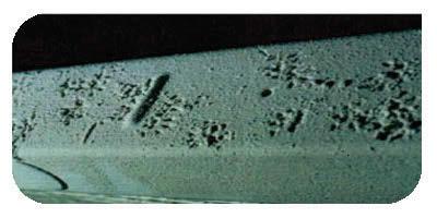
151 Gnawing damage Rat tracks 142
 born-risk generally has multiple causes and attempts to provide effective control by applying")
152 Rat droppings Integrated Pest Management Integrated pest management recognises that pest (in these case - rats) born-risk generally has multiple causes and attempts to provide effective control by applying multiple management strategies in response. Management strategies are selected to best fit the specific context of these risks and can be generally recognised as: Organisational Controls Cultural Controls Physical Controls Organisational Controls Organisational controls are most often developed at the macro level. They can include: Design specifications and construction and operational standards to eliminate rodent harbourage or resources in the first instance. Minimising concealed and/or inaccessible structure voids where rats may harbour; engineering specifications including rat proofing at the design stage; protocols for effective food storage and protection, waste management and cleaning; response protocols in the event of rat activity. Development and enforcement of regulations including IHA, food hygiene provisions, waste management provisions, immigration /border inspection provisions. Cross organisational matters including relationship building between Min of Health, port authorities and companies, importers/exporters, shipping companies. Reporting and evaluation of import health trends and case studies to provide a feedback loop to improving organisational controls. Cultural controls Cultural controls focus on the human factors that enable or prevent effective application of organisational controls. Having high quality operational protocols is of little point if they are 143
153 not followed by those responsible for their implementation. There are several key elements to getting the best out of cultural controls. These include: Knowledge of the technical issues around pest action and control. Motivation to act on that knowledge. Capacity to act. Without resources nothing happens. Collective responsibility for the pest management outcomes. Physical Controls Maintenance and sanitation: Ship cargo spaces, tank top ceilings and other parts of the ship should be kept in a good state of repair to avoid infestation. Cleanliness, or good housekeeping, is an important means of controlling pests on a ship. Since pests in general on ships become established and multiply in debris, much can be done to prevent their increase by simple, thorough cleaning. Box beams and stiffeners, for example, become filled with debris during discharge of cargo and unless kept clean can become a source of heavy infestation. It is important to remove thoroughly all cargo residue from deckhead frames and longitudinal deck girders at the time of discharge, preferably when the cargo level is suitable for convenient cleaning. Where available, industrial vacuum cleaners are of value for the cleaning of cargo spaces and fittings. Physical control aim to prevent and eliminate rat infestations at the operational level subject to actual interaction with the physical environment. A number of sub-controls can be considered with operational actions aimed at managing rat risks. Some of these may include: Exclusion of Rats Preventing rat entering ships at port. Rat proofing. Food protection. Cargo protection. Detection of Rats Interview Captain or senior crew regarding any reports or signs of rat activity. Perform inspection of the vessel for signs of rat activity. Think 3 dimensions high low and hidden spaces. Light dark areas by torch or other artificial means. Check food and waste storages in particular. Tracking powder can reveal low levels of rat activity. Elimination of Rats Correct structural defects Correct procedural defects Control current rat population Rodenticides (use needs to be considered in context with cargo, risk of crosscontamination and competing food-sources. Traps. Can also be used for detection. Rodenticide application When the situation permits, rodenticides usually provide the most cost-effective approach to rodent control. Select a rodenticide with an active ingredient and formulation that works well for the particular environment. Correct bait placement is key to an effective integrated pest 144
154 management program. Proper placement insures rapid rodent control and protects children, pets and non-target animals from bait contact. Two primary types of rodenticide bait are available non-anticoagulants (acute) and anticoagulants. Non-anticoagulants. Bromethalin and zinc phosphide based products are examples of acute baits which have no antidote. Palatability is generally low with products containing these active ingredients. Non-anticoagulants are considered single-feed baits because rodents typically stop feeding after one meal. If a lethal dose is ingested, rodents usually die within 24 hours. If a sub-lethal dose is eaten, rodents tend to develop bait shyness. Rodenticide Application Tips Neophobia the fear of new objects makes roof rats and Norway rats extremely nervous about changes in their territory. It takes several days for rats to accept a new object in their environment, including bait stations. Place rodenticides in areas inaccessible to children and non-target animals, preferably in properly installed, tamper-resistant bait stations. Bait stations not only provide added security for children and non-target animals, but also protect bait from the elements and provide a comfortable place for rodents to feed and groom. Use the proper rodenticide for the target rodent and the best formulation for the environment. Wax block types are best in wet areas. Using information obtained during the inspection process, place baits in rodent runways as close to their nest as possible. Use a sufficient amount of product to assure an uninterrupted supply of bait between service visits. In areas of identified mice activity, rodenticide bait placements should be no further than 2-4 meters apart due to their limited home range. Place control material as lose to the nest as possible, and between the nest and food source. In areas of identified rat activity, rodenticides should be placed every 5-10 meters. Pre-baiting is the process of placing non-toxic bait prior to toxic bait in order to increase product acceptance. This practice generally is used for acute baits (e.g., zinc phosphide) with low palatability. Trapping Tips In sensitive areas where rodenticide use is not permitted, traps are especially useful. Traps also prevent rodent deaths in inaccessible areas. After rodents and their patterns have been identified, follow the appropriate trapping methods. Trapping methods: Store snap traps away from insecticides and chemicals that may impart a flavour. Remember, rodents have a keen sense of taste. Bait snap traps with food that is more attractive than other readily available food sources, such as gumdrops, peanut butter, bacon, nutmeats or dried fruit (raisins). Secure bait to the snap trap trigger a length of thread works well. For rats, fish (tuna) and meat (cat/dog food) may be used to bait traps. Glue boards can be baited, if necessary, with non-oily foods. The use of peanut butter, bacon and other oily, greasy foods will cause the glue to lose its stickiness. 145
155 Bait some mouse snap traps with nesting materials, such as cotton or dental floss, with a drop of vanilla. Mice constantly look for nesting material. Place mechanical or snap traps and glue boards in areas unsuitable for rodenticide applications. Position snap traps and glue boards to intercept rodents in runways. Place snap traps with the trigger toward the runway generally along a wall, in corners, behind and under objects and near abundant tracks and droppings. Snap traps also may be attached to pipes and beams used as runways. More traps are better than fewer traps. Pre-bait traps until rodents, especially rats, overcome their fear and take bait readily. This may take several days for mature rats. Glue boards shouldn t be used in areas with excessive dust or wetness both elements make glue boards ineffective. Check glue boards frequently to prevent rodents from escaping. For mice, repeating or automatic mechanical traps may be used. Watch for tracks in the dust on the top of low-profile traps, which indicate mice are running over the top of them. 146
156 11. Sampling Methodologies Although there are general techniques for sampling for vectors which we will cover through this section it should always be remembered that there are ship specific considerations to be factored into surveillance while conducting ship inspections for vectors. From WHO Training Toolkit for Ship Inspection under IHR 2005 and issuance of Ship Sanitation Certificates Planning Previous visits in ports known to have infected vectors or endemic to vector born diseases should be taken into consideration by inspectors and a more thorough inspection for such vectors would be essential in this case. Different vectors can be found on board ships such as: cockroaches, flies, mosquitoes, bedbugs, fleas, bees, mites, ants, beetles, pests of stored products, fruit flies and rodents. Each of these vectors has different biology and behaviour. Inspectors should take consideration of biology and behaviour of vectors in order to identify harbourage places when inspecting ships and be able to recognize evidence for their presence. It is possible to find vectors at all areas of the ship and therefore, all areas should be inspected: Food areas (preparation, service, dishwashing, refrigerators and other equipment, utensil storage, food stores) Garbage room (between and under ragbags and food packaging) Cabins (mattress, under bed and other furniture, carpets, coatings, lockers) Engine room (drainage systems, joints of decks and bulkheads, among pipe lines) Open decks (lifeboats and any standing water) Ventilation system Laundry Cargo holds However, some areas are most probable to be infested by specific species. Examples of places that each vector can be found are shown below: Location Cabin mattress, curtains Lifeboats, open decks Garbage, underneath of refrigerators Food stores Dining room Garage Cargo holds Vector Bedbugs Mosquito eggs and larvae Cockroaches Fruit flies, rodents, pests of stored products House fly Rodents, mosquitoes Rodents, mosquitoes Moreover, specific loads can be associated with specific vectors for examples mosquito eggs can be found in used tyres. 147
157 Inspectors should check in all areas of the ship for: presence of vectors at all stages of their life cycle (e.g. eggs, larva, pupa and adult mosquitoes, eggs, nymphs, adult bedbug); other evidence for their presence such as droppings, faeces, dead vectors, cast skins; conditions supporting vector harbourage such as inadequate deck drainage, pooling of water, accumulated garbage, poor hygiene, cracks and cervices in food preparation areas; entry points. Vector management plan A good vector management plan should contain the following: Training of management team Crew positions and responsibilities List of vectors that can be found on board Active surveillance (e.g. visual inspections using a flush light at night at high risk areas) Passive surveillance (placement of traps ) Schedule of visual inspections Schedule of trap placement (locations) representative of the ship Records of findings during Surveillance and control measures applied List of pesticides carried on board Revision as appropriate Rat guards Rat guards are required to be placed when visiting ports where plague is endemic. Moreover, the ship operator policy of the local regulation may require the placement of rat guards. The proper placement of rat guards are shown below: Rat guards should have a 36-inch (91.4 cm) minimum outside diameter, a cone angle of 30 degrees, and be made of 18 gauge steel or aluminum. Rat guards must be mounted with the point of the cone toward the ship on all tending lines, at least 6 feet (1.8m) from the pier and greater than 2 feet (0.6m) from the ship. Use rags to plug gaps, securing the rags tightly to prevent loosening or being pulled apart by the rat. Ensure stray lines are kept out of the water. If two lines are in close proximity to each other, either group the lines to pass through a single rat guard, or install the rat guards side-by-side or touching to prevent rats from jumping from one line to another, skirting the rat guards and making them ineffective. Source: US Navy shipboard pest control manual (2003). USA Department of the Navy and Navy Disease Vector Ecology and control Center (Bancor, Washington) reviewed by J.A. Corneil, Washington. Source: US Navy shipboard pest control manual (2003). USA Department of the Navy and Navy Disease Vector Ecology and control Center (Bancor, Washington) reviewed by J.A. Corneil, Washington. 148
158 Below is the part of the WHO Handbook check list that refers to vector management system and evidence for standing water. Code of areas Inspection results: evidence found, sample results, documents reviewed Control measures and corrective actions Required Recommended 13.1 Overall vector management system No rat-proof guard. Place rodent-proof guard to prevent rodents from boarding ships via the mooring lines No integrated vector management plan. Develop an integrated vector management plan No vector control inspection records and logs available (including pesticide application). Conduct routine surveillance on vectors and reservoirs; for example, deploy and check rodent traps and other devices. Develop vector control inspection records and logs, including pesticide application logs Standing water Evidence of standing water in different areas of the ship s open spaces (e.g. lifeboat covers, bilges, scuppers, awnings, gutters, airtreatment plants) that can hold insect larvae. Evidence of depressions or culverts that can collect standing water. Implement operational procedures to control public health risks to crew and passengers, and communities that could be affected by ships and cargoes arriving in ports Evidence of live vectors or their larvae in standing water inside lifeboats. Eliminate standing water and apply vector control measures. 149
159 Sampling mosquitoes There is no single method ideally suited to sampling mosquito populations, each technique employed will be more suitable for specific species and unsuitable for others. In any mosquito surveillance the programme should be developed bearing the desired outcome in mind. For example, if you are in an area where the primary concern was Aedes aegypti, you would not focus your efforts on light trapping at significant distances from the area of concern as Ae. aegypti has a short flight range and is not greatly attracted to light traps. Whereas if you were concerned with Ae. vigilax you would not waste resources sampling small containers in urban areas as it is a saltmarsh breeder. Most programmes will be concerned with a range of mosquito species and a suite of sampling methodologies should be employed Trapping Adults Ovitraps &Tyre Traps The following represents best practice for the construction and maintenance of ovitraps and tyre traps for the surveillance of exotic Aedes spp., and sampling of eggs, larvae (and adult mosquitoes if caught). (Adapted from guidelines prepared by P. Whelan, G. Hayes and J. Carter, Territory Health Services). Trap Equipment Tyre Trap Tyre, used car or small aviation type Aged water - tap water containing rabbit pellets (preferably lucerne-based) left to stand for a week or so Mosquito Trap Collection Sheets Map with exact locations of ovitraps to be setup/serviced White plastic tray 1 or 3ml pipette Sample Tubes with label Pencil Ovitrap One litre blackened, screw-top, glass/plastic jars (painted or inside black casing) 3 sets of numbered, masonite or wooden paddles; 16cm x 3cm Aged water - tap water containing rabbit pellets (preferably lucerne-based) left to stand for a week or so Trap Collection Sheets Map with exact locations of ovitraps to be setup/serviced Compartmentalised, plastic, paddle transport box Trap Transport Container Trap Construction Tyre Trap Used automobile or light aircraft tyres (<500mm diameter) can be placed outdoors in sheltered shaded areas, near vegetation if possible, in an upright position. The vertical alignment of the tyre should be marked so it can be replaced in the same aspect and filled with at least one litre of water to the same marked level (or to the drainage hole) on each occasion. This will ensure that eggs laid previously will be covered for hatching and not left away from or above the water line. Chaining or otherwise fixing the tyre in position for security purposes, can facilitate the correct positioning. 150


160 Tyre traps are typically old tyres with a drainage hole cut into the tread or side wall. Cutting into the side wall is easier in the field and can be done with a box cutter or other sturdy knife. The tyres may be left black or marked with red paint. Tyre traps remain in the field and need to be serviced regularly. They should be marked to aid placement, e.g. it is suggested that a weather-proof label be attached to the tyre that is: a. readily identifiable b. describes the purpose of the tyre e.g. Mosquito Surveillance Trap and requests that individuals desist from interfering with the trap c. includes appropriate name and contact information. When not in use, tyre traps should be stored in an area where they can t get wet and subsequently provide a breeding site for nuisance mosquitoes. Tyres should to be inspected weekly over the warmer months of the year and fortnightly at other times. All the water is drained from the tyre and into a white shallow tray for larval inspection. NB. It is important to carry out this step from week one, and not just from week two, as Culicine mosquitoes lay their eggs on the water surface and they may hatch within hours of oviposition. Any larvae detected should be carefully collected and transferred to labelled sample tubes. Ovitrap Ovitraps are typically made out of black, glass/plastic containers of approximately ml and which contain a paddle. They should be prepared in the laboratory or workroom prior to going out in the field. Each ovitrap should have a different number and each component (jar, paddle, black casing (if required)), should be numbered identically. Fill the clean ovitrap jars with aged water to depth of 10cm or near to the top (dependant on the size of jar used). Place the numbered, clean, masonite (or wooden) paddle (rough side up), in the corresponding numbered jar (See Photos). Rough side Paddle Types Paddle Position Place the jar into the black plastic casing (where required) and load the completed ovitraps into containers ready for transportation to the field sites. Trap Position Selection Traps should be deployed indoors or outdoors in relatively secluded, sheltered, shady, low to the ground (0-1m above ground) sites, near vegetation (where possible), protected from rain and animal disturbance, but near areas where there is regular human activity. They should not be placed near spider webs or inside very thick vegetation. 151
161 Ovitraps should be stabilised by situating between bricks or stones, or behind/under a suitable object such as a wash trough. Trap Placement Tyre Trap Ensure that each time the trap is serviced it is repositioned at the same orientation and same angle of lean as previously. Any relocation of trap positions should be recorded on sample sheets and the field map and the new positions allocated new site numbers. Fill the trap with water to the same level as previously filled and add a methoprene pellet or ~30 s- methoprene granules. S-methoprene does not act as a repellent in traps. Ovitrap Replacement ovitraps are positioned at the same time the already exposed ovitraps are collected. Any relocation of trap positions should be recorded on sample sheets and the field map and the new positions allocated new site numbers. Ensure that the replacement ovitrap jar, masonite paddle and black plastic casing have corresponding site numbers, that the water in the jar is at the correct level and top-up if required. Also that the paddle is placed rough side up and out (see photo). Place the ovitrap in the designated position and add a methoprene pellet or 10 methoprene granules. Trap Collection Tyre traps Empty the trap completely into a white tray (this may take more than one fill of the tray). Remove all larvae (instars 1-4), pupae and exuvia using plastic pipettes and transfer to a labelled sample tube. The label should be written in pencil and contain the trap sample number with the collector s initials and the collection date. NB. Larval skins (exuvia) can also be used for species identification. If any adults are present in the water, remove carefully and place into a separate labelled specimen tube. Complete the Trap Collection Sheet (positive or negative). Cross contamination between trays should be avoided by washing the pipettes (inside and out) between each. Give the inside of the trap a brief scrub with a brush and flush with water (if a suitable supply is at hand). Replace trap, (see trap placement above). Repeat for each trap. Ovitraps Exposed ovitraps are collected at the same time the replacement ovitraps are being positioned. Record any disturbance to the ovitrap in the additional information section of the Trap Collection Sheet, such as invasion of trap by ants or frogs, trap stolen or vandalised, no water left in ovitrap, trap tipped/blown over, paddle lost etc). Remove the paddle and place rough side up in the compartmentalised plastic paddle transport box (see photo). 152

.")
162 Paddle Transport Box Remove the trap, record the presence of larvae, pupae or dead adults (Additional Information section of the Trap Collection Sheet) and place in the Trap Transport Container. NB. The presence of any pupa skins indicates that inspections have been too far apart and the ovitrap servicing period needs to be shortened. This aspect should be brought to the attention of the supervisor immediately on return from the field. Ovitrap Processing On returning from the field: 1. Remove the paddle from the plastic transport box and examine for eggs. They should be near the water mark on the paddle (see photo). Use a dissecting microscope or magnifying glass if available to examine the paddles more closely. 2. If eggs are visible, wooden paddles should be packaged into individual slide mailers for transport. Wooden paddle with eggs at watermark and close up of eggs Cut off the lower part of the paddle just below the water line using scissors and trim off the top. Be sure not to cut off any eggs. 153


, pupae, and exuvia using plastic pipettes and transfer to a labelled sample tube.")
. 9.")
163 Lay the remaining part onto a piece of tissue and write on the sample number and date. Wrap the tissue round the paddle and place into a slide mailer. Close the slide mailer and make sure it is labelled correctly. 3. Tip all the water from the jar into a white plastic tray. 4. The trap jars should be examined by eye for mosquito eggs that may have been laid at the water surface. If present, attempt to remove the eggs without damaging them, so they can be sent through to the laboratory. If this is not possible, sterilise the jar with boiling water. 5. Examine the water in the tray for the presence of larvae, pupae, exuvia (skins) or adults. 6. Remove all larvae (instars 1-4), pupae, and exuvia using plastic pipettes and transfer to a labelled sample tube. The label should be written in pencil, contain the trap sample number with the collector s initials and the collection date. NB. Larval skins can also be used for species identification. 7. If any adults are present in the water, remove carefully and place into a separate labelled specimen tube. 8. Complete the corresponding Trap Collection Sheet (positive or negative). 9. Repeat for each trap. Cross contamination between trays should be avoided by washing the pipettes (inside and out) between each. NB. At least two sets of jars and paddles are required for each trap site and should be used on a rotational basis. One set is in use, while another set is being sterilised and dried. NB. Additional paddles will be required if any are sent through to the laboratory for hatching. 154


164 Trap Cleaning Tyre trap As mentioned earlier, tyre traps should be regularly brushed and flushed in the field. If the trap becomes very dirty and there are no facilities for cleaning it on site, transport it back to base and clean properly. Ovitrap All trap parts (jars, paddles, casings) should be cleaned with boiling water, whether mosquitoes were observed or not. Do not use detergent as this can make the traps too clean and less attractive to ovipositing females. Scrubbing of the jars and paddles will be required to remove any old hatched egg cases. Use a dedicated scrubbing brush, which has not been used for washing with detergent. Wooden paddles may be reused, providing there was no evidence of mosquito eggs before cleaning. It should be noted however, that these will not last long and a supply of new paddles should be available in advance. Lethal Ovitrap Lethal ovitraps are becoming more commonly used for removing the adult mosquito population from an area as part of control operations. Standard lethal ovitraps employ a strip of fabric impregnated with a residual insecticide. The idea is that gravid females are attracted to the trap to oviposit and are killed when alighting on the strip, thereby preventing further breeding. Biodegradable versions of this trap have been developed by the Tropical Public Health Unit (TPHU) in Cairns so they need not be recollected. This technology is of use in control but is not so useful when assessing mosquito populations. As the removal of the adult mosquitoes from the environment may be advantageous, but also knowing what has been removed is required. Sticky Ovitraps Sticky ovitraps are constructed using a standard ovitrap and inserting an acetate film coated with a glue. Mosquito adults attracted to the trap end up stuck to the glue surface and when the adhesive strip is collected form the trap may it may be viewed under the microscope to identify the specimen. In Cairns, Australia this method is employed for trapping bloodfed adult mosquitoes which may be sent for PCR analysis to identify presence of arboviruses. Sticky ovitraps capture the gravid females as they attempt to lay eggs, they could be considered as Gravid Traps. Sticky adhesive strip 155

165 Gravid Traps Standard gravid traps employ a combination of ovitrap attractants and light trap mechanics. The trap consists of a body of water similar to a large ovitrap which attract females ready to oviposit. A fan and collection pot system is mounted over the water and is designed to suck the females up into the collection pot as they fly over the water body. As with lethal ovitraps this system removes the mosquitoes from the environment so is effectively also a form of control. Light Traps Adult traps utilising light, carbon dioxide (CO 2) and, if appropriate, octenol are used for attracting and capturing adult mosquitoes in the field. There are several types of light trap available; generally using dry ice or a CO 2 gas cylinder. Traps baited only with light usually collect a variety of insects. To target host seeking insects such as adult female mosquitoes, CO 2 baited light traps can be employed. These traps attract adults from a considerable area through a screened funnel into a killing jar or mesh bag suspended below the trap. Experiments have shown that CO 2 used in conjunction with other items may increase the number of mosquitoes and the range of species collected. Other chemicals such as Octenol have been shown to a useful attractant for certain species. NB. The use of other attractants (such as Octenol) can also act as a repellent for some mosquito species. 156

166 Positioning and Erecting a Light Trap The ideal positioning of light traps will vary depending on the species being targeted but in general: Within sight of potential habitat Trees provide shelter from wind and rain and sometimes provide food for adult mosquitoes. If there are none suitable nearby, erect a tripod or other suitable hanger Avoid too much competition from other light sources. Avoid direct competition from other sources of blood feeds, a trap placed in the middle of a room full of people will catch a much lower proportion of mosquitoes. Light Trap Servicing Before you leave the base, organize charged batteries and other equipment needed (listed above) for current servicing run and place in vehicle. NB. Ensure that as many clean collection cups as are required, as well as a couple of spares, are taken on each trip and the stockings do not have any holes. At the trap site; 1. Always remove collection cup before disconnecting the battery. [Disconnecting the battery will stop the fan that prevents any captured mosquitoes from escaping]. Tie the collection cup stocking in a loop, not a knot and ensure the cup is labelled correctly (date of collection). All collection cups removed from the field should be stored in a chilly bin or similar, and placed in the freezer on arrival back at base. 2. Check the fan housing is clear of debris such as insects and clean with a toothbrush if necessary. Recheck the operation of the equipment after this. 3. Change the battery. 4. Check octenol wick is wet. If dry, dip wick slightly into octenol container. 5. Attach the collection cup at base of trap. Ensure the stocking does not have any holes in it and that the rubber band is sufficient to hold the cup in place. 6. Attach trap connections to battery Red to positive terminal and Black to negative terminal. 7. Change gas bottle and reconnect the regulator. 8. Record any changes in habitat or location at which light trap is placed on the National Mosquito Trap Collection Sheet. Significant changes to the location could reduce the likelihood of the trap collecting mosquitoes. 9. Before leaving the trap, ensure that the gas is on, the fan is blowing, the light is going, and that the light source is not blocked by any object. 10. Respect private property. 11. Ensure the Mosquito Trap Collection Sheet is completed before leaving each site. On return to base; 1. Place collection cups in freezer to kill mosquitoes these need to stay there overnight. 2. Recharge batteries as necessary (Refer to battery charging protocol). Keep uncharged batteries in designated spot, away from those already charged. Light Trap Maintenance 157
.")
167 Unscrew the motor and fan Apply CRC to the joints and screws Ensure the fan is unclogged and the fan housing is clean. Check the battery clamps are clean. Ensure that the regulator and all parts and leads are working and the regulator is calibrated to release the appropriate amount of gas over the duration of the trap placement (e.g. one week). The gas bottle needs to be near empty so you can determine the trap has been working when a trap is serviced (change in weight). Battery Maintenance Please refer to your batteries instructions for direction on recharging. Contact the supplier of the batteries if you are unsure about the battery charging requirements. Be aware that you may need protective clothing or other safety equipment when working with some kinds of battery. BG Sentinel Trap The BG sentinel trap was designed to attract Aedes aegypti but has since been found to be effective at attracting a range of other mosquitoes. It utilises a fan system to draw mosquitoes into a collection pot in a similar way to a light trap however it is the way the attractants are emitted that is different. The trap mimics convection currents created by a human body releasing attractants through a large surface area. The trap generally employs a BG-Lure, which releases a combination of other attractants: ammonia, lactic acid, and fatty acids as an attractant, however you may use CO2 in conjunction with the lure. One of the main drawbacks is that the trap has been designed for indoor use and its current design is not suitable for use outdoors in inclement conditions Larval sampling Groundwater Habitats - For shipping inspections and usual port work groundwater is not usually a problem, however a few ports will have some groundwater habitat. If you do not have groundwater in your port this section may not be relevant. 158


168 Groundwater sampling incorporates most of the habitat categories excluding container type habitats and will include field drains and runnels, swamps, marshes, mangrove, ponds, lake edges etc. For field sampling of ground water habitats, it is important to walk around the entire margin of the site to determine the entry or exit points and possible source of the water. Permanent sample points may be chosen after initial sampling if longer term surveillance is likely to occur. In a larger site, this may include a point in each vegetation or water type. These permanent sample points should be at sites where there is year-round access. All sample sites should be marked on a map. These permanent sample points will provide an assessment of how the breeding habitat, species type and numbers of mosquito larvae change over time as well as potentially important factors in that habitat that may lead to fluctuations in mosquito numbers. Additional larval samples should also be taken at different points throughout the habitats during each visit to make sure the permanent sample points are efficient indicators of larval breeding sites. The following procedures relate to sampling for mosquitoes in natural water bodies such as ponds, marshlands, drains, oxidation ponds and so on, but may also apply to containers being checked for the presence of container-breeding mosquito larvae. Though a few mosquito species can live in water with a gentle current or flow, larvae of most species will be found in still water. Mosquito larvae are usually found where surface vegetation or debris is present. In larger bodies of water, larvae are confined to marginal areas or floating surface materials (e.g. weed mats). Examine maps and aerial photographs when available, for vegetation patterns and likely areas of mosquito breeding. Plan the access route and plan your specific search sites. When searching for mosquito larvae be prepared to walk and push through thick vegetation into the selected sites chosen on the aerial photographs. There is no substitute for leg work and perseverance. Please, however, beware of your personal safety at all times. When approaching a margin of a water body it is important to note the vegetation patterns. The different types of grass, reeds or other vegetation may be clues to deciding exactly where the mosquitoes are likely to be and which habitats must be sampled. When you have selected particular habitats, look at the water before disturbing it with the feet, ladle or shadow. Note the presence of fish and other predators, and look for larval activity. Remember that wind may cause larvae to gather at the down-wind end of a pool. Procedures for Sampling Groundwater Habitats 1. Do not cast your shadow over the water as this will disturb the larvae. Look through the water carefully for wriggling or hanging larvae before you disturb the water with your feet or ladle. Quickly skim the larvae into your ladle. NB. Aedes larvae generally dive to the bottom of the water (and usually they swim down at an angle) when disturbed. Some species spend a lot of time near the bottom of the water column, this should be factored in when sampling any habitat. 159


169 2. Begin sampling at the waters edge. Dip into any grass or vegetation and let the water run into the dipper (ladle). Scoop the dipper up just before it completely fills with water. If there is a chance that larvae have been disturbed prior to dipping then wait for approximately 30 seconds for larvae to resurface then continue with sampling. If the habitat allows, move forward three paces, stop and take another two samples and so on. You can cover a greater area by zigzagging as you move through the habitat. 3. Take time to search for 1st instar larvae. A "visual dip is not completely reliable on its own, so you may have to look for a shadow of movement in the bottom of the dipper as they are very small and are very difficult to see. 4. If you find larvae transfer them to a specimen tube using a plastic pipette. Number the tube and complete the sampling form. Where there is dense, floating vegetation or debris, use the following method: 1. Disturb the water, causing the larvae and pupae to sink below the surface. 2. Clear away the vegetation or debris with the dipper and wait a few minutes for the larvae and pupae to return to the surface. 3. Collect the larvae and pupae with the dipper 4. Pipette all larvae collected from one site into a single sampling tube where possible. If no larvae are present return with light traps and collect some adults. 5. Always keep sample tubes and sheets together in the field. 6. Record the number of dips made, this information will give a guide as to how big any future adult populations may be. Normally ten dips are made at each site and the results for each dip recorded, even the negative ones. Record the number of larvae, instar and pupae on a data collection sheet. Transfer the larvae to the vial with the aid of the pipette. If no larvae are found it is still important that the sample is recorded. After sampling and finding no larvae complete the sampling form. 7. Upon return to base, follow the procedure for sample handling. Artificial and Natural Container Habitats Container sampling incorporates artificial and natural container habitats, and arguably also includes artificial subterranean habitat. Natural containers such as tree holes, leaf axils and coconut shells etc, and artificial containers such as discarded rubbish, tyres, tin cans, plastic sheeting, oil drums, buckets and guttering etc. 160

170 1. Examine the area, for any containers and note the type of container and the presence of water or larvae in each container. 2. Each container should be sampled for larvae. 3. The first step in sampling larvae is to look carefully at the surface of the water for larvae or pupae. 4. With a ladle, quietly lower the ladle as deep into the container as possible so that larvae can be seen again the white background of the ladle. The ladle can then be slowly extracted with the larvae and water. 5. Depending on size, the container can be emptied carefully into a white tray for further examination. 6. Tyres can be prised open and the water can be scooped with a ladle or net, ensuring that the ladle rim makes contact with the bottom of the tyre. 7. Pipette ALL larvae collected from one site into ONE sampling tube. DO NOT collect excessive numbers, ideally a maximum of 20 fourth instar larvae or pupae, or 50 early instar larvae, per tube. Do not collect pupae, if pupae are present try to find some larvae. If no larvae are present return with light traps and collect some adults. 8. ALWAYS keep sample tubes and sheets together in the field. 9. If no larvae are found it is still important that the sample is recorded. After sampling and finding no larvae complete the sampling form. 10. Upon return to the office follow the procedure for sample handling Biting/Landing Collections The bait subject rolls up his shirtsleeves or trouser legs and sits quietly for minutes, collecting the mosquitoes that settle on the exposed skin. Various techniques are used such as test tubes and small vacuum devices. Note: For medical/ethical reasons, this method should only be used under the supervision of senior environmental health staff. There is no legal barrier to collecting samples from personnel who are fully clothed. 161
171 Aspirating Adults Adults on the wing or resting on a surface may be collected using an aspirator, often called a pooter. It is a device for collecting small insects or spiders using light suction. Motorised versions are available but normally the suction produced is through the lungs. A mouth-operated aspirator When using the type of aspirator depicted above, air is sucked through the thick end of the tube while the thinner end is held near the mosquito. The mosquito is sucked into the thin tube as far as the join with the larger tube where movement is stopped by a mesh barrier. The mosquito may then be blown into a collection tube and processed. Another type of aspirator is shown in section 8.4 Sampling mites below Collecting Mosquitoes using a test-tube Hold the mouth of the tube directly over the mosquito. When the mosquito is disturbed it will fly into the tube. Close the mouth of the tube with your index finger or thumb. Remove your finger and push a plug of cotton wool into the tube. Push the plug down until the mosquito is trapped in the bottom 2 cm of the tube. Collect a second mosquito as described above and insert a second plug to trap it in the next 2 cm of the tube Sampling ticks Excerpts from Baker, Mites and ticks of domestic animals: an identification guide and information source. The Stationery Office, London. 240pp. Ticks are usually large enough to be seen with the naked eye, but it is advisable to use a hand lens in the field and a dissecting microscope in the laboratory to search for small species and immature stages. Straight-shafted forceps are useful for collecting nymphal and adult ticks, but tick larvae are more easily picked up on the end of a moistened tip of a fine paintbrush. A fine probe or minute spatula or similar is a useful tool for manoeuvring specimens. 162
172 Removing ticks from hosts The safest and most effective way to remove an attached tick is to grasp it behind the mouthparts with fine forceps and pull gently and steadily away from the skin until the tick releases its hold. Do not twist, jerk or crush the tick's body as this may release body fluids harbouring pathogens, directly into the wound. Barbed mouthparts of the tick help to anchor it in the flesh of its host and ticks secrete compounds in their saliva that help to cement them in the feeding wound. Pulling too strongly or twisting while attempting to remove a tick may result in tearing of the tick, leaving the hypostome embedded in the skin. This can lead to bacterial infection in the feeding wound. Do not attempt to remove an attached tick with caustic chemicals or by applying heat. This can kill the tick before it disengages its mouthparts. It can also cause the tick to regurgitate into the feeding wound and therefore increase the chance of transmitting a pathogen. The removed tick should always be saved for identification, place into a sealed container and place in the freezer or add ethanol to the container. The attachment site of the tick should be washed thoroughly with warm soapy water and rubbing alcohol to remove any possible pathogens. Wash your hands as well as the tweezers or any other object the tick (or fluids from the tick) may have contacted. Objects used to remove or dispose of ticks as well as the site of the tick bite should be disinfected Free-living ticks Ticks on vegetation or in a pasture can be collected by dragging, i.e. pulling a flannel sheet approximately 1.5mx1m slowly across the ground. The tick attaches to the sheet, mistaking it for a passing host. These can then be removed using forceps. Material can be removed from host environments such as nests and burrows to search for ticks in a white tray. Ticks can be recovered from the loose material by gentling shaking the material in a tray or sieve. This action can also stimulate the ticks to right themselves and move to a more protected location making them more visible amongst the debris. Rocks adjacent to nests and burrows should also be overturned as some species prefer to live in this type of habitat Trapping Ticks Soft ticks can be readily collected via dry ice traps. Blocks of dry ice emit large amounts of carbon dioxide, a host seeking stimulant. Traps are set in and around nesting areas of animal hosts. Soft ticks can be observed running along the surface of the ground towards the trap and are collected by hand, or inside a collection chamber in the trap. Where dry ice is not readily available or there is inappropriate storage facilities it is possible to make dry ice snow using a CO 2 bottle and a Sno Pack. The liquid faction is run out of the bottle through the Sno Pack and a 500g compressed CO2 block is produced. 163
.")

173 11.3 Sampling Sandflies Sand flies are easiest to collect as adults in traps. Recent studies conducted by the United States Military in Tallil Air Base in Iraq have assessed the effectiveness of several traps in collecting sand flies (Appendix 19.2). Two of the more effective traps for collecting sand flies they examined, are discussed below CDC light trap The CDC light traps used are very similar to the adult traps discussed in 8.1 for collecting mosquitoes though they have been run without CO 2 when collecting, CO2 use may increase the effectiveness of the traps. These traps have been effective but the study showed that their effectiveness was improved if the bulb was replaced with a UV bulb. This type of trap collects the largest number of sand flies but also a large amount of other bicatch. Less bicatch will be collected using a standard bulb but also less sand flies, the reasoning for trapping should be considered when choosing trap types. Traps should be placed about 0.5 m above the ground and 30 m or more apart Sticky traps The other trap used was a 20cm by 7cm sticky cockroach trap and was found to be more effective when used in conjunction with a chemical light stick. All colours resulted in an increased catch, but red and blue sticks seemed to be the most effective. The sticky trap / light sticks should be placed on the ground with the sticky surface and light stick facing up Sampling mites Excerpts from Baker, Mites and ticks of domestic animals: an identification guide and information source. The Stationery Office, London. 240pp Attached mites Because most parasitic mites exist predominantly on the host, the best way to sample them is to target any hosts in the area. Samples should be scraped from the skin of any symptomatic hosts 164
174 and analyzed under a microscope. Mites can occur all over a hosts body, but some species prefer particular areas. For example, on mammals, the ears, muzzle chin and ventral midline are commonly affected areas, while on birds, the scaly areas of the legs and face, inside of feather quills etc. Dislodged specimens are easier to see when combing or brushing is carried out over a white sheet or tray. If the host is dead, the results may be improved by first anaesthetizing any mites still living by leaving the body in a box with a wad of ether-soaked cotton wool for about 30 minutes. An alternative method is flotation. Specimens can be separated from dead hosts by shaking the body in a bowl or plastic bag containing a weak detergent solution and then filtering the liquid. Acari have a water repellent cuticle and the detergent helps to free the specimens by reducing the surface tension. Additional methods may be required to separate specimens that are either too firmly attached or are actually buried in the skin. A simple method which can work for skin scrapings: mount tissues in a drop of 10% KOH or NaOH on a flat glass microscope slide add a coverslip and clear by warming for 5-10 minutes apply gentle pressure to the coverslip to separate the specimens from the softened tissues Unattached mites Unattached mites can be sucked up into an aspirator (often called a pooter). This particularly effective for catching rapidly moving mites without harming them. Specimens can be collected directly into preservative such as 70% ethanol, however this is not recommended for mouth operated aspirators as the user will inhale ethanol fumes. The entire aspirator can be placed in the freezer for a few minutes to slow down the specimens and then transfer them directly to a tube containing preservative or remove the top of the aspirator and fasten a lid to the vial and replace in the freezer. A mouth-operated aspirator A white tray is a useful piece of equipment for collecting mites as they show up well on a white background. Also when dropped on the ground, trays can act as a lure to some larval trombiculid mites. Medium or heavy gauge plastic bags should be used to transport small dead animals or habitat samples for further investigation. 165
175 11.5 Sampling fleas The direct collection of fleas generally uses the mimicking of a blood sources movements to attract the flea to jump to the source. Inserting a roll of flannel into a rodents burrow and moving it around for several seconds before retrieving will cause any fleas to become excited and jump onto the material. The flannel may then be removed and sealed into a bag or container and frozen. When the fleas are incapacitated they may be removed from the material into appropriate containers Alternatively, a yellow-coloured plate coated with a tacky substance is mounted above and below the teeth of a rake or between rollers and passed over an area infested by fleas. The vibrations and motion caused by the movement of the teeth or the rollers against the supporting surface excite and attract the insects which jump toward the brightly coloured plate where they become entrapped into the tacky substance. The apparatus may be used to either determine the nature and extent of the infestation prior to treatment. Collection of fleas through rodent hosts is another method. Unfortunately flea behaviour is such that they will quickly abandon their host if it dies, so the use of sticky rat boards or some form of humane trap is required for this method. With the sticky trap placement along known rodent corridors is the most ideal option. They need to be well anchored and regularly collected / inspected. One benefit of the sticky board is that if fleas leave the animal they may still become trapped in the glue. Other humane traps tend to be baited and should be placed appropriately. In both cases the host will be alive when collected and will need to be dispatched appropriately, bearing in mind that the fleas will abandon the body shortly afterwards Sampling Lice Because most lice exist predominantly on the host, the best way to sample them is to target any hosts in the area. Samples should be plucked or scraped from the skin or brushed from the hair of any symptomatic hosts, using a fine comb, and analyzed under a microscope. lice can occur all over a hosts body, but some species prefer particular areas, e.g. Hair, eyebrows, beard, armpits, groin etc Sampling Bedbugs Advice to be given before inspection Bed bug inspections need to include thorough searches behind several objects in a property (behind pictures, headboards, under floorboards, paintchips etc. It is important, therefore, that the Pest Manager explains the inspection processes in detail to the client and should provide; Instructions that it will be necessary to inspect the room, including looking through cupboards and drawers. Instructions that it will be necessary to remove bed heads, lift carpets and dismantle other items to access all bed bug harbourages. Instructions on any activities the client will be required to undertake prior to the inspection 166
176 Advice to the client that follow up inspections after the initial inspection and treatment will be necessary Surveillance Tools and Equipment Useful tools for a bed bug inspection include: A powerful torch A 10x magnifying lens (to inspect for live bed bugs and eggs) Collection bottles (for gathering bed bugs for later confirmation of identity, sticky tape can also be used for gathering bugs) Fine tipped forceps (for picking up bed bugs) Gloves Screwdrivers and spanners for dismantling items An inspection mirror Plastic bags (large and small) to hold bottles, tape, infested items, etc. Notepad, for recording details of the infestation A digital camera (for recording infested sites, the digital images or printouts can also be given to the client in a report or provided as part of an educational package) Indications of a Bed Bug Infestation Indicators of infestation include: Live or dead bed bugs, and cast skins. Live bed bugs will confirm that the infestation is currently active. Faecal spotting. This is digested blood defaecated by the bed bugs. It may be initially observed on the sheets, but will be commonly noticed along the mattress seams and other places where bed bugs hide. On light coloured surfaces individual faecal marks appear as small dark round spots,however the spotting may be in colour from cream, through grey to almost black. Generally the spotting, will occur in groups and appear as splotches of dark marks. Note that the faeces of nymphal cockroaches appear similar, however bed bug blood spotting tends to occur in groups as the insect by nature aggregates. Red blood coloured spots or smears on the sheets may occur which can be the result of bed bugs passing sera, or engorged bugs being squashed by movements of the sleeping host. Eggs (cream in colour with a slight bend, approx. 1mm, which tend to be laid in crevices in dark areas. A bed bug smell sometimes described as sickly sweet but is akin to that of stink bugs. This is usually only noticed in heavy infestations, if close to the bugs or during the treatment process. There are specially trained dogs for detecting infestations. 167


177 Adults, nymphs, eggs, shed skins, and fecal spots on a mattress. Dark spots on mattress and box spring are a telltale sign of bed bugs Bed bugs often reside along baseboards. Photo at right shows eggs, nymphs, adults and fecal spots near a carpet edge. 168


178 Bed bugs also congregate along seams of sofas and recliners. Photo at right shows bugs hiding near a recessed screw under a night stand (note the presence of fecal spots). Bed bugs on a bed showing blood spots (Harold Harlan, DPMIAC, Armed Forces Pest Management Board) 169
 Bed bugs")

179 Bed bugs and faeces, shed skins, and eggs in fold of pillowtop mattress in heavy infestation (Photo by Richard Cooper, Cooper Pest Solutions) Bed bugs clustering on ceiling- wall intersection in a very heavy infestation; note smear marks from residents crushing some with a broom (Larry Pinto, Pinto and Associates) 170
")
180 Bed bug Eggs glued in hole made by Furniture staple (Stephen Doggett, Dept of Medical Entomology, ICPMR) 171
")
181 Bed bugs behind apartment baseboard in heavy infestation (Jeff White, Cooper Pest Solutions) 172
 Bed bugs,")

182 Bed bugs between slat and frame of an infested bed (Richard Cooper, Cooper Pest Solutions) Bed bugs, eggs, and shed skins in seam and under label of a soft-sided suitcase (Richard Cooper, Cooper Pest Solutions) 173

183 Close up of bed bugs infesting running shoe (Stephen Doggett Dept of Medical Entomology, ICPMR) Other methods used For Detection Specialised sniffer dogs can be used to detect the sweet smell which infestations produce Traps are available but many are impractical (Large, require electricity or a carbon dioxide bottle, not viable for hotel rooms where guests may be deterred from staying). As yet no traps have been devised which are particularly effective (Sticky traps placed in heavily infested areas often catch no bed bugs) so are not advised as a sampling tool at this stage Important Information Bed bugs spread readily & any live stage that is transported has the potential to start a new infestation elsewhere, Therefore: No infested items should be removed from the property before they have been thoroughly treated or encased. Otherwise bedbugs can easily be spread throughout the building or to other buildings Any item removed from the property must be properly disposed of or treated to kill all stages which may be present Samples should be properly bagged/placed in secure tubes for identification 11.8 Sampling Cockroaches 174


184 Trapping can determine resting areas and infestation severity, monitor effectiveness of chemical controls, and detect population increases which may then require insecticide treatment. Several types of traps can be purchased. Most are about the size of a large matchbox, have openings at both ends, and have the inside surface covered with a very sticky adhesive and slow-release food attractant. Cockroaches detect the food odor, enter the trap, and become immobilized by the adhesive. Traps can also be made from deep glass jars with a layer of petroleum jelly on the inside to prevent escape, and either commercially available bait or a piece of fruit as an attractant. Traps should be positioned with (both) ends open and accessible to intercept cockroaches as they travel to and from harbourage and feeding areas. For maximum efficiency they should be placed in dark areas such as along bulkheads and in tight spaces. Traps should be left out for a minimum of 24 hours including an overnight period. A suitably placed trap can catch numerous cockroach adults and/or nymphs daily. Traps are relatively inexpensive, convenient to use, disposable, and do not need to contain toxic chemicals. If two or more cockroaches are caught within a 24 hour period this may signal that a pesticide control operation is required. Any live cockroaches still in traps can be killed with a 3% solution of dishwashing liquid in water. Cockroaches may also be detected by physically searching resting sites. Commonly inhabited areas on ships include around false bulkheads, holes for electrical wiring and plumbing, in lagging and torn insulation, behind bulletin boards, around supports in serving lines and around other kitchen equipment and fittings, behind fridges, in deck drains, ventilation grating, fuse boxes, food stores and on coming food supplies or other products that may be brought on to the ship and already containing infested materials such as wood and paper products. Looking for signs of cockroach faeces is also a good way to spot past or present cockroach activity. A screwdriver for opening grates and hatchings will be required, as is a keen eye, and a flashlight for illuminating dim or unlit areas. Cockroaches will oftentimes be disturbed by the light and run away, making them even easier to spot. A flushing agent, usually a pyrethroid may also be used to check for cockroach activity. This is typically sprayed into a harbourage area and monitored for 3-5 minutes for any signs of cockroaches. Because of the repellant properties in these agents they should not be used in areas where traps or insecticide controls are going to be used Sampling Rats 175


.")
185 Also see control and surveillance section 9.5 Types of Traps Cage traps Snap traps Repeating traps Humane traps Glueboards General Rules for Trap & Bait Station Placement Pre-bait traps (unset) for 1-2 days. Use food not in competition with what is present in the area. Set traps close to wall, in dark corners, behind and under objects. Set traps perpendicular to walls so that the catch bar snaps towards wall. Set glue boards parallel with walls. Force animals to pass over or into traps or glue boards by placing boxes or boards to form barriers. Check glue boards every 24hrs. Use more traps than animals. Rodents will become trap shy if they survive. Put out more bait placements than you think necessary. Under baiting with an acute rodenticide leads to bait shyness. Bait stations for rats placed near exterior entrances and along runways/tracks (every 15m). Bait stations for mice placed near interior entrances and along runways (every 6m) Preparation for Transport If specimens are to be sent to New Zealand for identification, further work such as genetic analysis or storage, they need to be packaged appropriately so they arrive in good condition. 176

186 No live specimens must be sent, they will not be allowed into the country (see section 9.4) Mosquito Larvae All water should be removed from the sample tube with a plastic Pasteur pipette, taking care not to remove or damage any larvae. For specimens not required for genetic analysis, the preferred killing method is to place the specimens in boiled water for 1-5 minutes to allow all larvae to die and their proteins to be denatured. The boiled water should then be removed using a pipette and replaced with 70-95% ethanol. For specimens intended for molecular biology, this boiling water step should be skipped and the specimens placed directly into ethanol. DO NOT put cotton wool, tissue or any other padding in the sample tube with the larvae as they can get trapped in the padding and become damaged or desiccated. The tube should contain larvae and ethanol ONLY (see photo). Sample tube containing larvae Mosquito Adults All adults should be handled carefully to ensure no wings or legs break off and as few scales are removed as possible as these are important features for accurate species identification. If necessary to handle an adult specimen, use fine forceps to carefully hold the femur of the middle leg on either side of the body. This part of the mosquito is quite robust, compared to e.g. the wings, and is not usually required for identification, as is for example, the hind femur. Live adults must not be posted. Where possible freeze live adult specimens in the container in which they were trapped, for 24 hours before transferring them to a sample tube for dispatch. In the absence of a freezer use carbon dioxide gas or 2-3 dry ice pellets (10 minutes exposure, depending on size of container) to kill the specimens before removing from their containers. Wet adults must be carefully removed and placed to air dry on blotting paper. Place the specimens out of direct sunlight and in a draft-free area and leave for half an hour before packaging for dispatch. [Do not attempt to wipe or blot any excess moisture as this will remove scales and damage the specimens.] Do not add any alcohol. Place the specimens in a sample tube of the appropriate size for the number of specimens present (small, medium or large). Use multiple tubes for one sample (all accurately labelled) where required. Carefully position tissue paper in the top of the tube to help prevent the adults being shaken around the tubes too much during transit. DO NOT squash the tissue paper down onto the adult specimens (see photo). 177

187 Sample tube containing adult specimen Ticks and Mites Ticks and mites can be transported successfully while still alive, however live exotic organisms are not permitted to be imported into New Zealand without the appropriate permissions and therefore only dead organisms should be sent. Therefore these organisms should be preserved in 70 percent ethanol in small containers or vials with leak-proof lids Sandflies Sand fly adults should be processed as per Mosquito adults (see section ) Fleas Fleas should be stored in 70% ethanol for their preservation and in preparation for identification Bed Bugs Bed bugs should be stored in 70% ethanol for their preservation and in preparation for identification Cockroaches Cockroaches should be stored in 70% ethanol for their preservation and in preparation for identification Rats Rats may be preserved by freezing them, however a simple ID should be possible on site allowing for destruction rather than preservation unless the specimen needs to be kept for any reason. Please never forward rats to the MCS Laboratory Packaging for Transport All samples must be securely packaged and carefully labelled for transport. 178
Understanding Epidemics Section 3: Malaria & Modelling
 Understanding Epidemics Section 3: Malaria & Modelling PART B: Biology Contents: Vector and parasite Biology of the malaria parasite Biology of the anopheles mosquito life cycle Vector and parasite Malaria
Fight The Bite. Mosquito Control on Woodlots. Introduction and Overview. History. Vector. Mosquitoes and Flies
 Fight The Bite Mosquito Control on Woodlots Introduction and Overview Josh Jacobson Assistant Biologist Theresa Micallef Overview District Background/History Mosquito Biology What We Do West Nile Virus
Insect vectors. Dr. Carmen E. Rexach Micro 1 Mt SAC Biology Department Internet version
 Insect vectors Dr. Carmen E. Rexach Micro 1 Mt SAC Biology Department Internet version Biological vs mechanical transmission Mechanical Pathogen is picked up from a source and deposited on another location
Introduction. Description. Mosquito
 Introduction Mosquito There are about 82 species of mosquitoes in Canada and over 2,500 species throughout the world. The entire cycle from egg to adult of some Canadian species can take less than 10 days,
Medical and Veterinary Entomology
 Medical and Veterinary Entomology An eastern treehole mosquito, Aedes triseriatus, takes a blood meal. Urbana, Illinois, USA Alexander Wild Photography Problems associated with arthropods 1) Psychological
Mosquito Reference Document
 INTRODUCTION Insects (class Insecta) are highly diverse and one of the most successful groups of animals. They live in almost every region of the world: at high elevation, in freshwater, in oceans, and
 Kraichat.tan@mahidol.ac.th 1 Outline Vector Borne Disease The linkage of CC&VBD VBD Climate Change and VBD Adaptation for risk minimization Adaptation Acknowledgement: data supported from WHO//www.who.org
COOPERATIVE EXTENSION SERVICE
 COOPERATIVE EXTENSION SERVICE Some Common or Important Kentucky Mosquitoes By Lee Townsend, Extension Entomologist Kentucky is home to more than 50 species of mosquitoes. This publication provides information
2017 REPORT OF VECTOR CONTROL ACTIVITIES
 Ventura County Environmental Health Division 800 S. Victoria Ave., Ventura CA 93009-1730 TELEPHONE: 805/654-2813 or FAX: 805/654-2480 Internet Web Site Address: www.vcrma.org/envhealth 2017 REPORT OF VECTOR
Chapter 5: Aquatic Plants, Mosquitoes and Public Health
 Chapter 5: Aquatic Plants, Mosquitoes and Public Health James P. Cuda: University of Florida, Gainesville FL; jcuda@ufl.edu Introduction Approximately 200 species of aquatic plants are classified as weeds
Their Biology and Ecology. Jeannine Dorothy, Entomologist Maryland Department of Agriculture, Mosquito Control Section
 Their Biology and Ecology Jeannine Dorothy, Entomologist Maryland Department of Agriculture, Mosquito Control Section Mosquito Biology 60+ species in Maryland in 10 genera 14 or more can vector disease
Urbani School Health Kit. A Malaria-Free Me. Urbani School Health Kit TEACHER'S RESOURCE BOOK
 Urbani School Health Kit TEACHER'S RESOURCE BOOK A Malaria-Free Me A Campaign on the Prevention and Control of Malaria for Health Promoting Schools Urbani School Health Kit World Health Organization Western
PLASMODIUM MODULE 39.1 INTRODUCTION OBJECTIVES 39.2 MALARIAL PARASITE. Notes
 Plasmodium MODULE 39 PLASMODIUM 39.1 INTRODUCTION Malaria is characterized by intermittent fever associated with chills and rigors in the patient. There may be enlargement of the liver and spleen in the
Biology and Control of Insects and Rodents Workshop Vector Borne Diseases of Public Health Importance
 Vector-Borne Diseases of Public Health Importance Rudy Bueno, Jr., Ph.D. Director Components in the Disease Transmission Cycle Pathogen Agent that is responsible for disease Vector An arthropod that transmits
Mosquito Control Matters
 Mosquito Control Matters Community Presentation: FIGHT THE BITE Mosquitoes and West Nile Virus Prevention Luz Maria Robles Public Information Officer Sacramento Yolo Mosquito & Vector Control District
Lowering The Risk of Mosquito-Borne Diseases. How do you avoid a mosquito-borne disease?
 Presentation to: Presented by: Date: Lowering The Risk of Mosquito-Borne Diseases Tiffany Nguyen, PhD MPH How do you avoid a mosquito-borne disease? Don t get bit by a mosquito! If only it were that easy.
HEALTHY TONGA TOURISM A GUIDE TO CONTROLLING MOSQUITO-BORNE DISEASES FOR TOURIST ACCOMMODATION BUSINESSES IN TONGA
 HEALTHY TONGA TOURISM A GUIDE TO CONTROLLING MOSQUITO-BORNE DISEASES FOR TOURIST ACCOMMODATION BUSINESSES IN TONGA Contents 1. Purpose of guide 1 2. Vector-borne diseases and control planning 1 Mosquito
Urbani School Health Kit. A Dengue-Free Me. Urbani School Health Kit TEACHER'S RESOURCE BOOK
 Urbani School Health Kit TEACHER'S RESOURCE BOOK A Dengue-Free Me A Campaign on the Prevention and Control of Dengue for Health Promoting Schools Urbani School Health Kit World Health Organization Western
Rural Training Center Thailand (RTC-TH) REEEPP
 REEEPP") Rural Training Center Thailand (RTC-TH) REEEPP An innovative, non-traditional community-based environmental education program integrating math, science, geography, English language, and technology lessons
Rural Training Center Thailand (RTC-TH) REEEPP An innovative, non-traditional community-based environmental education program integrating math, science, geography, English language, and technology lessons
Mosquito Activity Book
 Mosquito Activity Book Enter Exit A Public Education Program North Shore Mosquito Abatement District The Mosquito Mosquitoes are aquatic insects with a head, thorax, abdomen, two wings and six legs. They
Blood protozoan: Plasmodium
 Blood protozoan: Plasmodium Dr. Hala Al Daghistani The causative agent of including Plasmodium vivax P. falciparum P. malariae P. ovale. malaria in humans: four species are associated The Plasmodium spp.
Guide to the Twelve Most-Important Oklahoma Mosquitoes of Concern for Pest Control Specialists October 2017
 Oklahoma Cooperative Extension Service EPP-7335 Guide to the Twelve Most-Important Oklahoma Mosquitoes of Concern for Pest Control Specialists October 2017 David L. Bradt Entomology and Plant Pathology
County of San Diego Vector Control Program. Mosquitoes, Rats, Ticks and More!
 County of San Diego Vector Control Program Mosquitoes, Rats, Ticks and More! What is a Vector? Any organism capable of carrying and transferring a disease Common vectors: Mosquitoes Ticks Rats Flies What
University Park Mosquito Repression Program
 University Park Mosquito Repression Program When it comes to those pesky mosquitos, here is what you need to be aware of for this summer, and what you can do to stop them from causing you trouble. 1 Take
West Nile Virus. Mosquito Control and Personal Protection. West Nile Virus Information - Mosquito Control and Personal Protection
 West Nile Virus Mosquito Control and Personal Protection Objective of the Presentation Description of West Nile Virus Transmission of West Nile Virus Life Cycle of Mosquitoes Controlling Breeding Areas
BIO Parasitology Spring 2009
 BIO 475 - Parasitology Spring 2009 Stephen M. Shuster Northern Arizona University http://www4.nau.edu/isopod Lecture 10 Malaria-Life Cycle a. Micro and macrogametocytes in mosquito stomach. b. Ookinete
Blood protozoan: Plasmodium
 Blood protozoan: Plasmodium The causative agent of including Plasmodium vivax P. falciparum P. malariae P. ovale. malaria in humans:four species are associated The Plasmodium spp. life cycle can be divided
Rain and the mosquitoes they bring! Justin Talley, Extension Livestock Entomologist Bruce Noden, Medical/Veterinary Entomologist
 Entomology and Plant Pathology, Oklahoma State University 127 Noble Research Center, Stillwater, OK74078 405.744.5527 Vol. 13, No. 20 http://entoplp.okstate.edu/pddl/ Jun 20, 2014 Rain and the mosquitoes
MEDICAL VECTORS. - An Education guide for Border Health and Integrated Pest management - Prepared by NZ BioSecure - Southern Monitoring Services
 MEDICAL VECTORS - An Education guide for Border Health and Integrated Pest management - Prepared by NZ BioSecure - Southern Monitoring Services Purpose Medical Vectors are an ongoing Public Health concern
Pesky Ectoparasites. Insecta fleas, lice and flies. Acari- ticks and mites
 Pesky Ectoparasites Parasite control should be at the forefront of every pet owner s life as all animals have the propensity to contract numerous ones at one stage or another. They are a challenge to the
A. Effect upon human culture 1. Control of malaria has contributed to world=s population explosion 2. Africans brought to U.S.
 VI. Malaria A. Effect upon human culture 1. Control of malaria has contributed to world=s population explosion 2. Africans brought to U.S. because they were resistant to malaria & other diseases 3. Many
Their Biology and Ecology. Jeannine Dorothy, Entomologist Maryland Department of Agriculture, Mosquito Control Section
 Their Biology and Ecology Jeannine Dorothy, Entomologist Maryland Department of Agriculture, Mosquito Control Section Mosquito Biology 60- plus species in Maryland in 10 genera 10 or more can vector disease
Activity Book. Morgan Freeborni OUCH! (530)
") Activity Book OUCH! Verna Vexans Morgan Freeborni Shasta Mosquito District & Vector Control www.shastamosquito.org (530) 365-3768 Facts About mosquitoes Page 2 World-wide more than 2,700 types of mosquitoes
Activity Book OUCH! Verna Vexans Morgan Freeborni Shasta Mosquito District & Vector Control www.shastamosquito.org (530) 365-3768 Facts About mosquitoes Page 2 World-wide more than 2,700 types of mosquitoes
Mosquito Madness A few Dawn H. Gouge. about. Mosquitoes can breed in..
 Mosquito Madness A few Dawn H. Gouge about Mosquitoes can breed in.. A. natural water catchments, and manmade containers, but not in irrigated lawns B. in man-made containers holding more than 1 pint of
INVASIVE MOSQUITO SPECIES ALERT Aedes aegypti
 INVASIVE MOSQUITO SPECIES ALERT Aedes aegypti The Aedes aegypti mosquito has been found in several areas throughout California. Help us protect public health by educating yourself on how to identify and
VECTORS AND DISEASE. LTC Jason H. Richardson Walter Reed Army Institute of Research. Sand flies Ticks. Mosquitoes. Fleas. Chigger Mites Lice.
 VECTORS AND DISEASE Sand flies Ticks Mosquitoes Fleas Chigger Mites Lice Tsetses LTC Jason H. Richardson Walter Reed Army Institute of Research HIT LIST RISK Predeployment, area specific, risk assessment.
Mosquitoes and the diseases they spread. An Independent District Protecting Public Health since 1930
 Mosquitoes and the diseases they spread An Independent District Protecting Public Health since 1930 Berkeley City Council Presentation 12/13/2016 What we ll talk about today Overview of ACMAD Mosquito
Which came first, The Mosquito. Or the Egg?
 Which came first, The Mosquito Or the Egg? No one really knows for sure. But what we do know is that mosquitoes go through four stages of growth: Eggs hatch into larva, which curl up into pupa, which then
Mosquitoes in Your Backyard Diversity, life cycles and management of backyard mosquitoes
 Mosquitoes in Your Backyard Diversity, life cycles and management of backyard mosquitoes Martha B. Reiskind, PhD & Colleen B. Grant, MS North Carolina State University, Department of Applied Ecology, Raleigh,
Public Health Problem Related to Mosquito
 Public Health Problem Related to Mosquito Goutam Chandra Ph.D., D.Sc. Professor of Zoology Mosquito, Microbiology and Nanotechnology Research Units Parasitology Laboratory The University of Burdwan West
Santa Clara County Vector Control District Operations and Surveillance Report January 2019
 Page 1 Santa Clara County Vector Control District Operations and Surveillance Report January 2019 District Mission Table of Contents page Manager s Message 1 Operation Report 2 Professional Development
Santa Clara County Vector Control District Operations and Surveillance Report February 2018
 Page 1 Santa Clara County Vector Control District Operations and Surveillance Report February 2018 District Mission Table of Contents page Manager s Message 1 Operations Report: Curbs and Catchbasins 2
Extension Notes. Mosquitoes and the Zika Virus. Beth Wilson Pulaski County Extension Office
 Extension Notes Beth Wilson Pulaski County Extension Office Mosquitoes and the Zika Virus According to the CDC Zika webpage (http://www.cdc.gov/zika/geo/united states.html), 691 travelassociated cases
Malaria. This sheet is from both sections recording and includes all slides and diagrams.
 Malaria This sheet is from both sections recording and includes all slides and diagrams. Malaria is caused by protozoa family called plasmodium (Genus) mainly affect blood system specially RBCs and each
Desplaines Valley. Mosquito Abatement District. Prepared by the Desplaines Valley Mosquito Abatement District PROVISO LYONS OAK PARK RIVERSIDE
 Desplaines Valley Mosquito Abatement District PROVISO RIVER FOREST OAK PARK RIVERSIDE LYONS Prepared by the Desplaines Valley Mosquito Abatement District THE DESPLAINES VALLEY Mosquito Abatement District
Santa Clara County Vector Control District Operations and Surveillance Report October 2018
 Page 1 Santa Clara County Vector Control District Operations and Surveillance Report October 2018 Table of Contents page Manager s Message 1 District Mission To detect and minimize vector-borne diseases,
Vector-Borne Disease Status and Trends
 Vector-Borne Disease Status and Trends Vector-borne Diseases in NY 2 Tick-borne Diseases: Lyme disease Babesiosis Ehrlichiosis/Anaplasmosis Rocky Mountain Spotted Fever Powassan Encephalitis STARI Bourbon
Vector Control in emergencies
 OBJECTIVE Kenya WASH Cluster Training for Emergencies Oct 2008 3.06 - Vector Control in emergencies To provide practical guidance and an overview of vector control in emergency situations It will introduce
Ticks and tick-borne diseases
 Occupational Diseases Ticks and tick-borne diseases Ticks Ticks are small, blood sucking arthropods related to spiders, mites and scorpions. Ticks are only about one to two millimetres long before they
A:Malaria (Plasmodium species) Plasmodium falciparum causes malignant tertian malaria P. malariae: causes Quartan malaria P. vivax: causes benign
 Plasmodium falciparum causes malignant tertian malaria P. malariae: causes Quartan malaria P. vivax: causes benign") A:Malaria (Plasmodium species) Plasmodium falciparum causes malignant tertian malaria P. malariae: causes Quartan malaria P. vivax: causes benign tertian malaria P. ovale: causes benign tertian malaria
A:Malaria (Plasmodium species) Plasmodium falciparum causes malignant tertian malaria P. malariae: causes Quartan malaria P. vivax: causes benign tertian malaria P. ovale: causes benign tertian malaria
MOSQUITO REPELLENTS. South Dakota mosquitoes FS 920
 FS 920 P e r s o n a l MOSQUITO REPELLENTS Michael A. Catangui, Ph.D. associate professor and Extension entomologist James A. Wilson, Extension pesticide education coordinator Personal mosquito repellents
MOSQUITO CONTROL & CHANGES TO THE WEST NILE VIRUS PROGRAM 2012
 MOSQUITO CONTROL & CHANGES TO THE WEST NILE VIRUS PROGRAM 2012 OVERVIEW What is West Nile Virus? What is the difference between a WNV and a Nuisance Mosquito Control Program? What did the program look
Ticks and Lyme Disease
 Ticks and Lyme Disease Get Tick Smart Know the bug Know the bite Know what to do Know the Bug Ticks are external parasites Arachnid family Feed on mammals and birds Found Worldwide Two groups hard and
Israel Journal of Entomology Vol. XXIII(1989) pp
 pp") Israel Journal of Entomology Vol. XXIII(1989) pp. 51-57 THE PROSPECT OF BACILLUS THURINGIENSIS VAR. ISRAELENSIS AND BACILLUS SPHAERICUS IN MOSQUITO CONTROL IN THAILAND SOMSAK PANTUWATANA Department of
Israel Journal of Entomology Vol. XXIII(1989) pp. 51-57 THE PROSPECT OF BACILLUS THURINGIENSIS VAR. ISRAELENSIS AND BACILLUS SPHAERICUS IN MOSQUITO CONTROL IN THAILAND SOMSAK PANTUWATANA Department of
MISSION DEBRIEFING: Teacher Guide
 Activity 3: Vector Villains The students will learn how some insects act as vectors that carry a disease. They will then create a Wanted Poster, complete with a rap sheet of details for their fictitious
SOLUTIONS TO ANIMAL PEST CONTROL STUDY QUESTIONS For the TDA Commercial/Non-Commercial Exam
 1 SOLUTIONS TO ANIMAL PEST CONTROL STUDY QUESTIONS For the TDA Commercial/Non-Commercial Exam INSTRUCTIONS: As you study through the text, look for the answers to the following questions and mark them
Parasitology Amoebas. Sarcodina. Mastigophora
 Parasitology Amoebas Sarcodina Entamoeba hisolytica (histo = tissue, lytica = lyse or break) (pathogenic form) o Trophozoite is the feeding form o Life Cycle: personfeces cyst with 4 nuclei with thicker
Leader s Guide Safety & Health Publishing
 1714 TICK BITE PREVENTION & RESPONSE Leader s Guide Safety & Health Publishing TICK BITE PREVENTION & RESPONSE PROGRAM SYNOPSIS: If you spend time in the outdoors in North America, you stand a good chance
Mosquito Information (Tom Floore, American Mosquito Control Asociation, 2000)
") Mosquito Information (Tom Floore, American Mosquito Control Asociation, 2000) Introduction Mosquitoes are insects belonging to the order Diptera, the True Flies. Like all True Flies, they have two wings,
Mosquito Information (Tom Floore, American Mosquito Control Asociation, 2000) Introduction Mosquitoes are insects belonging to the order Diptera, the True Flies. Like all True Flies, they have two wings,
Mosquitoes are among the most important
 B-6119 6-02 Mosquitoes and the Diseases they Transmit J. A. Jackman and J. K. Olson* Mosquitoes are among the most important insect pests affecting the health of people and animals. Biting female mosquitoes
MALARIA A disease of the developing world
 MALARIA A disease of the developing world Introduction Malaria is an infectious disease and is found mainly in the world s poorest tropical areas, such as Africa, South America and South East Asia. The
Rabbits, companion animals and arthropod-borne diseases
 Vet Times The website for the veterinary profession https://www.vettimes.co.uk Rabbits, companion animals and arthropod-borne diseases Author : Glen Cousquer Categories : RVNs Date : December 1, 2013 Glen
Taking your pets abroad
 Taking your pets abroad Your guide to diseases encountered abroad Produced by the BVA Animal Welfare Foundation www.bva-awf.org.uk BVA AWF is a registered charity (287118) Prevention is better than cure!
Our Offer to Investors
 THE 21 CENTURY HAS FINALLY BROUGHT THE MUCH NEEDED QUANTUM LEAP IN APPLICABLE TECHNOLOGY FOR IMPROVING PUBLIC HEALTH AND PROVIDING ALTERNATIVE TECHNOLOGY We, the founders of ZEROPIC, are proud to be part
The Increase and Spread of Mosquito Borne Diseases. Deidre Evans
 The Increase and Spread of Mosquito Borne Diseases Deidre Evans Mosquito Borne Diseases A rise in temperature is one on of the most common factors contributing to the increase of mosquito borne diseases.
Insect Bite Avoidance
 Insect Bite Avoidance Introduction Many tropical diseases are transmitted by insects, such as malaria, yellow fever, Japanese encephalitis, dengue, West Nile virus, and leishmaniasis. In some instances
Encephalomyelitis. Synopsis. Armando Angel Biology 490 May 14, What is it?
 Encephalomyelitis Armando Angel Biology 490 May 14, 2009 Synopsis What is it? Taxonomy Etiology Types- Infectious and Autoimmune Epidemiology Transmission Symptoms/Treatments Prevention What is it? Inflammation
Goals for this presentation: 1. Become familiar with the natural wetland ecosystem at NBVC Pt. Mugu. 2. Know the types of water sources for
 Goals for this presentation: 1. Become familiar with the natural wetland ecosystem at NBVC Pt. Mugu. 2. Know the types of water sources for mosquitoes in the area 3. Learn the 2 most prevalent mosquito
Species of Mosquitoes in Nevada
 Category 10: Mosquito Pest Control Mosquito Pest Control Learning Objectives After studying this section, you should be able to: Describe the life cycle of mosquitoes and the best time during their life
Mosquitoes. The handy guide for busy families, concerned citizens, community leaders, and businesses
 Mosquitoes in our communities The handy guide for busy families, concerned citizens, community leaders, and businesses These SMALL insects have LARGE impacts on our way of life in Los Angeles County Get
Understanding Ticks, Prevalence and Prevention. Tim McGonegal, M.S. Branch Chief Mosquito & Forest Pest Management Public Works
 Understanding Ticks, Prevalence and Prevention Tim McGonegal, M.S. Branch Chief Mosquito & Forest Pest Management Public Works Outline Brief overview of MFPM program Tick Biology Types of ticks and disease
FIRST DEFENCE. Permanent insect protection, woven right in, means NosiLife offers protection from bites and complete piece-of-mind.
 DISCOVER YOUR WORLD FIRST DEFENCE Permanent insect protection, woven right in, means NosiLife offers protection from bites and complete piece-of-mind. The world s first and only permanent insect protective
Greater Los Angeles County Vector Control District
 Name Distributed to elementary school students within the boundaries of... Greater Los Angeles County Vector Control District District Headquarters: 12545 Florence Avenue, Santa Fe Springs, CA 90670, 562-944-9656
Mosquitoes in a changing environment
 Mosquitoes in a changing environment Anders Lindström National Veterinary Institute Sweden Tree hole mosquito, Aedes geniculatus The One health concept is the realization that we are connected to our environment
Medical Section, TIFR
 Overview It is nicknamed as Break-bone fever About 390 million dengue infections occur every year world- wide Dengue fever is caused by Aedes aegypti mosquitoes It gets under your skin and into your blood
Altona Mosquito Control Policy 2016
 TOWN OF ALTONA MOSQUITO CONTROL POLICY The Town of Altona Public Works and Parks Departments recognize their important role in controlling the adult mosquito population within the limits of the Town of
Chikungunya. A mosquito-borne disease
 A mosquito-borne disease Chikungunya is a disease caused by a virus transmitted by mosquitoes It is also called contorted fever and that which bends up The virus is called Chikungunya Virus The virus is
A Survey of Mosquito Populations in the Bryan-College Station Area
 A Survey of Mosquito Populations in the Bryan-College Station Area Sarah Choi, Katie Poulter, Amber Ramirez, and Zeb Thomas Texas A&M University - College Station Edited by Robin Callahan Abstract: Some
9 Parasitology 9 EXERCISE EQA. Objectives EXERCISE
 0696T_c09_81-90.qxd 07/01/2004 23:19 Page 81 EXERCISE 9 Parasitology Exercise Pre-Test Attempt to answer the following questions before starting this exercise. They will serve as a guide to important concepts.
EBA Series FOOTHILL ABORTION UPDATE: PART I: THE TICK
 EBA Series FOOTHILL ABORTION UPDATE: PART I: THE TICK Foothill abortion in cattle, also known as Epizootic Bovine Abortion (EBA), is a condition well known to beef producers who have experienced losses
EXHIBIT E. Minimizing tick bite exposure: tick biology, management and personal protection
 EXHIBIT E Minimizing tick bite exposure: tick biology, management and personal protection Arkansas Ticks Hard Ticks (Ixodidae) Lone star tick - Amblyomma americanum Gulf Coast tick - Amblyomma maculatum
Frequently Asked Questions
 Frequently Asked Questions Q. What attracts female mosquitoes to humans? A. Carbon Dioxide (CO2), Hormones, Pheromones Q. Why can't mosquito control programs spray during the day? A. Mosquitoes are more
Zoonotic Diseases. Risks of working with wildlife. Maria Baron Palamar, Wildlife Veterinarian
 Zoonotic Diseases Risks of working with wildlife www.cdc.gov Definition Zoonoses: infectious diseases of vertebrate animals that can be naturally transmitted to humans Health vs. Disease Transmission -
Greatly enlarged drawing of the mosquito pupa that transmits yellow fever, Aedes aegypti L. How Mosquitoes Live
 -'-"-THE L-625 The Mosquito John G. Thomas and Weldon Newton* Mosquitoes are a problem in all parts of Texas. Varied conditions in the State provide an environment for many different kinds of mosquitoes.
ANIMAL PEST CONTROL Study Questions to help you prepare for the TDA Commercial/Non-Commercial Exam
 1 ANIMAL PEST CONTROL Study Questions to help you prepare for the TDA Commercial/Non-Commercial Exam INSTRUCTIONS: As you study through the text, look for the answers to the following questions and mark
Life Cycle of Malaria for Primary Schools
 Life Cycle of Malaria for Primary Schools This lesson provides the teacher with material to teach the life cycle of malaria in a basic way. It may therefore be appropriate for primary school classes, or
Know Thy Enemy. Enemy #1. Tick Disease. Tick Disease. Integrated Pest Management. Integrated Pest Management 7/7/14
 Enemy #1 Know Thy Enemy Understanding Ticks and their Management Matt Frye, PhD NYS IPM Program mjf267@cornell.edu www.nysipm.cornell.edu 300,000 cases of Lyme Disease #1 vector- borne disease in US http://animals.howstuffworks.com/arachnids/mite-
UNDERSTANDING THE TRANSMISSION OF TICK-BORNE PATHOGENS WITH PUBLIC HEALTH IMPLICATIONS
 UNDERSTANDING THE TRANSMISSION OF TICK-BORNE PATHOGENS WITH PUBLIC HEALTH IMPLICATIONS A. Rick Alleman, DVM, PhD, DABVP, DACVP Lighthouse Veterinary Consultants, LLC Gainesville, FL Tick-transmitted pathogens
Aquaculture and human health
 Aquaculture and human health Jimmy Turnbull Institute of Aquaculture University of Stirling Scotland UK 1 Introduction zoonosis The transmission of a disease from an animal or nonhuman species to humans.
Public Health Pest Control Learning Objectives. Category 8, Public Health Pest Control. After studying this section, you should be able to:
 Category 8: Public Health Pest Control Public Health Pest Control Learning Objectives After studying this section, you should be able to: Describe the concepts and significance of host, reservoir and vector
4. List 3 characteristics of all arthropods. a. b. c. 5. Name 3 main arthropod groups.
 Arthropod Coloring Worksheet Arthropods (jointed appendages) are a group of invertebrate animals in the Kingdom Animalia. All arthropods have a hard exoskeleton made of chitin, a body divided into segments,
Dry season survival of Aedes aegypti eggs in various breeding sites
 SURVIVAL OF A. AEGYPTI EGGS 433 Dry season survival of Aedes aegypti eggs in various breeding sites in the Dar es Salaam area, Tanzania * M. TRPI 1 Abstract In field experiments in different breeding sites
Phylum:Apicomplexa Class:Sporozoa
 Phylum:Apicomplexa Class:Sporozoa The most characteristic features of sporozoa are 1-unique appearance of most protozoa makes it possible for knowledge able person to identifiy them to level of genus and
On People. On Pets In the Yard
 *This information is provided by the Center for Disease Control as part of the public domain. Avoiding Ticks Reducing exposure to ticks is the best defense against Lyme disease, Rocky Mountain spotted
Disease Carrying Insects
 Protecting Yourself from Disease Carrying Insects A Reference Guide to Mosquitoes, West Nile Virus and Repellents Your logo here Date Customers letter, Cover Photos Courtesy of CDC Inside this Guide West
Pest Solutions. A Strategy for Flea Control
 Pest Solutions A Strategy for Flea Control A Strategy for Flea Control Fleas are a continuing problem in public health and cases of incomplete control following insecticide treatment are occasionally reported
Insects, Rodents and Global Climate Change
 Insects, Rodents and Global Climate Change Marc L. Lame, Indiana University, School of Public and Environmental Affairs 1 1 C C C C C C C C News to us W. Kenya Malaria spread from 3 to 13 districts Sweden
ECHINOCOCCOSIS. By Dr. Ameer kadhim Hussein. M.B.Ch.B. FICMS (Community Medicine).
.") ECHINOCOCCOSIS By Dr. Ameer kadhim Hussein. M.B.Ch.B. FICMS (Community Medicine). INTRODUCTION Species under genus Echinococcus are small tapeworms of carnivores with larval stages known as hydatids proliferating
ECHINOCOCCOSIS By Dr. Ameer kadhim Hussein. M.B.Ch.B. FICMS (Community Medicine). INTRODUCTION Species under genus Echinococcus are small tapeworms of carnivores with larval stages known as hydatids proliferating
What are Ticks? 4/22/15. Typical Hard Tick Life Cycle. Ticks of the Southeast The Big Five and Their Management
 Ticks of the Southeast The Big Five and Their Management LT Jeff Hertz, MSC, USN PhD Student, Entomology and Nematology Dept., University of Florida What are Ticks? Ticks are MITES.really, really ig mites.
Intraspecific relationships extra questions and answers (Extension material for Level 3 Biology Study Guide, ISBN , page 153)
") i Intraspecific relationships extra questions and answers (Extension material for Level 3 Biology Study Guide, ISBN 978-1-927194-58-4, page 153) Activity 9: Intraspecific relationships extra questions
i Intraspecific relationships extra questions and answers (Extension material for Level 3 Biology Study Guide, ISBN 978-1-927194-58-4, page 153) Activity 9: Intraspecific relationships extra questions