Development of the retinal tapetum lucidum of t he wa l leye (Stizostedion vitreum vitreum)
|
|
|
- Kimberly Waters
- 6 years ago
- Views:
Transcription

1 Histol Histopath (1989) 4: Histology and Histopathology Development of the retinal tapetum lucidum of t he wa l leye (Stizostedion vitreum vitreum) C.R. Braekevelt', D.B. Mclntyre2 and F.J. Ward2 'Department of Anatomy and *Department of Zoology, The University of Manitoba, Winnipeg, Manitoba R3E OW3, Canada Summary. The development of the retinal tapetum lucidum within the cells of the retinal pigment epithelium (RPE) has been investigated by both light and electron microscopy in the walleye (Stizostedion vitreum vitreum) in specimens ranging in total length from mm. In addition changes in the arrangement of the photoreceptors (both rods and cones) in both light and darkadaptation have also been studied. At 25 mm no evidence of a tapetum is present. At about 30 mm it makes its initial appearance as granular bodies formed within the apical smooth endoplasmic reticulum (SER) cisternae of the RPE cells in the superior temporal fundus. The developing tapetum then spreads peripherally and continues to thicken in existing areas. By 90 mm it is well established throughout the fundus but always appears better developed in the superior fundus. By mm it is essentially adult in appearance. At mm the rods and cones begin to form bundles producing macroreceptors of photoreceptors. In dark-adaptation the rod bundles are retracted and have one or more cone cells centrally located in each bundle, with the bundles separated from one another by melanosomes. Initially when no tapetal material is present, post-larva1 walleye are positively phototactic and feed on zooplankton. In the adult condition when a tapetum lucidum and large macroreceptors are present, the walleye is negatively phototactic and feeds almost exclusively on larger organisms such as other fish. Key words: Tapetum lucidum, RPE, Development, Walleye lntroduction The tapetum lucidum of the vertebrate eye is a Offprint requests to: Dr. C.R. Braekevelt, Department of Anatomy, The University of Manitoba, 730 William Avenue, Winnipeg, ManitobaR3E OW3, Canada reflective layer situated behind (scleral to) the photoreceptors of the retina which by reflecting light back on to the photoreceptors, provides these lightsensitive cells with a second opportunity for stimulation, thereby enhancing retinal sensitivity (Duke-Elder, 1958; Walls, 1967; Rodieck, 1973). The tapetum lucidum is often located in the choroid immediately adjacent to the retina. In this location it consists of either a large collection of specialized cells containing a wide variety of reflective materials and referred to as a tapetum cellulosum or a large array of closely arranged extracellular collagen which is termed a tapetum fibrosum (Pedler, 1963; Hebel, 1969; Braekevelt, 1981,1983; Lesiuk and Braekevelt, 1983). The reflective material of a tapetum lucidum may also be located within the cells of the retinal epithelium in which case it is referred to as a retinal tapetum. This type of tapetum is most common amongst teleosts although it has been reported in other taxa as well (Pirie, 1966; Arnott et al., 1970; Braekevelt, 1976, 1977, 1984). The adult appearance of the retinal tapetum has been described in several species of fish including the walleye (Moore, 1944; Nicol et al., 1973; Zyznar and Ali, 1975; Ali and Anctil, 1976; Braekevelt, 1980a, 1980b, 1982b) but little is known concerning its development. This study employs light and electron microscopy to describe the initial appearance and subsequent development of the retinal tapetum in the walleye ((Stizostedion vitreum vitreum) and correlates these findings with changes in photoreceptor arrangement and with the size of the specimens studied. Materials and methods For this study the eyes of walleyes (Stizostedion vitreum vitreum) ranging in total length from 25 mm to 140 mm were examined by light and electron microscopy. At al1 stages a minimum of two lightadapted eyes were studied. In addition several specimens were dark-adapted to obseme retinomotor changes.
 the eye was fixed intact while in larger specimens (50-140 mm) the globe was opened at the equator prior to fixation.")
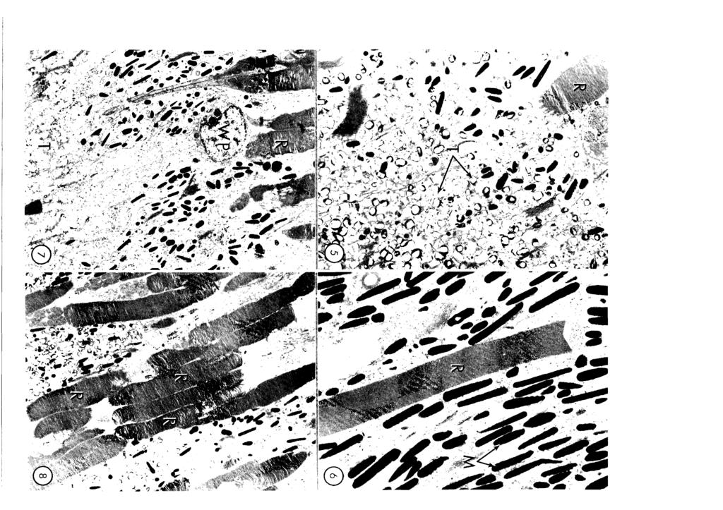
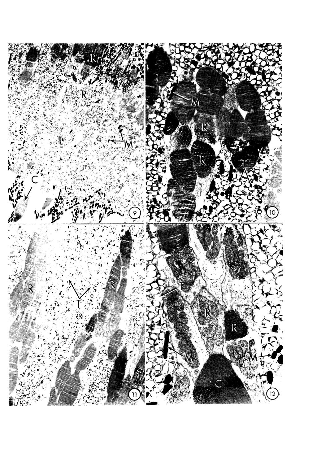
2 64 Tape tal development in the walleye The fish were decapitated and the eyeballs enucleated. In the case of small specimens (25-40 mm) the eye was fixed intact while in larger specimens ( mm) the globe was opened at the equator prior to fixation. The eyes were fixed for 5 hrs in 5% glutaraldehyde buffered to ph 7.3 with 0.1M Sorensen's phosphate buffer. The posterior half of the eyeball was then removed, washed in 5% sucrose in 0.1 M Sorensen's buffer (ph 7.3) and further divided into pieces less than 1 mm2. In specimens over about 30 mm in length the retina was separated into superior and inferior portions and processed separately. The material was then post fixed for 2 hrs in 1% osmium tetroxide in the same phosphate buffer, dehydrated in graded ethanols to propylene oxide and embedded in Araldite. Pieces of plastic-embedded tissue were reorientated to desired angles by means of a wax mount. Thick sections (0.5 pm) were cut, stained with toluidine blue and examined by light microscopy. Thin sections (60-70 nm) of selected areas were then cut on an LKB ultratome and collected on copper grids. The grids were stained in aqueous uranyl acetate and lead citrate and examined and photographed in a Philips EM 201 electron microscope. Results The retinal pigment epithelium (RPE) of the walleye consists of a single layer of cells which are initially roughly cuboidal in shape but which elongate to become columnar as the fish becomes larger. Basally the cells of the RPE abut on Bruch's membrane (complexus basalis) and show a few basal infoldings (Fig. 1). Apically the epithelial cells display numerous villous processes which enclose the photoreceptor outer segments (Figs. 1, 2,7, 8). Wandering phagocytes which appear to be a fairly constant feature of the teleost retina are also noted in this species (Fig. 7). Internally the RPE cells show plentiful smooth endoplasmic reticulum (SER). sparse rough endoplasmic reticulum (RER), numerous basally located mitochondria, severa1 myeloid bodies and a pleomorphic vesicular nucleus (Fig. 1). Melanin pigment granules (melanosomes) occupy the tips of the apical processes and are scattered throughout the body of the RPE cells in light-adaptation (Figs. 1, 6). In dark-adaptation while the melanosomes in the body of the epithelial cells move to the basal region of the RPE cells, the melanosomes in the tips of the apical processes remain in place (Fig. 12). In specimens 25 mm in length there is no indication of the presence of any reflective (tapetal) material in any location within the RPE cells (Figs. 1, 2). At about 30 mm of length in the superior temporal fundus, the first indication of tapetal development is seen. In the apical portion of the RPE cell body at the leve1 of the photoreceptor outer segments, the SER begins to show small dilations which are initially filled with flocculent or granular material (Fig. 3). These granular sites become more numerous and gradually also become more electron dense. When they attain a size of approximately 0.2 pm in diameter, they usually display a square or rectangular electron-lucent center due to the loss or chipping out of the presumed crystalline center during sectioning of the material (Figs. 3-5). The granules of tapetal material while of much the same size as the melanosomes, never attain the very electron-dense appearance of melanin before the center is lost in sectioning (Figs. 3-5). Once the beginning of the tapetum is established, the accumulation of tapetal material continues and the tapetum spreads laterally in al1 directions in the fundus as well as in an apical-basa1 fashion within the RPE cells (Fig. 4). By 40 mm the tapetum occupies a band about 20 mm wide in the superior temporal fundus (Fig. 5). The production of new tapetal material occurs both at the Fig. l. Electron micrograph of the retinal pigment epithelium (RPE) from a light-adapted 25 mm specimen. Bruch's membrane (6) an RPE nucleus (N) and rod outer segments (ROS) are indicated. No tapetal material is seen. x 6,600 Fig. 2. Electron micrograph of the retinal pigment epithelium (RPE) from a light-adapted 25 mm specimen. The rod (R) photoreceptorsare cut in cross-section. A myeloid body (My) and melanosomes (M) are indicated. No tapetal material is present. x 9,900 Fig. 3. Electron micrograph of the RPE from the superior fundus of a 30 mm light-adapted specimen. Tapetal material is beginning to form. Rod photoreceptors (R) and melanosomes (M) are also indicated. x 9,500 Fig. 4. Electron micrograph of the superior RPE from a light-adapted 35 mm specimen. Tapetal material O is more plentiful. Rod (R) and cone (C) photoreceptors are indicated. x 6,400 Fig. 5. Electron micrograph of the RPE from the superior fundus of a 40 mm specimen (light-adapted) to indicate a thickening of thetapetal region m. A rod (R) is indicated. x 6,400 Fig. 6. Electron micrograph of the RPE from the inferior fundus of a light-adapted 50 mm specimen. No tapetal material is present. Melanosomes (M) are plentiful and a rod (R) is indicated. x 9,900 Fig. 7. Electron micrograph of the RPE from the superior fundus of a 50 mm specimen. The tapetal region O reaches almost to the rod (R) outer segments in this light-adapted specimen. A wandering phagocyte (WP) is also indicated. x 4,200 Fig. 8. Electron micrograph of the RPE from the superior fundus of a 60 mm light-adapted specimen. The rods (R) are beginning to form bundles and the tapetal material has spread around the rod outer segments. x 3,900 Fig. 9. Electron micrograph of the tapetal region in the inferior fundus of a 90 mm light-adapted specimen. Melanosomes (M) are not plentiful within the tapetum O. 60th rods (R) and cones (C) are indicated. x 2,700 Fig. 10. Electron micrograph of a bundle of rods (R) from a 125 mm light-adapted specimen taken from the superior fundus. While rod bundles are mainly isolated by tapetum O scattered melanosomes (M) are also present. x 7,300 Fig. 11. Electron micrograph of rod bundles (R) from a dark-adapted 140 mm specimen taken from the inferior fundus. No melanosomes are noted amongst the tapetal material O. x 4,100 Fig. 12. Electron micrograph of a rod bundle (R), from the superior fundus of a dark-adapted 140 mm specimen. A cone (C) photoreceptor is indicated as are melanosomes (M) located in the apical process of the RPE cellsand which separate the photoreceptor bundles. Tapetal material is also indicated. x 9,900

3
4
5
.")
6 Tapetal development ín the walleye advancing front of the tapetum as well as within the thickness of the established tapetum to give a very dense accumulation of tapetal material that soon outweighs the melanosomes in both numbers and volume (Fig. 5). As more tapetal material is formed and the tapetum widens within the RPE cells, it comes to surround the photoreceptor inner and outer segments with the apical melanin granules being displaced into a thin layer between cone photoreceptors. During this time the RPE cells gradually become taller (more columnar) to accommodate the increase of tapetal material. By 50 mm of length, while the tapetum is well established in the superior fundus (Fig. 7) it has not yet spread to the inferior fundus which still shows only an abundance of melanosomes (Fig. 6). At about 60 mm the tapetum in the superior fundus has increased in thickness such that the inner and outer segments of the lightadapted rods are now separated by both tapetal material and melanosomes, the latter of which have been almost excluded from the main tapetal mass (Fig. 8). Also at approximately mm the first indications of the grouping of rod photoreceptors into bundles of cells, which will be the condition in the adult retina. are first noted (Fig. 8). By 90 mm the tapetum has spread further peripherally and is now also well established in the inferior fundus as well (Fig. 9). While a good deal of variability is noted within s~ecimens of similar len~th. at al1 intervals L. sampled, the tapetum in the superior fundus is always thicker and more ~rominenthan in the inferior fundus. and thus we canno't confirm the observation made by ~ l i and Anctil (1977) that the tapetum is more highly developed in the ventral region of the walleye's retina. By 125 to 140 mm the superior funda1 region is essentially adult in appearance with a large tapetum lucidum and the rods being arranged in definite bundles of cells with these bundles being separated from one another by tapetal material (Fig. 10). In the lightadapted state when the rod bundles are most fully extended into the RPE cells, melanosomes are noted between the rod bundles (Fig. 10) whereas in the darkadapted state when the rod bundles retract they are completely surrounded by tapetal material with no melanosomes present in the body of the tapetum (Fig. 11). While the rods of the walleye undergo extensive retinomotor or photomechanical movements, the cones (both single and twin) appear to move but little in response to environmental lighting. In light-adaptation when rods and cones are most completely separated, no spatial relationships are noted between the two photoreceptor types (Fig. 9). However, in dark-adaptation when the rod bundles are retracted and are in close proximity to the cones it can be appreciated that each rod bundle has in its central region one or more large cone photoreceptors (Fig. 12). In dark-adaptation, while the vast majority of melanosomes within the RPE migrate to th6 basa1 region of these cells, those melanosomes located in the apical processes remain in place and serve to isolate the bundles of rods or «macroreceptors» from one another (Fig. 12). In the dark therefore the tapetum is devoid of melanosomes and light passing to the rod outer segments would best gain acess to the photoreceptors by passing along the macroreceptors. Also as the mass of tapetal material is very large in proportion to the melanosomes present, it is unlikely that these melanosomes can completely occlude or mask the tapetum during lightadaptation in this species. Discussion The basic morphology of the retinal epithelium (RPE) of the walleye (Stizostedion vitreum vitreum) is essentially similar to that described for other vertebrates (Nguyen-Legros, 1978; Braekevelt. 1980a, 1982a). Although the retinal epithelial cells in teleosts are usually columnar (Braekevelt, d). in those species such as the walleye and goldeye (Hiodon alosides) with a retinal tapetum, the RPE cells become extremely tal1 (Zyznar and Ali, 1975; Braekevelt, 1982b). This is perhaps due to the very large accumulation of reflective material within these cells and also the necessity for these cells to accommodate the movement of the photoreceptors and interna1 structures during retinomotor movements. The wandering phagocytes at the photoreceptor-retina1 epithelial interface noted in the walleye have been reported in most other teleost species examined (Braekevelt, 1980c, 1985). As these phagocytes are present in apparently non-pathological retinas, they are felt to be a normal constituent of the teleost retina. Fish often have long photoreceptor outer segments and it may be that there is an accelerated rate or volume of outer segment shedding necessitating the presence of a second group of phagocytic cells in addition to the retinal epithelium itself. A tapetum lucidum is often present in the eye of species whose habitat is ordinarily poorly illuminated. The design of a tapetum is basically quite simple, consisting of a reflecting layer located scleral to the photoreceptor cells. This reflecting layer is most usually located in the choroid immediately externa1 to the retinal epithelium (choroidal tapetum). The retinal epithelium overlying such a tapetum is unpigmented to allow light to reach to and be reflected back from the tapetal area (Walls, 1967; Rodieck, 1973; Braekevelt, 1982a, 1983). In comparatively fewer species the tapetal material is located within the retinal epithelial cells themselves (retinal tapetum). Amongst the vertebrates reported with retinal tapeta lucida are a number of teleosts (Nicol et al., 1973; Braekevelt, 1980a,b, 1982b) the Crocodrilians (Laurens and Detwiler, 1921; Braekevelt, 1977), the fruit bat Pteropus cynopterus (Walls, 1967) the opossum Didelphis virginiana (Walls, 1967; Braekevelt, 1976) and the nighthawk Chordeiles minor (Braekevelt, 1984). The reflecting material within a tapetum lucidum can vary widely in chemical composition from species to species. Amongst the compounds reported are guanine, cholesterol, zinc cysterne, riboflavin, pteridine and various lipids (Pirie, 1966; Arnott et al., 1970; Nicol et al., 1973). The reflective bodies in the walleye are reported to be 7, 8-dihydroxanthopterin, a reduced
 in the tapetum, the crystals are very precisely arranged so as to reflect light almost straight back along the incoming path (Nicol,")
7 Tapetal development in the walleye 69 pteridine (Zyznar and Ali, 1975). In most species with crystalline reflecting material (eg guanine) in the tapetum, the crystals are very precisely arranged so as to reflect light almost straight back along the incoming path (Nicol, 1969; Locket, 1974). Such a tapetum is normally choroidally located and probably reflects light most effectively. In species such as the walleye with a retinal tapetum however the reflecting material is usually more haphazardly scattered within the RPE cells (Braekevelt, 1976, 1977, 1980a,b). Perhaps the closer proximity of the reflecting material to the photoreceptors compensates for the more diffuse reflectance that would be produced by a retinal tapetum. The first indication of the presence of a retinal tapetum in the walleye is noted in the superior fundus of specimens about 30 mm in length where it appears to be formed as isolated granules within the smooth endoplasmic reticulum. It would not appear to be functional at this time as walleye at this stage are positively phototactic and feed exclusively on zooplankton (Houde and Forney, 1970; Bulkowski and Meade, 1983). As the tapetum continues to thicken up and spread peripherally in the fundus the young walleye becomes increasingly negatively phototactic (Ryder, 1977). As subadults and adults they are active after sunset when they often return to surface waters to feed (Kelso and Ward, 1977), while in turbid waters walleye are often active during the day (Ryder, 1977). Concomitant with the formation of a functional tapetum lucidum and beginning about mm in length is the arrangement of the rod photoreceptors into bundles of to form «macroreceptors». Grouped or bundled photoreceptors have been reported in severa1 teleost species but to date in no other animal groups (Moore, 1944; Locket, 1970; Meyer-Rochow, 1972; Ali and Anctil, 1976; Munk, 1977). Al1 teleost species which display grouped photoreceptors inhabit either turbid or deep-sea waters where the leve1 of illumination is low. Some of these species also show a prominent retinal tapetum lucidum (Ali and Anctil, 1976; Braekevelt, 1982b). The bundling of photoreceptors results in the close apposition of the inner and outer segments of numerous photoreceptors. Since each photoreceptor bundle is isolated and closely invested by a reflective layer (retinal tapetum) it is likely that each bundle has a great deal of interna1 reflectance and probably functions as a wave-guide structure to funnel light to the closely packed and extremely long outer segments of the rods. The melanosomes which remain in the apical processes of the RPE cells in darkadaptation in the walleye and goldeye (Braekevelt, 1982b) would further serve to isolate each bundle of photoreceptors or macroreceptor and enhance its waveguide function. Zyznar and Ali (1975) have shown that transmitted light passes most effectively through the macroreceptors of the walleye retina. Retinomotor responses or photomechanical movements which refer both to the movement of melanosomes within RPE cells and the lengthening or shortening of photoreceptor cells in response to environmental illumination are a common feature in teleosts, amphibians and some birds (Walls, 1967; Ali 1971, 1975; Rodieck, 1973; Burnside and Laties, 1979). It is believed that retinomotor responses occur to adapt the eye for day or night vision by shielding or unmasking photoreceptor outer segments to incident light (Walls, 1967). With a few exceptions photomechanical movements appear to replace active pupillary responses (Burnside and Laties, 1979). Al1 teleost species in which photomechanical movements have been reported showed marked movements of the rod photoreceptors (Engstrom, 1963; Braekevelt, 1975). In most species, cones also change length to some extent in response to environmental illumination. although some species including the goldeye and walleye appear to have no cone movement (Zyznar and Ali, 1977; Braekevelt, 1982b). As both the goldeye and walleye have prominent bundled photoreceptors, perhaps the non-movement of the cones in some way is connected with the maintenance of the bundles during dark-adaptation. While the majority of the melanosomes of the RPE cells move basally in dark-adaptation, the melanosomes of the apical region always remain in position. In lightadaptation when the melanosomes in the body of the RPE cells become more widely dispersed, they probably cannot effectively mask the tapetal material and hence even during the day the tapetum would be functional to some extent as light would continue to reach the rod outer segments via the large cone photoreceptor cell bodies which would remain as pathways through the apically located melanosomes of the RPE cells. The negative phototaxis of the adult walleye would tend to support this view. The development of a functional tapetum lucidum and the subsequent formation of macroreceptors during the first summer of life is consistent with changes in response to environmental illumination and changes in feeding behavior. Walleye larvae are positively phototactic prior to the development of a functional tapetum lucidum, but become increasingly negatively phototactic at lengths greater than 30 mm (Ryder, 1977; Bulkowski and Meade, 1983). Initially walleye feed on small organisms, principally zooplankton. (Houde, 1967) but a transition to selecting larger organisms such as minnows takes place at a walleye length of about mm (Smith and Noyle, 1945). This transition occurs when photoreceptors are bundling to form macroreceptors. Bundling may enhance perception or larger objects such as forage fish in dim light in clear water or in turbid water. Acknowledgements. The excellent technical assistance of D.M. Love is gratefully acknowledged. This work was supported in part by funds from the Natural Sciences and Engineering Research Council of Canada and the Medical Research Council of Canada. References Ali M. (1971). Les responses retinomotrices: caracteres et
. Retinas of Fishes: An Atlas. Springer- Verlag. Berlin. Ali M. (1977). Retinal structure and function in the walleye (Stizostedion vitreum vitreum) and sauger (S. canadense). J. Fish. Res. Bd.")
8 Tapetal development in the walleye rnechanisrnes. Vision Res. 1 1, Ali M. (1975). Retinornotor responses. Vision in Fishes. Ed. M.A. Ali, Plenum. New York. pp Ali M. and Anctil N. (1976). Retinas of Fishes: An Atlas. Springer- Verlag. Berlin. Ali M. (1977). Retinal structure and function in the walleye (Stizostedion vitreum vitreum) and sauger (S. canadense). J. Fish. Res. Bd. Can. 34, Arnott H.J., Maciolek N.J. and Nicol J.A.C. (1970). Retinal tapetum lucidum: a novel reflecting system in the eye of teleosts. Science. 169, Braekevelt C.R. (1974). Fine structure of the retinal pigment epithelium, Bruch's membrane and choriocapillaris in the northern pike Esox lucius. J. Fish. Res. Bd. Can. 31, Braekevelt C.R. (1975). Photoreceptor fine structure in the northern pike. J. Fish. Res. Bd. Can. 32, Braekevelt C.R. (1 976). Fine structure of the retinal epithelium and tapetum lucidum of the opossum (Didelphis virginiana) J. Morph. 150, Braekevelt C.R. (1977). Fine structure of the retinal epithelium of the spectacled caiman (Caiman sclerops). Acta anat. 97, Braekevelt C.R. (1980a). Fine structure of the retinal epithelium and tapetum lucidum in the giant danio (Danio malabaricus) (Teleost). Anat. Embryol. 158, Braekevelt C.R. (1980b). The fine structure of the retinal epithelium in the scissortail (Rasbora trilineata)(teleost). Anat. Anz. 148, Braekevelt C.R. (1980~). Wandering phagocytes at the retinal epithelium - photoreceptor interface in the teleost retina. Vision Res. 20, Braekevelt C.R. (1980d). Fine structure of the retinal pigment epithelium in the mud minnow (Umbra liml). Can. J. Zool. 58, Braekevelt C.R. (1981). Fine structure of the tapetum lucidum of the domestic ferret. Anat. Embryol. 163, Braekevelt C.R. (1982a). Fine structure of the retinal epithelium, Bruch's membrane (complexus basalis) and choriocapillaris in the domestic ferret. Acta. Anat. 113, Braekevelt C.R. (1982b). Fine structure of the retinal epithelium and retinal tapetum lucidum of the goldeye (Hiodon alosoides). Anat. Embryol. 1 64, Braekevelt C.R. (1983). Morphology of the tapetum fibrosum in the eye of the domestic sheep. Can. J. Zool. 61, Braekevelt C.R. (1 984). Retinal pigment epithelial fine structure in the nightawk (Chordeiles minor). Ophthalmologica. 188, Braekevelt C.R. (1985). Further observations on the presence of wandering phagocytes within the teleostean retina. Anat. Anz. 160, Bulkowski L. and Meade J.W. (1983). Changes in phototaxis during early development of walleye. Trans. Amer. Fish. Soc. 11 2, Burnside B. and Laties A.M. (1979). Pigment movement and cellular contractility in the retinal pigment epithelium. In: The Retinal Pigment Epithelium. Edited by K.M. Zinn & M.F. Marmor. Harvard Univ. Press. Cambridge. pp Duke-Elder Sir S. (1 958). System of Ophthalmology Vol. l. The Eye in Evolution. Henry Kimpton. London. Engstrom K. (1963). Structure organization and ultrastructure of the visual cells in the teleost family Labridae. Acta Zool. 44, Hebel R. (1969). Light and electron microscope investigations of the cells of the tapetum lucidum in the dog. Z. Anat. Entwick.- Gesch. 129, Houde E.D. (1967). Food of the pelagic young of the walleye, Stizostedion vitreum vitreum, in Oneida Lake, New York. Trans. Amer. Fish. Soc. 96, Houde E.D. and Forney J.L. (1970). Effects of water currents on distribution of walleye larvae in Oneida Lake, New York. J. Fish. Res. Bd. Can. 27, Kelso J.R.M. and Ward F.J. (1977). Unexploited percid populations of West Blue Lake, Manitoba and their interactions. J. Fish. Res. Bd. Can. 34, Laurens H. and Detwiler S.R. (1921). Studies on the retina. The structure of the retina of Alligator mississipiensis and its photomechanical changes. J. Exp. Zool. 32, Lesiuk T.P. and Braekevelt C.R. (1983). Fine structure of the canine tapetum lucidum. J. Anat. 136, Locket N.A. (1970). Retinal structure in a deep-sea fish Sternophyx diaphana (Herman). Exp. Eye Res. 9, Locket N.A. (1974). The choroidal tapetum lucidum in Latimeria chalumnae. Proc. Roy. Soc. B. 186, Meyer-Rochow V.B. (1972). The larval eye of the deep-sea fish Cataetyx memoriabilis. Z. Morph. Tiere 72, Moore G.A. (1944). The retinae of two North American teleosts with special reference to their tapeta lucida. J. Comp. Neurol. 80, Munk 0. (1977). The visual cells and retinal tapetum of the foveate deep-sea fish Scopelosaurus lepidus (Teleostei). Zoomorph. 87, Nicol J.A.C. (1969). The tapetum lucidum of the sturgeon. Contr. Mar. Sci. 4,518. Nicol J.A.C., Arnott H.J. and Best. A.C.G. (1973). Tapeta lucida in bony fishes (Actinopefygi): a survey. Can. J. Zool. 51, Nguyen-Legros J. (1978). Fine structureof the pigment epithelium in the vertebrate retina. Int. Rev. Cytol. Suppl. 7, Pedler C. (1963). The fine structure of the tapetum cellulosum. Exp. Eye Res. 2, Pirie A. (1966). The chemistry and structure of the tapetum lucidum in animals. In: Aspects of Comparative Opthalmology. Edited by O. Graham-Jones. Pergammon Press. Oxford. pp Rodieck R.W. (1973). The Vertebrate Retina. Principles of Structure and Function. W.H. Freeman. San Francisco. Ryder R.A. (1977). Effects of ambient light variations on behaviour of yearling, subadult and adult walleyes. Stizostedion vitreum vitreum. J. Fish. Res. Bd. Can. 34, Smith L.L. and Noyle J.B. (1945). Factors influencing production of yellow pike-perch (Stizostedion vitreum vitreum) in Minnesota rearing ponds. Trans Arner. Fish. Soc. 73, Walls G.L. (1967). The Vertebrate Eye and its Adaptive Radiation. Hafner Publ. Co. New York. pp 786. Zyznar F.S. and Ali M.A. (1975). An interpretative study of the organization of the visual cells and tapetum lucidum of Stizostedion. Can. J. Zool. 53, Accepted July 14, 1988
タイ産カタクチイワシの網膜反射板 Retinal Tapetum
 タイ産カタクチイワシの網膜反射板 Retinal Tapetum 誌名 水産増殖 = The aquiculture ISSN 03714217 著者 Awaiwanont, K. 巻 / 号 49 巻 1 号 掲載ページ p. 29-34 発行年月 2001 年 3 月 農林水産省農林水産技術会議事務局筑波産学連携支援センター Tsukuba Business-Academia Cooperation
Both Rod and Cone Disc Shedding ore Related to Light Onset in the Cat
 Both Rod and Cone Disc Shedding ore Related to Light Onset in the Cat Sreven K. Fisher,* Bruce A. Pfeffer.f and Don H. Anderson* Nineteen domestic cats were entrained to a 12-hr light/12-hr dark lighting
Progressive Retinal Atrophy in the Abyssinian Cat
 Progressive Retinal Atrophy in the Abyssinian Cat Electron Microscopy Kristina Narfstr6m*t and Sven Erik Nilsson* Seven adult Abyssinian cats at different stages of a recessively inherited retinal degenerative
Electron Microscopy of the Tapetum Lucidum of the Cat*
 Published Online: 25 January, 1959 Supp Info: http://doi.org/10.1083/jcb.5.1.35 Downloaded from jcb.rupress.org on December 26, 2018 Electron Microscopy of the Tapetum Lucidum of the Cat* By MAURICE H.
A comparison of placental tissue in the skinks Eulamprus tympanum and E. quoyii. Yates, Lauren A.
 A comparison of placental tissue in the skinks Eulamprus tympanum and E. quoyii Yates, Lauren A. Abstract: The species Eulamprus tympanum and Eulamprus quoyii are viviparous skinks that are said to have
Veterinary Ophthalmology
 Veterinary Ophthalmology Eyelids Protect the eye Provides part of and spreads the tear film Regulates the amount of light that enters the eye Clears foreign material Third Eyelid Protects the cornea by
Master Thesis in Biology (60 ECTS)
") Master Thesis in Biology (60 ECTS) Seasonal changes in the Tapetum lucidum as an adaptation to winter darkness in reindeer (Rangifer tarandus tarandus) Sandra Katharina Christiane Siefken May 2010 University
Master Thesis in Biology (60 ECTS) Seasonal changes in the Tapetum lucidum as an adaptation to winter darkness in reindeer (Rangifer tarandus tarandus) Sandra Katharina Christiane Siefken May 2010 University
A Scanning Electron Microscopic Study of Eggshell Surface Topography of Leidynema portentosae and L. appendiculatum (Nematoda: Oxyuroidea)
") The Ohio State University Knowledge Bank kb.osu.edu Ohio Journal of Science (Ohio Academy of Science) Ohio Journal of Science: Volume 88, Issue 5 (December, 1988) 1988-12 A Scanning Electron Microscopic
The Ohio State University Knowledge Bank kb.osu.edu Ohio Journal of Science (Ohio Academy of Science) Ohio Journal of Science: Volume 88, Issue 5 (December, 1988) 1988-12 A Scanning Electron Microscopic
THE TAPETUM IN SCYLIORHINUS CANICULA
 J. mar. biol. Ass. U.K. (I96I) 4I, 27I-277 Printed in Great Britain THE TAPETUM IN SCYLIORHINUS CANICULA By J. A. C. NICOL The Plymouth Laboratory (Text-figs. 1-3) Elasmobranchs are peculiar in having
Although owls can t move their eyes, many other adaptations help these raptors spot prey.
 This website would like to remind you: Your browser (Apple Safari 7) is out of date. Update your browser for more security, comfort and the best experience on this site. Media Spotlight Bird s Eye View
Anatomy and Embryology Department, Faculty of Veterinary Medicine, Zagazig University, 44511, Egypt
 Slov Vet Res 2018; 55 (Suppl 20): 263 72 DOI 10.26873/SVR-653-2018 Original Research Article ABSENCE OR PRESENCE OF TAPETUM LUCIDUM: MACRO AND MICROSCOPIC INVESTIGATIONS IN DONKEY (EQUUS ASINUS), CAT (FELIS
Reductions in Taurine Secondary to Photoreceptor Loss in Irish Setters with Rod-Cone Dysplasia
 Reductions in Taurine Secondary to Photoreceptor Loss in Irish Setters with Rod-Cone Dysplasia S. Y. Schmidr*t and G. D. Aguirre$ These studies show that onset of photoreceptor cell degeneration preceded
Mesosomes are a definite event in antibiotic-treated Staphylococcus aureus ATCC 25923
 Tropical Biomedicine 24(1): 105 109 (2007) Mesosomes are a definite event in antibiotic-treated Staphylococcus aureus ATCC 25923 Santhana Raj, L. 1*, Hing, H.L. 2, Baharudin Omar 2, Teh Hamidah, Z. 1,
Seasonal Variations of yeso sika Deer Skin and its Vegetable Tanned Leather
 Seasonal Variations of yeso sika Deer Skin and its Vegetable Tanned Leather Shigeharu Fukunaga, Akihiko Yoshie, Ikuo Yamakawa, Fumio Nakamura Laboratory of Animal By-product Science, Graduate School of
Liver and Gallbladder Morphology of the juvenile Nile crocodile, Crocodylus niloticus (Laurenti, 1768)
") Liver and Gallbladder Morphology of the juvenile Nile crocodile, Crocodylus niloticus (Laurenti, 1768) by ERNA VAN WILPE Submitted in partial fulfilment of the requirements for the degree MSc DEPARTMENT
Liver and Gallbladder Morphology of the juvenile Nile crocodile, Crocodylus niloticus (Laurenti, 1768) by ERNA VAN WILPE Submitted in partial fulfilment of the requirements for the degree MSc DEPARTMENT
UC Davis UC Davis Previously Published Works
 UC Davis UC Davis Previously Published Works Title Evolution of the tapetum Permalink https://escholarship.org/uc/item/3jz996d5 Journal Transactions of the American Ophthalmological Society, 100 ISSN 0065-9533
SUPPLEMENTARY INFORMATION
 doi:10.1038/nature11046 Supplementary Figure 1: Images of PB-positive cells in the subepidermal region (a-i) Representative images of PB positive cells in the subepidermis of the upper beak of the pigeon.
Formoguanamine-induced blindness and photoperiodic responses in the Japanese quail, Coturnix coturnix japonica
 J. Biosci., Vol. 19, Number 4, October 1994, pp 479-484. Printed in India. Formoguanamine-induced blindness and photoperiodic responses in the Japanese quail, Coturnix coturnix japonica 1. Introduction
SCANNING electron - microscopy has
 Characteristics of the Absorptive Surface of the Small Intestine of the Chicken from 1 Day to 14 Weeks of Age 1 R. C. BAYER, C. B. CHAWAN, F. H. BIRD AND S. D. MUSGRAVE Department of Animal and Veterinary
How the eye sees. Properties of light. The light-gathering parts of the eye. 1. Properties of light. 2. The anatomy of the eye. 3.
 How the eye sees 1. Properties of light 2. The anatomy of the eye 3. Visual pigments 4. Color vision 1 Properties of light Light is made up of particles called photons Light travels as waves speed of light
BEAK AND FEATHER DYSTROPHY IN WILD SULPHUR-CRESTED COCKATOOS (CACATUA GALERITA)
") BEAK AND FEATHER DYSTROPHY IN WILD SULPHUR-CRESTED COCKATOOS (CACATUA GALERITA) Author(s): Steven McOrist, Douglas G. Black, David A. Pass, Peter C. Scott, and John Marshall Source: Journal of Wildlife
BEAK AND FEATHER DYSTROPHY IN WILD SULPHUR-CRESTED COCKATOOS (CACATUA GALERITA) Author(s): Steven McOrist, Douglas G. Black, David A. Pass, Peter C. Scott, and John Marshall Source: Journal of Wildlife
Time of Day. Teacher Lesson Plan Nocturnal Animals Pre-Visit Lesson. Overview
 Teacher Lesson Plan Nocturnal Animals Pre-Visit Lesson Duration: 40-50 minutes Minnesota State Science Standard Correlations: 3.4.1.1.2. Wisconsin State Science Standard Correlations: B 4.6, C.4.1, C.4.2
Title. CitationJapanese Journal of Veterinary Research, 24(1-2): 37. Issue Date DOI. Doc URL. Type. File Information
: 37. Issue Date DOI. Doc URL. Type. File Information") Title DISTRIBUTION OF LYMPHATIC TISSUES IN DUCK CAECA Author(s)KITAMURA, Hirokazu; SUGIMURA, Makoto; HASHIMOTO, Yos CitationJapanese Journal of Veterinary Research, 24(1-2): 37 Issue Date 1976-05 DOI 10.14943/jjvr.24.1-2.37
Title DISTRIBUTION OF LYMPHATIC TISSUES IN DUCK CAECA Author(s)KITAMURA, Hirokazu; SUGIMURA, Makoto; HASHIMOTO, Yos CitationJapanese Journal of Veterinary Research, 24(1-2): 37 Issue Date 1976-05 DOI 10.14943/jjvr.24.1-2.37
Anat. Labor. of Prof. H. SETO, Tohoku University, On the Sensory Terminations Formed along the Ductus
 Anat. Labor. of Prof. H. SETO, Tohoku University, Sendai. On the Sensory Terminations Formed along the Ductus Pancreaticus in Cat. The existence of PACINIan bodies in the pancreas of mammals, especially
HISTOLOGY OF MAMMARY GLAND DURING LACTATING AND NON-LACTATING PHASES OF MADRAS RED SHEEP WITH SPECIAL REFERENCE TO INVOLUTION
 International Journal of Science, Environment and Technology, Vol. 5, No 3, 2016, 991 996 ISSN 2278-3687 (O) 2277-663X (P) HISTOLOGY OF MAMMARY GLAND DURING LACTATING AND NON-LACTATING PHASES OF MADRAS
Proceeding of the SEVC Southern European Veterinary Conference
 www.ivis.org Proceeding of the SEVC Southern European Veterinary Conference Oct. 17-19, 2008 Barcelona, Spain http://www.sevc.info Reprinted in the IVIS website with the permission of the SEVC www.ivis.org
The oriental fruit fly, Bactrocera dorsalis
 Zoological Studies 37(2): 95-101 (1998) Morphology and Ultrastructure of the Alimentary Canal of Oriental Fruit Fly Bactrocera dorsalis (Hendel) (Diptera: Tephritidae) (I): The Structure of the Foregut
Criconemoides similis 1 G. W. BIRD ~
 Somatic Musculature of Trichodorus porosus and Criconemoides similis 1 G. W. BIRD ~ Abstract: The somatic musculature of Trichodorus porosus is transversely striated, and that of Criconemoides similis
Chemical and ultrastructural changes in tapetum of beagles with a hereditary abnormality
 Chemical and ultrastructural changes in tapetum of beagles with a hereditary abnormality Guang Y. Wen, John A. Sturman, Henryk M. Wisniewski, A. MacDonald, and Wendell H. Niemann We have defined the defect
Morphology of Shells From Viable and Nonviable Eggs of the Chinese Alligator (Alligator sinensis)
") ~ JOURNAL OF MORPHOLOGY 222:103-110 (1994) Morphology of Shells From Viable and Nonviable Eggs of the Chinese Alligator (Alligator sinensis) CAROLE S. WINK AND RUTH M. ELSEY Department of Anatomy, Louisiana
~ JOURNAL OF MORPHOLOGY 222:103-110 (1994) Morphology of Shells From Viable and Nonviable Eggs of the Chinese Alligator (Alligator sinensis) CAROLE S. WINK AND RUTH M. ELSEY Department of Anatomy, Louisiana
Lacerta viridis. Functional anatomy of the lungs of the green lizard, (Accepted 18 February 1977)
") J. Anat. (1978), 125, 2, pp. 421-431 421 With 9 figures Printed in Great Britain Functional anatomy of the lungs of the green lizard, Lacerta viridis C. MEBAN Department of Anatomy, The Queen's University
J. Anat. (1978), 125, 2, pp. 421-431 421 With 9 figures Printed in Great Britain Functional anatomy of the lungs of the green lizard, Lacerta viridis C. MEBAN Department of Anatomy, The Queen's University
,,, THE MORPHOLOGY AND MORPHOMETRY OF THE PECTEN OCULI IN DIURNAL AND NOCTURNAL BIRDS: A
 ,,, THE MORPHOLOGY AND MORPHOMETRY OF THE PECTEN OCULI IN DIURNAL AND NOCTURNAL BIRDS: A COMPARATIVE STUDY" BY llijama, S.G., B. V. M. (NBI), Department of Veteri nary Anatomy, University of I\Jairobi.
Analysis of Sampling Technique Used to Investigate Matching of Dorsal Coloration of Pacific Tree Frogs Hyla regilla with Substrate Color
 Analysis of Sampling Technique Used to Investigate Matching of Dorsal Coloration of Pacific Tree Frogs Hyla regilla with Substrate Color Madeleine van der Heyden, Kimberly Debriansky, and Randall Clarke
International Journal of Science, Environment and Technology, Vol. 5, No 5, 2016,
 International Journal of Science, Environment and Technology, Vol. 5, No 5, 2016, 3249 3253 ISSN 2278-3687 (O) 2277-663X (P) HISTOPATHOLOGICAL STUDY OF PULMONARY ANTHRACOSIS IN SHEEP Amaravathi M* 1, Satheesh
HISTOPATHOLOGY. Introduction:
 Introduction: HISTOPATHOLOGY Goats and sheep are the major domestic animal species in India. Much of the economy of the country has been depend upon the domestication of these animals. Especially economy
Coturnix coturnix coturnix
 Coturnix coturnix coturnix L. Coturnix coturnix coturnix L. 8 circadian clock 0 9 Galliformes Coturnix coturnix coturnix Phasianidae L. 0 9 8 7 Photoneuroendocrine gland Transmission TEM electron microscope
Development of the Intestinal Villi Associated
 Development of the Intestinal Villi Associated with the Increased Epithelial Cell Mitosis in Chickens Koh-en YAMAUCHI, Eiji NAKAMURA and Yutaka ISSHIKI Laboratory of Animal Science, Faculty of Agriculture,
RETINITIS PIGMENTOSA*
 Brit. J. Ophihal. (1955), 39, 312. ABNORMAL FUNDUS REFLEXES AND RETINITIS PIGMENTOSA* BY R. P. CRICK Royal Eye Hospital, London THE normal variation of the fundus reflex which gives a " shot-silk" appearance
Postilla PEABODY MUSEUM OF NATURAL HISTORY YALE UNIVERSITY NEW HAVEN, CONNECTICUT, U.S.A.
 Postilla PEABODY MUSEUM OF NATURAL HISTORY YALE UNIVERSITY NEW HAVEN, CONNECTICUT, U.S.A. Number 117 18 March 1968 A 7DIAPSID (REPTILIA) PARIETAL FROM THE LOWER PERMIAN OF OKLAHOMA ROBERT L. CARROLL REDPATH
DLS Sample Preparation Guide
 DLS Sample Preparation Guide The Leica TCS SP8 DLS is an innovative concept to integrate the Light Sheet Microscopy technology into the confocal microscope. Due to its unique optical architecture samples
2008/048 Reducing Dolphin Bycatch in the Pilbara Finfish Trawl Fishery
 2008/048 Reducing Dolphin Bycatch in the Pilbara Finfish Trawl Fishery PRINCIPAL INVESTIGATOR: Prof. N.R. Loneragan ADDRESS: Centre for Fish and Fisheries Research Biological Sciences and Biotechnology
Key words: Coccidia, Choleoeimeria rochalimai, fine structure, gall bladder epithelium, Hemidactylus mabouia, Brazil
 FOLIA PARASITOLOGICA 47: 91-96, 2000 Ultrastructural study of meronts and gamonts of Choleoeimeria rochalimai (Apicomplexa: Eimeriidae) developing in the gall bladder of the gecko Hemidactylus mabouia
PSY 2364 Animal Communication. Elk (Cervus canadensis) Extra credit assignment. Sad Underwing (Catocala maestosa) 10/11/2017
 Extra credit assignment. Sad Underwing (Catocala maestosa) 10/11/2017") PSY 2364 Animal Communication Elk (Cervus canadensis) Kingdom: Phylum: Class: Order: Family: Genus: Species: Animalia Chordata Mammalia Artiodactyla Cervidae Cervus canadensis Extra credit assignment Sad
PSY 2364 Animal Communication Elk (Cervus canadensis) Kingdom: Phylum: Class: Order: Family: Genus: Species: Animalia Chordata Mammalia Artiodactyla Cervidae Cervus canadensis Extra credit assignment Sad
DRAFT TANZANIA STANDARD
 Hatching eggs Specification DRAFT TANZANIA STANDARD TANZANIA BUREAU OF STANDARDS 1 Hatching eggs Specification TBS/AFDC 22 (5271) P3 0 FOREWORD This Tanzania standard was developed due to rapid increase
A Lymphosarcoma in an Atlantic Salmon (Salmo salar)
") A Lymphosarcoma in an Atlantic Salmon (Salmo salar) Authors: Paul R. Bowser, Marilyn J. Wolfe, and Timothy Wallbridge Source: Journal of Wildlife Diseases, 23(4) : 698-701 Published By: Wildlife Disease
A Lymphosarcoma in an Atlantic Salmon (Salmo salar) Authors: Paul R. Bowser, Marilyn J. Wolfe, and Timothy Wallbridge Source: Journal of Wildlife Diseases, 23(4) : 698-701 Published By: Wildlife Disease
Effect of Sodium Hypochlorite on the Oocyst Wall of Eimeria tenella as Shown by Electron Microscopy1
 32 PROCEEDINGS OF THE HELMINTHOLOGICAL SOCIETY This alteration appeared similar to that observed by light microscopy (Figs. 5, 6). Literature Cited Dixon, K. E. 1966. The physiology of excystment of the
Key words: Plasmodium, Kentropyx calcarata, Brazil, merogony, gametocytes, ultrastructure
 FOLIA PARASITOLOGICA 49: 2-8, 2002 Fine structure of erythrocytic stages of a Plasmodium tropiduri-like malaria parasite found in the lizard Kentropyx calcarata (Teiidae) from north Brazil Ilan Paperna
A NEW TYPE OF BRYOZOAN GIZZARD, WITH REMARKS ON THE GENUS BUSKIA.
 A NEW TYPE OF BRYOZOAN GIZZARD, WITH REMARKS ON THE GENUS BUSKIA. RAYMOND C. OSBURN AND RUTH M. VETH Department of Zoology and Entomology, Ohio State University A certain few of the Ctenostome Bryozoa
DEVELOPMENT OF THE HEAD AND NECK PLACODES
 DEVELOPMENT OF THE HEAD AND NECK Placodes and the development of organs of special sense L. Moss-Salentijn PLACODES Localized thickened areas of specialized ectoderm, lateral to the neural crest, at the
EXOSTOSIS OF THE MANDIBLE OF THE CHICKEN
 EXOSTOSIS OF THE MANDIBLE OF THE CHICKEN COMPLICATING EDEMA OF THE WATTLES GEORGE MILTON SMITH, M.D.1 (AnutomioaZ Laboratory, Yale School of Medicine, New Haven, Connecticut) During the past year opportunity
SCANNING ELECTRON MICROSCOPY OF THE MUCOSAL SURFACE OF THE FORESTOMACHS AND ABOMASA OF GREY, WHITE AND BLACK KARAKUL LAMBS
 OnderstepoortJ. Vet. Res., 59, 167-174 (1992) SCANNING ELECTRON MICROSCOPY OF THE MUCOSAL SURFACE OF THE FORESTOMACHS AND ABOMASA OF GREY, WHITE AND BLACK KARAKUL LAMBS H. B. GROENEWALD, Department of
THE VISUAL MECHANISMS OF TENEBRIO MOLITOR: VARIATIONS TAKING PLACE IN THE ERG OF PUPA AND ADULT DURING DEVELOPMENT
 J. Exp. Biol. (1969), 51. 635-641 635 With 5 text-figures Printed in Great Britain THE VISUAL MECHANISMS OF TENEBRIO MOLITOR: VARIATIONS TAKING PLACE IN THE ERG OF PUPA AND ADULT DURING DEVELOPMENT BY
Early-Onset, Autosomal Recessive, Progressive Retinal Atrophy in Persian Cats METHODS
 Early-Onset, Autosomal Recessive, Progressive Retinal Atrophy in Persian Cats HyungChul Rah, 1 David J. Maggs, 2 Thomas N. Blankenship, 3 Kristina Narfstrom, 4 and Leslie A. Lyons 1 PURPOSE. An early-onset
examnined when three weeks old. Not one of the eyes showed
 A NOTE ON THE DOG'S TAPETUM IN EARLY LIFE* BY C. H. USHER ABERDEEN THIS note is written for the purpose of pointing out that the tapetum of the dog is not recognizable ophthalmoscopically for several weeks
Effects of Natural Selection
 Effects of Natural Selection Lesson Plan for Secondary Science Teachers Created by Christine Taylor And Mark Urban University of Connecticut Department of Ecology and Evolutionary Biology Funded by the
Response to SERO sea turtle density analysis from 2007 aerial surveys of the eastern Gulf of Mexico: June 9, 2009
 Response to SERO sea turtle density analysis from 27 aerial surveys of the eastern Gulf of Mexico: June 9, 29 Lance P. Garrison Protected Species and Biodiversity Division Southeast Fisheries Science Center
THE EXPERIMENTAL MODIFICATION OF THE OESTROUS CYCLE IN THE FERRET BY DIFFER- ENT INTENSITIES OF LIGHT IRRADIATION AND OTHER METHODS
 THE EXPERIMENTAL MODIFICATION OF THE OESTROUS CYCLE IN THE FERRET BY DIFFER- ENT INTENSITIES OF LIGHT IRRADIATION AND OTHER METHODS BY F. H. A. MARSHALL School of Agriculture, Cambridge {Received 12 December
Sensory Setae of the First Tarsi and Palps of the Mite Macrocheles muscaedomesticae1.2
 $ Sensory Setae of the First Tarsi and Palps of the Mite Macrocheles muscaedomesticae1.2 ~, 'r L. B. COONS ANDR. C. AXTELL Department of Entomology, North Carolina State University, Raleigh 27607 By scanning
THE LARVA OF ROTHIUM SONORENSIS MOORE & LEGNER. BY IAN MOORE Department of Entomology, University of California, Riverside, California 92521
 THE LARVA OF ROTHIUM SONORENSIS MOORE & LEGNER WITH A KEY TO THE KNOWN LARVAE OF THE GENERA OF THE MARINE BOLITOCHARINI (COLEOPTERA STAPHYLINIDAE) BY IAN MOORE Department of Entomology, University of California,
Your Eye, My Eye, and the Eye of the Aye Aye: Evolution of Human Vision from 65 Million Years Ago to the Present
 # 75 Your Eye, My Eye, and the Eye of the Aye Aye: Evolution of Human Vision from 65 Million Years Ago to the Present Dr. Christopher Kirk December 2, 2011 Produced by and for Hot Science - Cool Talks
Electron Microscopic Observations on Ciliated Epithelium of Tracheal Organ Cultures Infected with Bordetella bronchiseptica
 Microbiol. Immunol. Vol. 33 (2), 111-121, 1989 Electron Microscopic Observations on Ciliated Epithelium of Tracheal Organ Cultures Infected with Bordetella bronchiseptica Kachiko SEKIYA,*,1 Yutaka FUTAESAKU,2
Central Marine Fisheries Research Institute, Mandapam Camp
 w«r n Mar. biol. Ass. India, 1961, 3 (1 & 2): 92-95 ON A NEW GENUS OF PORCELLANIDAE (CRUSTACEA-ANOMURA) * By C. SANKARANKUTTY Central Marine Fisheries Research Institute, Mandapam Camp The specimen described
Laboratory 7 The Effect of Juvenile Hormone on Metamorphosis of the Fruit Fly (Drosophila melanogaster)
") Laboratory 7 The Effect of Juvenile Hormone on Metamorphosis of the Fruit Fly (Drosophila melanogaster) (portions of this manual were borrowed from Prof. Douglas Facey, Department of Biology, Saint Michael's
Laboratory 7 The Effect of Juvenile Hormone on Metamorphosis of the Fruit Fly (Drosophila melanogaster) (portions of this manual were borrowed from Prof. Douglas Facey, Department of Biology, Saint Michael's
Light, Scanning and Transmission Electron Microscopical Study on the Oviduct of the Ostrich (Struthio
 Light, Scanning and Transmission Electron Microscopical Study on the Oviduct of the Ostrich (Struthio camelus) A.S.Saber*, S.A.M.Emara*, O.M.M.AboSaeda** * Faculty of Veterinary Medicine, Sadat City Branch,
Lesson 7. References: Chapter 6: Chapter 12: Reading for Next Lesson: Chapter 6:
 Lesson 7 Lesson Outline: Embryonic Origins of the Dermis Specializations of the Dermis o Scales in Fish o Dermal Armour in Tetrapods Epidermal/Dermal Interactions o Feathers o Hair o Teeth Objectives:
Amphibians. Land and Water Dwellers
 Amphibians Land and Water Dwellers Amphibians Most amphibians do not live completely in the water or completely on land and most must return to water to reproduce http://potch74.files.wordpress.com/2007/09/amphibians.jpg
Retinal Degeneration Basics
 Retinal Degeneration Basics OVERVIEW Retinal refers to the retina; the retina is the innermost lining layer (located on the back surface) of the eyeball; it contains the light-sensitive rods and cones
cyst&' appeared to be of two kinds-one smaller and Smnith "is inclined to regard these epithelial cell parasites as
 COCCIDIA IN SUBEPITHELIAL INFECTIONS OF THE INTESTINES OF BIRDS PHILIP B. HADLEY From the Agricultural Experiment Station of the Rhode Island State College' Received for publication, July 10, 1916 In an
EYE CONDITIONS IN THE DOMESTIC FERRET
 EYE CONDITIONS IN THE DOMESTIC FERRET Several conditions can impact the eyes of domestic ferrets. The following conditions are the most common: cataracts, glaucoma, uveitis, infections, nutritional or
Importance of Electron Microscopy to reveal species-specific characteristics of gland secretion
 mportance of Electron Microscopy to reveal species-specific characteristics of gland secretion Gabriella Chieffi Baccari 1, Alessandra Santillo 1, and Sergio Minucci 2 1 Department of Life Sciences, Second
26. The Relationships between Oxygen Consumption and Duration o f Pupal-Adult Development in the Silkworm Bombyx mandarina
 134 Proc. Japan Acad., 69, Ser. B (1993) [Vol. 69(B), 26. The Relationships between Oxygen Consumption and Duration o f Pupal-Adult Development in the Silkworm Bombyx mandarina By Weide SHEN and Kunikatsu
HaloGLS, HaloCandle and HaloSpherical lamps
 GE Lighting HaloGLS, HaloCandle and HaloSpherical lamps Halogen Lamps HaloGLS 20W, 30W, 42W, 53W, 70W and 100W HaloCandle 20W, 30W and 42W HaloSpherical 20W, 30W and 42W DT SHEET information GE s Retrofit
Transformed centrioles In adult and aged cat pinealocytes
 Transformed centrioles In adult and aged cat pinealocytes J. L. Calvo. J. Boya*. J. E. Garcia-Mauriño and D. Rancaño Department of Histology. Faculty of Medicine. University Complutense, 28040 Madrid.
Vol. XIV, No. 1, March, The Larva and Pupa of Brontispa namorikia Maulik (Coleoptera: Chrysomelidae: Hispinae) By S.
 By S.") Vol. XIV, No. 1, March, 1950 167 The Larva and Pupa of Brontispa namorikia Maulik (Coleoptera: Chrysomelidae: Hispinae) By S. MAULIK BRITISH MUSEUM (NATURAL HISTORY) (Presented by Mr. Van Zwaluwenburg
Vol. XIV, No. 1, March, 1950 167 The Larva and Pupa of Brontispa namorikia Maulik (Coleoptera: Chrysomelidae: Hispinae) By S. MAULIK BRITISH MUSEUM (NATURAL HISTORY) (Presented by Mr. Van Zwaluwenburg
Overall structure is similar to humans, but again there are differences. Some features that are unique to mammals: Found in eutherian mammals.
 Mammalian anatomy and physiology (part II): Nervous system: Brain: Sensory input: Overall structure is similar to humans, but again there are differences. Some features that are unique to mammals: Smell:
MORPHOLOGICAL DESCRIPTION OF THE DEVELOPING OSTRICH EMBRYO: A TOOL FOR EMBRYONIC AGE ESTIMATION
 ISRAEL JOURNAL OF ZOOLOGY, Vol. 47, 2001, pp. 87 97 MORPHOLOGICAL DESCRIPTION OF THE DEVELOPING OSTRICH EMBRYO: A TOOL FOR EMBRYONIC AGE ESTIMATION ERAN GEFEN* AND AMOS AR Department of Zoology, Tel Aviv
Body Parts and Products (Sessions I and II) BROWARD COUNTY ELEMENTARY SCIENCE BENCHMARK PLAN
 BROWARD COUNTY ELEMENTARY SCIENCE BENCHMARK PLAN") activities 22&23 Body Parts and Products (Sessions I and II) BROWARD COUNTY ELEMENTARY SCIENCE BENCHMARK PLAN Grade K Quarter 3 Activities 22 & 23 SC.F.1.1.1 The student knows the basic needs of all living
activities 22&23 Body Parts and Products (Sessions I and II) BROWARD COUNTY ELEMENTARY SCIENCE BENCHMARK PLAN Grade K Quarter 3 Activities 22 & 23 SC.F.1.1.1 The student knows the basic needs of all living
A case of achromatopsia. Perceptual Colour Space. Spectral Properties of Light. Subtractive Colour Mixture. Additive Colour Mixture
 A case of achromatopsia The wrongness of everything was disturbing, even disgusting he turned increasingly to black and white foods to black olives and white rice, black coffee and yoghurt. These at least
Dry season survival of Aedes aegypti eggs in various breeding sites
 SURVIVAL OF A. AEGYPTI EGGS 433 Dry season survival of Aedes aegypti eggs in various breeding sites in the Dar es Salaam area, Tanzania * M. TRPI 1 Abstract In field experiments in different breeding sites
Perception & Attention Course. George Mather
 Perception & Attention Course George Mather A case of achromatopsia The wrongness of everything was disturbing, even disgusting he turned increasingly to black and white foods to black olives and white
Satintone Specialty Extenders for Use in Coatings Applications
 Satintone Specialty Extenders for Use in Coatings Applications Satintone Specialty Extenders for Use in Coatings Applications Calcined kaolin products by BASF have become universally used in latex and
By H. G. JOHNSTON, Ames, Iowa.
 Dec., 19930 Bulletin of the Brooklyn Entomological Society 295 FOUR NEW SPECIES OF MIRIDAE FROM TEXAS (HEMIPTERA).* By H. G. JOHNSTON, Ames, Iowa. Phytocoris conspicuus n. sp. This species is readily distinguished
Distributed by: Scotch-Brite Bristle Discs and Brushes. Patented abrasives. Safer operation and conformability in contoured areas.
 Distributed by: Scotch-Brite Bristle Discs and Brushes Patented abrasives. Safer operation and conformability in contoured areas. Why choose the revolutionary Scotch-Brite Bristle Discs and Brushes? Long
Module Egg. MODULE NO. 25: Internal Quality of Egg
 Module Egg MODULE NO. 25: Internal Quality of Egg Quality Quality : Degree of excellence Those conditions and characteristics that consumers want, and are willing to pay for, are, in a broad sense, factors
The effect of environmental temperature on the growth of vertebrae in the tail of the mouse
 /. Embryol. exp. Morph. Vol. 24, 2, pp. 405-410, 1970 405 Printed in Great Britain The effect of environmental temperature on the growth of vertebrae in the tail of the mouse By JANET F. NOEL 1 AND E.
Temperature Gradient in the Egg-Laying Activities of the Queen Bee
 The Ohio State University Knowledge Bank kb.osu.edu Ohio Journal of Science (Ohio Academy of Science) Ohio Journal of Science: Volume 30, Issue 6 (November, 1930) 1930-11 Temperature Gradient in the Egg-Laying
Development, comparative morphology and cornification of reptilian claws in relation to claws evolution in tetrapods
 Contributions to Zoology, 78 (1) 25-42 (2009) Development, comparative morphology and cornification of reptilian claws in relation to claws evolution in tetrapods Lorenzo Alibardi 1, 2 1 Dipartimento di
INVESTIGATIONS ON THE SHAPE AND SIZE OF MOLAR AND ZYGOMATIC SALIVARY GLANDS IN SHORTHAIR DOMESTIC CATS
 Bulgarian Journal of Veterinary Medicine (2009), 12, No 4, 221 225 INVESTIGATIONS ON THE SHAPE AND SIZE OF MOLAR AND ZYGOMATIC SALIVARY GLANDS IN SHORTHAIR DOMESTIC CATS Summary A. A. MOHAMMADPOUR Department
DRAFT PUBLIC SPACES MASTER PLAN. POPS Advisory Committee October 30, 2017
 PUBLIC SPACES MASTER PLAN NOTE: This presentation is a working document, and some recommendations or ideas may have evolved or changed based on continued discussions and additional analyses. POPS Advisory
Do blue-eyed white cats have normal or abnormal retinofugal pathways? R. W. Guillery, T. L. Hickey, and P. D. Spear
 Do blue-eyed white cats have normal or abnormal retinofugal pathways? R. W. Guillery, T. L. Hickey, and P. D. Spear Three white cats that had blue eyes and no tapetum were studied by behavioral, electrophysiological,
INTER-FAMILY DOMINANCE IN CANADA GEESE
 INTER-FAMILY DOMINANCE IN CANADA GEESE BY HAROLD C. HANSON SEVERAL factors combine to make the social habits of geese among the most interesting and complex in bird life: the slowness with which individuals
NATIONAL BIORESOURCE DEVELOPMENT BOARD Dept. of Biotechnology Government of India, New Delhi
 NATIONAL BIORESOURCE DEVELOPMENT BOARD Dept. of Biotechnology Government of India, New Delhi MARINE BIORESOURCES FORMS DATA ENTRY: Form- 1(general ) (please answer only relevant fields;add additional fields
Pre-natal construction of neural circuits (the highways are genetically specified):
:") Modification of Brain Circuits as a Result of Experience Chapter 24, Purves et al. 4 th Ed. Pre-natal construction of neural circuits (the highways are genetically specified): (1/6/2010) Mona Buhusi Postnatal
Modification of Brain Circuits as a Result of Experience Chapter 24, Purves et al. 4 th Ed. Pre-natal construction of neural circuits (the highways are genetically specified): (1/6/2010) Mona Buhusi Postnatal
The Ocular Fundus of the Horse
 EQUINE VETERINARY JOURNAL 17 The Ocular Fundus of the Horse K. c. BARNETT M.A., PH.D., B.SC., M.R.C.V.S. Department of Veterinary Clinical Studies. School of Veterinary Medicine, University of Cambridge
ANIMAL HUSBANDARY AND VETERINARY SCIENCE (CODE NO. 02) PAPER - I
 PAPER - I") ANIMAL HUSBANDARY AND VETERINARY SCIENCE (CODE NO. 02) PAPER - I 1. Animal Nutrition Metabolism of carbohydrates, proteins and fats, Requirements for maintenance, growth and production of milk, meat,work,
ANIMAL HUSBANDARY AND VETERINARY SCIENCE (CODE NO. 02) PAPER - I 1. Animal Nutrition Metabolism of carbohydrates, proteins and fats, Requirements for maintenance, growth and production of milk, meat,work,
Morphology and Ultrastructure of Possible Integumentary Sense Organs in the Estuarine Crocodile (Crocodylus porosus)
") JOURNAL OF MORPHOLOGY 229:315-324 (1996) Morphology and Ultrastructure of Possible Integumentary Sense Organs in the Estuarine Crocodile (Crocodylus porosus) KATE JACKSON, DAVID G. BUTLER, AND JOHN H.
JOURNAL OF MORPHOLOGY 229:315-324 (1996) Morphology and Ultrastructure of Possible Integumentary Sense Organs in the Estuarine Crocodile (Crocodylus porosus) KATE JACKSON, DAVID G. BUTLER, AND JOHN H.
Exploring simvastatin, an antihyperlipidemic drug, as a potential topical antibacterial agent
 Supplementary materials Exploring simvastatin, an antihyperlipidemic drug, as a potential topical antibacterial agent Shankar Thangamani 1, Haroon Mohammad 1, Mostafa Abushahba 1, Maha Hamed 1, Tiago Sobreira
THE PRETRIGEMINAL CAT AS AN INSTRUMENT FOR INVESTIGATION OF THE OCULAR FIXATION REFLEX
 ACTA NEUROBIOL. EXP. 1980, 40: 381-385 Lecture delivered at the Warsaw Colloquium on Instrumental Conditioning and Brain Research May 1979 THE PRETRIGEMINAL CAT AS AN INSTRUMENT FOR INVESTIGATION OF THE
SEMESTER ONE 2007 INFECTION and IMMUNITY GRADUATE ENTRY PROGRAMME PARASITOLOGY PRACTICAL 9 Dr TW Jones NEMATODES
 SEMESTER ONE 2007 INFECTION and IMMUNITY GRADUATE ENTRY PROGRAMME PARASITOLOGY PRACTICAL 9 Dr TW Jones NEMATODES Objectives After this class I expect you to be able to: 1. Describe and recognise the range
A quantitative study of hair growth using mouse and rat vibrissal follicles
 /. Embryol. exp. Morph. Vol. 72, pp. 209-224, 1982 209 Printed in Great Britain Company of Biologists Limited 1982 A quantitative study of hair growth using mouse and rat vibrissal follicles I. Dermal