Inhibitory mechanisms in the LGN: A possible substrate for amblyopia?
|
|
|
- Vivian Gilmore
- 5 years ago
- Views:
Transcription
1 Pacific University CommonKnowledge College of Optometry Theses, Dissertations and Capstone Projects Inhibitory mechanisms in the LGN: A possible substrate for amblyopia? Fred Narzisi Pacific University Recommended Citation Narzisi, Fred, "Inhibitory mechanisms in the LGN: A possible substrate for amblyopia?" (1982). College of Optometry This Thesis is brought to you for free and open access by the Theses, Dissertations and Capstone Projects at CommonKnowledge. It has been accepted for inclusion in College of Optometry by an authorized administrator of CommonKnowledge. For more information, please contact CommonKnowledge@pacificu.edu.
2 Inhibitory mechanisms in the LGN: A possible substrate for amblyopia? Abstract Inhibitory mechanisms in the LGN: A possible substrate for amblyopia? Degree Type Thesis Rights Terms of use for work posted in CommonKnowledge. This thesis is available at CommonKnowledge:
3 Copyright and terms of use If you have downloaded this document directly from the web or from CommonKnowledge, see the Rights section on the previous page for the terms of use. If you have received this document through an interlibrary loan/document delivery service, the following terms of use apply: Copyright in this work is held by the author(s). You may download or print any portion of this document for personal use only, or for any use that is allowed by fair use (Title 17, 107 U.S.C.). Except for personal or fair use, you or your borrowing library may not reproduce, remix, republish, post, transmit, or distribute this document, or any portion thereof, without the permission of the copyright owner. [Note: If this document is licensed under a Creative Commons license (see Rights on the previous page) which allows broader usage rights, your use is governed by the terms of that license.] Inquiries regarding further use of these materials should be addressed to: CommonKnowledge Rights, Pacific University Library, 2043 College Way, Forest Grove, OR 97116, (503) inquiries may be directed to:. copyright@pacificu.edu This thesis is available at CommonKnowledge:

4 ... ' Jr./ d r7-/j i F' INHIBITORY MECHANISMS IN THE LGN: A ~ POSSIBLE SUBSTRATE FOR AMBLYOP-IA?/ / Research Project As Partial Fulfillment of The Doctor of Optometry Degree at PACIFIC UNIVERSITY COLLEGE OF OPTOMETRY February 1982 Presented By \ Fred}Narzisi... ~... Advisor: Steven J. Cool, Ph.D. C, 6._ + s, -,5v. f 5 <"'( ~') Ccl-\-.::. ~~~. -~_ : '\,


5 TABLE OF CONTENTS I. Introduction 1 II. Methods A. Faci lity Pr eparati ons Animal Care Facility Laboratory Facility 2 5 B. Experimental Proced ures 1. Surgical Preparations a. General. 31 b. Craniotomy. 32 c. Tracheotomy Immobilization/Vital Signs Refraction. ~ Single Cell Recording and Receptive Field Mapping III. Preliminary Results 42 IV. Discussion.. 48 v. Bibliography 50
6 ACKNOWLEDGEMENT I would l i ke t o thank Doctor Steve Cool f o r t he patience he showed at var ious times whi l e organizing the laboratory and for his elucidation of the hypothesis and experimental procedures.

7 I. INTRODUCTION Amblyopia c omes from the Greek words amblys meaning blunt or dull and ops meaning eye. Amplyopia is a term applied to an eye with a visual acuity that cannot be corrected with conventional lens to better than 20/40. It is particularly disturbing due to the fact that there seems to be no detectable eye disease associated with the reduced visual acuity. It is usually monocular but can affect both eyes. Also, an individual with an amblyopic eye will usually have difficulty in pointing and moving that eye. Detecting amblyopia is not always easy since the patient can frequently call out some of the smallest letters. They tend to skip letters in the middle of rows but correctly call out those on the ends. There are many etiologically classified amblyopias but this paper will deal only with functional amblyopia. Amblyopia ex anopsia (amblyopia attributable to nonuse or prolonged suppression). It is usually associated with strabismus or anisometropia and is sometimes identified as disuse amblyopia, obligatory amblyopia or suppression amblyopia. -1-


8 Amblyopia is treatable in most cases even though the exact mechanisms of its etiology are unknown. The first question to consider is whether the prevalence of amblyopia warrants t he expenditure of valuable manpower and research funds in an effort to determine its mechanism. Flom and Neuma ierl have reported f rom numerous studies that the prevalence of amplyopia (20 / 40 or worse ) is approximately 1.8% of the population. This would mean that in the United States alone that more than 4,000,000 people h ave some form of amblyopia. This i s certai nly a significant number and, I feel, worthy of the scien tific community's efforts to find a solutiori. It was not until the middle and lat e 1950's that experimental neurophysiology had advanced to a stage where individual visual cortical cell activit y could be monitored while presenting various visual s t imuli.2-6 Rubel and Wiesel in 1959 reported the following findings from single neuron recordings in the cat striate cortex: ''1. Recordings were made from single cells in the striate cortex of lightly anaesthetized cats. The retinas were sti mulated separately or simultaneously with light spots of various sizes and shapes. -2-

 covering the whole receptive field, or diffuse illumination of the whole retina, was relatively ineffective in driving most units, owning to")
9 2. In the light-adapted state cortical cells were active in the absence of additional light stimulation. I ncreasing the depth of anaesthesia tended to suppress this maintained activity. 3. Restr icted retinal areas which on illumination influenced the firing of s ingle cortical units were called receptive fields. These fields were usually subdivided into mutually antagonistic excitatory and inhibitory r egions. 4. A light stimulus (approximately one second duration) covering the whole receptive field, or diffuse illumination of the whole retina, was relatively ineffective in driving most units, owning to mutual antagonism between excitatory and inhibitory regions. 5. Excitatory and inhibitory regions, as mapped by stationary stimuli, were arranged within a receptive field in a side-by-side fashion with a central area of one type flanked by antagonistic areas. The centers of receptive fields could be either excitatory or inhibitory. The flanks were often asymmetrical, in that a given stationary stimulus gave unequal responses in corr~sponding portions of the flanking areas. In a few fields only two regions could be demonstrated, located side-by-side. Receptive fields could be oriented in a vertical 1 horizontal or oblique manner. -3-

10 6. Effective driving of a unit required a stimulus specific in form, size, position and orientation, bas8d on the arrangement of excitatory and inhibitory regions within receptive fields. 7. A spot of light gave greater responses for some directions of movement than for others. Responses were often stronger for one direction of movement than for the opposite; i n some units these asymmetries could be interpreted i n terms of receptive field arrangements. 8. Of the forty-five units studied, t hirty-six were driven from only one eye, fifteen from the ipsilateral eye and twenty-one from the contralateral; the remaining nine coul d be d r iven from the two eyes independently. In some binocular units the two eyes were equally effective,.in others various degrees of dominance of one eye over the other were seen. 9. Binocularly activated units were driven from roughly homologous regions i n the two retinas. For each unit the fields mapped for the two eyes were similar in size, form and orientation, and when stimulated with moving spots, showed similar directional preferences. -4-


11 10. In a binocular unit excitatory and inhibitory regions of the two receptive fields interacted, and summation and mutual antagonism could be shown just as within a single receptive field." 3 This study provided information about how the normal visual cortex was organized which provided a sensitive means of de tecting and studying any mod ification of the visual input to the v i sual cortex. Furt her s t udies into lateral geniculate and nonstriate visual areas (18/19) of the cat4-6 also provided a means of. studying the changes that might take place in these structures and the onset of such changes relative to each other. It has been well established by Rubel and Wiesel in their studies with cats that there is a critical period of visual development during which amblyopia may be induced if an eye is sufficiently deprived of visual 1nput. The initial experiments leading to this discovery were first directed at establishing what cortical cell organization existed in kittens and how early in life it was operational. the following: A Rubel and Wiesel study in 1963 reported "Responses of single cells to visual stimuli were studied in the striate cortex of very young kittens. Two animals, aged 8 to 16 days, had had no previous exposure to patterned stimuli. Responses of cortical cells in these animals were strikingly similar to those of adult cats. --5-

12 Fields were simple or complex, with a clear receptive-fiel d orientation. Cells with similar orientations appeared to be grouped in columnar regions. The majority of cells were driven by the two eyes, with patterns of binocular interaction that were similar to those in the adult. Compared with cells in the mature cat, those in young kittens responded somewhat more sluggishly to visual stimuli, and receptivefield orientations tended to be not quite so well defined. In two other kittens, one monocularly depri ved by translucent occluder from birth for 19 days, the other a normal 20-day old, responses to patterned stimulation of either eye were entirely nor mal by adult standards. It is concluded that many of the connections responsible for the highly organized behavior of cells in the striate cortex must be present at birth or within a few days of it. The development of these connections occurs even in the absence of patterned visual experience." 7. A similar study by Wi esel reported the following about the lateral geniculate body: "1. Kittens were subjected to deprivation of form and light in one eye, at various ages and for various periods. Deprivation was accomplished either by suturing the l i ds together or by placing a translucent contact occluder over the cornea. -6-

13 2. In kittens with the lids of one eye sutured from birth for three months, most geniculate cells with input from the deprived eye had normal receptive fields, with an on-center and an off-periphery, or the reverse. The normal process by which the peripheral suppression demonstrable in retinal ganglion cells is increased at the geniculate level was observed. The over-all activity of cells in layers fed by the deprived eye was, however, diminished, and a few cells had sluggish responses and receptive fields with abnormally large centers. 3. Marked histological changes were present in layers fed by the deprived eye. Mean cell areas were decreased by about 40% for the dorsal and middle layers and 25% for the ventral layer, and nuclei and nucleoli were also shrunken. No obvious histological changes were found in the retinas, optic nerves, superior colliculi, or straite cortex. 4. Lid closure for comparable periods in two month old, visually experienced kittens produced similar but less severe histological changes in the lateral geniculate bodies. No changes were seen in adult cats visually deprived by lid suture of one eye for three months. -7-

14 5. A translucent contact occluder placed over one eye from b i rth for two to two and one half months produced similar histological changes, but again these.;ere less marked, with 10-15% reduction in mean cell area, in the appropriate dorsal and middle layers. In one kitten a translucent occuluder was placed over one eye at five weeks for a three month period; there was no atrophy of geniculate cells. 6. Geniculate cells measured in a newborn kitten are smaller than those in the adult, and are even smaller than cells in the atrophic layers of kittens deprived from birth by lid suture for three months, indicating that some growth of cells occurs subsequent to birth, in spite of visual deprivation." 8 The next logical step was to conduct an experiment to see if monocular deprivation would produce alterations in cortical cell activity and/or their receptive fields. From thei r previous work Hubel and Wiesel knew this about striate cortex cells: "1. Four-fifths are binocularly influenced. 2. For any given cell the receptive fields mapped in the two eyes are similar in arrangement and occupy corresponding retinal positions, but with some cells responding preferentially to the contralateral eye while others preferred the ipsilateral eye. - 8-

15 3. That ocular-dominance was predominately contralateral, see ocular dominane histogram below. Ocular Dominance Histogram 60 l 40 ' I 20 I - I I I' Contralateral Equal Ipsilateral Ocular Dominance 4. And that very young kittens resembled adults in all of these respects."4-9 -


16 With this information in hand they conducted their next experiment in the sequence and reported the following: "1. Single-unit recordings were made from striate cortex of kittens in which one eye had been deprived of vision eit her from birth or subsequently, and for various periods of time. 2. Kittens deprived from birth for two to three months showed profoundly defect ive vision i n the deprived eye. Visual placing and following reactions were absent, and there was no hint of any ability to perceive form. Pupillary light reflexes were nevertheless normal. 3. In kittens deprived from birth, either by suturing the lid of one eye or by covering the cornea with a translucent contact occluder, the great majority of cortical cells were actively driven from the normal eye, with normal receptive fields. On the other hand, only one cell out of 84 was at all influenced by the deprived eye, and in that cell the receptive fields in the two eyes were abnormal. A few cells could not be driven from either eye. 4. In one two month old kitten monocularly deprived with a translucent contact occluder, the corneal electroretinograms were normal in the two eyes. On flashing a light in the previously occluded eye the slow-wave -10-

17 potentials evoked in the visual cortex of the two hemispheres were highly abnormal, compared with responses from the normal eye. 5. One to two months of normal visual experience prior to monocular deprivation by lid suture or with a translucent occluder reduced the severity of the physiological defect. Even though the ability of the deprived eye to influence cortical cells was still well below normal. On the other hand, three months of deprivation by lid closure in an adult c at produced no detectable physiological abnormality. 6. We conclude that monocular deprivation in kittens can lead to unresponsiveness of cortical cells to stimulation of the deprived eye, and that the effect is most severe in animals deprived from birth. The relative normality of responses in newborn kittens suggests that the physiological defect in the deprived kittens represents a disruption of connections that were present at birth." 9 We now have the first hint of a possible neurophysiological cause for amblyopia in kittens. At this point it appeared that early visual deprivation some how disrupted innately determined connections within or between the LGN and the striate cortex. In an effort to close tha loop, that is, if depriving one eye reduced its cortical responsiveness, then depriving both eyes should produce an almost -11-


18 total unresponsivenes s of cortical cells to stimulation of either eye. Strangely enough this did not occur. "Responsive cells were found throughout the greater part of all penetrations, and over half of t hese cells seemed perfectly normal. The cortex was nevertheless not normal in that many cells responded abnormally, and many were completely unresponsive. In the fifth kitten an eye was opened and vision tested. The pupillary response was normal but the animal from its b e havior appeared to be blind. Histologically the lateral geniculate body showed changes similar to those found after monocular deprivation, but they occurred throughout all layers bilaterally: the. Nissl-stained cells appeared pale, cross-sectional areas of cell bodies were reduced by about 40%, and the pale substance between cell nests was greatly reduced in volume. There were no obvious changes in retinas or cortex. It thus appears that at the cortical level the results of closing one eye depend upon whether the other eye is also closed. The damage produced by monocular closure may therefore not be caused simply by disuse, but may instead depend to a large extent on interaction of the two pathways."ll We now have a second variable - (interaction between the two eyes) and the first one, that of early visual deprivation, as possible causes of amblyopia. -12-


19 In an effort to get closer to producing an amblyopia in cats that more closely paralled the human situation, an artificial strabismus was produced in kittens by cutting an extraocular muscle. This procedure would also provide for an intact visual system from the retina to the cortex which was not possible with suturing or occlusion. This study reported the following: " In four kittens the right medial rectus was severed at about the time of normal eye opening, producing an obvious divergent squint. The animals were raised under normal conditi ons for periods of three months to one year. When the t wo eyes were then t ested I, separately, no behavioral visual defects were seen. Recordings from the striate cortex were normal, except for a marked decrease in the proportion of binocularly driven cells: instead of about 80%, only 20% could be influenced from the two eyes. The cortex appeared normal microscopically. In a given penetration there was a marked tendency for cells driven from a particular eye to occur in long uninterrupted, sequences. These results suggest that the strabismus caused cells to shift in their ocular dominance, a given cell coming to favor more and more the eye that dominated it at birth, ultimately losing all connections with the nondominant eye. We conclude that a lack of synergy in the input from the two eyes is sufficient to cause a profound disruption in the connections that subserve binocular interaction. In two kittens an opaque contact occluder was placed over one eye one day and the other eye the next, alternating eyes each day from shortly after birth to an age of ten weeks. -13-


20 This kept the eyes from working together without introducing the possibility of antagonisti c interaction between them. Vision in either eye seemed normal. Penetrations in the striate cor tex gave resul ts similar to those obtained in squint anaimals; if anything, the shift in ocular dominance was more extreme, 91% of cells being driven by only one eye. Again cells were spati ally aggregated according to ocular dominance. Recordings from normal adult cats indicate that besides being grouped according to receptive-field orientation, cells in the striate cortex are grouped by ocular dominance into regions o f ipsilateral contralateral, and mixed dominance. The exaggeration of eye dominance or individual cells, in animals raised with squint or alternating monocular occlusion, produces an accentuation of these cortical subdivisions." 10 They had now produced an amblyopic model that more closely paralled what is found in the human amblyope aside from the fact that there is still a huge step to take if one is to draw a concl usion concerning the human amblyope. Their nex t study was to determine if deprivation by occlusion was transient or permanent. This study was summarized as follows : "In kittens, monocular or binocular deprivation by lid suture for the first three months of life leads to virtual blindness, marked morphological -14~


21 changes in the lateral geniculate body, and a severe deterioration of innate cortical connections. In seven kittens. whose eyes had been sutured at birth for three months, s i x unilaterally and one bilaterally, an attempt was made to assess the extent of recovery by reopening an eye and allowing the animals to l i ve f or another three to fifteen months. In two of the monocular closures the deprived eye wa$ opened and t he normal eye closed. In all kittens there was some slight behavioral recovery during the first three months, but the animals remained severely handicapped and never learned to move freely using visual cues. There was no morphological improvement in the lateral geniculate body. Our previous impression that atrophy can develop with deprivation beginning at three months was confirmed. In monocularly deprived animals a few cells in the striate cortex may have recovered responses to stimulation to the originally deprived eye, but in many of these cells the responses were abnormal. In the binocularly deprived kitten there was a marked increase in the proportion of cells responding abnormally to the eye that was reopened, without any obvious increase in the total number of cells responding to that eye. ~ve conclude that the animal's capacity to recover from the effects of early monocular and binocular visual deprivation, whether measured behaviorally, morphologically, or in terms of single-cell cortical physiology, is severely limited, even for recovery periods of over a...?'


22 year." 1 2 The last paragraph of this report, unfortunately, was adopted by many in the ophthalmologic, optometric and textbook writing fields. It has been shown 13 that deprivation amblyopia, is treatable and it is not age dependent. It was thought at this time that the visual system of the cat was not similar enough to the human visual system to make any statements about the mechanism of deprivation amblyopia in humans from the foregoing data. So the next logical step was to p r oduce deprivation amblyopia in a sub-human primate whose visual system more closely matched that of man's. "The rhesus monkey was chosen since its visual system is virtually identical to that of man." 6-7" 14 Von Noorderr14 conducted essentially the same experiment as WieseJ and Hubel12 except that he used rhesus monkeys and from this study he concluded the following: "As to the site of the l esion in unilateral deprivation amblyopia, it becomes necessary to refer to data collected since t he beginning of this century in lower animals, since no such information is available in pr1mates. With the exception of one group of investigators who found a decreas~ of acetylcholinesterase in the r etina of rats with pattern vision deprivation, 14 all authors agree on normalcy of the retina and optic nerve in such studies. The most profound morphologic changes have been described in the lateral geniculate body associated with the deprived eye and consist of a marked decrease of cells on that side. 2,lS


23 At this t ime we can only conclude from our study that the defect is not in the dista l part of the retina since the ERG's of the normal and the depri ved eye were indistinguishabl e. Normalcy of the ERG was also r eported by Wiesel and Hubel3 in kittens after pattern vision deprivation. However, Ganz, et a found a 40% reduction of the b wave after pattern vision deprivation in kittens. Ophthalmoscopic anomal ies, such as optic atrophy, described by Chow a nd his coworkers18 in chimpanzees reared in complete darkness, were not found in our amblyopic monkeys. "14 Hubel and WiesellO were unable to produce amblyopia in artificially induced e xotropia i n cats. Von Noorden, however, was able to produce amblyopia in artificially induced esotropia in rhesu s monkeys, but again not in exotropic monkeys.15 At this point in his investi gations Von Noorden was very inter~sted i n est ablishing what the critical time period was during development when deprivation or strabismic amblyopia could be induced and what duration of occlusion or strabismus during this critical period was necessary to produce the amblyopia. He was anxious to e stablish thi s because he felt that the rhesus monkey was a very good model from which he could extr apolate to the h uman system with a high degree of confidence. He suspected that some of the amblyopia he observed in his human clinical population may have been due to inadvertent occlusion during the human critical period. -17-

24 Von Noorden reported "The finding that only brief periods of unilateral occlusion during visual immaturity may cause severe, irreversible behavioral defects in the adult monkey is of special interest. Alerted from our monkey experiments, we have recently observed three patients in whom a similar mechanism may have been a factor in causing the amplyopia."l6 Upon histological studies of these monkeys he reported "Since only diffuse l ight can enter the eye after lid closure, the only common factor shared by deprivation and strabismus appears to be the dissimilarity of input received by the two eyes. In the case of visual deprivation this may lead to competition, and perhaps interaction on a geniculate or cortical level, between the input received by the occluded eye and the non-occluded eye. With strabismus, the visual message received by the deviated eye perhaps competes in a similar manner with that of the fixing eye."l7 Van Noorden, now, has arrived at the same conclusion is his experiments with rhesus monkeys as Hubel and Wiesel had in their experiments with cats, that is, that there appears to be an inhibitory effect initiated by the seeing eye on the amblyopic eye. For the next five years several investigatorslb-20 corroborated the data previously collected by Hubel-Wiesel and Von Noorden. The next logical step then would be to design an experiment that might inhibit the inhibitory action of the seeing eye on the amblyopic eye. Duffy attempted just such an experiment -18-
 properties of normal visual neurones.")

25 and reported his findings: "Several lines of evidence suggest that inhibition in the cat visual system may be mediated by Y-aminobutyric acid (GABA). Of particular importance are the reports that bicuculline, a GABA-receptor blocker,4 is able to alter receptive field (RF) properties of normal visual neurones. 5-8 We, therefore, attempted to restore binocularity in monocularly deprived cats by intravenous administration of bicuculline. This drug was able to restore binocular input to more than half the cortical neurones tested. We postulate that the restoration of binocular input by bicuculline occurs by reduction of inhibitory mechanisms by way of GABA-receptor blockade. This mechanism is compatible with available information about the cat visual system. First, inhibitory input has been proposed as an important determinant of the normal response characteristics of Vlsual neurones. Second, GABA has been implicated as a visual inhibitory transmitter Finally, bicuculline alters the receptive fields of normal visual neurons in a manner consistent with reduction of inhibition 5-8. If our interpretation is correct, then the total dominance by the normal eye results from active inhibition of the relatively intact input from the amblyop.ic eye and suggests that feline amblyopia may not involve a permanent, irreversible loss of visual funct.ion."21 Duffy admitted that his experiment was not definitive enough to indicate where b icuculline was acting
 must occur transynaptically through the DLG since the locus of inhibition woul d be principally in the'striate cortex")
26 Kratz and Spear 22 r 23 attacked the binocular inhibition question in a different way. They reared monocularly deprived kittens and then after the critical period, enucleated the seeing eye. Kratz reported the following: "In this case, the release (due to enucleation) must occur transynaptically through the DLG since the locus of inhibition woul d be principally in the'striate cortex itself. It is possibl e t hat some inhibition could occur in the DLG, contributing to the marked reduction in cells in the deprived laminae which have Y-type responsive properties. 49 However, the major component of the inhibition would have to occur in striate cortex since many DLG cells receiving inputs from the deprived eye continue to respond normallyl8,19,49,50,56". 22 Spear reports, "The results suggest that the reduced cell-size may be secondary to the changes in striate cortex which I have described. However, the precise relationship between the striate cortex and the lateral geniculate cell-size changes still are not fully understood. For example, we do not know the relationship between which cells change in size and which cells maintain functional, but suppressed, inputs to striate cortex. We also do not yet know the relationships between t:.he suppressed inputs to striate cortex and the X- and Y-cell functional classes in the dorsal lateral geniculate."

27 It appears that this is not the end of the saga because Maffei and Fiorentini reported, "We conclude that visual deprivation of one eye during the early period of life in cats impairs, on the average, the spatial resolution of the neurones in the LGN o n to which the deprived eye projects." 24 Upon recording from the optic tract of the same animal they found : "The data for penet ration 8 in Table 1 were obtained by recording from optic tract fibers project ing to a restricted area of the visual field. They look very much like the statistics evaluated for similar samples of neurons of the LGN, suggesting that the retinal neurones of the deprived eye might be involved in the loss of visual acuity observed at the LGN level. A similar hypothesis was proposed previously.3 Further recordings from retinal fibers would be required to assess this point."24 At the present time we know that there are abnormal receptive fields and action potentials 'all the way from the optic tract to the striate cortex, that there is a binocular inhibitory effort by the seeing eye on the deprived eye, and that the LGN shows a reduction in cell size in the layers associated with the deprived eye. -21-

28 It woul d seem to me that the next s tep would be to determine where in the visual system these abnormal fields and action potentials initi ate and what role the LGN plays in the inhibition exhibited by t he s eeing eye. -22-

29 II. METHODS A. Facility Preparations In order to investigate the possible neural mechanisms associated wi t h amblyopia as discussed in the previous section, it is necessary to begin wi th an animal model. As the previous studies showed, the cat, is a good animal to use. Their visual syst em is quite similar to the human visual syst em, they are relatively inexpensive, easy to handle, propogate sufficiently and are a relatively easy anima'l to maintain. Equipment and instrumentation to completely immobilize the animal and provide for most of its vital functions is required. These include maintaining its core body temperature, respiration, monitoring its heart rate and function, providing for focusing and alignment of its eyes during the recording portion of the experiment. A craniotomy must be performed to expose the proper portion of the brain to be recorded from, a tracheotomy must be performed to provide for artificial respiration. A screen of some type is needed to project a visual stimulus and also to map out receptive f ields. But even before any of the above can be actually put into practice, an approved animal housing facility must be available. -23-

30 1. Animal Care Facility What was referred to as the animal quarters when I first started working with Doctor Cool was nothing more than four filthy rooms above the third floor on t he northeast wing of Jefferson Hall. They were used mostly for storage of apparatus or equipment from previous experiments. One room was used to house a colony of RCS rats. In one room we put in a false wall to seal off openings t o an exercise area t hat had been used in the past. We removed all the stored equipment, replaced a sink, fixed the plumbing, disman tled old and dirty cages, fixed locks on doors, shaved off t h e bottoms of doors to equalize pressure to the different rooms. We also plastered and painted the floors and walls i n all the rooms including the ceiling in the main room. We cleaned out previously clogged floor drains, removed, rewired and added lighting fixtures, repaired the timing mechanisms on the room lighting sw itches~ We sealed off windows to provide for a darkroom in which to dark rear cats. An int ercom system was installed to the ma i n lab. Las t but not l east, we had i nstalled a heating and air conditionin g unit with medical filters to control the environment in the animal quarters. At the same time that these renovations were being accomplished, Doctor Cool designed a cat breeding cage, -24-

31 12' x 8' x 4' to be built out of 500 pounds of 1/4" angle iron and fencing wire. All the angle iron was ordered in pre-determined lengths from solid stock. Every piece had to be hand cleaned, drilled, deburred and painted before it could be assembled. All of this seems very straight forward, but at every turn there were unique problems presented that required a great deal of patience and sometimes an ingenious solution. Only after all this was accomplished, could a federal inspector be called in to inspect and approve the facility. A licensed Veter.incirian 's services are also required by law to ensure that any sick.or diseased animals are properly cared for. All of this is necessary because according to federal law, any a nimal that is to participate in an experiment, must be held for twentyfour hours in an approved facility before it can be used. 2. Laboratory Facility The laboratory where the actual recordings were to take place required a considerable amount of modification to accommodate such an experiment. The instrumentation to be used to monitor, record and power the experiment had to be housed in a concise, organized and convenient manner. It had to be convenient in that the electrical connections would not be remote and that all monitors and control switches were easy to access. (See Figures 1 and 2.) -25-


32 Figure 1 Rack and Equipment Used for Biomedical Monitoring Figure 2 Microcomputer and Audio Equipment -26-


33 During a recording session the electrical activity of the brain can be obscured by other electrical activity in the room or building so it is necessary to have a separate area in the room electrically shielded from the building and the rest of the room. (See Figure 3.) Figure 3 Electrically Shielded Room It is also necessary to isolate the animal during- a recording session from any external building or room vibrations. Therefore, the animal is affixed to a stereotaxic apparatus which is secured to a 2 x 3 foot square piece of 1/2" boiler plate which rests on multiple layers of foam rubber which itself is on top of a 700 pound marble table resting on a 2 x 4 foot -27-


34 piece of 1/2" ~lywood on a 1/2" thick rubber floor covering. (See Figure 4.) Figure 4 Vibration Dampening Table Numerous cabinets were ordered, assembled and mounted to accommodate the many vials, containers, towels, chemicals and miscellaneous parts and equipment that any well and self-sufficient lab must have. Additional workbenches were ordered, assembled and wired with electrical outlets. (See Figures 5 and 6.) -28-

35 Figure 5 Workbenches and Racks for Miscellaneous Parts Figure 6 Storage Cabinets -29-


 Figure 7 Fume Hood and Electrode Etching Equipment There were, of")
36 Since professionally manufactured recording electrodes are expensive and sometimes unreliable, it was decided that we would make our own tungsten electrodes. This required a fume hood and a piece of equipment designed and built by Doctor Cool to properly etch and coat the electrodes. We used the procedures to etch and coat the electrodes as outlined by Cool and Crawford. 25 (See Figure 7.) Figure 7 Fume Hood and Electrode Etching Equipment There were, of course, other almost innumerable small projects and activities that supported these major laboratory modifications. -30-


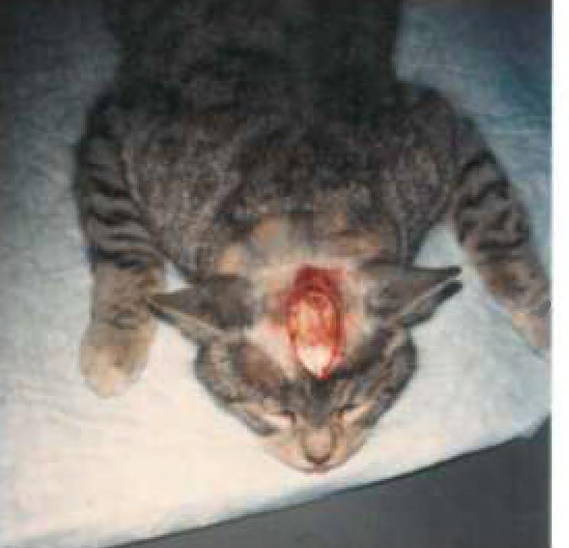
37 B. Experimental Procedures 1. Surgical Preparations a. General The procedures and equipment used in the actual data gathering process are the same as those outlined by Crawford and Coo1 26 When an experiment is to be run, the animal is deprived of food and water approximately twelve hours before the experiment is to begin. The experiment is begun by retrieving the animal from the animal facility, weighing it and anesthetizing with an intraperitoneal injection of 40 mg. of Nembutal per kiolgram of body weight. When the animal is sedated the top of its head, its throat, two spots on its chest just lateral to the anterior nipples, and the inside of both thighs are shaved. (see Figures 8 and 9.) Figure 8 Top of Head Shaved -31-


38 Figure 9 Ventral Shaved Areas This is to accommodate respectively the craniotomy, tracheotomy, cardiac monitoring and access to the femoral vein for an intravenous anesthesia to be administered for the duration of the experiment. b. Craniotomy Following this, an incision is made down the midline of the skull and the tissues separated sufficiently to allow for an opening through the skull of approximately 10 mm in diameter. This opening is made over the posterior lateral gyrus which reveals the posterior termination of the sulcus lateralis laterally and the central sinus medially. This allows access to the -32-


 Figure 10 Mid Line Incision")
39 visual cortex and lateral geniculate nucleus. This opening is gently filled in with surgical bone wax to maintain the integrity of the brain in an intact skull. (See Figures 10, 11 and 12.) Figure 10 Mid Line Incision With Skull Exposed -33-


40 Figure rnm Trephined Craniotomy -34-


41 Figure 12 Craniotomy After Being Filled With Surgical Bone Wax Two holes are then drilled into the frontal sinuses, one in each sinus. These openings allow two screws to secure a bracket to the skull. This bracket provides for an attachment to the stereotaxic apparatus which is the sole support and alignment mechanism for the head and eyes. (See Figure 13.) -35-


42 Figure 13 Support and Alignment Bracket in Place c. Tracheotomy Next an incision is made down the midline of the animal's throat and the tissues separated to expose the trachea. An incision is then made down the midline of the trachea to accommodate the insertion of a tube which will allow for artifical respiration. (See Figure 14.) - 36-


43 Figure 14 Intubated Trachea 2. Immobilization -Vital Signs The animal is then secured and aligned in the stereotaxic apparatus which is inside the electrically shielded room. The tubes from the artificial respirator are secured to the tracheal insertion. (See Figure 15.) -37-


 is inserted in the femoral vein; this is a")
44 Figure 15 Immobilized Animal Secured to Stereotaxic Apparatus The EKG electrodes are inserted under the skin on the chest and taped to hold them in place. An anal thermometer is inserted to record core body temperature. The thermometer is connected to a recording device which automatically turns a heating pad on or off as the animal s core temperature deviates from normal core temperature. An IV of Flaxedil in lactated ringers (5 mg/cc) is inserted in the femoral vein; this is a synthetic curare and completely immobilizes the animal. When normal respiration stops, the artifical respirator -38-


45 is turned on. Vagus innervation to the heart is slowly replaced by the heart's own pacemaker. Under optimum conditions, the animal can be maintained in this condition for forty-eight hours. 3. Refraction Plano contact lenses are placed on the animal's corneas after it has been dilated with atropisol (1%), a nd efricel (10%). Retinoscopy is performe d to establish the animal's refractive status. (See Figure 16. ) Figure 16 Retinoscopy -39-


46 Once t his is determined, the appropriate power contact lens is placed on each corne a t o fix the anima l 's far point at one meter. An op thalmoscope with a plan o lens in place is s e c ured t o a holder and its light is d i rected at the optic disc of each eye which is then projected onto a scre en one meter away. The details of this procedure are outlined by Fernald, R., and Chase, R.27 and Pettigrew, J.D., Cooper, M.L., Blasdel, G.G. 28 If the animal's far point is truly one meter, the disc will be in sharp focus on the screen. Its borders and accompanying vessel s a re traced out on the back s ide of the screen. From here area centralis of each eye can be mapped out on the s creen. (See Figure 17.) Figure 17 Optic Discs, Retinal Vessels and Areas Centra l is Mapped out on Tangent Screen -40-

.")
47 4. Single Cell Recording and Receptive Field Mapping A tungsten electrode is then secured to a DKI 607w hydraulic microdrive. The electrode is positioned above the bone wax which is covering the opening of the skull. (See Figure 18.) Figure 18 Electrode Secured to Microdrive The microdrive (as the name implies) can be advanced or retracted in micrometer increments (one millionth of a meter). The electrode is electronically connected to an oscilliscope which has an analog display of any electrical activity near the electrode. The electrode -41-


48 is advanced into the brain while a moving light is bein9 displayed on the screen in front of the animal. This is continued until a large enough signal can be detected and i ts receptive area mapped out on the screen. The signal may be of a binocular nature and then two separate receptive fields are mapped out representing corresponding retinal areas on the two retinas. I I I. PREI,IMINARY RESULTS At the time of this writing, we have run six separate preparations and each a nimal contributed to more efficient and better organized surgical and laboratory procedures. They also allowed us to work out any e l ectronic problems associated with the equipment, shielding and the electrods. In the first animal we were unable to elicite any e l ectrical activity in the brain. This was due to faulty electrodes which were made in our laboratory. In the second animal, three probes were made and we got a spike at a depth of mm. (See Figure 19.)


49 F i gure 1 9 Animal Number Two - Depth mm Two good responses were e licited from t h e third animal. The first r e sponse was at a depth of mm and had a small and difficult t o define recepti ve. field. (See Figure 20.) -43 -


50 Figure 20 Animal Number Three - Depth mm On the same penetration, but at a depth of mm, an on-off binocular response was elicited and mapped out. (See Figure 21.) Figure 21 Animal Number Three - Depth mm - 44-

 Figure 22 Animal Number Five- Depth.")
51 The next anima l experienced a cardic arrest early in the recording session and apparently wa s severe enough to depress most cortical activity. However, we did get good isolation of optic radiation f ibers. The fifth animal y ielded t wo responses 6n a single penetration, one at.759 mm; its r eceptive field was superior t emporal and binocular. (See Figure 22.) Figure 22 Animal Number Five- Depth.759 mm The second was at a depth of nwi its receptive field was also superior temporal and binocular. (See Figure 23.) -45-


52 Figure 23 Animal Number Five - Depth L 063 mm The. sixth and last a n i mal yielded some very interesting data. On t he twelft h penetration at a depth of.179 rom in the brain, two cells were activated. Cell number one and cell number two had amplitudes of 100 ~V a nd 135 ~V and durations of.25 msec and.125 msec r espectively. Their receptive fields were binocular. The right eye's receptive fie ld was mapped out as b eing 4.5 t emporal f rom the center o f the temporal disc margin and 14 superior from the center of the temporal disc margin. The left eye's receptive was 14.5 temporal from the temporal disc margin and 12.5 superior from the center of the temporal disc margin. A bar of l ight 2.5 long and.25 wide was moved from lower right to upper left in the receptive field. Both cells showed a strong right eye dominance. (See F i gure 24.) -46 -

53 Figure 24.Animal Number Six - Depth.179 rom A response from a third cell on the same penetration at a depth of rom was recorded. It was a hypercomplex cell with its receptive field in the center of area centralis of the right eye. It was extremely sensitive to a vertical bar approximately.5 long and very narrow. (See Figure 25.) -47-


54 Figure 25 Animal Number Six - Depth mm IV. Discussion All of the previously recorded activity was made from the visual cortex or optic radiation fibers. Next it will have to be demonstrated that these laboratory set-ups and procedures are adequate to record from the lateral geniculate nucleus (LGN). A breeding colony will have to be started so that kittens will be available to be reared under special conditions that will make them amblyopic. When the research reaches this stage, it will then start addressing the possibility of inhibitory mechanisms in the LGN as they relate to deprivation amblyopia. -48-


55 It is ver y e xciting to activate and map out a recept ive f ield under these conditions. It is o ne t h i ng to read a bout how the visual cortex is organized and how certain cells will respond to a particular shape of ligh t, o r d i r ection and speed of movement, o r a simple on- off response, but it really drives horne the fac t wh e n you can actually make the cell respond by manipula t i ng the light yourself. This has been a very valuable.experience for me to learn more abou t amblyopia and to be involved wi th the many detail s associated with setting up a laboratory in order to i n vestigate a hypot hesi s that involves animal models. I feel that optometry should do more of this t ype o f res ear ch. PUCO can n ow offer students a first-hand l ook at this kind of research and this exposure may d r aw those students who have a predilection f o r research into the field. -49-
 :338-354, August 1957. 3. Hubel, D.H., Wiesel, T. N.: Receptive Fields of Single Neurones in the Cat's Striate Cortex. J. Physiol 148(3) :574-59 1, October 1959. 4. Hubel, D.H., Wiesel, T.N.: Integrative Action in the Cat's Lateral Geniculate Body.")
56 Refer ences 1. F1om, M.C., Neumaier, R. W.. : Prevalence of Amblyopia. Public Health Reports 34(4), April Barlow, H.B., Fitzhugh, R., Kuffler, S.W.: Change of Organization in the Recepti v e Field s of the Cat's Retina During Dark Adaptation. J. Physiol 137(3) : , August Hubel, D.H., Wiesel, T. N.: Receptive Fields of Single Neurones in the Cat's Striate Cortex. J. Physiol 148(3) : , October Hubel, D.H., Wiesel, T.N.: Integrative Action in the Cat's Lateral Geniculate Body. J. Physiol 155(2): , February Hubel, D.H., Wiesel, T.N.: Receptive Fields, Binocular Interaction and Functional Architecture in the eat's Visual Cortex. J. Physiol 160(1) :1 06 ~154, January Hubel, D.H., Wiesel, T.N.: Receptive Fields and Functional Architecture in Two Nonstriate Visual Areas (18 and 19) of the Cat. J. Neurophysiol 28: , March Hubel, D.H., Wiesel, T.N. : Receptive Fields of Cells in Striate Cortex of Very Young Visually Inexperienced Kittens. J. Neurophysio l. 26: , November Wiesel, T.N., Hubel, D.H.: Effects of Visual Deprivation on Morphology and Physiology of Cells in the eat's Lateral Geniculate Body. J. Neurophysiol 26: , November Wiesel, T.N., Hubel, D.H.: Single Cell Responses in Striate Cortex of Ki ttens Deprived of Vi sion in One Eye. J. Neurophysiol 26: , November Hubel, D.H., Wiesel, T.N.: Binocular Interaction in Striate Cortex of Kittens Reared with Artificial Squint. J. Neurophysiol 28: , November Wiesel, T.N., Hubel, D.H.: Comparison of the Effects of Unilateral arid Bilateral Eye Closure on Cortical Unit Responses in Kittens. J. Neurophysiol 28: , November
, May 14. Von Noorden, G.K., Dowling-, J.E.")
 :215-220, August 1970. 16. Von Noorden, G.K.: Experimental Amblyopia in Monkeys; Further Behavioral Observations and Clinical Correlations. Invest Ophth 12(10) :721-726, October 1973.")
57 12. Wiesel, T.N., Hubel, D.H.: Extent of Recovery from the Effects of Visual Deprivation in Kittens. J. Neurophysiol 28: , November Birnbaum, Koslmve, and Sanet: Therapy as a Function of Ag-e: Am. J. of Optom. and Physiol Success in Amblyopia A Literature Survey. Optics, 54(5), May 14. Von Noorden, G.K., Dowling-, J.E., Ferg-uson, D.C.: Experimental Amblyopia in Monkeys. I: Behavioral Studies of Stimulus Deprivation Amblyopia. Arch Ophth 84 (2): , Augus t Von Noorden, G. K., Dowling, J. E.: Experimental Amblyopia in Monkeys. II: Behavioral Studies in Strabismic Amblyopia. Arch Opth 84(2) : , August Von Noorden, G.K.: Experimental Amblyopia in Monkeys; Further Behavioral Observations and Clinical Correlations. Invest Ophth 12(10) : , October Von Noorden, G.K. : Histological Studies of the Visual System in Monkeys with Experimental Amblyopia. Invest Ophth 12(10) : , October Baker, F.H., Grigg, P., Von Noorden, G.K.: Effects of Visual Deprivation and Strabismus on the Response of Neurons in the Visual Cortex of the Monkey, Including Studies on the Striate and Prestriate Cortex in the Normal Animal. Brain Res 66(2) : , Crawford, M.L.J., et al.: Physiological Consequences in Unilateral and Bilateral Eye Closure in Macaque Monkeys: Some Further Observations. Brain Res 84 (1): , Blakemore, C., Van Sluyters, R.C.: Experimental Analysis of Amblyopia and Strabismus. Brit J. Ophth 58(3): , March Duffy, F.H., Burchfiel, J.L., Conway, J.L.: Bicuculline Reversal of Deprivation Amblyopia in the Cat. Nature 260: , March 18, Kratz, K.E., Spear, P.D.: Postcritical-Period Deprivation on Striate Cortex Cells in the Cat. J. Neurophysiol 39(3) : , May Spear, P.D.: Role of Binocular Interactions in Visual System Development in the Cat, in Cool, S.J., Smith, E.L., (Ed.): Frontiers in Visual Science. New York, Springer-Verlag, 1978, pp

58 24. Maffei, L., Fiorentini, A.: Monocular Deprivation in Kittens Impairs the Spatial Resolution of Geniculate Neurones. Nature 264:754, Cool, S.J., Crawford, M.L.J. and Scheer, I.J.: Microel ectrode Preparations for CNS Recording. Review of Scientific Instruments, Vol. 41, No r October Tungsten The 10, 26. Crawford,.1. L. J., Cool, S. J. : Binocular Stimulation a nd Response Variability o f Striate Cort e x Units in the Cat. Vision Res. Vol. 10, pp , Fernald, R., Chase, R.: An Improved Method for Plotting Re tinal -Landmarks and Focusing the Eyes. Vision Res. Vol. II, pp Pettigrew, J.D., Cooper, M.L., Blasdel, G.G.: Improved use of Tapetal Reflection for Eye-Position Monitoring Invest Ophth 18(5) : , May
Experimental analysis of amblyopia
 Brit. J. Ophthal. (I974) 58, I76 Experimental analysis of amblyopia and strabismus COLIN BLAKEMORE AND RICHARD C. VAN SLUYTERS The Physiological Laboratory, Cambridge In the past few years physiological
Pre-natal construction of neural circuits (the highways are genetically specified):
:") Modification of Brain Circuits as a Result of Experience Chapter 24, Purves et al. 4 th Ed. Pre-natal construction of neural circuits (the highways are genetically specified): (1/6/2010) Mona Buhusi Postnatal
Modification of Brain Circuits as a Result of Experience Chapter 24, Purves et al. 4 th Ed. Pre-natal construction of neural circuits (the highways are genetically specified): (1/6/2010) Mona Buhusi Postnatal
THE POSTNATAL DEVELOPMENT OF THE VISUAL CORTEX AND THE INFLUENCE OF ENVIRONMENT
 THE POSTNATAL DEVELOPMENT OF THE VISUAL CORTEX AND THE INFLUENCE OF ENVIRONMENT Nobel lecture, 8 December 1981 by TORSTEN N. WIESEL Harvard Medical School, Department of Neurobiology, Boston, Massachusetts,
M. uch interest has recently been focused. Visual development in cats. 394 Pettigrew Investigative Ophthalmology. S.
 394 Pettigrew Investigative Ophthalmology May 1972 The one third of recordable cells in three-monthold binocularly sutured animals which you describe as "normal" could only be so called if one used the
Differential Effects of Early Monocular Deprivation on Binocular and Monocular Segments of Cat Striate Cortex
 J~uRNALOFNEUROPH YSIOLOGY Vol. 40, No. 4, July 1977. Printed in U.S.A. Differential Effects of Early Monocular Deprivation on Binocular and Monocular Segments of Cat Striate Cortex J. R. WILSON AND S,
Consequences of alternating monocular deprivation on eye alignment and convergence in cats. Randolph Blake, M. L. ]. Crawford, and Helmut V. B.
![Consequences of alternating monocular deprivation on eye alignment and convergence in cats. Randolph Blake, M. L. ]. Crawford, and Helmut V. B.](/thumbs/94/120773134.jpg "Consequences of alternating monocular deprivation on eye alignment and convergence in cats. Randolph Blake, M. L. ]. Crawford, and Helmut V. B.") Consequences of alternating monocular deprivation on eye alignment and convergence in cats Randolph Blake, M. L. ]. Crawford, and Helmut V. B. Hirsch Four kittens were raised with an opaque contact lens
Consequences of alternating monocular deprivation on eye alignment and convergence in cats Randolph Blake, M. L. ]. Crawford, and Helmut V. B. Hirsch Four kittens were raised with an opaque contact lens
Effects of Early Monocular Lid Suture on Spatial and Temporal Sensitivity of Neurons in Dorsal Lateral Geniculate Nucleus of the Cat
 JOURNALOF NEUROPHYSIOLOGY Vol. 43, No. 2, February 1980. Printed in U.S.A. Effects of Early Monocular Lid Suture on Spatial and Temporal Sensitivity of Neurons in Dorsal Lateral Geniculate Nucleus of the
abnormal lateral geniculate body. His anatomical study suggested that chiasm instead of remaining uncrossed. They thus reach the wrong hemispheres,
 J. Physiol. (1971), 218, pp. 33-62 33 With 1 plate and 9 text-figures Printed in Great Britain ABERRANT VISUAL PROJECTIONS IN THE SIAMESE CAT BY D. H. HUBEL AND T. N. WIESEL From the Department of Neurobiology,
deprived eye (reverse occlusion). beyond 1 year of age; only two of six animals recovered sufficient vision to enable
. beyond 1 year of age; only two of six animals recovered sufficient vision to enable") Journal of Physiology (1988), 395, pp. 639-66 639 With 8 text-figures Printed in Great Britain THE EXTENT OF VISUAL RECOVERY FROM EARLY MONOCULAR OR BINOCULAR VISUAL DEPRIVATION IN KITTENS BY DONALD E.
Journal of Physiology (1988), 395, pp. 639-66 639 With 8 text-figures Printed in Great Britain THE EXTENT OF VISUAL RECOVERY FROM EARLY MONOCULAR OR BINOCULAR VISUAL DEPRIVATION IN KITTENS BY DONALD E.
preferring rightward movement. A changeover later than 5 weeks of age peak of the critical period for directional deprivation may occur earlier
 J. Physiol. (1976), 257, pp. 155-170 155 With 5 text-figures Printed in Great Britain KITTENS REARED IN A UNIDIRECTIONAL ENVIRONMENT: EVIDENCE FOR A CRITICAL PERIOD BY N. W. DAW AND H. J. WYATT* From the
geniculate nucleus of kittens raised with convergent squint in one eye,
 J. Phyaiol. (1977), 270, pp. 345-366 345 With 1 plate and 9 text-ftgure8 Printed in Great Britain NASAL FIELD LOSS IN KITTENS REARED WITH CONVERGENT SQUINT: NEUROPHYSIOLOGICAL AND MORPHOLOGICAL STUDIES
Do blue-eyed white cats have normal or abnormal retinofugal pathways? R. W. Guillery, T. L. Hickey, and P. D. Spear
 Do blue-eyed white cats have normal or abnormal retinofugal pathways? R. W. Guillery, T. L. Hickey, and P. D. Spear Three white cats that had blue eyes and no tapetum were studied by behavioral, electrophysiological,
Effects of Convergent Strabismus on the Development of Physiologically Identified Retinogeniculate Axons ih Cats
 THE JOURNAL OF COMPARATIVE NEUROLOGY 28922-212 (1989) Effects of Convergent Strabismus on the Development of Physiologically Identified Retinogeniculate Axons ih Cats P.E. GARRAGHTY, A.W. ROE, Y.M. CHINO,
PATTERN EVOKED RESPONSE DEFICIENCY IN PATTERN DEPRIVED CATS 1
 Electroencephalography and Clinical Neurophysiology, 1973, 35: 569-573 Elsevier Scientific Publishing Company, Amsterdam - Printed in The Netherlands 569 PATTERN EVOKED RESPONSE DEFICIENCY IN PATTERN DEPRIVED
CLARSBISHOP AREA IN THE CAT: LOCATION AIVD RETINOTOPICAL PROJECTION
 ACTA NEUROBIOL. EXP. 1975, 35: 179488 CLARSBISHOP AREA IN THE CAT: LOCATION AIVD RETINOTOPICAL PROJECTION Krzysztof TURLEJSKI and Andrzej MICHALSKI Department of Neurophysiology, Nencki Institute of Experimental
spider monkeys by recording extracellularly from single units and stimulating
 J. Physiol. (1968), 195, pp. 215-243 215 With 3 plates and 14 text-figures Printed in Great Britain RECEPTIVE FIELDS AND FUNCTIONAL ARCHITECTURE OF MONKEY STRIATE CORTEX By D. H. HUBEL AND T. N. WIESEL
injected eve. (Received 1 November 1977) with electrolytic lesions. A good correspondence was found between the location of
 with electrolytic lesions. A good correspondence was found between the location of") J. Physiol. (1978), 281, pp. 267-283 267 With 6 plates and 3 text-figures Printed in Great Britain OCULAR DOMINANCE IN LAYER IV OF THE CAT'S VISUAL CORTEX AND THE EFFECTS OF MONOCULAR DEPRIVATION By CARLA
J. Physiol. (1978), 281, pp. 267-283 267 With 6 plates and 3 text-figures Printed in Great Britain OCULAR DOMINANCE IN LAYER IV OF THE CAT'S VISUAL CORTEX AND THE EFFECTS OF MONOCULAR DEPRIVATION By CARLA
Binocular Impulse Blockade Prevents the Formation of Ocular Dominance Columns in Cat Visual Cortex
 The Journal of Neuroscience August 1986, f?(8): 2117-2133 Binocular Impulse Blockade Prevents the Formation of Ocular Dominance Columns in Cat Visual Cortex Michael P. Stryker and William A. Harris Department
Binocular Exposure causes Suppression of the Less Experienced Eye in Cats Previously Reared with Unequal Alternating Monocular Exposure
 Binocular Exposure causes Suppression of the Less Experienced Eye in Cats Previously Reared with Unequal Alternating Monocular Exposure Nino Tumosa,* Stacy Nunberg, Helmut V. B. Hirsch, and Suzannah Bliss
THE PRETRIGEMINAL CAT AS AN INSTRUMENT FOR INVESTIGATION OF THE OCULAR FIXATION REFLEX
 ACTA NEUROBIOL. EXP. 1980, 40: 381-385 Lecture delivered at the Warsaw Colloquium on Instrumental Conditioning and Brain Research May 1979 THE PRETRIGEMINAL CAT AS AN INSTRUMENT FOR INVESTIGATION OF THE
Binocular Interactions in Striate Cortical Neurons of Cats Reared with Discordant Visual Inputs
 The Journal of Neuroscience, August 1994, 14(8): 55-567 Binocular Interactions in Striate Cortical Neurons of Cats Reared with Discordant Visual Inputs Yuzo M. Chino, Earl L. Smith III, Kazuyuki Yoshida,
The contralateral impairment of the orienting ocular-following reflex after lesions of the lateral suprasylvian cortex in cats
 The contralateral impairment of the orienting ocular-following reflex after lesions of the lateral suprasylvian cortex in cats Boguslaw ~ernicki and Maciej Stasiak Department of Neurophysiology, Nencki
Area Centralis Position Relative to the Optic Disc Projection in Kittens as o Function of Age
 Investigative Ophthalmology & Visual Science, Vol. 29, No. 8, August 1988 Copyright Association.for Research in Vision and Ophthalmology Area Centralis Position Relative to the Optic Disc Projection in
Effects of Retinal Image Degradation on Ocular Growth in Cats
 Effects of Retinal Image Degradation on Ocular Growth in Cats J. Nathan, 5. G. Crewrher,* D. P. Crewrher,* and P. M. Kielyf High-powered negative and positive contact lenses have been used to produce a
Spatial and Temporal Sensitivity of Normal and Amblyopic Cats
 JOURNALOF NEUROPHYSIOLOGY Vol. 48, No. 2, August 1982. Printed in U.S.A. Spatial and Temporal Sensitivity of Normal and Amblyopic Cats STEPHEN LEHMKUHLE, KENNETH E. KRATZ, AND S. MURRAY SHERMAN Department
Active sensing. Ehud Ahissar
 Active sensing Ehud Ahissar 1 Active sensing Passive vs active sensing (touch) Comparison across senses Basic coding principles -------- Perceptual loops Sensation-targeted motor control Proprioception
RETINITIS PIGMENTOSA*
 Brit. J. Ophihal. (1955), 39, 312. ABNORMAL FUNDUS REFLEXES AND RETINITIS PIGMENTOSA* BY R. P. CRICK Royal Eye Hospital, London THE normal variation of the fundus reflex which gives a " shot-silk" appearance
Development of Neuronal Response Properties in the Cat Dorsal Lateral Geniculate Nucleus During Monocular
 JOURNALOF NEUROPHYSIOLOGY Vol. 5, No. 1, July 1983. Printed in U.S.A. Development of Neuronal Response Properties in the Cat Dorsal Lateral Geniculate Nucleus During Monocular Deprivation STUART C. MANGEL,
DREXEL UNIVERSITY COLLEGE OF MEDICINE ANIMAL CARE AND USE COMMITTEE POLICY FOR PREOPERATIVE AND POSTOPERATIVE CARE FOR NON-RODENT MAMMALS
 DREXEL UNIVERSITY COLLEGE OF MEDICINE ANIMAL CARE AND USE COMMITTEE POLICY FOR PREOPERATIVE AND POSTOPERATIVE CARE FOR NON-RODENT MAMMALS OBJECTIVE: This policy is to ensure that appropriate provisions
Morphology of Retinogeniculate X and Y Axon Arbors in Cats Raised With Binocular Lid Suture
 JOURNALOFNEUROPHYSIOLOGY Vol. 60, No. 6, December 1988. Printed Morphology of Retinogeniculate X and Y Axon Arbors in Cats Raised With Binocular Lid Suture DENIS RACZKOWSKI, DANIEL J. UHLRICH, AND S. MURRAY
The Role of Early Experience in the Development and Maintenance of Orientation Selectivity in the Cat's Visual Cortex: M. Stryker
 Reprinted from Neurosciences Research \rogram Bulleti~ VOl~e IS, Number 3, Neuronal mechan1sms 1n visual perception E. P~p~el, R. Held & J.E. Dowling, edito;s (Cambr1dge, Mass.: MIT Press, 1977) Pages
THE JOURNAL OF COMPARATIVE NEUROLOGY 233: (1985)
") THE JOURNAL OF COMPARATIVE NEUROLOGY 233:190-212 (1985) Termination Patterns of Individual XI and Y-Cell Axons in the Visual Cortex of the Cat: Projections to Area 18, to the 17/18 Border Region, and to
THE JOURNAL OF COMPARATIVE NEUROLOGY 233:190-212 (1985) Termination Patterns of Individual XI and Y-Cell Axons in the Visual Cortex of the Cat: Projections to Area 18, to the 17/18 Border Region, and to
lowering of the visual acuity. When closure was extended through the first by varying the age at eye closure. Waiting until 1 month of age
 J. Physiol. (1970), 206, pp. 437-455 437 With 6 text-ftgure8 Printed in Great Britain CONSEQUENCES OF MONOCULAR DEPRIVATION ON VISUAL BEHAVIOUR IN KITTENS BY P. B. DEWS AND T. N. WIESEL From the Laboratory
Cortical Cell Orientation Selectivity Fails to Develop in the Absence of ON-Center Retinal Ganglion Cell Activity
 The Journal of Neuroscience, March 1, 2000, 20(5):1922 1930 Cortical Cell Orientation Selectivity Fails to Develop in the Absence of ON-Center Retinal Ganglion Cell Activity Barbara Chapman and Imke Gödecke
Recommended Resources: The following resources may be useful in teaching
 Unit D: Egg Production Lesson 1: Producing Layers Student Learning Objectives: Instruction in this lesson should result in students achieving the following objectives: 1. Discuss the materials and equipment
The Critical Period for Ocular Dominance Plasticity in the Ferret s Visual Cortex
 The Journal of Neuroscience, August 15, 1999, 19(16):6965 6978 The Critical Period for Ocular Dominance Plasticity in the Ferret s Visual Cortex Naoum P. Issa, Joshua T. Trachtenberg, Barbara Chapman,
Lens luxation when the lens gets wobbly
 Lens luxation when the lens gets wobbly Introduction The lens what is it there for? The lens - anatomy Lens luxation What does that mean? Lens luxation - what to look out for? Lens luxation How can it
Expression of a Surface-Associated Antigen on Y-Cells in the Cat Lateral Geniculate Nucleus Is Regulated by Visual Experience
 The Journal of Neuroscience, March 1988, 8(3): 874-882 Expression of a Surface-Associated Antigen on Y-Cells in the Cat Lateral Geniculate Nucleus Is Regulated by Visual Experience Mriganka Sur, Douglas
S Fault Indicators. S.T.A.R. Type CR Faulted Circuit Indicator Installation Instructions. Contents PRODUCT INFORMATION
 Fault Indicators S.T.A.R. Type CR Faulted Circuit Indicator Installation Instructions Service Information S320-75-1 Contents Product Information..........................1 Safety Information............................2
Regional Variation in the Representation of the Visual Field in the Visual Cortex of the Siamese Cat
 THE JOURNAL OF COMPARATIVE NEUROLOGY 193:237-253 (1980) Regional Variation in the Representation of the Visual Field in the Visual Cortex of the Siamese Cat MICHAEL LEE COOPER AND GARY G. BLASDEL Division
(Received 29 June 1972)
") J. Physiol. (1973), 228, pp. 115-137 115 With 9 text-figures Printed in Great Britain CONTRASTS IN SPATIAL ORGANIZATION OF RECEPTIVE FIELDS AT GENICULATE AND RETINAL LEVELS: CENTRE, SURROUND AND OUTER
J. Physiol. (1973), 228, pp. 115-137 115 With 9 text-figures Printed in Great Britain CONTRASTS IN SPATIAL ORGANIZATION OF RECEPTIVE FIELDS AT GENICULATE AND RETINAL LEVELS: CENTRE, SURROUND AND OUTER
A Comparison of Visual Pathways in Boston and Midwestern Siamese Cats
 A Comparison of Visual Pathways in Boston and Midwestern Siamese Cats CARLA SHA'TZ2 Department of Neurobiology, Harvard Medical School, 25 Shattuck Street, Boston, Massachusetts 021 15 ABSTRACT A genetic
1250 Reports. Axial lengths and refractive errors in kittens reared with an optically induced anisometropia. EARL L. SMITH, III, GREGORY W.
 1250 Reports Invest. Ophthalmol. Vis. Sci. September 1980 the existence of this arterial ring based upon sections obtained from man and monkey. 6 " 8 Although there are reports demonstrating a well-developed
Pet-Temp PT-300 Ear Thermometer Frequently Asked Questions
 Pet-Temp PT-300 Ear Thermometer Frequently Asked Questions 1) Is the Pet-Temp accurate? Yes, the Pet-Temp has a laboratory (in vitro) accuracy of 0.2 C (0.3 F). Clinical studies have verified the accuracy
Veterinary Ophthalmology
 Veterinary Ophthalmology Eyelids Protect the eye Provides part of and spreads the tear film Regulates the amount of light that enters the eye Clears foreign material Third Eyelid Protects the cornea by
log no. VNS23011 Ocular dominance columns in strabismus VNS23~6! :31 pm
 VNS23~6! 23011 1011 07007006 2:31 pm log no. VNS23011 Visual Neuroscience ~2006!, 23, 1 11. Printed in the USA. Copyright 2006 Cambridge University Press 0952-5238006 $16.00 DOI: 10.10170S0952523806230116
David H. Hubel. A Biographical Memoir by Robert H. Wurtz
 David H. Hubel 1926 2013 A Biographical Memoir by Robert H. Wurtz 2014 National Academy of Sciences. Any opinions expressed in this memoir are those of the author and do not necessarily reflect the views
UNIVERSITY OF PITTSBURGH Institutional Animal Care and Use Committee
 UNIVERSITY OF PITTSBURGH Institutional Animal Care and Use Committee Policy: Surgical Guidelines EFFECTIVE ISSUE DATE: 2/21/2005 REVISION DATE(s): 2/14/15; 3/19/2018 SCOPE To describe guidelines and considerations
1Ila and V. Canberra, A.C.T. 2601, Australia (Received 21 March 1979)
") J. Physiol. (1980), 302, pp. 483-505 483 With 2 plate and 9 text-ftigurew Printed in Great Britain THE AFFERENT CONNEXIONS AND LAMINAR DISTRIBUTION OF CELLS IN AREA 18 OF THE CAT BY A. R. HARVEY* From
J. Physiol. (1980), 302, pp. 483-505 483 With 2 plate and 9 text-ftigurew Printed in Great Britain THE AFFERENT CONNEXIONS AND LAMINAR DISTRIBUTION OF CELLS IN AREA 18 OF THE CAT BY A. R. HARVEY* From
UTILITY OF THE NEUROLOGICAL EXAMINATION IN RATS
 ACTA NEUROBIOL. ELW. 1980, 40 : 999-3 Short communication UTILITY OF THE NEUROLOGICAL EXAMINATION IN RATS David E. TUPPER and Robert B. WALLACE Laboratory of Developmental Psychobiology, University of
My recollections of Hubel and Wiesel and a brief review of functional circuitry in the visual pathway
 J Physiol 587.12 (2009) pp 2783 2790 2783 TOPICAL REVIEW My recollections of Hubel and Wiesel and a brief review of functional circuitry in the visual pathway Jose-Manuel Alonso Department of Biological
NUMBER: R&C-ARF-10.0
 1. PURPOSE PAGE 1 OF 6 This policy describes the procedures for keeping and maintaining animal medical records. This procedure is approved by the Creighton University Institutional Animal Care and Use
examination, the slight resistance encountered being sufficient By J. HERBERT PARSONS.
 PROCEEDI NGS OF THE PHYSIOLOGICAL May 10, 1902. SOCIETY, A method of measuring a visual illusion. By HORACE DARWIN and W. H. R. RIVERS. The instrument we show is designed for the quantitative study of
HALE SECURITY PET DOOR CAT GUARDIAN patent pending
 HALE SECURITY PET DOOR CAT GUARDIAN patent pending The Cat Guardian is an electronics package that can be added to a Hale Pet Door door or wall model of at least 1 3 / 8 thick to allow dogs free passage
Research with Animals
 Research with Animals Matthew Olugbenga Oyeyemi momattyemi@gmail.com +2348038059952 Research with Animals 1 Objectives Describe situations when animals may be research subjects Identify laws and regulations
JUDGING RABBITS 4-H LEADER MANUAL EM4502E WHY JUDGE? HOW TO JUDGE
 EM4502E 4-H LEADER MANUAL JUDGING RABBITS WHY JUDGE? Judging is an art practiced every time you say, I like this one better than that one. Judging helps you organize your thoughts and make a decision.
NUMBER: /2005
 Purpose PAGE 1 OF 7 The purpose of this policy is to describe the procedures for keeping and maintaining animal medical records. This procedure is approved by the Creighton University Institutional Animal
BUYING GUIDE ALGOT. Storage across the home. DESIGN Francis Cayouette
 BUYING GUIDE ALGOT Storage across the home DESIGN Francis Cayouette QUALITY Designed for use throughout the home. ALGOT shelves are hardwearing, stain resistant and easy to keep clean. Brackets are made
Department of Optometry, Oxford Eye Hospital Vision Related Electrodiagnostic Tests
 Oxford University Hospitals NHS Trust Department of Optometry, Oxford Eye Hospital Vision Related Electrodiagnostic Tests Information for patients Page 2 This leaflet is tells you about the test(s) for
(Received 22 November 1984) studies were made on twenty such pairs; eight X on-centre, seven Y on-centre, two
 studies were made on twenty such pairs; eight X on-centre, seven Y on-centre, two") J. Physiol. (1985), 369, pp. 249-268 249 With 12 text-ftgures Printed in Great Britain A COMPARISON OF VISUAL RESPONSES OF CAT LATERAL GENICULATE NUCLEUS NEURONES WITH THOSE OF GANGLION CELLS AFFERENT
J. Physiol. (1985), 369, pp. 249-268 249 With 12 text-ftgures Printed in Great Britain A COMPARISON OF VISUAL RESPONSES OF CAT LATERAL GENICULATE NUCLEUS NEURONES WITH THOSE OF GANGLION CELLS AFFERENT
PetSpy Premium Dog Training Collar, Models M919-1/M919-2
 PetSpy Premium Dog Training Collar, Models M919-1/M919-2 What is in the Package: M919-1/M919-2 Remote Transmitter Receiver Collar / E-Collar Radio Frequency: 900 Mhz Built-in Batteries information: Transmitter:
Express Train Electronic Radio Dog Training Collar
 Express Train Electronic Radio Dog Training Collar OPERATING INSTRUCTIONS Product Safety Statements Proper Collar Use: Express Train Radio Electronic Dog Training Collars are intended for the sole purpose
The Laminar and Size Distribution of Commissural Efferent Neurons in the Cat Visual Cortex*
 Arch. histol. jap., Vol. 42, No. 2 (1979) p. 119-128 The Laminar and Size Distribution of Commissural Efferent Neurons in the Cat Visual Cortex* Kazuhiko SHOUMURA Department of Anatomy (Prof. S. DEURA),
THE VISUAL MECHANISMS OF TENEBRIO MOLITOR: VARIATIONS TAKING PLACE IN THE ERG OF PUPA AND ADULT DURING DEVELOPMENT
 J. Exp. Biol. (1969), 51. 635-641 635 With 5 text-figures Printed in Great Britain THE VISUAL MECHANISMS OF TENEBRIO MOLITOR: VARIATIONS TAKING PLACE IN THE ERG OF PUPA AND ADULT DURING DEVELOPMENT BY
Perioperative Care of Swine
 Swine are widely used in protocols that involve anesthesia and invasive surgical procedures. In order to ensure proper recovery of animals, preoperative, intraoperative and postoperative techniques specific
SINGLE ANNUAL IMPLANT
 Manage pet ferret adrenal cortical disease with a SINGLE ANNUAL IMPLANT NOT APPROVED BY FDA Legally marketed as an FDA Indexed Product under MIF 900-013. FOR USE IN FERRETS ONLY. Extra-label use is prohibited.
Animal, Plant & Soil Science
 Animal, Plant & Soil Science Lesson C5-9 Veterinary Terminology Interest Approach Gather some common veterinary tools (e.g., scissors, forceps, and scalpels). Ask the students what each item is and for
Phylum Platyhelminthes Flatworms
 Phylum Platyhelminthes Flatworms The Acoelomates The acoelomates are animals that lack a coelom. Acoelomates lack a body cavity, and instead the space between the body wall and the digestive tract is filled
AnOn. Behav., 1971, 19,
 AnOn. Behav., 1971, 19, 575-582 SHIFTS OF 'ATTENTION' IN CHICKS DURING FEEDING BY MARIAN DAWKINS Department of Zoology, University of Oxford Abstract. Feeding in 'runs' of and grains suggested the possibility
2/5/2016. Military Tourniquet PFN:SOMTRL0B. Terminal Learning Objective. Reason. Hours: 0.5
 Military Tourniquet PFN:SOMTRL0B Hours: 0.5 Slide 1 Terminal Learning Objective Action: Communicate knowledge about the military tourniquet Condition: Given a lecture in a classroom environment Standard:
Effects of Cage Stocking Density on Feeding Behaviors of Group-Housed Laying Hens
 AS 651 ASL R2018 2005 Effects of Cage Stocking Density on Feeding Behaviors of Group-Housed Laying Hens R. N. Cook Iowa State University Hongwei Xin Iowa State University, hxin@iastate.edu Recommended
Ultra Min No-Bark Training Collar Ultra Small Ultra Powerful Ultra Control
 No-Bark Dog Training Device Owner s Manual Ultra Min-e 2090 TM No-Bark Training Collar Ultra Ultra Ultra Small Powerful Control D.T. Systems, Inc. 1 Congratulations and thank you for purchasing our Ultra
Temperature Gradient in the Egg-Laying Activities of the Queen Bee
 The Ohio State University Knowledge Bank kb.osu.edu Ohio Journal of Science (Ohio Academy of Science) Ohio Journal of Science: Volume 30, Issue 6 (November, 1930) 1930-11 Temperature Gradient in the Egg-Laying
Unit D: Egg Production. Lesson 4: Producing Layers
 Unit D: Egg Production Lesson 4: Producing Layers 1 1 Terms broodiness caged layer production floor production layers 2 2 3 I. Layers are chickens that are used to produce large quantities of eggs. A.
Spatial Analysis of Ocular Dominance Patterns in Monocularly Deprived Cats
 Spatial Analysis of Ocular Dominance Patterns in Monocularly Deprived Cats Kerstin E. Schmidt, Michael Stephan, Wolf Singer and Siegrid Löwel 1 Max-Planck-Institut für Hirnforschung, Neurophysiologische
Cages for Raising Pullets. Our Experience. Your Success.
 Cages for Raising Pullets Our Experience. Your Success. Modular Manure Belt (MMB ) Pullet System 3-High to 8-High Systems 16.24 [412.5] Cages That Set The Standard 25.34 [643.6] 30.00 [762.0] Chore-Time
striking it with unsheathed claws, was accompanied
 JOURNAL OF THE EXPERIMENTAL ANALYSIS OF BEHAVIOR TRANSFER OF AN ESCAPE RESPONSE FROM TAIL SHOCK TO BRAIN- STIMULA TED ATTACK BEHAVIOR' DAVID ADAMS AND JOHN P. FLYNN YALE UNIVERSITY SCHOOL OF MEDICINE VOLUME
Serendipity and the Siamese Cat: The Discovery That Genes for Coat and Eye Pigment Affect the Brain. Jon H. Kaas
 Serendipity and the Siamese Cat: The Discovery That Genes for Coat and Eye Pigment Affect the Brain Jon H. Kaas Abstract One day in the late 1960s, Ray Guillery was examining brain sections through the
texp. Biol. (196a), 39,
, 39,") texp. Biol. (196a), 39, 239-242 ith 1 plate Printed in Great Britain INNERVATION OF LOCOMOTOR MOVEMENTS BY THE LUMBOSACRAL CORD IN BIRDS AND MAMMALS BY J. TEN CATE Physiological Laboratory, University
texp. Biol. (196a), 39, 239-242 ith 1 plate Printed in Great Britain INNERVATION OF LOCOMOTOR MOVEMENTS BY THE LUMBOSACRAL CORD IN BIRDS AND MAMMALS BY J. TEN CATE Physiological Laboratory, University
Teaching Assessment Lessons
 DOG TRAINER PROFESSIONAL Lesson 19 Teaching Assessment Lessons The lessons presented here reflect the skills and concepts that are included in the KPA beginner class curriculum (which is provided to all
DLS Sample Preparation Guide
 DLS Sample Preparation Guide The Leica TCS SP8 DLS is an innovative concept to integrate the Light Sheet Microscopy technology into the confocal microscope. Due to its unique optical architecture samples
Fiber-Optic Accessories
 Fiber-Optic Accessories Various Accessories Expand the Usefulness of the Fiber-Optic Cables Lenses extend sensing distances Side-view attachments provide space savings Flexible stainless steel tubes protect
F1 IN THE NAME OF GOD
 F1 IN THE NAME OF GOD Slide 1 F1 FEIKO.IR.SOFT; 2011/07/06 Lid Laceration Conjunctival Hemorrhage a) No therapy is necessary b) Usually resolve in 7-12 days. Subconjunctival Hemorrhage Corneal Abrasion
Mass Delivery of Nonsurgical Sterilants
 Neutersol - What Works? What Targets? What Next? Nonsurgical Sterilization Presentation By: Sean Hawkins, President November 11, 2006 ACC&D Annual Conference Mass Delivery of Nonsurgical Sterilants Fun
$? 479 THE FUNCTION OF M. DEPRESSOR CAUDAE AND M. CAUDOFEMORALIS IN PIGEONS
 Oct.1 $? 479 THE FUNCTION OF M. DEPRESSOR CAUDAE AND M. CAUDOFEMORALIS IN PIGEONS BY HARVEY I. FISHER THE usual method of determining the function of a muscle is by gross dissection and study of attachments.
STANDARD OPERATING PROCEDURE #110 MOUSE ANESTHESIA
 STANDARD OPERATING PROCEDURE #110 MOUSE ANESTHESIA 1. PURPOSE This Standard Operating Procedure (SOP) describes methods for anesthetizing mice. 2. RESPONSIBILITY Principal Investigators (PIs) and their
STANDARD OPERATING PROCEDURE #111 RAT ANESTHESIA
 STANDARD OPERATING PROCEDURE #111 RAT ANESTHESIA 1. PURPOSE This Standard Operating Procedure (SOP) describes methods for anesthetizing rats. 2. RESPONSIBILITY Principal Investigators (PIs) and their research
Australian and New Zealand College of Veterinary Scientists. Membership Examination. Small Animal Surgery Paper 1
 Australian and New Zealand College of Veterinary Scientists Membership Examination June 2018 Small Animal Surgery Paper 1 Perusal time: Fifteen (15) minutes Time allowed: Two (2) hours after perusal Answer
Half Moon Aquarium Kit
 WARNING Buyer/User assumes all responsability for safety and proper use not in accordance with the directions and sately labels. Half Moon Aquarium Kit * Half moon shape aquarium kit * Equipped with a
FOOD AND AGRICULTURE ORGANIZATION OF THE UNITED NATIONS Rome, 1983
 ii by Soterios Economides FAO Consultant FOOD AND AGRICULTURE ORGANIZATION OF THE UNITED NATIONS Rome, 1983 The designations employed and the presentation of material in this publication do not imply the
Preparing for an AAALAC (and IACUC) Site Visit
 Site Visit") Preparing for an AAALAC (and IACUC) Site Visit Marcel Perret-Gentil, DVM, MS Neal Guentzel, PhD Updated 5/2013 About This Presentation Help you prepare for AAALAC site visit (as well as IACUC inspections)
Preparing for an AAALAC (and IACUC) Site Visit Marcel Perret-Gentil, DVM, MS Neal Guentzel, PhD Updated 5/2013 About This Presentation Help you prepare for AAALAC site visit (as well as IACUC inspections)
ANIMAL BEHAVIOR. Laboratory: a Manual to Accompany Biology. Saunders College Publishing: Philadelphia.
 PRESENTED BY KEN Yasukawa at the 2007 ABS Annual Meeting Education Workshop Burlington VT ANIMAL BEHAVIOR Humans have always been interested in animals and how they behave because animals are a source
Refinement Issues in Animal Research. Joanne Zurlo, PhD Institute for Laboratory Animal Research National Academy of Sciences
 This work is licensed under a Creative Commons Attribution-NonCommercial-ShareAlike License. Your use of this material constitutes acceptance of that license and the conditions of use of materials on this
SUMMARY OF PRODUCT CHARACTERISTICS
 SUMMARY OF PRODUCT CHARACTERISTICS 1. NAME OF THE VETERINARY MEDICINAL PRODUCT NOSEDORM 5 mg/ml Solution for injection for dogs and cats [DE, ES, FR, PT] 2. QUALITATIVE AND QUANTITATIVE COMPOSITION Each
Laminar and Columnar Distribution of Geniculo-cortical Fibers in the Macaque Monkey
 Laminar and Columnar Distribution of Geniculo-cortical Fibers in the Macaque Monkey DAVID H. HUBEL AND TORSTEN N. WIESEL Department of Neurobiology, Harvurd Medical School, 25 Shattuck Street, Boston,
Neuroscience Letters
 Neuroscience Letters 437 (2008) 65 70 Contents lists available at ScienceDirect Neuroscience Letters journal homepage: www.elsevier.com/locate/neulet Weakened feedback abolishes neural oblique effect evoked
Permanent Alterations in Muscarinic Receptors and Pupil Size Produced by Chronic Atropinization in Kittens
 No. 2 Reports 239 Permanent Alterations in Muscarinic Receptors and Pupil Size Produced by Chronic Atropinization in Kittens Earl L. Smith III,* Dianna A. Redburn,f Ronald 5. Harwerrh,* and Gregory W.
XL³ 800 IP 55metal distribution cabinets, freestanding enclosures and cable compartments
 XL³ 800 IP 55metal distribution cabinets, freestanding 87045 LIMOGES Cedex Telephone : 05 55 06 87 87 Fax : 05 55 06 88 88 Cat. No(s) : 20451/52/53/54/56/57/58/59/73/74 CONTENTS PAGE 1. General characteristics...
Pixie-7P. Battery Connector Pixie-7P Fuse* Motor. 2.2 Attaching the Motor Leads. 1.0 Features of the Pixie-7P: Pixie-7P Batt Motor
 1.0 Features of the Pixie-7P: Microprocessor controlled Low Resistance (.007 ohms) High rate (2800 Hz) switching (PWM) Up to 7 Amps continuous current (with proper air flow) High Output (1.2amp) Battery
Previous experiments on ferrets which were designed to determine the way in
 425 J. Physiol. (I95I) II3, 425-433 RELATION OF RETINAL STIMULATION TO OESTRUS IN THE FERRET BY A. P. D. THOMSON From the Department of Anatomy, University of Birmingham (Received 31 July 1950) Previous